RESEARCH ARTICLE
Human Cardiac Mesenchymal Stromal Cells
with CD105
+
CD34
-
Phenotype Enhance the
Function of Post-Infarction Heart in Mice
Justyna Czapla1☯, Sybilla Matuszczak1☯, Ewa Wiśniewska2, Magdalena Jarosz-Biej1,
Ryszard Smolarczyk1, Tomasz Cichoń1, Magdalena Głowala-Kosińska1, JoannaŚliwka2, Marcin Garbacz2, Mateusz Szczypior2, Tomasz Jaźwiec2, Agnieszka Langrzyk1,
MichałZembala2, Stanisław Szala1*
1Center for Translational Research and Molecular Biology of Cancer, Maria Skłodowska-Curie Memorial Cancer Center and Institute of Oncology, Gliwice Branch, Gliwice, Poland,2Department of Cardiac Surgery and Transplantology, Silesian Center for Heart Diseases, Zabrze, Poland
☯These authors contributed equally to this work.
Abstract
Aims
The aim of the present study was to isolate mesenchymal stromal cells (MSC) with CD105+ CD34-phenotype from human hearts, and to investigate their therapeutic potential in a mouse model of hindlimb ischemia and myocardial infarction (MI). The study aimed also to investigate the feasibility of xenogeneic MSCs implantation.
Methods and Results
MSC isolated from human hearts were multipotent cells. Separation of MSC with CD105+ CD34-phenotype limited the heterogeneity of the originally isolated cell population. MSC secreted a number of anti-inflammatory and proangiogenic cytokines (mainly IL-6, IL-8, and GRO). Human MSC were transplanted into C57Bl/6NCrl mice. Using the mouse model of hindlimb ischemia it was shown that human MSC treated mice demonstrated a higher capil-lary density 14 days after injury. It was also presented that MSC administrated into the ischemic muscle facilitated fast wound healing (functional recovery by ischemic limb). MSC transplanted into an infarcted myocardium reduced the post-infarction scar, fibrosis, and increased the number of blood vessels both in the border area, and within the post-infarction scar. The improvement of left ventricular ejection fraction was also observed.
Conclusion
In two murine models (hindlimb ischemia and MI) we did not observe the xenotransplant rejection. Indeed, we have shown that human cardiac mesenchymal stromal cells with CD105+CD34-phenotype exhibit therapeutic potential. It seems that M2 macrophages are essential for healing and repair of the post-infarcted heart.
a11111
OPEN ACCESS
Citation:Czapla J, Matuszczak S, Wiśniewska E, Jarosz-Biej M, Smolarczyk R, CichońT, et al. (2016) Human Cardiac Mesenchymal Stromal Cells with CD105+CD34-Phenotype Enhance the Function of
Post-Infarction Heart in Mice. PLoS ONE 11(7): e0158745. doi:10.1371/journal.pone.0158745
Editor:Shree Ram Singh, National Cancer Institute, UNITED STATES
Received:March 24, 2016
Accepted:June 21, 2016
Published:July 14, 2016
Copyright:© 2016 Czapla et al. This is an open access article distributed under the terms of the
Creative Commons Attribution License, which permits unrestricted use, distribution, and reproduction in any medium, provided the original author and source are credited.
Data Availability Statement:All relevant data are within the paper and its Supporting Information files.
1. Introduction
In late 1970s the Friedenstein group first isolated and described the basic features of mesenchy-mal cells present in the bone marrow of rodents [1–4].
Until now, mesenchymal stromal cells (MSC) are the subject of many studies worldwide. The existing data suggest that MSC can participate in a variety of therapeutic processes [5,6]. MSC may be involved in: (1) bone, cartilage, and muscle regeneration [7]; (2) improvement of the function of the post-infarction heart [8], regeneration of damaged liver and wound healing [9]; (3) inhibi-tion of abnormal immune response [10]; (4) or may serve as carriers of therapeutic genes [11].
MSC are attributed to four primary mechanisms of action [12,13]: (1) MSC are able to home to the inflammatory areas; (2) MSC are able to transdifferentiate into, for example, cardiomyocytes, endothelial cells, smooth muscles cells; (3) MSC secrete factors that stimulate regeneration and repair of damaged tissues and organs; (4) MSC possess immunomodulatory properties (they inhibit, among others, inflammation). For this reason, MSC are called„guardians of inflammation”[14].
The aim of our study was to find an answer to questions: Do human MSC with CD105+ CD34-phenotype possess therapeutic properties, in other words, do they facilitate the func-tional recovery of ischemic hindlimb and infarcted mouse heart? Do therapeutic properties of MSC may be tested in xenogeneic system?
In the study we investigated therapeutic potential of MSC with CD105+CD34-phenotype. The choice to use these cells may be explained by four reasons. (1) CD105 antigen (endoglin, TGFβcoreceptor) is a characteristic marker for mesenchymal cells [15]. (2) MSC selected for the presence of CD105 exhibited significantly better therapeutic properties than unselected cells [16]. Endoglin activates pathways involved in scar remodeling, and protects endothelial cells against apoptosis induced by hypoxia [17]. (3) A purified CD105+cells population has a reduced heterogeneity, which may influence its potency, safety, tissue specific efficacy and mechanism of action [18]. (4) It has also been proven that cells isolated from hearts have a greater therapeutic potential than cells isolated from bone marrow [19].
Our data confirm therapeutic properties of human MSC with CD105+CD34-phenotype. Human MSC stimulate angiogenesis in mouse ischemic tissues, reduce the post-infarction scar and fibrosis, and increase the left ventricular ejection fraction in a model of murine MI. More-over, we did not observe MSC rejection. Additionally, MSC secrete factors which may polarize macrophages toward anti-inflammatory M2 phenotype [20]. It seems that M2 macrophages are essential for healing and repair of the post-infarcted heart [21,22].
2. Materials and Methods
An expanded Methods section can be found in the supplementary material online (S1 Text).
2.1. Ethical statement
The experiments were performed in accordance with the Declaration of Helsinki, with the approval of the Local Committee on Bioethics in Katowice. All patients provided written informed consent for the collection of excised heart and subsequent analysis.
Mice (6- to 8-week-old, C57Bl/6NCrl males) were bred in our animal facility house. The experimental protocol was approved by the Local Ethics Commission (Medical University of Silesia, Katowice, Poland).
2.2. Isolation of CD105
+CD34
-cells
The tissue collected from human hearts was minced into fragments of approximately 1mm3 and digested with collagenase using a modification of as previously described protocol [19].
within the framework of Innovative Economy Operational Programme 2007-2013.
After obtaining 90–100% confluence of MSC, the cells were incubated with antibodies directed against human antigens: CD105 and CD34. The population of CD105+CD34-cells was separated using a BD FACSAria™III cell sorter (BD Biosciences). The cells from the 1stto 3rdpassages were used for further experiments.
2.3. Immunophenotypic analysis and differentiation of MSC with
CD105
+CD34
-phenotype
The phenotype of CD105+CD34-cells (1st passage) was determined using a flow cytometer (BD FACSCanto™BD Biosciences). The cells were incubated with appropriate antibodies directed against the following human antigens: CD29–FITC (eBioscience), CD105–APC, CD73–PE, CD90–PE-Cy7, CD44–FITC, CD34–PE-Cy7, CD31–FITC, CD146–PE, KDR– PE, LIN–FITC, CD45–PE, HLA-DR–PE-Cy7 (BD Pharmingen), or isotype-matched control antibodies were used. The Human Mesenchymal Stem Cell Functional Identification Kit (R&D Systems, Minneapolis, MN, USA) was used to differentiate CD105+CD34-cells into adipocytes, osteoblasts, and chondroblasts. The procedure was performed in accordance with the manufac-turer’s instructions.
2.4. Analysis of the secretome of CD105
+CD34
-cells
The type and quantity of cytokines and growth factors secreted by CD105+CD34-cells, was assessed using the Human Cytokine Antibody Array C5 kit (RayBiotech, Norcross, GA, USA). The analysis was conducted in accordance with the manufacturer’s instructions.
2.5. Mouse model of hindlimb ischemia
Unilateral femoral artery ligation was performed as described [23]. An hour after ligation, 106 [23] of CD105+CD34-cells in 100μL of PBS-or 100μL of PBS-were administered into the C57Bl/6NCrl male mice muscle. The function of the limb were assessed at the following time points: 2, 5, 7, 9, 12 and 14 days after artery ligation. Limb function were assessed on a scale of 0–3 points according to the following criteria [24]: 3–most severe, unable to use the foot; 2– no dragging, ability to flex the angle, able to use the limb as weight support; 1–positive plantar flexion, some problems can be observed as compared to the healthy limb; 0–no difference between the ligated limb and the healthy (control) limb can be observed.
2.6. Induction of myocardial infarction (MI) and CD105
+CD34
-cells
implantation
MI was induced by ligation of the left anterior descending coronary artery (LAD) in 8-10-week-old C57Bl/6NCrl male mice kept under anesthesia. 7 days after LAD ligation, second thoracotomy was performed and 0.5x10⁶CD105+CD34-cells suspended in 10μl of PBS-or 10μl of PBS-(control), were injected into the border area of the post-infarction scar. The sham group consisted of mice injected with PBS-without LAD ligation. Hearts were collected 42 days after PBS-or CD105+CD34-cells injection [16].
2.7. Immunohistochemistry
Formalin-fixed heart were embedded in paraffin or frozen in liquid nitrogen and sectioned into 5–8μm slices. Viable tissue and collagen were visualized by incubation with a 0.1% solu-tion of Fast Green and a 0.1% solusolu-tion of Sirius Red (Sigma) in 1.2% picric acid (1h, RT). Blood vessels were detected by incubation with either lectin conjugated with FITC (Lycopersicon
conjugated secondary antibody. Macrophages were stained with appropriate antibodies (CD206 or iNOS; Abcam) and fluorochrome-conjugated secondary antibodies. Human cells in a mouse tissue were identified by human lamin A+C, Nuclear Envelope Marker (Abcam). IL -6 was stained with anti-IL-6 antibody (Abcam) and Texas Red-conjugated secondary antibody. Nuclei were counterstained by DAPI.
2.8. Identification of M1 and M2 macrophages
Flow cytometry was used to determine macrophages subset in the post-infarcted hearts. At the following time points: 1, 3 and 7 days, hearts were collected, minced and digested with collage-nase solution.
2.9. Immunophenotypic analysis of BMDM
BMDM (Bone Marrow-Derived Macrophages) were isolated and differentiated as previously described [25,26]. Seven days after isolation, BMDM were incubated for 48h with conditioned medium from CD105+CD34-cells (MSC-CM), control medium (IMDM medium supple-mented with 20% FBS, L-glutamine and antibiotics) or control medium with lipopolysaccha-ride (LPS; 1μg/mL [27,28], eBioscience). Flow cytometry were used to determine the
phenotype of BMDM from each group.
2.10 Statistics
Data were presented as mean ± standard deviation and compared between groups using Stu-dent’s t-test, or by one-way analysis of variance (ANOVA), using Tukeys’multiple comparison test for post hoc analysis, with p<0.05 considered statistically significant. Non-parametric
testing (Kruskal-Wallis test, U Mann Whitney test) were used in the case of data that do not have a normal distribution. Statistical analysis was performed using Statistica 10 software.
3. Results
3.1. MSC isolated from the heart
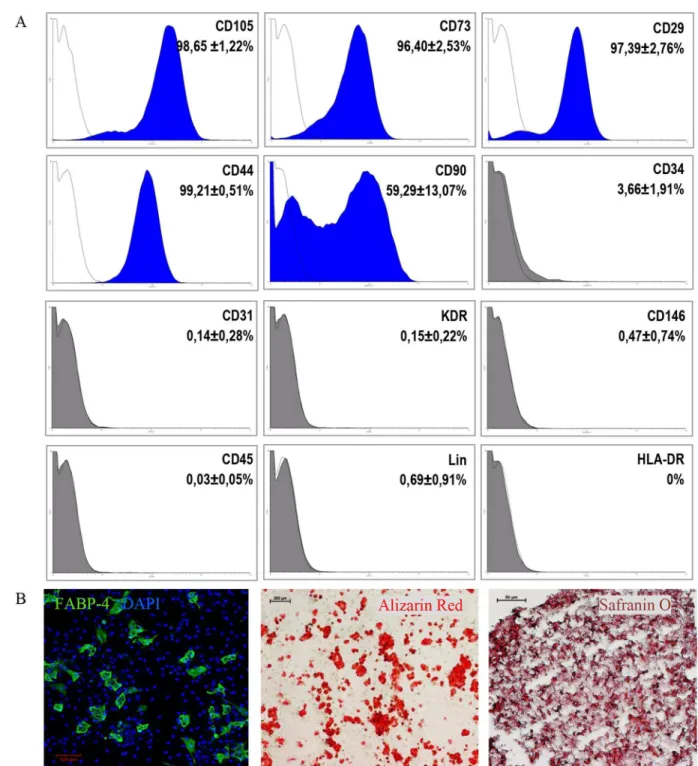
The culture of MSC was established for 19 tissue fragments derived from the right ventricle. After obtaining 90–100% MSC confluence, a population of CD105+CD34-cells was sorted. Cell surface antigen phenotyping of CD105+CD34-cells (1st passage) indicated that over 96% of the cells expressed markers characteristic for mesenchymal cells, such as CD105, CD29, CD73, and CD44 (Fig 1A). The exception was the CD90 marker, whose percentage of cells equaled on average 59.3%. CD105+CD34-cells in culture were devoid of (<1%) hematopoietic
marker (CD45) and mature blood cells markers (LIN), and they did not express molecules of the major histocompatibility complex class II (HLA-DR). Among CD105+CD34-cells there were no cells expressing a marker specific for mature endothelial cells (CD31), progenitor cells of the vessels (KDR), or CD146+cells. We have shown previously that MSC isolated from human adult hearts lack c-Kit antigen [29]. CD105+CD34-cells were able to differentiatein vitrointo adipocytes, osteoblasts, and chondroblasts (Fig 1B).
3.2. Secretome of CD105
+CD34
-cells
Fig 1. Phenotype and differentiation potential of CD105+CD34-cells selected from the MSC population(A) Phenotype and differentiation potential of CD105+CD34-cells selected from the MSC population isolated from fragments of heart tissue. Over 96%
of the cells express markers characteristic for mesenchymal cells: CD105, CD29, CD73, and CD44, with the exception of the CD90 marker (~59.3%) (n = 11). CD105+CD34-cells in culture do not express the following antigens (<1%): CD45, Lin, HLA-DR, CD31,
KDR, and CD146. Despite the selection of the CD105+CD34-population with high specificity of the process (P = 97.68±1.52%,
n = 11), the presence of CD105+CD34+cells (~3.7%) was still observed in the culture. (B) The cellsin vitrodifferentiate into
adipocytes (FABP4, green, n = 6, magn. 10x), osteoblasts (Alizarin Red, red, n = 6, magn. 4x), and chondroblasts (Safranin O, dark red, n = 4, magn. 20x).
doi:10.1371/journal.pone.0158745.g001
3.3. Mouse model of hindlimb ischemia
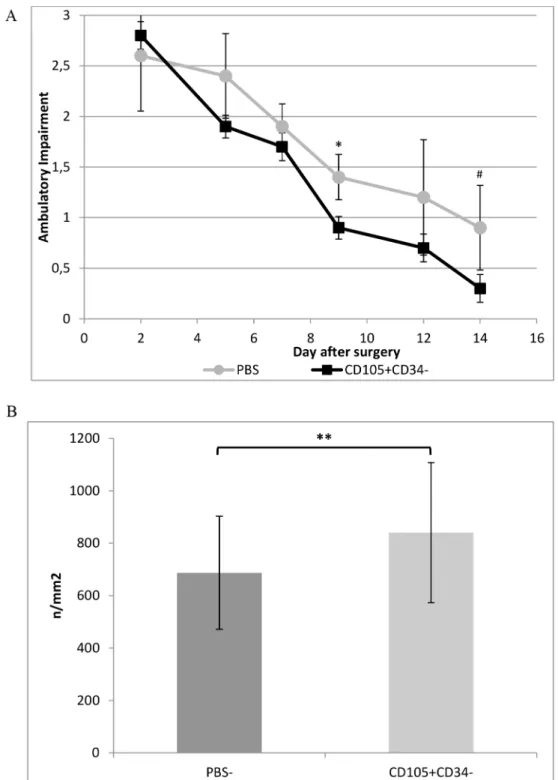
Mouse model of hindlimb ischemia allowed to assess proangiogenic features of CD105 +-CD34-cells. Injection of one million CD105+CD34-cells into the ischemic muscle facilitated fast wound healing (functional recovery by ischemic limb) (Fig 3A). Such an effect were not observed in the control group (mice that received PBS-). After 28 days, the mice in both groups are recovering the full functionality in ischemic limb [23]. After injection of CD105 +-CD34-cells into ischemic muscle the increase in the number of blood vessels (840±247.1/ mm2) in comparison with the control group (687.3±215.8/mm2) was observed (p<0.01)
(Fig 3B).
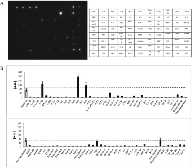
Fig 2. Cytokines and growth factors secreted by CD105+CD34-cellsin vitro.(A) An exemplary image of a membrane used for the analysis of 80 cytokines and growth factors secreted by CD105+CD34-cells. (B) Densitometric analysis of 80 cytokines
and growth factors secreted by CD105+CD34-cells indicates that CD105+CD34-cells secrete mainly IL-6, IL-8, and GRO
molecules (n = 5).
Fig 3. Therapeutic potential of CD105+CD34-cells tested in a mouse model of hindlimb ischemia.(A) CD105+CD34-cells treated mice demonstrated improved functional outcomes compared to the control mice
(PBS-) (n = 5; experiment repeated twice). (B) The number of blood vessels was higher at day 14 in mice after
administration of CD105+CD34-cells compared to the control mice after administration of PBS-(n = 10; 10 muscles per group were analyzed, in each muscle 10 pictures were taken).*p<0.05 #p = 0.056**p<0.01 by the Mann-Whitney U test.
doi:10.1371/journal.pone.0158745.g003
3.4. CD105
+CD34
-cells affect the increase in the number of blood
vessels after MI
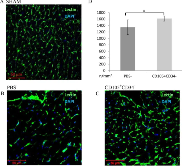
The assessment of the number of blood vessels was performed 6 weeks after MI. In the border area of the post-infarction scar, the vessels were stained with lectin (Fig 4). After 6 weeks a statistically significant increase in the density of capillaries in mice after administration of CD105+CD34-cells (1616±39/mm2) compared to the control group (mice that received PBS-) (1347±114/mm2) (p<0.05) was observed (Fig 4D). The number of vessels located
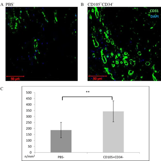
within the area of the post-infarction scar was determined using anti-CD31 antibody (Fig 5). A statistically significant increase in the number of vessels in mice after administration of CD105+CD34-cells (343±44/mm2) compared to the control group (178±32/mm2) (p<0.01)
was observed (Fig 5C).
Fig 4. Capillary density in the border area of the post-infarction scar 7 weeks after MI.(A B C) Representative images presenting the number of blood vessels in the border area of the post-infarction scar (magn. 20x) (A) in a Sham group, (B) in mice after administration of PBS-, (C) in mice after administration of CD105+CD34-cells. (D) In the area bordering the
post-infarction scar a significant increase in the number of blood vessels in mice after administration of CD105+CD34-cells
compared to the control mice after administration of PBS-was observed. n = 7;
*p<0.05 by the Student’s t-test.
3.5. CD105
+CD34
-cells influence the reduction in the size of the
post-infarction scar and fibrosis
Fig 6A–6Cpresents representative images of histochemical staining of a post-infarction scar 6
weeks after administration of CD105+CD34-cells or PBS-. In mice injected with CD105+CD34 -cells 7 days after MI, a statistically significant reduction in the size of post-infarction scar (22.53±5.55%) compared to the control PBS-group (32.84±3.76%; p<0.01) was observed (Fig 6D).
Fig 5. Capillary density within the area of the post-infarction scar 7 weeks after MI.(A B C) Representative images presenting the number of blood vessels within the area of the post-infarction scar. Magn. 20x. (A) in mice after administration of PBS-(B) in mice after administration of CD105+CD34-cells. (C) Within the scar, a significant increase in the number of blood
vessels in mice after administration of CD105+CD34-cells compared to the control mice after administration of PBS-was
observed. n = 7;**p<0.01 by the Student’s t-test.
doi:10.1371/journal.pone.0158745.g005
The deposition of collagen between the muscle fibers in the border area of the post-infarc-tion scar is associated with remodeling of the post-infarcpost-infarc-tion heart.Fig 6E–6Gshows
represen-tative images of collagen staining deposited between the cells of the muscle tissue in the border area of the post-infarction scar. In mice injected with CD105+CD34-cells a statistically signifi-cant reduction in the amount of deposited collagen (14.23±3.02%), compared to the control PBS-group (24.29±4.95%) (p<0.01) was observed (Fig 6H).
3.6. CD105
+CD34
-cells improve the left ventricular ejection fraction after
MI
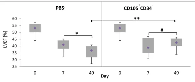
The ultrasound examination of left ventricle was performed: before LAD ligation, 7 days after LAD ligation, before administration of the cells or PBS-, and 42 days after administration of CD105+CD34-cells or PBS-. The measured parameter was left ventricular ejection fraction (LVEF). An improvement of functional parameters of infarcted heart was observed only in a group of mice injected with CD105+CD34-cells. The myocardial contractility increased from 38.8% LVEF (after MI) to 42.5% (p = 0.0946). A statistically significant increase of LVEF was observed between a group of mice treated with CD105+CD34-cells and a group of mice injected with PBS-(36.8% vs 42.5%, respectively, p<0.01) (Fig 7).
Fig 6. Changes in a size of the scar and fibrosis in the post-infarction heart of a mouse 7 weeks after MI. (A B C) Representative images illustrating a post-infarction scar in the mouse heart in a (A) Sham group or after administration of (B) PBS-or (C) CD105+CD34-cells. The images are composed of several shots (magn. 4x). (D)
The area of the post-infarction scar is significantly smaller after administration of CD105+CD34-cells compared
to the control group, in which PBS-was administered (n = 14);
**p<0.01 by the U Mann–Whitney test. (E F G) Representative images illustrating collagen (pink) deposited between the fibers of the muscle tissue (green) in the mouse heart in a (E) Sham group or after administration of (F) PBS-, or (G) CD105+CD34-cells. The sections with the greatest ratio of post-infarction scar to the whole stained area were used for examination (magn. 40x). (H) A statistically significant reduction of fibrosis was observed in the border area of the post-infarction scar after administration of CD105+CD34-cells compared to the control group after administration of PBS-(n = 14);
**p<0.01 by the U Mann–Whitney test.
doi:10.1371/journal.pone.0158745.g006
Fig 7. Changes in left ventricular ejection fraction (LVEF) 6 weeks after the cells administration.LVEF increased by 5.75% between treated groups at 49 day. Day 0–LVEF (baseline); day 7–LVEF seven days after MI induction, the day of administration of CD105+CD34-cells or PBS-: day 49
–LVEF on the day of the collection of the hearts. PBS-: n = 16;
CD105+CD34-: n = 18;**p<0.01;*p<0.05; # p = 0.0946. Comparisons between groups and within groups were performed by the one-way analysis of variance (ANOVA) with repeated measures with post-hoc Tukey tests.
3.7. IL-6 secretion after CD105
+CD34
-cells administration and the
presence of macrophages with M2 phenotype in ischemic mouse heart
tissue
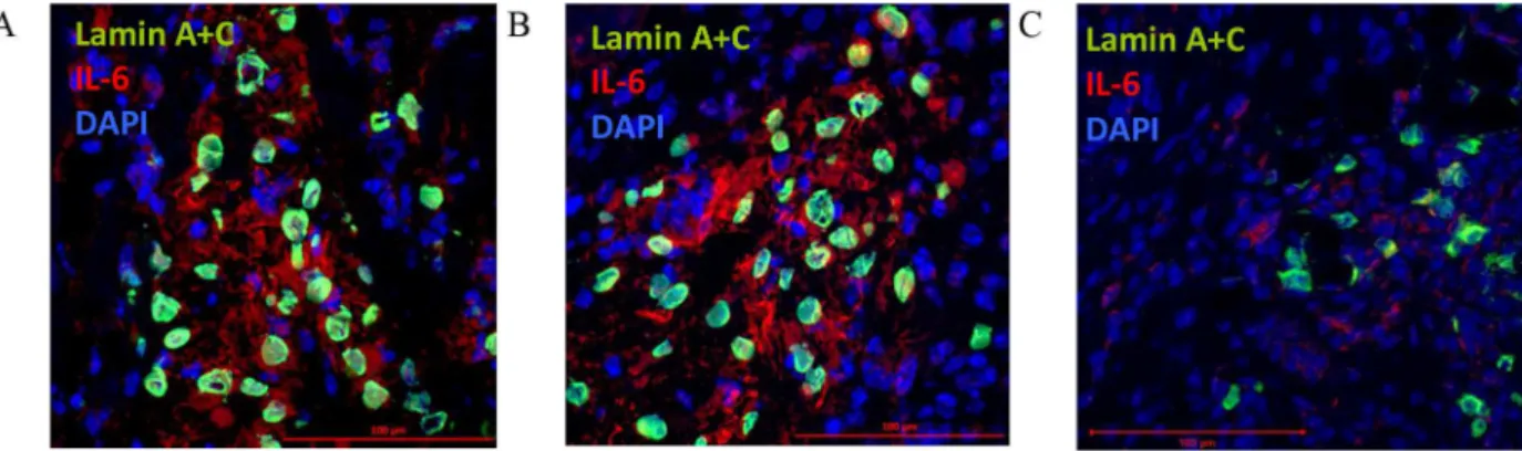
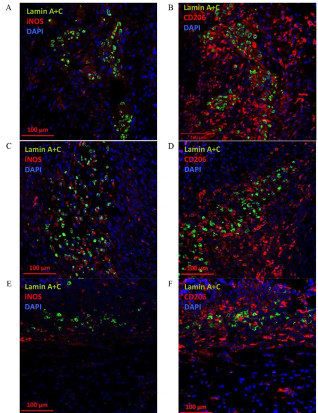
When CD105+CD34-cells were administrated into the border area of the post-infarction scar they retain their ability to IL-6 secretion (Fig 8). IL-6 is secreted at the first and third day after CD105+CD34-cells administration (Fig 8A and 8B). The cytokine secretion is related with the cells presence in the tissue, and at the day seventh CD105+CD34-cells and IL-6 were slightly detected (Fig 8C). Additionally, around the implanted human cells an increase in the number of anti-inflammatory, and proangiogenic macrophages with the M2 phenotype (CD206, C mannose receptor type 1) at all time points was observed (Fig 9). However, small amounts of pro-inflammatory macrophages with the M1 phenotype was also observed (iNOS, enzyme, inducible nitric oxide synthase). A trace amount of CD105+CD34-cells were still observed in the tissue of post-infarcted heart up to 7 days after the cells administration (Fig 9E and 9F).
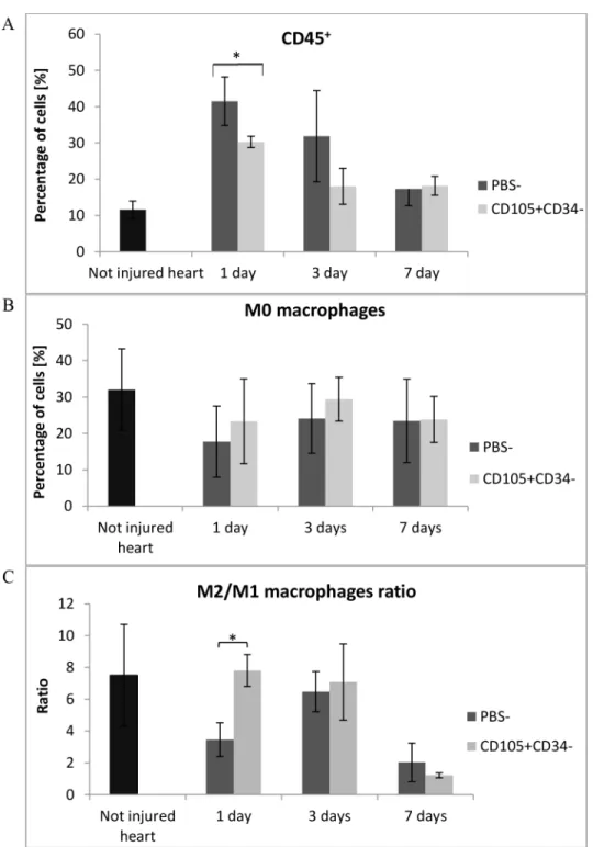
To verify and compare the obtained results with the control group, the flow cytometry anal-ysis of whole collagenase–digested mice hearts were performed. We characterized both cells of the immune system and cardiac macrophage subsets in the infarcted heart at the certain points after MI. Leukocytes were stained with anti-CD45 antibody and gated with CD45 fluorescence versus side scatter. The number of CD45+cells infiltrating the post infarcted tissue was signifi-cantly lower (p<0.05) in hearts collected 1 day after CD105+CD34-cells injections in
compari-son with the control group (Fig 10A). The number of CD45+cells was over 3-fold lower (average: 1.8x10⁶vs. 0.5x10⁶cells). The level of macrophages M0 (CD45+F480+cells) was higher in the in mice injected with CD105+CD34-cells (Fig 10B). Two macrophage subsets were identified: M1 (CD45+F480+CD86+cells) and M2 (CD45+F480+CD206+cells). The per-centage of M1 macrophages was relatively constant (<5%) during first days after the cells or
PBS-injections and did not differ between groups (data not shown). However, the percentage of M2 macrophages was higher in mice injected with CD105+CD34-cells 1 and 3 days after injections. Moreover, in those mice 1 day after the cells implantation the M2/M1 ratio was sig-nificantly higher compared to the control group (7.8 vs. 3.5) (p<0.05) (Fig 10C).
3.8. MSC-CM induced the expression of CD206 in BMDM
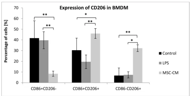
To directly assess if CD105+CD34-cells induce an M2 phenotype in macrophages we deter-mined the expression of mannose receptor (CD206), a well-accepted marker for M2
Fig 8. CD105+CD34-cells secrete IL-6 after administration into the post-infarction mouse heart.CD105+CD34-cells (lamin A+C,
green) administrated into the border area of the post-infarction scar secreted IL-6 (IL-6, red) after 1 day (A), 3 days (B). After 7 days a trace amount of CD105+CD34-cells were observed and IL-6 was slightly secreted (C) (magn. 40x).
Fig 9. The presence of M2 macrophages after administration of CD105+CD34-cells into the post-infarction mouse heart.(A) 1 day after implantation of CD105+CD34-cells (lamin A+C, green) into the border area of the
post-infarction scar, small amounts of pro-inflammatory macrophages with the M1 phenotype (iNOS, red) around the transplanted human cells were observed (magn. 20x). (B) 1 day after CD105+CD34-cells (lamin A+C, green) implantation an increase in the number of anti-inflammatory, and proangiogenic macrophages with the M2 phenotype (CD206, red) in their surroundings was observed. The similar phenomenon was observed: (C) (D) 3 days after implantation of CD105+CD34-cells (lamin A+C, green) and (E) (F) 7 days after implantation of CD105+CD34-cells
macrophages, in BMDM using flow cytometry. The incubation of MSC-CM with BMDM greatly increased the percentage of CD206+CD86+and CD206+CD86-macrophages (46% and Fig 10. MSC with CD105+CD34-phenotype modulate inflammation after administration into infarcted mouse heart.(A) The number of leukocytes (CD45+cells) infiltrating the post-infarcted heart were
significantly lower 1 day after CD105+CD34-cells administration; n = 3. (B) The level of M0 macrophages (%
of CD45+F480+cells) accumulated in the infarcted heart was higher after CD105+CD34-cells administration;
n = 3. (C) CD105+CD34-cells significantly increased the M2/M1 ratio 1 day after the cells administration;
n = 3*p<0.05 compared to the control (PBS-) group by the Student’s t-test.
32.3%, respectively) than in the control BMDM (30.4% and 6.7%, respectively) and in BMDM incubated in medium with LPS (19.6% and 7.4%, respectively) (Fig 11). All analyzed BMDM expressed F4/80 antigen, a characteristic marker for macrophages (data not shown).
4. Discussion
The aim of the present study was to investigate therapeutic potential of human MSC with CD105+CD34-phenotype in a mouse model of hindlimb ischemia and myocardial infarction. We aimed also to investigate whether MSC may be transplanted into fully immunocompetent recipients without immunosuppression. The unique immune privileged nature of MSC may prevent rapid rejection in a xenogeneic environment and preserve their ability to promote the repair of injured tissues [30,31].
Population of cardiac cells with CD105+CD34-phenotype was selected with the use of FACS (Fluorescence-Activated Cell Sorting) thereby were characterized by specific immuno-phenotype. Theoretically, the cells population should exhibit reduced heterogeneity. The popu-lation of these cells, however, expresses all antigens characteristic for MSC and differentiatein vitrointo adipocytes, chondroblast and osteoblasts (Fig 1).
CD105+CD34-cells in the culture conditions secrete mainly: IL-6, IL-8, GRO, OPG and HGF. However, the predominant cytokine is IL-6 (Fig 2). Some papers suggest that IL-6 may induce cardiomyocytes hypertrophy, fibroblasts proliferation and elevated fibrosis, contribut-ing to adverse myocardial remodelcontribut-ing [32,33]. IL-6 participates also in immune reactions. Depending on the microenvironment the cytokine may be pro- or anti-inflammatory one [34]. IL-6 is involved in polarization of M1 macrophages towards macrophages with M2 phenotype [35,36]. M1 are pro-inflammatory macrophages. M1 secrete a toxic effector molecules (ROS and NO) and pro-inflammatory cytokines (IL-1β, TNF, IL-6) [37]. M2 are anti-inflammatory Fig 11. MSC-CM increased the expression of CD206 in BMDM.The incubation of MSC-CM with BMDM greatly increased the percentage of CD206+CD86+and CD206+CD86-macrophages in comparison with control BMDM cells and BMDM cells incubated in medium with LPS; n = 6*p<0.05;**p<0.01 by one-way analysis of variance (ANOVA), using Tukeys’multiple comparison test for post hoc analysis for the groups: CD86+CD206-and CD86+CD206+; by
Kruskal–Wallis one-way analysis of variance, using multiple comparison of mean ranks for all groups, for CD86-CD206+
group.
doi:10.1371/journal.pone.0158745.g011
cytokine (a member of the tumour necrosis factor receptor (TNFR) superfamily) stimulates the secretion of IL-6 cytokine [38]. IL-8 may act as pro-inflammatory cytokine able to recruit monocytes and neutrophils. In addition to the pro–inflammatory cells recruitment, IL-8 serves to promote their activation [39,40]. On the other hand, IL-8 together with GRO, and HGF cytokines are associated with angiogenesis promotion. Therefore, the predominant cytokines secreted by cells with CD105+CD34-phenotype are cytokines that stimulate the formation of M2 phenotype in macrophages and cytokines with proangiogenic properties [41]. We have shown, both in a mouse model of hindlimb ischemia and myocardial infarction, that MSC with CD105+CD34-phenotype exhibit therapeutic properties. MSC facilitated fast functional recov-ery of ischemic limb and promoted angiogenesis (Fig 3). In the infarcted hearts MSC stimu-lated: (1) an increase of vascularity, both in the border area of the post-infarction scar, and within the scar (Figs4and5), (2) a reduction of the post-infarction scar and fibrosis (Fig 6), (3) an increase of the left ventricular ejection fraction (Fig 7).
How do human MSC exert their beneficial effect? MSC are short-lived cells. After intrave-nous infusion they are present in the lungs up to 24 hours [42]. MSC retention time in the heart is also short: 4 hours after intramyocardial administration there is 10% of injected MSC, 24 hours later only 1% [43]. In our experiments trace amount of CD105+CD34-cells were still observed 7 days after the cells injections directly into infarcted heart (Fig 8). Should we con-sider this relatively short retention time as sufficient to enable transplanted cells to transdiffer-entiate and to replace the damaged cells? It appears, that this time is adequate to stimulate an activity of other cells via MSC secreted factors (so-called“cell empowerment”described by Wang et al.[10]). The cells which are stimulated by MSC are mainly macrophages [28,44,45].
The primary mechanism of MSC action may be described as "hit and run" (see: Ankrum et al. [46]). Short-lived MSC secrete a various paracrine factors: cytokines and growth factors, which, among others, inhibit apoptosis, fibrosis, activity of immune cells, induce angiogenesis [43]. Among these factors there is also IL-6. IL-6 participates in polarization of M1 macro-phages towards M2 phenotype [35,36]. In the M1!M2 macrophage polarization may be
involved also other growth factors (e.g. VEGF, TGF-β, PLGF, GM-CSF), chemokines (CCL2) or interleukins (e.g. IL-4, IL-10, IL-21) [47].
Our study supports the hypothesis of macrophages "educated" by MSC [44]. CD105+CD34 -cells in the ischemic mouse heart tissue secreted IL-6, a cytokine known for macrophages polarization (Fig 8). In the area of post-infarction scar, where CD105+CD34-cells were trans-planted, we observed the influx of M2 macrophages (cells with F4/80+CD206+phenotype) (Figs9and10C). Furthermore, in the culture of murine macrophages stimulated with human MSC (CD105+CD34-) conditioned medium, we noted an increase in the level of CD206 (a marker of M2 macrophages) (Fig 11).In vitroCD105+CD34-cells polarized macrophages towards M2 phenotype. We believe that secreted IL-6 may be the cytokine that contributes to M1!M2 polarization. M2 macrophages may be, in fact, the therapeutic cells. M2 cells
stimu-late, inter alia, angiogenesis [48].
Nevertheless, the transdifferentiation of CStC into the cardiomyocytes is a controversial event, rarely observedin vivo[49].
On the other hand Gaebel et al. [16] compared the therapeutic potential of human MSC (hMSC) derived from different sources (umbilical cord blood, adipose tissue, bone marrow). All isolated hMSC populations showed to a certain extent a therapeutic potential. Nevertheless, CD105+revealed overall a better myocardial performance. The intracardiac CD105+cells injec-tion showed a significant funcinjec-tional improvement of left ventricle. The pure fracinjec-tion of CD105+hMSC exhibited a favorable survival pattern of infarcted hearts which translated into a more robust preservation of cardiac formation [16].
Do xeno- or allografts may be recognized and rejected by host immune system? The success of therapy involving allografts indicates that it is unlikely (see: Ankrum et al. [42]). This doubt was attempted to explain by MSC immune privilege phenomenon [49]. MSC lack MHC class II molecules, express low levels of MHC class I, the cells lack the expression of costimulatory molecules CD80 and CD86. Hence, they are thought to be not recognized by the host immune system [50]. Nevertheless, under appropriate conditions, allogeneic MSC induce a memory T-cell response resulting in rejection of an allogeneic donor graft [51]. Also IL-2-activated NK cells effectively lyse autologous and allogeneic MSC [52].
In our study, both in the mouse model of hindlimb ischemia and myocardial infarction, we did not notice, however, rejection of transplanted human CD105+CD34-cells by the host organism. It may be that short-lived MSC are not able to induce an immune response. The number of mononuclear cells (MNC: lymphocytes, monocytes, and dendritic cells) isolated from infracted hearts was almost 3-times lower in mice which human CD105+CD34-cells were administrated compared to the control group (data not shown). And even if such a response appears, it is worth remembering that M2 macrophages are presumably the effector (therapeu-tic) cells, not MSC. We believe that allogeneic and xenogeneic grafts can be used to test on the properties of MSC and their sub-populations. Especially in therapeutic models with the use of small laboratory animals.
In summary, we have shown that the examined cells (xenogeneic transplants) have thera-peutic properties: they inhibit the inflammatory response, reduce fibrosis and infarct size, increase vascularization in the ischemic hindlimb and post-infarction heart, and increase left ventricular ejection fraction.
Supporting Information
S1 Text. A detailed description of the methodology.
(DOCX)
Author Contributions
Conceived and designed the experiments: JC SM SS MJB MZ RS TC. Performed the experi-ments: JC SM EW MJB RS TC MGK JŚMG MS TJ AL. Analyzed the data: JC SM EW MJB MG. Contributed reagents/materials/analysis tools: JC SM RS TC. Wrote the paper: JC SM SS MJB EW RS TC JŚMG MZ. Obtained permission for use of human tissue and mice: MZ TC RS SS. Work coordination and supervision: SS.
References
1. Friedenstein AJ, Chailakhjan RK, Lalykina KS. The development of fibroblast colonies in monolayer cultures of guinea-pig bone marrow and spleen cells. Cell Tissue Kinet 1970; 3:393 403. PMID:
2. Friedenstein AJ, Chailakhyan RK, Latsinik NV, Panasyuk AF, Keiliss-Borok IV. Stromal cells responsi-ble for transferring the microenvironment of the hemopoietic tissues. Cloning in vitro and retransplanta-tion in vivo. Transplantaretransplanta-tion 1974; 7:331–340.
3. Friedenstein AJ, Piatetzky-Shapiro II, Petrakova KV. Osteogenesis in transplants of bone marrow cells. J Embryol Exp Morphol 1966; 16:381–390. PMID:5336210
4. Owen M, Friedenstein AJ. Stromal stem cells: marrow-derived osteogenic precursors. Ciba Found Symp 1988; 136:42–60. PMID:3068016
5. de Girolamo L, Lucarelli E, Alessandri G, Avanzini MA, Bernardo ME, Biagi E, et al. Italian Mesenchy-mal Stem Cell Group. MesenchyMesenchy-mal stem/stroMesenchy-mal cells: a new ''cells as drugs'' paradigm. Efficacy and critical aspects in cell therapy. Curr Pharm Des 2013; 19:2459–2473. PMID:23278600
6. Sharma RR, Pollock K, Hubel A, McKenna D. Mesenchymal stem or stromal cells: a review of clinical applications and manufacturing practices. Transfusion 2014; 54:1418–1437. doi:10.1111/trf.12421
PMID:24898458
7. Wang X, Wang Y, Gou W, Lu Q, Peng J, Lu S. Role of mesenchymal stem cells in bone regeneration and fracture repair: a review. Int Orthop 2013; 37:2491–2498. doi:10.1007/s00264-013-2059-2PMID:
23948983
8. Gnecchi M, Danieli P, Cervio E. Mesenchymal stem cell therapy for heart disease. Vascul. Pharmacol 2012; 57:48–55. doi:10.1016/j.vph.2012.04.002PMID:22521741
9. Stoff A, Rivera AA, Sanjib Banerjee N, Moore ST, Numnum TM, Espinosa-de-Los-Monteros A, et al. Promotion of incisional wound repair by human mesenchymal stem cell transplantation. Exp. Dermatol 2009; 18:362–369. doi:10.1111/j.1600-0625.2008.00792.xPMID:18803656
10. Wang Y, Chen X, Cao W, Shi Y. Plasticity of mesenchymal stem cells in immunomodulation: pathologi-cal and therapeutic implications. Nat Immunol 2014; 15:1009–1016. doi:10.1038/ni.3002PMID:
25329189
11. Uchibori R, Tsukahara T, Ohmine K, Ozawa K. Cancer gene therapy using mesenchymal stem cells. Int. J. Hematol 2014; 99:377–382. doi:10.1007/s12185-014-1537-7PMID:24578184
12. Meirelles Lda S, Fontes AM, Covas DT, Caplan AI. Mechanisms involved in the therapeutic properties of mesenchymal stem cells. Cytokine Growth Factor Rev 2009; 20:419–427. doi:10.1016/j.cytogfr.
2009.10.002PMID:19926330
13. Horwitz EM, Dominici M. How do mesenchymal stromal cells exert their therapeutic benefit? Cytother-apy 2008; 10:771–774. doi:10.1080/14653240802618085PMID:19089685
14. Prockop DJ, Oh JY. Mesenchymal stem/stromal cells (MSCs): role as guardians of inflammation. Mol Ther 2012; 20:14–20. doi:10.1038/mt.2011.211PMID:22008910
15. Dominici M, Le Blanc K, Mueller I, Slaper-Cortenbach I, Marini F, Krause D, et al. Minimal criteria for defining multipotent mesenchymal stromal cells. The International Society for Cellular Therapy position statement. Cytotherapy 2006; 8:315–317. PMID:16923606
16. Gaebel R, Furlani D, Sorg H, Polchow B, Frank J, Bieback K, et al. Cell origin of human mesenchymal stem cells determines a different healing performance in cardiac regeneration. PLoS One 2011; 6: e15652. doi:10.1371/journal.pone.0015652PMID:21347366
17. Nassiri F, Cusimano MD, Scheithauer BW, Rotondo F, Fazio A, Yousef GM, et al. Endoglin (CD105): a review of its role in angiogenesis and tumor diagnosis, progression and therapy. Anticancer Res 2011; 31:2283–2290. PMID:21737653
18. Watt SM, Gullo F, van der Garde M, Markeson D, Camicia R, Khoo CP, et al. The angiogenic properties of mesenchymal stem/stromal cells and their therapeutic potential. Br Med Bull 2013; 108:25–53. doi:
10.1093/bmb/ldt031PMID:24152971
19. Rossini A, Frati C, Lagrasta C, Graiani G, Scopece A, Cavalli S, et al. Human cardiac and bone marrow stromal cells exhibit distinctive properties related to their origin. Cardiovasc Res 2011; 89:650–660. doi:
10.1093/cvr/cvq290PMID:20833652
20. Dayan V, Yannarelli G, Billia F, Filomeno P, Wang XH, Davies JE, et al. Mesenchymal stromal cells mediate a switch to alternatively activated monocytes/macrophages after acute myocardial infarction. Basic Res Cardiol 2011; 106:1299–1310. doi:10.1007/s00395-011-0221-9PMID:21901289
21. de Couto G, Liu W, Tseliou E, Sun B, Makkar N, Kanazawa H, et al. Macrophages mediate cardiopro-tective cellular postconditioning in acute myocardial infarction. J Clin Invest 2015; 125:3147–3162. doi:
10.1172/JCI81321PMID:26214527
23. Brenes RA, Jadlowiec CC, Bear M, Hashim P, Protack CD, Li X, et al. Toward a mouse model of hind limb ischemia to test therapeutic angiogenesis. J Vasc Surg 2012; 56:1669–1679. doi:10.1016/j.jvs.
2012.04.067PMID:22836102
24. Rahman MM, Subramani J, Ghosh M, Denninger JK, Takeda K, Fong GH, et al. CD13 promotes mes-enchymal stem cell-mediated regeneration of ischemic muscle. Front Physiol 2014; 4: doi:10.3389/ fphys.2013.00402
25. Zhang X, Goncalves R, Mosser DM. The isolation and characterization of murine macrophages. Curr Protoc Immunol 2008; 14.1: doi:10.1002/0471142735.im1401s83PMID:19016445
26. Cho DI, Kim MR, Jeong H, Jeong HC, Jeong MH, Yoon SH, et al. Mesenchymal stem cells reciprocally regulate the M1/M2 balance in mouse bone marrow-derived macrophages. Exp Mol Med 2014; 46:e70.
doi:10.1038/emm.2013.135PMID:24406319
27. Gao S, Mao F, Zhang B, Zhang L, Zhang X, Wang M, et al. Mouse bone marrow-derived mesenchymal stem cells induce macrophage M2 polarization through the nuclear factor-κB and signal transducer and
activator of transcription 3 pathways. 2014; 239:366–375. doi:10.1177/1535370213518169PMID:
24500984
28. Kim J, Hematti P. Mesenchymal stem cell-educated macrophages: a novel type of alternatively acti-vated macrophages. Exp Hematol 2009; 37:1445–1453. doi:10.1016/j.exphem.2009.09.004PMID:
19772890
29. Matuszczak S, Czapla J, Jarosz-Biej M, Wiśniewska E, CichońT, Smolarczyk R, et al. Characteristic of
c-Kit+ progenitor cells in explanted human hearts. Clin Res Cardiol 2014; 103:711–718. doi:10.1007/
s00392-014-0705-3PMID:24722830
30. Saito T, Kuang JQ, Bittira B, Al-Khaldi A, Chiu RC. Xenotransplant cardiac chimera: immune tolerance of adult stem cells. Ann Thorac Surg. 2002; 74:19–24. PMID:12118756
31. MacDonald DJ, Luo J, Saito T, Duong M, Bernier PL, Chiu RC, et al. Persistence of marrow stromal cells implanted into acutely infarcted myocardium: observations in a xenotransplant model. J Thorac Cardiovasc Surg. 2005; 130:1114–1121. PMID:16214528
32. Fredj S, Bescond J, Louault C, Delwail A, Lecron JC, Potreau D. Role of interleukin-6 in cardiomyocyte/ cardiac fibroblast interactions during myocyte hypertrophy and fibroblast proliferation. J Cell Physiol. 2005; 204:428–436. PMID:15717324
33. Meléndez GC, McLarty JL, Levick SP, Du Y, Janicki JS, Brower GL. Interleukin 6 mediates myocardial fibrosis, concentric hypertrophy, and diastolic dysfunction in rats. Hypertension. 2010; 56:225–231. doi:
10.1161/HYPERTENSIONAHA.109.148635PMID:20606113
34. Scheller J, Chalaris A, Schmidt-Arras D, Rose-John S. The pro- and anti-inflammatory properties of the cytokine interleukin-6. Biochim Biophys Acta 2011; 1813:878–888. doi:10.1016/j.bbamcr.2011.01.034
PMID:21296109
35. Glenn JD, Whartenby KA. Mesenchymal stem cells: Emerging mechanisms of immunomodulation and therapy. World J Stem Cells 2014; 6:526–539. doi:10.4252/wjsc.v6.i5.526PMID:25426250
36. Bernardo ME, Fibbe WE. Mesenchymal stromal cells: sensors and switchers of inflammation. Cell Stem Cell 2013; 13:392–402. doi:10.1016/j.stem.2013.09.006PMID:24094322
37. Italiani P, Boraschi D. From Monocytes to M1/M2 Macrophages: Phenotypical vs. Functional Differenti-ation. Front Immunol 2014; 5: doi:10.3389/fimmu.2014.00514PMID:25368618
38. Caidahl K, Ueland T, Aukrust P. Osteoprotegerin: a biomarker with many faces. Arterioscler Thromb Vasc Biol 2010; 30:1684–1686. doi:10.1161/ATVBAHA.110.208843PMID:20720194
39. Geiser T, Dewald B, Ehrengruber MU, Clark-Lewis I, Baggiolini M. The interleukin-8-related chemotac-tic cytokines GRO alpha, GRO beta, and GRO gamma activate human neutrophil and basophil leuko-cytes. J Biol Chem 1993; 268:15419–15424. PMID:8340371
40. Eggenhofer E, Benseler V, Kroemer A, Popp FC, Geissler EK, Schlitt HJ, et al. Mesenchymal stem cells are short-lived and do not migrate beyond the lungs after intravenous infusion. Front Immunol 2012; 3: doi:10.3389/fimmu.2012.00297
41. Apostolakis S, Vogiatzi K, Amanatidou V, Spandidos DA. Interleukin 8 and cardiovascular disease. Cardiovasc Res. 2009; 84:353–360. doi:10.1093/cvr/cvp241PMID:19617600
42. Qazi BS, Tang K, Qazi A. Recent advances in underlying pathologies provide insight into interleukin-8 expression-mediated inflammation and angiogenesis. Int J Inflam. 2011; 2011:908468. doi:10.4061/
2011/908468PMID:22235381
43. Williams AR, Hare JM. Mesenchymal stem cells: biology, pathophysiology, translational findings, and therapeutic implications for cardiac disease. Circ Res 2011; 109:923–940. doi:10.1161/
CIRCRESAHA.111.243147PMID:21960725
45. Hoogduijn MJ, Betjes MG, Baan CC. Mesenchymal stromal cells for organ transplantation: different sources and unique characteristics? Curr Opin Organ Transplant 2014; 19:41–46. doi:10.1097/MOT.
0000000000000036PMID:24275893
46. Ankrum JA, Ong JF, Karp JM. Mesenchymal stem cells: immune evasive, not immune privileged. Nat Biotechnol 2014; 32:252–260. doi:10.1038/nbt.2816PMID:24561556
47. Chen P, Bonaldo P. Role of macrophage polarization in tumor angiogenesis and vessel normalization: implications for new anticancer therapies. Int Rev Cell Mol Biol 2013; 301:1–35. doi:
10.1016/B978-0-12-407704-1.00001-4PMID:23317816
48. Jetten N, Verbruggen S, Gijbels MJ, Post MJ, De Winther MP, Donners MM. Anti-inflammatory M2, but not pro-inflammatory M1 macrophages promote angiogenesis in vivo. Angiogenesis 2014; 17:109–
118. doi:10.1007/s10456-013-9381-6PMID:24013945
49. Zheng G, Ge M, Qiu G, Shu Q, Xu J. Mesenchymal Stromal Cells Affect Disease Outcomes via Macro-phage Polarization. Stem Cells International 2015; 2015:1–11.
50. Hoogduijn MJ. Are mesenchymal stromal cells immune cells? Arthritis Res Ther 2015; 17: 88 doi:10.
1186/s13075-015-0596-3PMID:25880839
51. Nauta AJ, Westerhuis G, Kruisselbrink AB, Lurvink EG, Willemze R, Fibbe WE. Donor-derived mesen-chymal stem cells are immunogenic in an allogeneic host and stimulate donor graft rejection in a non-myeloablative setting. Blood 2006; 108:2114–2120. PMID:16690970