Sofia Raquel Paulo Rebelo
Mestre em Biologia Evolutiva e do Desenvolvimento
Development of 3D
in vitro
models for
prediction of hepatic metabolism and toxicity
Dissertação para obtenção do Grau de Doutor em
Bioengenharia
Orientador: Paula M. Alves, PhD, ITQB-UNL
Co-orientador: Catarina Brito, PhD, ITQB-UNL
i
Development of 3D
in vitro
models for prediction of hepatic
metabolism and toxicity
Copyright
Sofia Raquel Paulo Rebelo
Faculdade de Ciências e Tecnologia
–
Universidade Nova de
Lisboa
A Faculdade de Ciências e Tecnologia e a Universidade Nova de Lisboa têm o direito, perpétuo e sem limites geográficos, de arquivar e publicar esta dissertação através de exemplares impressos reproduzidos em papel ou de forma digital, ou por qualquer outro meio conhecido ou que venha a ser inventado, e de a divulgar através de repositórios científicos e de admitir a sua copia e distribuição com objectivos educacionais ou de investigação, não comerciais, desde
iii I would like to express my gratitude to all the people who have contributed directly or indirectly to this thesis.
To my supervisor Dr. Paula Alves for the opportunity to join the animal cell technology unit and for the scientific support, knowledge and experience shared with me throughout these years. Also for the challenges, which have prompted me to go further, and the example of commitment to the group, that builds an environment of excellence.
To my co-supervisor Dr. Catarina Brito for engaging in this journey with me and for the support through it. Thanks for promoting my independence and broadening my scientific perspectives, which made me grow as a scientist. Also, for supporting the 3D team, understanding our needs and potentiating our skills.
To Professor Manuel Carrondo for truly being an entrepreneur and creating iBET, for the inspiring attitude and good advices concerning science and beyond.
To Dr. Paulo Marcelino and Dr. Sílvia Silva from Hospital Curry Cabral and Dr. Henrique Alexandrino and Prof. José Tralhão from Hospitais Universitários de Coimbra for making the work with human hepatocytes possible. Also, for the effort of bridging the gap between clinics and research, despite all the difficulties.
To Dr. Valery Shevchenko, Dr. Ruoya Li and Dr. Christophe Chesné from Biopredic for providing tools to perform some of the work developed in this thesis and for sharing knowledge.
To the MIT-Portugal program for a year of intense learning and experiences, which stimulated my interest in engineering. Thanks to my professors and colleagues.
To the financial support provided by Fundação para a Ciência e Tecnologia (SFRH/BD/70264/ 2010 and PTDC/EBB-BIO/112786/2009).
To my animal cell technology unit colleagues, from whom I learned from different fields and for the help provided; in particular to Marcos Sousa for the support with bioprocess operations.
To Rita Costa for sharing with me the hurdles and joys of working with human hepatocytes.
To the 3D team, Marta E., Catarina P., Daniel, Marta S. Ana Paula, Rita, Francisca A. and Vítor for truly being a team. For the discussions and critical suggestions, ideas and excitement shared and also for being supportive and making my daily life in the lab happier. You are much more than coworkers!
To all the people who contributed to the great environment in (and outside) the lab, especially to João Vidigal, Mafalda, Tiago, Fabiana, João Sá, Ana Oliveira, Paulo and Francisca. Thanks for all the great moments!
Pessoalmente gostaria de agradecer,
Aos meus amigos Maria João, Emília, Mara, Neto por estarem sempre por perto. Ao Miguel pela partilha, por me fazer rir e por estimular a minha criatividade.
À Jus e ao Jerónimo, pelo exemplo e pelo vosso papel na minha formação, que é muito maior do que imaginam.
v O uso de modelos celulares humanos para prever a função hepática em culturas in vitro permite compreender os mecanismos metabólicos de toxicidade e doença. A sua relevância biológica, benefícios económicos e fácil manipulação para múltiplos testes pode contribuir para aumentar a eficiência do desenvolvimento de novos fármacos na indústria farmacêutica.
A mimetização da função hepática em cultura é o maior desafio associado ao uso de modelos celulares hepáticos e requer a aplicação de estratégias de cultura avançadas, como cultura 3D, co-culturas e biomateriais. Contudo, estas etratégias estão muitas vezes associados a baixa robustez e baixa compatilidade com plataformas de ensaios celulares e com escalas maiores. Neste trabalho, foram usadas várias estratégias para desenvolver modelos celulares avançados baseados em esferóides de hepatócitos humanos e de células HepaRG, usando sistemas de cultura agitados. No capítulo 2, o isolamento de hepatócitos humanos a partir de tecido proveniente de hepatectomias foi implementado e o método de perfusão do tecido foi optimizado, resultando num protocolo de isolamento compatível com cultura 3D, com melhoria da eficiência do isolamento e agregação de hepatócitos. No capítulo 3, os hepatócitos humanos foram co-cultivados com células estaminais mesenquimais e o fenótipo dos dois tipos celulares foi caracterizado, mostrando que as células estaminais mesenquimais adquirem um papel de suporte de função hepática, funcionando como estroma. Os hepatócitos têm maior viabilidade em co-cultura e mantêm as funções diferenciadas, com elevada actividade das enzimas de detoxificação. No capítulo 4, foi implementada uma estratégia para a diferenciação das células HepaRG baseada em cultura 3D e microencapsulação em alginato, resultando numa maior eficiência de diferenciação em hepatócitos, com elevada actividade das enzimas de detoxificação e aumento da actividade biossintética.
O trabalho desenvolvido nesta tese resultou em novas estratégias para a cultura de modelos hepáticos humanos em 3D que são reprodutíveis, escaláveis e compatíveis com métodos de caracterização, tendo estes permitido ganhar conhecimento do fenótipo dos modelos desenvolvidos. Os modelos celulares hepáticos de origem humana podem contribuir para aumentar a eficiência da fase pré-clinica de desenvolvimento de fármacos, estudar doenças hepáticas e para desenvolver terapias celulares para a falência hepática.
vii The development of human cell models that recapitulate hepatic functionality allows the study of metabolic pathways involved in toxicity and disease. The increased biological relevance, cost-effectiveness and high-throughput of cell models can contribute to increase the efficiency of drug development in the pharmaceutical industry.
Recapitulation of liver functionality in vitro requires the development of advanced culture strategies to mimic in vivo complexity, such as 3D culture, co-cultures or biomaterials. However, complex 3D models are typically associated with poor robustness, limited scalability and compatibility with screening methods. In this work, several strategies were used to develop highly functional and reproducible spheroid-based in vitro models of human hepatocytes and HepaRG cells using stirred culture systems.
In chapter 2, the isolation of human hepatocytes from resected liver tissue was implemented and a liver tissue perfusion method was optimized towards the improvement of hepatocyte isolation and aggregation efficiency, resulting in an isolation protocol compatible with 3D culture. In chapter 3, human hepatocytes were co-cultivated with mesenchymal stem cells (MSC) and the phenotype of both cell types was characterized, showing that MSC acquire a supportive stromal function and hepatocytes retain differentiated hepatic functions, stability of drug metabolism enzymes and higher viability in co-cultures. In chapter 4, a 3D alginate microencapsulation strategy for the differentiation of HepaRG cells was evaluated and compared with the standard 2D DMSO-dependent differentiation, yielding higher differentiation efficiency, comparable levels of drug metabolism activity and significantly improved biosynthetic activity.
The work developed in this thesis provides novel strategies for 3D culture of human hepatic cell models, which are reproducible, scalable and compatible with screening platforms. The phenotypic and functional characterization of the in vitro systems performed contributes to the state of the art of human hepatic cell models and can be applied to the improvement of pre-clinical drug development efficiency of the process, model disease and ultimately, development of cell-based therapeutic strategies for liver failure.
ix
Chapter 1– Introduction………..1
Chapter 2– Optimization of the isolation of human hepatocytes from resected liver tissue towards 3D culture……… 25
Chapter 3– 3D co-culture of human hepatocytes and mesenchymal stem cells in bioreactors...39
Chapter 4– HepaRG microencapsulated spheroids in DMSO-free culture………...63
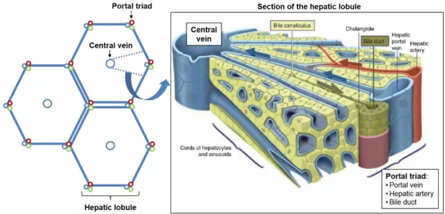
xi Figure 1.1 Schematic representation of the hexagonal-shaped liver lobules, with the central vein and portal triad.
Figure 1.2 Schematic representation of hepatocyte polarity.
Figure 1.3 Schematic representation of the liver acinus, with the major cell types of the liver and the periportal to pericentral zones represented.
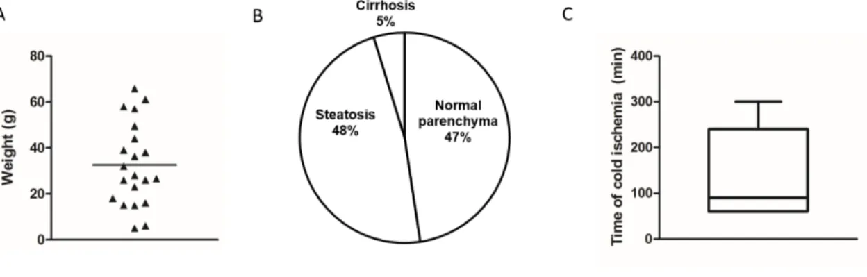
Figure 2.1 Sample characteristics and collection.
Figure 2.2 Comparison of perfusion process time between methods 1 and 2.
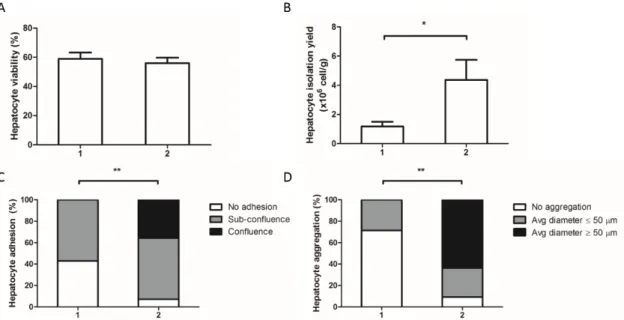
Figure 2.3 Effect of perfusion methods on isolation outcome variables.
Figure 2.4 2D and 3D culture of isolated hepatocytes.
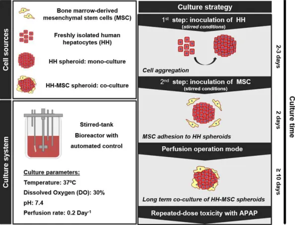
Figure 3.1 Schematic representation of the experimental design, including cell sources, culture system and strategy for the 3D co-culture of HH-MSC in perfusion stirred-tank bioreactors.
Figure 3.2 Morphology and viability of mono- and co-cultures in the bioreactor.
Figure 3.3 Characterization of hepatocytic and mesenchymal populations of spheroid co-cultures.
Figure 3.4 Biosynthetic metabolism.
Figure 3.5 Xenobiotic metabolism.
Figure 3.6 Repeated dose toxicity in bioreactors of co-cultures.
Figure 3.7 Immunofluorescence detection of A) Ki67 (green), nuclei (DAPI, blue) in cyrosections of HH-MSC spheroids at day 8 and B) Vimentin (red) and nuclei (DAPI, blue) in cyrosections of HH spheroids at day 6.
Figure 3.8 Immunofluorescence detection of F-actin (phalloidin, green), vimentin (red) and nuclei (DAPI, blue) in monocultures of BM-MSC after 6 days of 2D culture in WE medium.
Figure 3.9 Accumulation of CDF in the presence of the MRP2 inhibitor Indomethacin in co-cultured spheroids.
Figure 4.1 Characterization of encapsulated 3D HepaRG culture.
Figure 4.2 Gene expression profile of HepaRG cultures.
Figure 4.3 Phenotypic characterization of hepatic spheroids by immunolocalization of hepatic-specific markers.
Figure 4.4 Co-localization of the hepatocyte-specific marker HNF4α with DAPI nuclear staining in cryosections of 2D d28 and 3D cultures.
Figure 4.5 Excretory activity of HepaRG spheroids visualized by efflux of CDFDA.
Figure 4.6 Xenobiotic drug metabolism.
Figure 4.7 Homeostatic metabolism.
Figure 5.1 Schematic representation of the work performed in this thesis, comprising the cell sources and their bottlenecks as well as the aims, strategy and achievements of each chapter.
xiii LIST OF TABLES
Table 1.1 List of hepatic parameters that may be applied to evaluate the hepatic functionality of an
in vitro model.
Table 1.2 The available cell sources for the development of in vitro cellular hepatic models and their inherent advantages and disadvantages.
Table 2.1 Variables potentially affecting efficiency of hepatocyte isolation and culture, including controlled and non-controlled variables in this study.
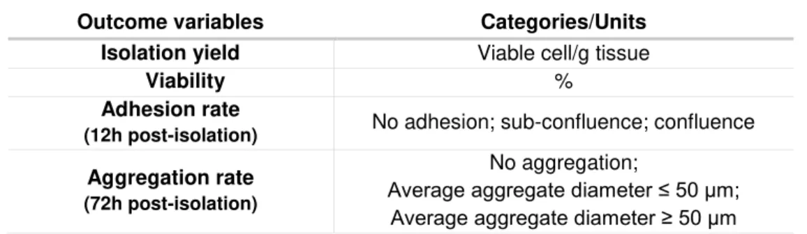
Table 2.2 Outcome variables used to assess the efficiency of isolation in the study.
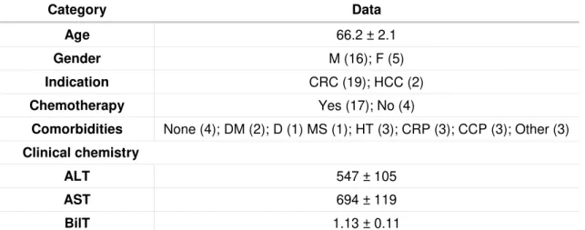
Table 2.3 Donor information and perioperative factors.
Table 3.1 Donor information.
Table 3.2 List of the genes analyzed for qRT-PCR and respective forward and reverse sequences.
xv
Abbreviation Full form
2D Two-dimensional
3D Three-dimensional
ALB Albumin
ALF Acute liver failure
APAP Acetaminophen
BAL Bioartificial liver
BLC Biliary-like cells
BNF β-Naphthoflavone
BRs Bioreactors
CDFDA 5-(and-6)-carboxy-2'-7'-dichlorofluorescein diacetate
CK18 Cytokeratin 18
COL I Collagen type I
CPS1 Carbamoyl phosphate synthase 1
CYP1A2 cytochrome P450 Family 1- Subfamily A- Polypeptide 2 CYP2C9 cytochrome P450 Family 2- Subfamily C- Polypeptide 9 CYP3A4 cytochrome P450 Family 3- Subfamily A- Polypeptide 4
CYP450 Cytochrome P450
DMSO Dimethyl Sulfoxide
ECM Extracellular Matrix
G6PC Glucose-6-phosphatase
GAPDH Glyceraldehyde 3- phosphate dehydrogenase
GS Glutamine Synthase
GSTA1 Glutathione S- transferase A1
HH Human hepatocytes
HLC Hepatocyte-like cells
HNF3β Hepatocyte nuclear factor 3 beta
HNF4α Hepatocyte nuclear factor 4 alpha
HSC Hepatic stellate cell
IL6 Interleukin 6
KC Kupffer cell
LSECs Liver sinusoidal endothelial cell
MRP2 Multidrug resistance protein 2
MSC Mesenchymal stem cell
NPLC Non parenchymal liver cells
PC Pericentral
PFA Paraformaldehyde
PP Periportal
PSC Pluripotent stem cells
PXR Pregnane X receptor
qRT-PCR Quantitative real time polymerase chain reaction
Rif Rifampicin
UGT Uridine 5'-diphospho-glucuronosyltransferase
VC Vehicle control
VIM Vimentin
1
C
HAPTER
1
Chapter 1
2
TABLE OF CONTENTS
1. Introduction ... 3 1.1 The need for humanhepatic cell models ... 3 1.2 In vivo liver microenvironment ... 4 1.3 In vitro evaluation of liver function ... 7 1.4 Hepatic cell sources ... 9 1.4.1 Human hepatocytes ... 9 1.4.2 Immortalized cell lines ... 10 1.4.3 Hepatocyte-like cells derived from pluripotent stem cells ... 11 1.5 Strategies for the culture of hepatic cell models ... 12 1.5.1 3D cultures ... 12 1.5.2 Biomaterials ... 13 1.5.3 Co-cultures ... 15 1.5.4 Bioreactors ... 16
2. Scope of the Thesis ... 18
3 1. Introduction
1.1 The need for humanhepatic cell models
The liver is a central organ in body homeostasis, comprising functions related with nutrient level maintenance, protein, bile and hormone synthesis and an important role in xenobiotic metabolism and detoxification. Due to the plethora of liver functions and, particularly, to the hepatic drug clearance, modeling the liver in vitro has applications in multiple areas, ranging from drug development to clinical applications.
The stringent pipelines of drug development and high costs associated with drug failure during clinical trials drive the need for an efficient prediction of efficacy and safety in pre-clinical development. It is estimated that approximately only 10% of the development paths that enter clinical development in phase I advance to market approval (Hay et al., 2014), resulting in a high R&D expenditure per drug approved. Although the use of animal models is essential to assess toxicity at the systemic level, the interspecies variability in drug metabolic mechanisms, the ethical issues associated with animal testing and the low throughput that animal testing allows are accountable for the inefficiency observed. Thus, the use of human liver models to evaluate safety at the pre-clinical stage could contribute to reduce the attrition rates observed in the industry, by improving predictability and therefore reducing costs throughout the drug development process. A relevant feature of hepatic in vitro models is the potential to mimic liver disease, allowing to depict cellular mechanisms in a more simplistic approach and higher relevance than alternative animal models. Disease modeling is not only important in the context of drug development, but also for basic and translational research, being particularly relevant for the study of liver damage (e.g. fibrosis, steatosis, cholestasis) caused by severe hepatic malignancies including drug induced liver injury (DILI), alcoholic liver disease, hepatitis B (HBV) and C (HCV), which may ultimately lead to acute liver failure (ALF).
Chapter 1
4
1.2 In vivo liver microenvironment
The design of hepatic tissues ex-vivo requires the comprehension of the liver microenvironment to ultimately seek its recreation. The liver is organized in hepatic lobules, which consist of microunits of extensively vascularized and compact layers of hepatocytes arranged radially around the central vein towards the portal triad, which comprises the portal vein, hepatic artery and bile duct (Figure 1.1). The liver parenchyma is composed of hepatocytes, which account for 60% of the cellular components of the liver and 80% of its total volume (LeCluyse et al., 2012). The remaining non-parenchymal liver cells (NPLC) include the biliary epithelial cells or cholangiocytes, the liver sinusoidal endothelial cells (LSECs), hepatic stellate cells (HSC), kupffer cells (KC) and pit cells.
Figure 1.1 Schematic representation of the hexagonal-shaped liver lobules, with the central vein and portal triad. Amplification of the lobule segment, showing the stratified parenchyma and the countercurrent flows of the bile and blood circulation. Adapted from Treyer and Müsch (2013).
5 Although previously not accounted to the hepatic functions, it is now widely accepted that the NPLC play an important role in supporting hepatocyte functionality and proliferation (Godoy et al., 2013). The cholangiocytes are epithelial cells which line the biliary ducts and are derived from the hepatoblasts, a common hepatocyte progenitor. They are involved in the bile secretion process, multiple liver diseases and have been implied in immunoregulation (Huang et al., 2006; Masyuk, Masyuk, & LaRusso, 2008). The LSECs are the fenestrated endothelial cells that compose the liver sinusoids. More than a barrier between blood and hepatocytes, LSECs actively participate in hepatic clearance, as the fenestrated endothelium acts as a selective barrier between the parenchyma and the circulatory system. LSECs can also act as scavengers due to the high receptor-mediated endocytic activity, clearing an array of physiological or foreign macromolecules from the blood (Braet & Wisse, 2002). Moreover, they are involved in inflammatory response and in the regeneration of hepatocytes after injury (DeLeve, 2013). Hepatic stellate cells (HSC) are perisinusoidal cells located between the endothelium and the liver parenchyma, in the space of Disse (Figure 1.3). These cells are responsible for the storage of vitamin A, control the production and homeostasis of extra-cellular matrix (ECM), regulate contractility of the sinusoids and secrete cytokines, thereby mediating the inflammatory response. Upon activation, HSC acquire a miofibroblastic phenotype, which impairs ECM regulation and ultimately leads to fibrosis (Puche et al., 2013). The Kupffer cells are the resident liver macrophages, with high endocytic and phagocytic activity and important mediators of the local and systemic inflammatory response through cytokine secretion (Dixon et al., 2013). The immune response is further mediated by the pit cells, which are intrahepatic leucocytes or natural killer cells.
Chapter 1
6
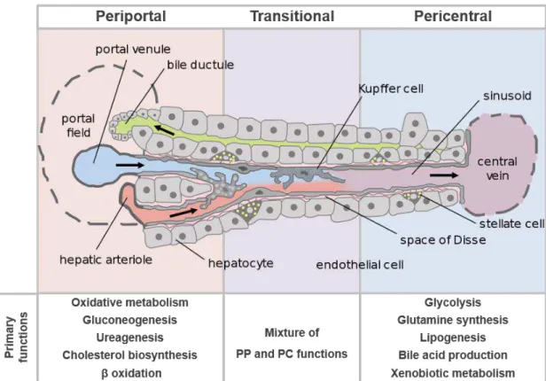
Figure 1.3 Schematic representation of the liver acinus, with the major cell types of the liver and the periportal to pericentral zones represented. The hepatocyte functions are listed in the figure, according to the position in the liver acinus. Adapted from Frevert et al. (2005).
While the normal liver is not particularly rich in ECM, its composition, topography and biomechanical properties strongly affect hepatocyte and NPLC phenotypes. ECM alterations are associated with severe pathologies such as fibrosis or cirrhosis. Matrix proteins are mostly present in the portal tracts, central veins and in the sinusoid walls, lining the frontier between the liver parenchyma and the blood (space of Disse). In the liver parenchyma, the most abundant ECM proteins are collagen type I and fibronectin whilst collagen type III, IV and laminin, are mostly present in the portal and central regions (Godoy et al., 2013). More than the ECM components per se, the matrix stiffness exerts a strong influence on the hepatocyte phenotype. The biomechanical forces are sensed by the hepatocytes through cell-surface receptors (e.g. integrins) and cytoskeleton, affecting intracellular signaling cascades (LeCluyse et al., 2012).
7 1.3 In vitro evaluation of liver function
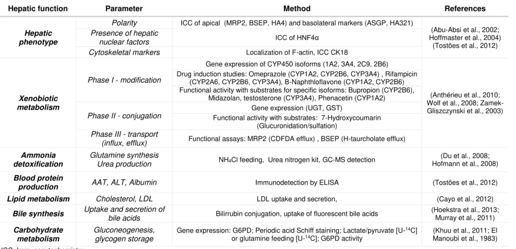
One of the challenges of applying in vitro models is to evaluate their performance and outlook its relevance to other models and, importantly, to the existent in vivo data. The standardization of methodologies and endpoints to assess functionality (e.g. activity levels, measurement units, etc.) and biomarkers would allow a more straightforward comparison of differentiation protocols, culture strategies and cell sources for a specific application. The parameters evaluated when characterizing a liver cell model include hepatic phenotype, xenobiotic metabolism and biosynthetic activity. In Table 1.1 an overview of the existent tools to evaluate in vitro models performance based on multiple parameters of liver functionality is presented.
Hepatic xenobiotic metabolism is a biotransformation process that encompasses 3 phases: I - modification, II - conjugation and III – excretion. Phase I is mostly performed by CYP450 complex, localized in the membrane of the endoplasmic reticulum, and includes reactions such as oxidation, reduction, hydrolysis, cyclization or decyclization. After modification, xenobiotics undergo conjugation with functional groups through methylation, sulphation, acetylation, glucoronidation, etc. to transform xenobiotics into hydrophilic metabolites that are actively transported towards the cell exterior. In phase III, metabolites may be further modified and are then excreted by specialized membrane transporters (e.g. MRP, ATP). Phase I enzymes are considered the most critical step of the biotransformation process, as they account for approximately 75% of the total drug metabolism and originate a high diversity of metabolites. The efflux activity is severely affected by hepatocyte polarization, which is required for the correct assembly of membrane transporters (Guengerich, 2008; Hewitt et al., 2007).
Table 1.1 List of hepatic parameters that may be applied to evaluate the hepatic functionality of an in vitro model.
Hepatic function Parameter Method References
Hepatic phenotype
Polarity ICC of apical (MRP2, BSEP, HA4) and basolateral markers (ASGP, HA321)
(Abu-Absi et al., 2002; Hoffmaster et al., 2004)
(Tostões et al., 2012) Presence of hepatic
nuclear factors ICC of HNF4α
Cytoskeletal markers Localization of F-actin, ICC CK18
Xenobiotic metabolism
Phase I - modification
Gene expression of CYP450 isoforms (1A2, 3A4, 2C9, 2B6)
(Anthérieu et al., 2010; Wolf et al., 2008; Zamek-Gliszczynski et al., 2003) Drug induction studies: Omeprazole (CYP1A2, CYP2B6, CYP3A4) , Rifampicin
(CYP2A6, CYP2B6, CYP3A4), Β-Naphthloflavone (CYP1A2, CYP2B6) Functional activity with substrates for specific isoforms: Bupropion (CYP2B6),
Midazolan, testosterone (CYP3A4), Phenacetin (CYP1A2)
Phase II - conjugation
Gene expression (UGT, GST)
Functional activity with substrates: 7-Hydroxycoumarin (Glucuronidation/sulfation)
Phase III - transport
(influx, efflux) Functional assays: MRP2 (CDFDA efflux) , BSEP (H-taurcholate efflux)
Ammonia detoxification
Glutamine synthesis
Urea production NH4Cl feeding, Urea nitrogen kit, GC-MS detection
(Du et al., 2008; Hofmann et al., 2008)
Blood protein
production AAT, ALT, Albumin Immunodetection by ELISA (Tostões et al., 2012) Lipid metabolism Cholesterol, LDL LDL uptake and secretion, (Cayo et al., 2012)
Bile synthesis Uptake and secretion of
bile acids Bilirrubin conjugation, uptake of fluorescent bile acids
(Hoekstra et al., 2013; Murray et al., 2011)
Carbohydrate metabolism
Gluconeogenesis, glycogen storage
Gene expression: G6PD; Periodic acid Schiff staining; Lactate/pyruvate [U-14C]
9 1.4 Hepatic cell sources
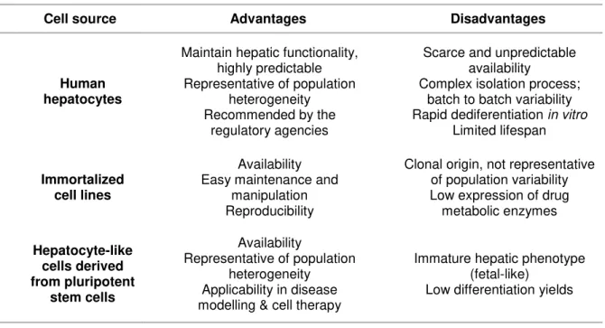
The available hepatic cell sources can be globally divided into three categories – primary cultures, immortalized cell lines and hepatocyte-like cells derived from pluripotent stem cells. The characteristics, applications and recent developments are summarized in Table 1.2.
Table 1.2. Available cell sources for development of cell hepatic models and their inherent advantages and disadvantages.
Cell source Advantages Disadvantages
Human hepatocytes
Maintain hepatic functionality, highly predictable Representative of population
heterogeneity Recommended by the
regulatory agencies
Scarce and unpredictable availability
Complex isolation process; batch to batch variability Rapid dediferentiation in vitro
Limited lifespan
Immortalized cell lines
Availability Easy maintenance and
manipulation Reproducibility
Clonal origin, not representative of population variability Low expression of drug metabolic enzymes Hepatocyte-like cells derived from pluripotent stem cells Availability
Representative of population heterogeneity Applicability in disease modelling & cell therapy
Immature hepatic phenotype (fetal-like)
Low differentiation yields
1.4.1 Human hepatocytes
Human hepatocytes isolated from liver tissue are the cell source with higher resemblance to in vivo in terms of hepatic functions, thus it is considered the gold standard for toxicity testing in pre-clinical drug development by the regulatory agencies (Guguen-Guillouzo and Guillouzo, 2010). Human hepatocytes express all the major enzymes and transporters and exhibit xenobiotic metabolic functions comparable to the liver tissue (Hart et al., 2010). Importantly, primary cultures are representative of the population variability caused by genetic polymorphisms or pathophysiology. Therefore, hepatotoxicity testing with primary cultures in pre-clinical phase may prevent idiosyncratic drug reactions and consequent drug retrieval from the market (Hewitt et al., 2007).
Chapter 1
10
batch-to-batch variability hamper its widespread adoption and drive the need for alternative cellular sources.
1.4.2 Immortalized cell lines
The availability, stable phenotype and unlimited life span that generally characterize immortalized cell lines make them a convenient alternative to primary cultures to model liver function in vitro. However, hepatic cell lines, either derived from human hepatomas or obtained by oncolytic immortalization, generally lack or have downregulated expression of metabolic enzyme families and fail to recapitulate liver metabolism at physiological levels, particularly regarding phase I enzymes. HepG2, the best characterized hepatic cell line, carries out most biosynthetic functions of the liver (e.g. albumin, plasminogen secretion) but has low activity of phase I and II enzymes (Westerink and Schoonen, 2007). Fa2N-4 is a non-tumorigenic immortalized cell line, which has been proposed to assess drug inducing mechanisms and has been applied by the industry. Nevertheless, direct comparison with human hepatocyte in terms of expression and activity levels of CYP450 have identified limitations of Fa2N-4 as a predictive hepatic model (Hariparsad et al., 2008).
HepaRG differs from other existent cell lines since it is a bipotent progenitor cell line that can be differentiated into hepatocyte-like cells (HLC) and biliary-like cells (BLC). Nevertheless, the 2D protocol to attain differentiated metabolic functions requires the supplementation of high Dimethyl sulfoxide (DMSO) concentrations. At the differentiated state, HLC exhibit activity of the CYP450 complex, phase II enzymes and express the gene regulatory proteins and liver specific proteins (Anthérieu et al., 2010; Le Vee et al., 2006). Moreover, the expression of phase I enzymes, particularly CYP3A4, was comparable to primary hepatocytes (Hart et al., 2010) and have been shown to express most of the hepatobiliary transporters (phase III) expressed in cultured human hepatocytes (Bachour-El Azzi et al., 2015), therefore it has been used to assess hepatotoxicity. Despite the unique characteristics and potential of HepaRG to be applied for hepatotoxicity studies, the high concentration of DMSO may interfere with detoxification pathways due to effects such as anti-apoptotic activity (Cerec et al., 2007) and limit its application for cell transplantation or extracorporeal liver support due to toxicity.
11 1.4.3 Hepatocyte-like cells derived from pluripotent stem cells
Chapter 1
12
1.5 Strategies for the culture of hepatic cell models
The use of 3D culture, biomaterials, co-cultures and bioreactors or all these strategies combined have provided advances in hepatic tissue engineering, which main methodologies and achievements are described below.
1.5.1 3D culture
Traditionally, hepatocytes were cultured as monolayers in adherent substrates coated with collagen. The rigidity of the substrate and the lack of cellular or 3D ECM support necessary to maintain the cuboidal shape and polarization resulted in dedifferentiation within 3 to 5 days, associated with downregulation of phase I enzymes and decrease of albumin production (Godoy et al., 2013). Media formulation (e.g. growth factor or DMSO supplementation; serum removal) has been used to improve cell survival and hepatic-specific functions (Hewitt et al., 2007). Moreover, 3D strategies that preserve the cuboidal shape achieve good outcomes in long-term functionality.
The collagen sandwich culture strategy, first implemented by Dunn and co-workers (1989), showed for the first time the impact of the ECM matrix in the culture of adult rat hepatocytes. The collagen sandwich consists in the culture of hepatocytes in between two gelled layers of collagen type I. Using this system, the cuboidal morphology of the hepatocytes is maintained and albumin secretion can be kept for several weeks, whereas hepatocytes without the collagen overlay cease albumin secretion within 1 week (Dunn et al., 1989).
The ex-vivo culture of liver tissue as precision cut slices is another 3D liver model, which principal advantage is the maintenance the original tissue architecture, ECM and cellular components. Thus, precision cut liver slices (PCLS) enable the study of multicellular processes, being applied in the study of liver physiology and in the metabolism and toxicity of xenobiotics. Despite the great advantage of sustaining liver architecture and microenvironment, PCLS often consist of several layers of cells and thick ECM, thus presenting diffusion problems, therefore cell viability can be maintained for short periods, typically up to 96h (de Graaf et al., 2010).
13 previously demonstrated by our group and others for rat (Miranda et al., 2009) and human (Tostões et al., 2012) primary cultures and also for immortalized hepatic cell lines (Chang and Hughes-Fulford, 2009). The effects of 3D culture in the differentiation of PSC are controversial: on one hand, it has been shown that 3D culture improves the efficiency of differentiation towards HLC, on the other hand, the 3D differentiation process may be more heterogenous which can be associated with unequal diffusion of soluble factors within spheroids (Schwartz et al., 2014).
1.5.2 Biomaterials
The use of biomaterials for the culture of hepatocytes has gained relevance in the past years, due to their capacity to elicit specific cellular functions, direct cell-cell interactions and support the 3D architecture. Biomaterials, either from natural or synthetic nature have inner characteristics such as chemical composition, porosity, permeability, degradability, biomechanical properties such as stiffness and elasticity and can be inert or biologically active, with adhesion motifs to ligands. Most of these characteristics can be used or tuned to suit the required architecture or composition of the liver (Jain et al., 2013).
The liver biomatrix is a natural type of biomaterial consisting of the native ECM recovered from decellularized livers, which can be used as a scaffold and re-populated. Importantly, the liver biomatrix is depleted of cellular components but ideally the original ECM composition remains intact as well as the tissue architecture and vasculature due to a sensitive decellularization procedure (Uygun and Yarmush, 2013). Liver decellularization was first described for rat livers, with rat hepatocytes maintaining viable and with high expression levels of albumin, urea and CYP450 genes up to 10 days after recellularization (Uygun et al., 2010). Baptista and coworkers, used the intact vasculature to reseed the scaffolds with human fetal liver and endothelial cells, which were capable of homing to their native niches within the scaffold (Baptista et al., 2011). The transplantation in rats after 90% hepatectomy has been successfully performed, despite the limited survival up to few days (Bao et al., 2011). Recellularization of porcine liver biomatrices using human fetal liver cells led to differentiated hepatic cells with high metabolic activity (Barakat et al., 2012), representing the major approach towards human liver transplantation with an engineered organ. Although the liver biomatrix represents the most promising solution for transplantation with engineered organs, its application for in vitro toxicity testing is hampered by the short-term survival (Uygun and Yarmush, 2013).
Chapter 1
14
Alginate, a linear polysaccharide copolymer of β-D-mannuronic acid and α-Lguluronic acid, is the most used biomaterial for cell immobilization and microencapsulation since it is inert, biocompatible and easy to use. Alginate crosslinking occurs upon addition of bivalent cations and all the procedure may be carried out in physiological conditions, with minimal effects to the cells (Lee and Mooney, 2012). The stiffness and porosity of alginate hydrogels may be tuned according to the composition of β-D-mannuronic acid, sequential structure, molecular size and calcium concentration (Martinsen et al., 1989). The hydrophilic porous network formed (approximately 90% of porosity) is compatible with 3D culture by allowing the establishment of cell-cell interactions. Moreover, alginate capsules are highly permeable, allowing the diffusion of oxygen, soluble factors and proteins (Lee and Mooney, 2012). The microencapsulation of rat hepatocyte spheroids allowed the long-term culture with maintenance of hepatic functions in stirred conditions for several weeks (Miranda et al., 2010; Tostões et al., 2011), denoting the role of alginate microencapsulation in the protection from shear stress. Moreover, alginate encapsulation of HepG2 C3A cells has been applied as a biocomponent for extracorporeal liver support (Yang et al., 2013). Importantly, alginate can be used in compliance with GMP guidelines and therefore potentially applied in cell-based therapy. Its potential use in cell transplantation has been shown in rats with ALF, that presented improved survival (Aoki et al 2005; Sgroi et al 2011; Jitraruch et al 2014)) with low immunogenicity when using ultrapure alginates. Nevertheless, the success after transplant differs substantially between studies due to the mechanical properties which may lead to capsule breakage (Santos et al., 2013). Thus, the improvement of the control on alginate mechanical properties may contribute to widespread the use of alginate for cell therapy applications.
15 1.5.3 Co-cultures
As described in section 1.2, the functional compartment of the liver (liver parenchyma) is supported by the non-parenchymal (NPLC) fraction, which contributes to provide the environmental cues to maintain hepatocyte function. Thus, the co-culture of hepatocytes and NPLC or other types of stromal cells ex-vivo has gained relevance in the past years as a strategy to prolong and enhance the hepatocyte specific functions in vitro (Godoy et al., 2013).
LSECs have an important role in supporting hepatocytes in vivo. However, maintaining LSECs phenotype in vitro is challenging since these cells require tight microenvironmental regulation to maintain their characteristics, namely extensive fenestrae and high scavenger activity (March et al., 2009). Moreover, LSECs complex isolation and purification procedure from liver tissue has hampered major developments for human in vitro systems (Cheluvappa, 2014). Even so, co-cultures of LSECs and hepatocytes from rat origin either in collagen sandwich co-culture (Bale et al., 2014) and layered collagen culture in transwells (Kang et al., 2013) have demonstrated that endothelial and hepatic phenotypes can be maintained in long term cultures up to 4 weeks, with a significant increase in albumin production and CYP3A4 activity. For the development of human cell models, the immortalized Human Umbilical Vein Endothelial Cell (HUVEC) line is routinely used, enhancing hepatocyte function (Salerno et al., 2011). Hepatic spheroids covered with HUVECs have also been applied to establish vascularized liver tissue as biocomponents of BAL devices (Inamori et al., 2009) and transplantation of co-encapsulated hepatocytes and HUVECs has extended survival and improved the liver functional biomarkers in rats with ALF (Qiu et al., 2012).
Co-cultures with hepatic stellate cells (HSC) have mostly focused on the mechanisms underlying disease, such as fibrosis or fatty acid accumulation (Giraudi et al., 2015; Puche et al., 2013). Nevertheless, the effect of HSC has been demonstrated to be beneficial for hepatocyte spheroids, mostly by supporting hepatocyte architecture and tight junctions (Lee et al., 2013; Thomas et al., 2005). Kupffer cells (KC) have been co-cultured with hepatocytes to incorporate the role of inflammation on drug metabolism and it has been demonstrated that there is differential down-regulation of CYP450 in rat hepatocytes upon interaction of inflammatory drugs, which translates into differential sensitivity to drugs (Milosevic et al., 1999; Tukov et al., 2006). The modulation of inflammation in human systems has been rarely addressed, but studies using hepatic and monocytic cell lines have explored the sensitivity to drugs such as Troglitazone in a co-culture model (Edling et al., 2009).
Chapter 1
16
In alternative to primary NPLC, the use of stromal cells to support hepatocyte function has been widely explored through the co-culture with fibroblasts or mesenchymal stem cells. The availability and easy manipulation of fibroblasts are among the characteristics that make them suitable alternatives for NPLC. Data from rodent studies consistently demonstrated that fibroblasts contribute to the stability of hepatocytic phenotype, which has been attributed to the soluble factors secreted by fibroblasts such as HGF or cell surface proteins such as N-cadherin (Khetani et al., 2004; Leite et al., 2011). The co-culture of human hepatocytes and fibroblasts has been the basis for the establishment of microscale culture in a micropatterned system, for drug development applications (Khetani and Bhatia, 2008a). The use of mesenchymal cells in co-culture has been mostly explored for cell therapy in cases of liver fibrosis (Kim et al., 2014) or ALF (Jung et al., 2013; Zhang et al., 2012), either through cell transplantation or use of conditioned media, which has been shown to alleviate the symptoms in rodent models. Moreover, one of the most significant steps towards the development of vascularized tissues in vitro has been achieved with triple co-cultures of IPS-derived hepatocyte-like cells, endothelial and mesenchymal stem cells with successful engraftment and functional vascular network, thus denoting the importance of MSC in the development of organ buds and vascularization (Takebe et al., 2013).
1.5.4 Bioreactors
Bioreactors (BRs) are devices engineered to support biological processes for multiple applications, ranging from production of biopharmaceuticals to tissue engineering. The key feature of these systems is the high level of control over the bioprocesses, which is achieved by on-line monitorization and automated regulation of environmental culture parameters, such as temperature, pH, partial pressure of oxygen (pO2), nutrient and metabolite concentration. Moreover, the dynamic conditions offered by bioreactors ensure efficient mass transfer in the culture vessel, which is a key factor to minimize oxygen and nutrient gradients and maintain a homogenous culture environment. The control, automation and efficient mass transfer simplify the transition from bench top bioreactors to larger scales, critical to meet the industrial requirements.
17 of nutrients and growth factors that contribute to elicit specific functions. It has previously been demonstrated that perfusion conditions elicit hepatic functions. In the MultiChamber Modular Bioreactor (McMB), primary cultures of human hepatocytes cultured in collagen-coated PDMS wells with constant perfusion had up-regulation of phase I, II and III enzymes and stable expression for longer periods than in static systems (Vinci et al. 2011). Several formats applying the same principle and presenting several adaptations are commercially available, such as the Minucell (Xia et al. 2009) which includes a collagen overlay to minimize the effects of shear stress. Although these represent the simplest formats of BRs, presenting an upgrade towards static culture by incorporating dynamic flow, they lack automated control and monitoring of culture parameters. Other monolayer-based BRs have been used to model physiological liver phenomena such as zonation. By controlling O2 tension, the gradient of oxygen tensions sensed
in vivo by pericentral and periportal hepatocytes was recapitulated (Allen et al., 2005).
Hollow-fiber bioreactors comprise an interwoven network of semipermeable membranes which are perfused by medium and oxygen, aiming to resemble blood capillaries in vivo. In between the capillary systems, the cells are arranged in compact 3D structures. Applying this principle, a number of bioreactors were designed for clinical application to support extracorporeal liver function in patients with liver failure: the Modular Extracorporeal Liver System (MELS), developed by Gerlach´s research group (Gerlach et al. 1994), and the AMC-bioreactor, by Chamuleau and coworkers (Flendrig et al. 1997). These systems have also been validated for pharmacological applications: in the miniaturized format of the MELS bioreactor, scaled down to 2 mL, major drug metabolizing P450 enzymes were preserved up to 23 days in primary cultures of human hepatocytes in co-culture with non-parenchymal cells (Zeilinger et al. 2011). More recently, this design has been applied for the differentiation of human pluripotent SC towards hepatocyte-like cells (Miki et al. 2011). The AMC-bioreactor has been validated using the hepatic cell line HepaRG, which presented phase I and II drug metabolism and production of bile salts (Nibourg et al. 2013). A major drawback of these systems is the inaccessibility to the cell compartment throughout the culture time, not allowing phenotypic monitoring and cell sampling. Furthermore, hollow fiber bioreactors fail to accurately control pH and pO2 within the fibers.
Chapter 1
18
design modular units with physiological relevance. With a design compatible with a 96 well plate, the system is composed of microunits of artificial liver acinus with an endothelial-like barrier, intended to simulate the mass transfer properties of the liver sinusoid. Primary cultures of human hepatocytes were maintained for 7 days in culture and were responsive to diclofenac toxicity at high concentrations (Lee et al. 2007). Khetani and Bhatia (Khetani and Bhatia, 2008b) developed a multiwell system containing micropatterned structures of PDMS for co-culture of fibroblasts and hepatocytes, which is compatible with robotic fluid handling and phenotypic screening tools. This co-culture system was validated for up to 6 weeks with maintenance of gene expression profile, phase I/II metabolism, canalicular transport, secretion of liver-specific products and susceptibility to hepatotoxins (Khetani and Bhatia 2008).
Stirred-tank bioreactors (STB), which have long been applied in industry for production of biopharmaceuticals, can also be used for in vitro cell models for pharmacological testing. In STB, cells are inoculated as cell suspension and the hydrodynamics of the bioreactor – determined by vessel, impeller type and agitation rate - is adjusted to elicit cell collisions and promote cell-cell contacts into aggregation. Dynamic parameters need to be balanced to guarantee diffusion through the aggregates, preventing the formation of necrotic centres, while the shear stress is minimized to avoid damage. Spheroid culture of rat hepatocytes has long been reported, resulting in increased albumin production and phase I-II activity (Abu-Absi et al. 2002) and maintenance of hepatocyte polarization (Miranda et al. 2009). More recently, primary cultures of human hepatocytes were maintained under physiological oxygen conditions and perfusion operation mode, extending culture viability and functionality for up to 3-4 weeks (Tostões et al. 2012). Hepatocytes in this system present a functional phenotype displaying bile canalicular networks, and inducible expression of CYP450. The use of biomaterials in STB has also been addressed, by alginate microencapsulation of rat hepatocyte (Miranda et al. 2010; Tostões et al. 2011), which represents a strategy to overcome eventual shear stress effects on stirred culture.
2. Scope of the thesis
The goal of this thesis was to develop culture strategies for the establishment of cellular models that recapitulate liver function in vitro, using human systems. These models made use of stirred conditions to promote cellular aggregation and were complemented with the use of biomaterials or co-cultures to overcome the existing caveats of hepatocyte culture by further enhancing the hepatic phenotype and/or long-term culture.
In chapter 1 a review of the needs for human hepatic in vitro systems, existent cell sources and culture strategies as well as their applications and limitations is provided.
19 In chapter 3, the co-culture of primary hepatocytes with bone marrow-derived mesenchymal stem cells as 3D spheroids is implemented. The results showed that the co-culture enhanced the hepatic phenotype and improved hepatocyte survival and functionality for long-term cultures, being the proof-of-concept of its application performed in repeated-dose toxicity testing with APAP.
In chapter 4, a strategy for the 3D culture of the hepatic cell line HepaRG is implemented, combining cell aggregation with alginate microencapsulation. The 3D strategy yielded a higher ratio of hepatocyte-like cells than the 2D DMSO-dependent protocol, with comparable levels of drug metabolic activity and enhanced biosynthetic metabolism.
In chapter 5, a general discussion about the work performed in this thesis is presented.
3. References
Abu-Absi SF, Friend JR, Hansen LK, Hu W-S. 2002. Structural polarity and functional bile canaliculi in rat hepatocyte spheroids. Exp. Cell Res.274:56–67.
Allen JW, Khetani SR, Bhatia SN. 2005. In vitro zonation and toxicity in a hepatocyte bioreactor. Toxicol. Sci.84:110–9.
Amour KAD, Bang AG, Eliazer S, Kelly OG, Agulnick AD, Smart NG, Moorman MA, Kroon E, Carpenter MK, Baetge EE, D’Amour K a. 2006. Production of pancreatic hormone-expressing endocrine cells from human embryonic stem cells. Nat. Biotechnol.24:1392–401.
Anthérieu S, Chesné C, Li R, Camus S, Lahoz A, Picazo L, Turpeinen M, Tolonen A, Uusitalo J, Guguen-Guillouzo C, Guillouzo A. 2010. Stable expression, activity, and inducibility of cytochromes P450 in differentiated HepaRG cells. Drug Metab. Dispos.38:516–525.
Aoki T, Jin Z, Nishino N, Kato H, Shimizu Y, Niiya T, Murai N, Enami Y, Mitamura K, Koizumi T, Yasuda D, Izumida Y, Avital I, Umehara Y, Demetriou AA, Rozga J, Kusano M. 2005. Intrasplenic transplantation of encapsulated hepatocytes decreases mortality and improves liver functions in fulminant hepatic failure from 90% partial hepatectomy in rats. Transplantation79:783–790. Bachour-El Azzi P, Sharanek A, Burban A, Li R, Le Guével R, Abdel-Razzak Z, Stieger B, Guguen-Guillouzo C, Guillouzo A. 2015. Comparative localization and functional activity of the main hepatobiliary transporters in HepaRG cells and primary human hepatocytes. Toxicol. Sci. Bale SS, Golberg I, Jindal R, McCarty WJ, Luitje M, Hegde M, Bhushan A, Usta OB, Yarmush ML. 2014. Long-Term Coculture Strategies for Primary Hepatocytes and Liver Sinusoidal Endothelial Cells. Tissue Eng. Part C. Methods.
Bañares R, Catalina M-V, Vaquero J. 2013. Liver support systems: will they ever reach prime time? Curr. Gastroenterol. Rep.15:312.
Bao J, Shi Y, Sun H, Yin X, Yang R, Li L, Chen X, Bu H. 2011. Construction of a portal implantable functional tissue-engineered liver using perfusion-decellularized matrix and hepatocytes in rats. Cell Transplant.20:753–766.
Baptista PM, Siddiqui MM, Lozier G, Rodriguez SR, Atala A, Soker S. 2011. The use of whole organ decellularization for the generation of a vascularized liver organoid. Hepatology53:604– 617.
Chapter 1
20
Cayo MA, Cai J, DeLaForest A, Noto FK, Nagaoka M, Clark BS, Collery RF, Si-Tayeb K, Duncan SA. 2012. JD induced pluripotent stem cell-derived hepatocytes faithfully recapitulate the pathophysiology of familial hypercholesterolemia. Hepatology56:2163–71.
Cerec V, Glaise D, Garnier D, Morosan S, Turlin B, Drenou B, Gripon P, Kremsdorf D, Guguen-Guillouzo C, Corlu A. 2007. Transdifferentiation of hepatocyte-like cells from the human hepatoma hepaRG cell line through bipotent progenitor. Hepatology45:957–967.
Chang TT, Hughes-Fulford M. 2009. Monolayer and spheroid culture of human liver hepatocellular carcinoma cell line cells demonstrate distinct global gene expression patterns and functional phenotypes. Tissue Eng. Part A15:559–567.
Chang TT, Hughes-Fulford M. 2014. Molecular mechanisms underlying the enhanced functions of three-dimensional hepatocyte aggregates. Biomaterials35:2162–71.
Cheluvappa R. 2014. Standardized isolation and culture of murine liver sinusoidal endothelial cells. Curr. Protoc. Cell Biol.65:2.9.1–8.
Chistiakov DA, Chistiakov PA. 2012. Strategies to produce hepatocytes and hepatocyte-like cells from pluripotent stem cells. Hepatol. Res.
Decaens C, Durand M, Grosse B, Cassio D. 2008. Which in vitro models could be best used to study hepatocyte polarity? Biol. Cell100:387–398.
DeLeve LD. 2013. Liver sinusoidal endothelial cells and liver regeneration. J. Clin. Invest. 123:1861–6.
Dixon LJ, Barnes M, Tang H, Pritchard MT, Nagy LE. 2013. Kupffer cells in the liver. Compr. Physiol.3:785–797.
Du Y, Han R, Wen F, Ng San San S, Xia L, Wohland T, Leo HL, Yu H. 2008. Synthetic sandwich culture of 3D hepatocyte monolayer. Biomaterials29:290–301.
Dunn JC, Yarmush ML, Koebe HG, Tompkins RG. 1989. Hepatocyte function and extracellular matrix geometry: long-term culture in a sandwich configuration. FASEB J.3:174–177.
Edling Y, Sivertsson LK, Butura A, Ingelman-Sundberg M, Ek M. 2009. Increased sensitivity for troglitazone-induced cytotoxicity using a human in vitro co-culture model. Toxicol. Vitr.23:1387– 1395.
Forbes SJ, Gupta S, Dhawan A. 2015. Cell therapy for liver disease: From liver transplantation to cell factory. J. Hepatol.62:S157–S169.
Frevert U, Engelmann S, Zougbédé S, Stange J, Ng B, Matuschewski K, Liebes L, Yee H. 2005. Intravital observation of plasmodium berghei sporozoite infection of the liver. PLoS Biol.3:1034– 1046.
Giraudi PJ, Barbero VJ, Marin V, Chavez-tapia NC, Tiribelli C, Rosso N. 2015. The importance of the interaction between hepatocyte and hepatic stellate cells in fi brogenesis induced by fatty accumulation. Exp. Mol. Pathol.98:85–92.
21 sources and non-parenchymal liver cells and their use in investigating mechanisms of hepatotoxicity, cell signaling and ADME. Arch. Toxicol. Vol. 87 1315-530 p.
Gonzalez SA, Keeffe EB. 2011. Chronic viral hepatitis: epidemiology, molecular biology, and antiviral therapy. Front Biosci16:225–250.
De Graaf IAM, Olinga P, de Jager MH, Merema MT, de Kanter R, van de Kerkhof EG, Groothuis GMM. 2010. Preparation and incubation of precision-cut liver and intestinal slices for application in drug metabolism and toxicity studies. Nat. Protoc.5:1540–1551.
Guengerich FP. 2008. Cytochrome P450 and chemical toxicology. Chem. Res. Toxicol.
Guguen-Guillouzo C, Guillouzo A. 2010. General review on in vitro hepatocyte models and their applications. Methods Mol. Biol.
Hariparsad N, Carr BA, Evers R, Chu X. 2008. Comparison of immortalized Fa2N-4 cells and human hepatocytes as in vitro models for cytochrome P450 induction. Drug Metab. Dispos. 36:1046–55.
Hart SN, Li Y, Nakamoto K, Subileau E, Steen D, Zhong X. 2010. A comparison of whole genome gene expression profiles of HepaRG cells and HepG2 cells to primary human hepatocytes and human liver tissues. Drug Metab. Dispos.38:988–994.
Hay M, Thomas DW, Craighead JL, Economides C, Rosenthal J. 2014. Clinical development success rates for investigational drugs. Nat. Biotechnol.32:40–51.
Hewitt NJ, Lechón MJG, Houston JB, Hallifax D, Brown HS, Maurel P, Kenna JG, Gustavsson L, Lohmann C, Skonberg C, Guillouzo A, Tuschl G, Li AP, LeCluyse E, Groothuis GMM, Hengstler JG. 2007. Primary hepatocytes: current understanding of the regulation of metabolic enzymes and transporter proteins, and pharmaceutical practice for the use of hepatocytes in metabolism, enzyme induction, transporter, clearance, and hepatotoxicity studies. Drug Metab. Rev.39:159– 234.
Hoekstra R, Nibourg G a a, Van Der Hoeven T V., Plomer G, Seppen J, Ackermans MT, Camus S, Kulik W, Van Gulik TM, Oude Elferink RP, Chamuleau R a FM. 2013. Phase 1 and phase 2 drug metabolism and bile acid production of HepaRG cells in a bioartificial liver in absence of dimethyl sulfoxide. Drug Metab. Dispos.41:562–567.
Hoffmaster KA, Turncliff RZ, LeCluyse EL, Kim RB, Meier PJ, Brouwer KLR. 2004. P-glycoprotein expression, localization, and function in sandwich-cultured primary rat and human hepatocytes: relevance to the hepatobiliary disposition of a model opioid peptide. Pharm. Res.21:1294–302. Hofmann U, Maier K, Niebel A, Vacun G, Reuss M, Mauch K. 2008. Identification of metabolic fluxes in hepatic cells from transient 13C-labeling experiments: Part I. Experimental observations. Biotechnol. Bioeng.100:344–54.
Huang BQ, Masyuk T V, Muff MA, Tietz PS, Masyuk AI, Larusso NF. 2006. Isolation and characterization of cholangiocyte primary cilia. Am. J. Physiol. Gastrointest. Liver Physiol. 291:G500–G509.
Inamori M, Mizumoto H, Kajiwara T. 2009. An approach for formation of vascularized liver tissue by endothelial cell-covered hepatocyte spheroid integration. Tissue Eng. Part A15:2029–2037. Jain E, Damania A, Kumar A. 2013. Biomaterials for liver tissue engineering. Hepatol. Int.8:185– 197.
Jitraruch S, Dhawan A, Hughes RD, Filippi C, Soong D, Philippeos C, Lehec SC, Heaton ND, Longhi MS, Mitry RR. 2014. Alginate Microencapsulated Hepatocytes Optimised for Transplantation in Acute Liver Failure. Ed. Valquiria Bueno. PLoS One9:e113609.
Jung J, Choi JH, Lee Y, Park J-W, Oh I-H, Hwang S-G, Kim K-S, Kim GJ. 2013. Human placenta-derived mesenchymal stem cells promote hepatic regeneration in CCl4 -injured rat liver model via increased autophagic mechanism. Stem Cells31:1584–96.
Chapter 1
22
Jungermann K, Kietzmann T. 2000. Oxygen: modulator of metabolic zonation and disease of the liver. Hepatology31:255–260.
Kang YBA, Rawat S, Cirillo J, Bouchard M, Noh HM. 2013. Layered long-term co-culture of hepatocytes and endothelial cells on a transwell membrane: toward engineering the liver sinusoid. Biofabrication5:045008.
Khetani SR, Bhatia SN. 2008a. Microscale culture of human liver cells for drug development. Nat. Biotechnol.26:120–126.
Khetani SR, Bhatia SN. 2008b. Microscale culture of human liver cells for drug development. Nat. Biotechnol.26:120–126.
Khetani SR, Szulgit G, Del Rio J a, Barlow C, Bhatia SN. 2004. Exploring interactions between rat hepatocytes and nonparenchymal cells using gene expression profiling. Hepatology40:545– 54.
Khuu DN, Scheers I, Ehnert S, Jazouli N, Nyabi O, Buc-Calderon P, Meulemans A, Nussler A, Sokal E, Najimi M. 2011. In vitro differentiated adult human liver progenitor cells display mature hepatic metabolic functions: a potential tool for in vitro pharmacotoxicological testing. Cell Transplant.20:287–302.
Kim M-D, Kim S-S, Cha H-Y, Jang S-H, Chang D-Y, Kim W, Suh-Kim H, Lee J-H. 2014. Therapeutic effect of hepatocyte growth factor-secreting mesenchymal stem cells in a rat model of liver fibrosis. Exp. Mol. Med.46:e110.
Kostadinova R, Boess F, Applegate D, Suter L, Weiser T, Singer T, Naughton B, Roth A. 2013. A long-term three dimensional liver co-culture system for improved prediction of clinically relevant drug-induced hepatotoxicity. Toxicol. Appl. Pharmacol.268:1–16.
LeCluyse EL, Witek RP, Andersen ME, Powers MJ. 2012. Organotypic liver culture models: Meeting current challenges in toxicity testing. Crit. Rev. Toxicol.
Lee KY, Mooney DJ. 2012. Alginate: Properties and biomedical applications. Prog. Polym. Sci. 37:106–126.
Lee PJ, Hung PJ, Lee LP. 2007. An artificial liver sinusoid with a microfluidic endothelial-like barrier for primary hepatocyte culture. Biotechnol. Bioeng.97:1340–1346.
Lee S-A, No DY, Kang E, Ju J, Kim D-S, Lee S-H. 2013. Spheroid-based three-dimensional liver-on-a-chip to investigate hepatocyte-hepatic stellate cell interactions and flow effects. Lab Chip 13:3529–37.
Leite SB, Teixeira AP, Miranda JP, Tostões RM, Clemente JJ, Sousa MF, Carrondo MJT, Alves PM. 2011. Merging bioreactor technology with 3D hepatocyte-fibroblast culturing approaches: Improved in vitro models for toxicological applications. Toxicol. Vitr.25:825–832.
Lutolf MP, Hubbell JA. 2005. Synthetic biomaterials as instructive extracellular microenvironments for morphogenesis in tissue engineering. Nat. Biotechnol.23:47–55.
El Manoubi L, Callikan S, Duee PH, Ferre P, Girard J. 1983. Development of gluconeogenesis in isolated hepatocytes from the rabbit. Am. J. Physiol.244:E24–30.
March S, Hui EE, Underhill GH, Khetani S, Bhatia SN. 2009. Microenvironmental regulation of the sinusoidal endothelial cell phenotype in vitro. Hepatology50:920–928.
Martinsen A, Skjåk-Braek G, Smidsrød O. 1989. Alginate as immobilization material: I. Correlation between chemical and physical properties of alginate gel beads. Biotechnol. Bioeng.33:79–89. Masyuk AI, Masyuk T V., LaRusso NF. 2008. Cholangiocyte primary cilia in liver health and disease. Dev. Dyn.
Milosevic N, Schawalder H, Maier P. 1999. Kupffer cell-mediated differential down-regulation of cytochrome P450 metabolism in rat hepatocytes. Eur. J. Pharmacol.368:75–87.
23 Miranda JP, Rodrigues A, Tostões RM, Leite S, Zimmerman H, Carrondo MJ, Alves PM. 2010. Extending hepatocyte functionality for drug-testing applications using high-viscosity alginate-encapsulated three-dimensional cultures in bioreactors. Tissue Eng. Part C, Methods 16:1223– 1232.
Murray JW, Thosani AJ, Wang P, Wolkoff AW. 2011. Heterogeneous accumulation of fluorescent bile acids in primary rat hepatocytes does not correlate with their homogenous expression of ntcp. Am. J. Physiol. Gastrointest. Liver Physiol.301:G60–8.
Pan XP, Li LJ. 2012. Advances in cell sources of hepatocytes for bioartificial liver. Hepatobiliary Pancreat. Dis. Int.
Puche JE, Saiman Y, Friedman SL. 2013. Hepatic stellate cells and liver fibrosis. Compr. Physiol. 3:1473–1492.
Qiu L, Wang J, Wen X, Wang H, Wang Y, Lin Q, Du Z, Duan C, Wang C, Wang C. 2012. Transplantation of co-microencapsulated hepatocytes and HUVECs for treatment of fulminant hepatic failure. Int. J. Artif. Organs35:458–65.
Salerno S, Campana C, Morelli S, Drioli E, De Bartolo L. 2011. Human hepatocytes and endothelial cells in organotypic membrane systems. Biomaterials32:8848–59.
Santos E, Pedraz JL, Hernández RM, Orive G. 2013. Therapeutic cell encapsulation: Ten steps towards clinical translation. J. Control. Release.
Schwartz RE, Fleming HE, Khetani SR, Bhatia SN. 2014. Pluripotent stem cell-derived hepatocyte-like cells. Biotechnol. Adv.
Sgroi A, Mai G, Morel P, Baertschiger RM, Gonelle-Gispert C, Serre-Beinier V, Buhler LH. 2011. Transplantation of encapsulated hepatocytes during acute liver failure improves survival without stimulating native liver regeneration. Cell Transplant.20:1791–1803.
Spence JR, Mayhew CN, Rankin SA, Kuhar M, Vallance E, Tolle K, Hoskins EE, Kalinichenko V V, Wells I, Zorn AM, Shroyer NF, Wells JM. 2011. Directed differentiation of human pluripotent stem cells into intestinal tissue in vitro. Nature470:105–109.
Takebe T, Sekine K, Enomura M, Koike H, Kimura M, Ogaeri T, Zhang R-R, Ueno Y, Zheng Y-W, Koike N, Aoyama S, Adachi Y, Taniguchi H. 2013. Vascularized and functional human liver from an iPSC-derived organ bud transplant. Nature499:481–4.
Thomas RJ, Bhandari R, Barrett DA, Bennett AJ, Fry JR, Powe D, Thomson BJ, Shakesheff KM. 2005. The effect of three-dimensional co-culture of hepatocytes and hepatic stellate cells on key hepatocyte functions in vitro. Cells. Tissues. Organs181:67–79.
Tostões RM, Leite SB, Miranda JP, Sousa M, Wang DI, Carrondo MJ, Alves PM. 2011. Perfusion of 3D encapsulated hepatocytes--a synergistic effect enhancing long-term functionality in bioreactors. Biotechnol. Bioeng.108:41–49.
Tostões RM, Leite SB, Serra M, Jensen J, Bjorquist P, Carrondo MJ, Brito C, Alves PM. 2012. Human liver cell spheroids in extended perfusion bioreactor culture for repeated-dose drug testing. Hepatology55:1227–1236.
Treyer A, Müsch A. 2013. Hepatocyte polarity. Compr. Physiol.3:243–287.
Tukov FF, Maddox JF, Amacher DE, Bobrowski WF, Roth RA, Ganey PE. 2006. Modeling inflammation-drug interactions in vitro: A rat Kupffer cell-hepatocyte coculture system. Toxicol. Vitr.20:1488–1499.
Uygun BE, Soto-Gutierrez A, Yagi H, Izamis M-L, Guzzardi MA, Shulman C, Milwid J, Kobayashi N, Tilles A, Berthiaume F, Hertl M, Nahmias Y, Yarmush ML, Uygun K. 2010. Organ reengineering through development of a transplantable recellularized liver graft using decellularized liver matrix. Nat. Med.16:814–820.
Chapter 1
24
Le Vee M, Jigorel E, Glaise D, Gripon P, Guguen-Guillouzo C, Fardel O. 2006. Functional expression of sinusoidal and canalicular hepatic drug transporters in the differentiated human hepatoma HepaRG cell line. Eur. J. Pharm. Sci.28:109–117.
Westerink WMA, Schoonen WGEJ. 2007. Cytochrome P450 enzyme levels in HepG2 cells and cryopreserved primary human hepatocytes and their induction in HepG2 cells. Toxicol. In Vitro 21:1581–1591.
Wolf KK, Brouwer KR, Pollack GM, Brouwer KLR. 2008. Effect of albumin on the biliary clearance of compounds in sandwich-cultured rat hepatocytes. Drug Metab. Dispos.36:2086–92.
Yang Y, Li J, Pan X, Zhou P, Yu X, Cao H, Wang Y, Li L. 2013. Co-culture with mesenchymal stem cells enhances metabolic functions of liver cells in bioartificial liver system. Biotechnol. Bioeng.110:958–68.
Zamek-Gliszczynski MJ, Xiong H, Patel NJ, Turncliff RZ, Pollack GM, Brouwer KLR. 2003. Pharmacokinetics of 5 (and 6)-carboxy-2’,7'-dichlorofluorescein and its diacetate promoiety in the liver. J. Pharmacol. Exp. Ther.304:801–809.
25
C
HAPTER
2
Chapter 2
26
TABLE OF CONTENTS
1. Introduction ... 28
2. Material and Methods ... 30 2.1 Biological material source and collection ... 30 2.2 Hepatocyte isolation ... 30 2.2.1 Perfusion - First step ... 30 2.2.2 Perfusion - Second step ... 30 2.2.3 Tissue disruption ... 30 2.2.4 Hepatocyte purification ... 31 2.3 Hepatocyte culture ... 31 2.3.1 2D culture ... 31 2.3.2 3D culture ... 31 2.4 Efficiency of isolation ... 31 2.4.1 Cell viability ... 31 2.4.2 Isolation yield ... 31 2.4.3 Hepatocyte adhesion ... 31 2.4.4 Hepatocyte aggregation ... 31 2.5 Data analysis and statistics ... 31
3. Results ... 32 3.1. Characterization of the donor population ... 32 3.2 Isolation process optimization ... 33
4. Discussion ... 35
27 ABSTRACT
Human resected liver tissue is a potential source of mature hepatocytes, which retain most hepatic features and are representative of the population variability, being useful for in vitro culture. The culture of hepatocytes as three-dimensional spheroids (3D) has been proven to be an efficient strategy to sustain long term functions of hepatocytes. However, most of the protocols available have been developed for monolayer culture and may not be adjusted for the aggregation of hepatocytes. Moreover, the factors affecting the efficiency of isolation are controversial, being important the selection of the best candidates for isolation.
This chapter describes the optimization of the isolation of hepatocytes from resected liver tissue towards 3D culture of hepatocyte spheroids. 21 liver samples obtained from patients undergoing liver resection were processed according to a modified two-step collagenase perfusion. A descriptive analysis of the most critical variables for the quality of the isolation and hepatocytes obtained was performed. For perfusion optimization 4 variables were manipulated. The yield of hepatocytes after isolation, hepatocyte viability, adhesion and aggregation in stirred systems were assessed to evaluate the perfusion performance. An average yield of 4.4±1.4 x 10⁶ hepatocytes per gram of tissue perfused and an average viability of 55.3±3.8% were obtained with the optimized protocol. Additionally, hepatocyte aggregation significantly improved, showing the optimized protocol is adequate for the 3D culture of hepatocytes in stirred conditions.