Muscle Cells Derived from Human Embryonic Stem Cells
Helena Vaza˜o1,2, Ricardo Pires das Neves1,2, Ma´rio Gra˜os2, Lino Ferreira1,2*
1CNC - Center of Neurosciences and Cell Biology, University of Coimbra, Coimbra, Portugal,2Biocant - Center of Innovation in Biotechnology, Cantanhede, Portugal
Abstract
In this study we demonstrate that CD34+cells derived from human embryonic stem cells (hESCs) have higher smooth muscle cell (SMC) potential than CD342cells. We report that from all inductive signals tested, retinoic acid (RA) and platelet derived growth factor (PDGFBB) are the most effective agents in guiding the differentiation of CD34+cells into smooth
muscle progenitor cells (SMPCs) characterized by the expression of SMC genes and proteins, secretion of SMC-related cytokines, contraction in response to depolarization agents and vasoactive peptides and expression of SMC-related genes in a 3D environment. These cells are also characterized by a low organization of the contractile proteins and the contractility response is mediated by Ca2+, which involves the activation of Rho A/Rho kinase- and Ca2+/calmodulin (CaM)/myosin light chain kinase (MLCK)-dependent pathways. We further show that SMPCs obtained from the differentiation of CD34+cells with RA, but not with PDGFBB, can be maturated in medium supplemented with endothelin-1 showing at the end
individualized contractile filaments. Overall the hESC-derived SMCs presented in this work might be an unlimited source of SMCs for tissue engineering and regenerative medicine.
Citation:Vaza˜o H, Neves RPd, Gra˜os M, Ferreira L (2011) Towards the Maturation and Characterization of Smooth Muscle Cells Derived from Human Embryonic Stem Cells. PLoS ONE 6(3): e17771. doi:10.1371/journal.pone.0017771
Editor:Niels Caˆmara, Universidade de Sao Paulo, Brazil
ReceivedJuly 28, 2010;AcceptedFebruary 14, 2011;PublishedMarch 10, 2011
Copyright:ß2011 Vaza˜o et al. This is an open-access article distributed under the terms of the Creative Commons Attribution License, which permits unrestricted use, distribution, and reproduction in any medium, provided the original author and source are credited.
Funding:This work was supported by a Marie Curie-Reintegration Grant, MIT-Portugal program, Crioestaminal, Associac¸a˜o Viver a Cieˆncia and Fundac¸a˜o para a Cieˆncia e a Tecnologia (PTDC/SA-BEB/098468/2008 and PTDC/CTM/099659/2008 to L.F.; SFRH/BD/40077/2007 to H.V.). The funders had no role in study design, data collection and analysis, decision to publish, or preparation of the manuscript.
Competing Interests:The authors have declared that no competing interests exist. * E-mail: [email protected]
Introduction
Vascular smooth muscle cells (VSMCs) have enormous applications in regenerative medicine [1,2,3]. Studies have demonstrated that smooth muscle-like cells (SMLCs) can be derived from bone marrow-[4,5], adipose-[6,7] and umbilical cord blood-derived stem cells [8]. Due to the easy expansion, human embryonic stem cells (hESCs) represent an alternative source of VSMCs particularly for old patients having stem cells with impaired function. Recent studies reported different strategies to differentiate hESCs into SMLCs by exposing a monolayer of undifferentiated hESCs to retinoic acid [9] or a combination of cell culture medium and extracellular matrix environment [10,11,12] either in single-hESC- [13], embryoid bodies (EBs)-[12] or stromal cell- [14] culture conditions. In one case, SMLCs transplanted subcutaneously in an animal model were able to contribute for the formation of functional blood microvessels [12]. Despite these advances, several issues remain poorly understood: (i) what hESC-derived population has the most SMC potential, (ii) the bioactive molecules involved in the differentiation process, (iii) the modulatory effect of 3D environments in SMLCs, (iv) the functionality of the differentiated SMLCs, and (v) the level of organization of the contractile protein filaments.
Here we evaluate the smooth muscle cell (SMC) differentiation of different cell populations isolated from human embryoid bodies grown in suspension for 10 days. The isolated cells were cultured in media supplemented with several inductive signals, including platelet-derived growth factor (PDGFBB), retinoic acid (RA),
transforming growth factor beta 1 (TGFb-1) or a combination of
PDGFBB with TGFb-1. We show that CD34+ cells have higher
SMC potential than CD342cells and PDGFBB and RA are the
most effective agents to drive the differentiation of hESCs into smooth muscle progenitor cells (SMPCs). We further demonstrate that these cells contract and relax in response to SMC agonists or inhibitors, respectively, and the effect is mediated by Rho A/Rho kinase- and Ca2+/CaM/MLCK-dependent pathways. In addition, cells encapsulated in 3D gel scaffolds further differentiate towards SMC lineage as confirmed by gene analysis. Finally, we show that Endothelin-1 induces the organization of the contractile protein filaments.
Materials and Methods
An expanded Materials and Methods section is provided in the online data supplement (Materials and Methods S1).
for 10 days at 37uC, 5% CO2in a humidified atmosphere, with
media changes every 3–4 days.
Isolation and differentiation of CD34+, CD342 and CD34+KDR2 cells
CD34+
cells were isolated from EBs at day 10 according to a protocol previously reported by us [12]. For some experiments, the CD34+cells were further separated in CD34+KDR2 cells. In this case, cells were labeled with anti-VEGF R2/KDR-PE antibody (R&D, http://www.rndsystems.com/), then conjugated with anti-PE antibody coupled with magnetic beads, and finally the magnetically labeled cells were separated into CD34+
KDR+
and CD34+ KDR2 using a MS-MACS column (Miltenyi Biotec, http://www.miltenyi-biotec.com). Isolated cells were grown on 24-well plates (1.56104
cells/cm2) coated with 0.1% gelatin and containing one of the following media: smooth muscle growth medium-2 (SMGM-2), endothelial growth medium-2 (EGM-2) or EGM-2 supplemented with PDGFBB(50 ng/mL, Prepotech, http://www.peprotech.com/)
or RA (1mM, Sigma, http://www.sigmaaldrich.com) or TGFb-1
(10 ng/mL, Prepotech) or a mixture of PDGFBB with TGFb-1
(50 ng/mL; 10 ng/mL). Human vascular smooth muscle cells (hVSMCs, isolated from the arteries of human umbilical cord, Lonza, http://www.lonza.com) were used as controls for the differentiation studies. Cells were cultured in SMGM-2 media (Lonza; until passage 6) being the medium changed every 2 days.
Immunofluorescence analysis
Cells were transferred to gelatin-coated slides containing differentiation medium, allowed to attach overnight, and then fixed with 4% paraformaldehyde (Electron Microscopy Sciences, http://www.emsdiasum.com/microscopy) for 15 min at room temperature. Cells were blocked with 1% (w/v) BSA and stained for 1 h with anti-human primary antibodies specific for smooth muscle a-actin (a-SMA, 1A4, Dako, http://www.dako.com/), smooth muscle myosin heavy chain (SM-MHC, SMMS-1, Dako) and calponin (CALP, Calponin1, Santa Cruz Biotec, http://www. scbt.com/). In each immunofluorescence experiment, an isotype-matched IgG control was used. Binding of primary antibodies to specific cells was detected with anti-mouse IgG Cy3 conjugate or anti-mouse IgG FITC (both form Sigma). Cell nuclei were stained with 49, 69-diamidino-2-phenylindole (DAPI) (Sigma) and the slides examined with either a Zeiss fluorescence microscope or Zeiss LSM 50 confocal microscope.
Reverse transcription-polymerase chain reaction (RT-PCR) analysis
Total RNA from experimental groups was isolated using a protocol with TRIzol (Invitrogen) and RNeasy Minikit (Qiagen, Valencia, http://www1.qiagen.com/). cDNA was prepared from 1mg total RNA using Taqman Reverse transcription reagents (Applied Biosystems, CA). Quantitative PCR (qPCR) was per-formed using Power SYBR Green PCR Master Mix and the detection using an ABI PRISM 7500 System (Applied Biosystems, http://www3.appliedbiosystems.com). Quantification of target genes was performed relative to the reference GAPDH gene: relative expression = 2[2(Ctsample2CtGADPH)]. The mean minimal
cycle threshold values (Ct) were calculated from quadruplicate reactions. Then, the relative gene expression in each experimental group was normalized to the relative gene expression found in hVSMCs. Primer sequences are published as supporting informa-tion (Table S1). For the RT2ProfilerTMPCR Array, cDNA was prepared from 1mg total RNA using the RT2 PCR Array first strand kit (SABiosciences, http://www.sabiosciences.com/).
RT-PCR assays were performed using the human extracellular matrix and adhesion molecules RT2 ProfilerTM PCR Array (SABios-ciences) on an ABI PRISM 7500 System. Data analysis was performed using analysis software provided by the kit manufacturer.
Intracellular Ca2+variation measurements
SMCs or hESC-derived cells were loaded with Fura-2 calcium fluorescent indicator [15] by incubation with 5mM of the membrane permeable acetoxymethyl (AM) derivative FURA-2/ AM (1 mM in DMSO, Molecular Probes, Invitrogen) and 0.06% (w/v) Pluronic F-127 (Sigma), using basal medium (M199, Sigma) as a vehicle (35ml/well, not supplemented with serum nor antibiotics), for 1 h at 37uC in 5% CO2and 90% humidity. Cells
were then stimulated with 100mM histamine (Sigma) [16],
1027M bradykinin (Calbiochem, http://www.merck-chemicals. com/) [17], 1025M angiotensin II (Calbiochem) [17], 1025M carbachol (AlfaAesar) [6] or 50 mM KCl (Merck, http://www. merck-chemicals.com) [16] by adding 1ml of a stock solution. A
detailed methodology for the fluorescence acquisition can be found on the online data supplement (Materials and Methods S1).
Contractility assays
Agonist-induced contractile activity of the differentiated cells was evaluated as previously described [6,12]. hECS-derived cells cultured for 3 passages were washed with DMEM and contraction was induced by incubating these cells with 1025M carbachol in DMEM (Sigma) medium for 30 min. Contraction was calculated as the difference in cell area between time zero and 30 minutes. The same microscopic fields were imaged before and after treatment for contraction analysis. In a distinct experiment, cell relaxation was induced by incubation with 1024M atropine (AlfaAesar) in DMEM for 1 h followed by contraction with 1025M carbachol. Contraction was calculated as before. hVSMCs (3rd passage) were used as controls.
Effect of RhoA/Rho kinase- and CaM/MLCK- agonists in cell contraction and maturation
hVSMCs or hESC-derived SMPCs were treated with the inhibitor W-7 (12mg/mL, Sigma) for 30 min and the agonist U46619 (1mM, a CAM kinase-agonist) (Cayman Chemicals, http://www.caymanchem.com) in serum-free M199 medium for 3 days. At the end, cells were characterized for the expression of SMC markers by immunostaining. Finally, the contraction of U46619-treated cells was evaluated by embedding the cells in fibrin gels (3.56104cells/50mL) and measuring gel size at time 0 and 14 h by microscopy. This methodology was repeated to evaluate the effect of Rho kinase-agonist in cell contraction and maturation. In this case, the cells were exposed to the inhibitor Y27632 (13mM) (Cayman Chemicals) and the agonist endothelin-1 (End-endothelin-1, endothelin-10 nM, Sigma).
Cytokine measurements
Cell culture supernatants were assayed for cytokines using a Bio-plex human 17-Bio-plex panel immunoassay kit (Bio-Rad, http:// www.bio-rad.com) and cytokines concentrations were determined using Bio-Plex Manager 5, according to manufacturer’s instruc-tions. Samples and controls were run in triplicate, standards and blanks in duplicate. A detailed methodology can be found on the online data supplement (Materials and Methods S1).
Co-culture of hESC-derived SMPCs with hVSMCs
dye, Sigma) were plated on top of mytomycin-inactivated hVSMC cultured on a 24 well plate for 2 days before use. This co-culture system was maintained for 5 days after which the overall cells were tripsinized, and fluorescent (PKH67+
cells) cells sorted by a FACS Aria (BD). The sorted cells were characterized by immunohisto-chemistry to evaluate the expression and organization ofa-SMA, SM-MHC and calponin filaments.
Culture of SMPCs in three-dimensional gels
Fibrin gels were obtained by the crosslinking of 20 mg/mL fibrinogen/TBS pH 7.4 in the presence of 50 U/mL thrombin/ TBS pH 7.4 (both from Sigma-Aldrich). Fibrin gels (50mL) were
prepared by mixing the following components: 10 mg/mL fibrinogen, 2.5 mM CaCl2, 2 U/mL thrombin and 0.01 mg/mL
aprotinin (Sigma-Aldrich). This solution was allowed to gel at 37uC in 100% relative humidity. hVSMC or hESC-derived SMPCs (36105) were encapsulated in fibrin gels (50ml). Cells were
centrifuged and resuspended in the fibrin gel precursor solution and included in 1 mL sterile syringes with cut tips. Polymerization was initiated at 37uC and allowed to proceed over 30 min. After polymerization, the cell constructs were removed from the syringe and placed in 24-well plates, containing specific medium, for up to 96 h.
Statistical analysis
An unpairedttest or one-way ANOVA analysis of variance with Bonferroni post test was performed for statistical tests using SigmaStat. Results were considered significant when P,0.05. Data are shown as mean6SEM.
Results
Effect of initial cell population and inductive signals on cell proliferation
We evaluated the SMC differentiation potential of three cell populations isolated from EBs at day 10: CD34+cells, CD342cells and CD34+KDR2 cells (mesenchymal origin [18]) (Figure 1A). The percentages of CD34+and CD34+KDR2 cells in EBs were approximately 2% and 1.7%, respectively, after MACS isolation. As evaluated by flow cytometry (Materials and Methods S1), the purity of the cell populations CD34+, CD342, and CD34+KDR2 was .80%, .98% and .98%, respectively (Figure 1B). These cells were plated on gelatin coated dishes, at low density (1.56104/cm2), and cultured on basal media (EGM-2
or SMGM-2) supplemented or not with different inductive signals to guide their SMC differentiation: PDGFBB (50 ng/mL)
[5,12,19], RA (1mM) [9,17,20], TGFb-1 (10 ng/mL) [5,21,22]
or a combination of TGFb-1(10 ng/mL) with PDGFBB (50 ng/
mL) (Figure 1A). These concentrations have been used previously in the differentiation of stem cells from different origins into vascular cells [5,9,21].
CD34+cells cultured on EGM-2 medium supplemented with PDGFBB presented the highest proliferation (more than 8
population doublings over 20 days), followed by the ones cultured in EGM-2 medium supplemented with RA (Figure 1C.1). CD34+
cells grown in EGM-2 medium without PDGFBBor RA
proliferated poorly over 40 days (Figure 1C.1). Interestingly, CD34+
cells cultured in medium supplemented with TGFb-1and
PDGFBB had low proliferation suggesting that TGFb-1 inhibited
the effect of PDGFBB. CD34+KDR2 cells cultured on EGM-2
medium without any supplements proliferated extensively, show-ing more than 8 population doublshow-ings over 20 days (Figure 1C.2). Similar proliferation potential was observed for CD34+
KDR2
cells grown in EGM-2 medium supplemented with RA, but not on medium supplemented with PDGFBB.
The proliferation rate of CD342cells was also assessed in the media formulations tested for CD34+ and CD34+KDR2 cells. CD342 cells cultured on EGM-2 medium supplemented with PDGFBB showed the highest proliferation, having more than 8
population doublings over an 18 days period (Figure 1C.3). In contrast, cells grown in EGM-2 medium supplemented with RA did show a poor proliferation over more than 20 days.
All taken together, the proliferation potential of the cells was dependent on the initial cell population and the supplements added to the basal media.
Effect of initial cell population and inductive signals on SMC differentiation
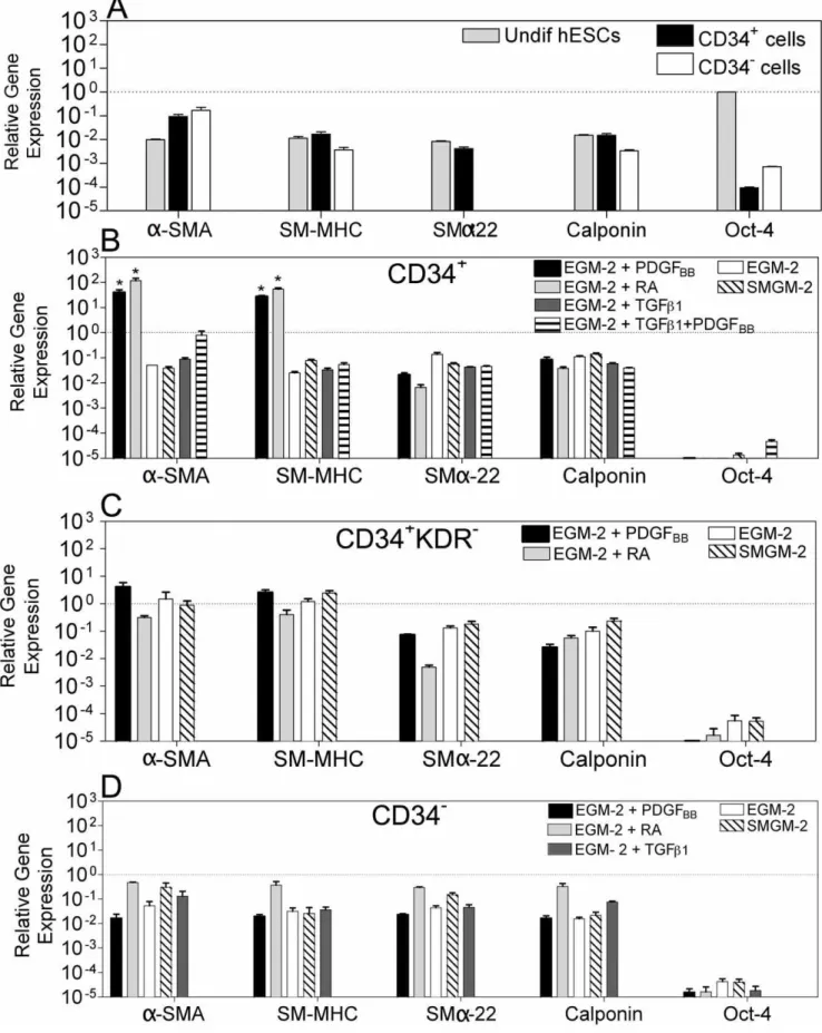
Next we evaluated the expression of SMC-specific genes in the hESC-derived cells (passage 4) by quantitative RT-PCR (Figure 2). The genes analyzed included:a-smooth muscle actin (a-SMA), an early marker of SMC differentiation and highly specific marker for SMCs in adult animals [23]; smooth muscle myosin heavy chain (SM-MHC), a later marker in SMC differentiation that seems to be highly restricted to SMCs [24]; calponin and smooth musclea-22, definitive SMC markers [25]. The gene expression in the hESC-derived cells was normalized by the corresponding gene expression in hVSMCs.
Undifferentiated CD34+
cells expressed low levels of SM-MHC (,2.0%), SMa-22 (,1%) and calponin (,2.0%), in most cases comparable to the levels found in undifferentiated hESCs, and moderate levels ofa-SMA (,10%) (Figure 2). Culture of these cells in the presence of EGM-2 medium supplemented with PDGFBBor RA contributed for the up-regulation of SMC genes,
as confirmed by the significant increase in the expression of a -SMA (.400-fold) and SM-MHC (.1,600-fold) when compared with undifferentiated CD34+
cells (p,0,05), and hVSMCS (a -SMA: .39-fold; SM-MHC: .27-fold) (Figure 2). Addition of TGFb-1, or TGFb-1plus PDGFBBincreased the cellular expression
ofa-SMA, SM-MHC, SMa-22 and calponin when compared to undifferentiated CD34+
cells; however, the expression was one order of magnitude lower than the one in hVSMCS, suggesting that the CD34+
-derived cells were not fully differentiated into SMCs.
CD34+
KDR2cells cultured in SMGM-2 medium or EGM-2 medium in the presence or absence of PDGFBBexpressed higher
levels of a-SMA (.42-fold), SM-MHC (.150-fold), SMa-22 (.18-fold) and calponin (.2-fold) than undifferentiated CD34+ cells (Figure 2). In addition, these cells showed similar levels of expression of a-SMA and SM-MHC to the one found in hVSMCs, suggesting that these cells shared features with SMCs.
In contrast to the CD34+
and CD34+
KDR2cells, CD342cells cultured in several media formulations had lower expression of SMC markers, indicating low efficiency of differentiation into SMCs (Figure 2). Of note, all the hESC-derived cells previously described had low expression of Oct-4 confirming their loss of pluripotency. In addition, hESC-derived cells expressing high levels of SMC markers had no expression of PECAM-1 (endothelial cell marker) and a-actinin (a marker of cardiomyocytes) (data not shown) confirming again their SMC differentiation.
Next, the expression and filament organization of contractile proteins was evaluated in cell populations having similar or highera -SMA and SM-MHC gene expression than hVSMCs, i.e., CD34+
RA, CD34+
PDGFBB, CD34+KDR2PDGFBB and CD34+KDR2
Individualized calponin filaments were observed in 16 to 60% of the overall cells; however, organizeda-SMA protein filaments were only observed in CD34+PDGF
BB (6.0%) and CD34+KDR2EGM-2
(6.1%) cells, and no organized SM-MHC filaments were observed in the hESC-derived populations (Figure S3). In contrast, hVSMCs expressed high levels of organized a-SMA (84.6%), SM-MHC (94.6%) and calponin (80.1%) filaments.
Collectively, gene and protein analyses indicate that several inductive signals (PDGFBB, or RA or EGM-2 basal medium) are
able to drive the SMC differentiation of CD34+
or CD34+ KDR2 cells, characterized by variable expression of SMC proteins and minimal assembly of thea-SMA protein into filaments.
Some hESC-derived cells present a secretion profile similar to hVSMCs
Using the multiplex cytometric bead array method we investi-gated the secretion of 17 analytes by hVSMC, CD34+PDGF
BB,
CD34+
RA, CD34+
KDR2PDGFBB, CD34+KDR2EGM-2 and
CD342PDGFBBcells. Out of the 17 analytes analyzed, hVSMCs
secreted IL-6, IL-7, IL-8, G-CSF, IFN-c, MCP-1 (MCAF) and TNF-a, above the detection limit (.0.2 pg/mL) (Figure 3). Cytokines IL-6 and IL-8 were the most secreted cytokines. CD34+PDGF
BBand CD34+KDR2EGM-2 cells secreted the same
analytes as hVSMCs. CD34+
KDR2PDGFBBand CD342PDGFBB
cells did not secrete IL-7, while CD34+
RA cells secreted all seven Figure 1. The effect of initial cell population and inductive signals on cell proliferation.(A) Protocols adopted to drive the differentiation of CD34+, CD34+KDR2and CD342cells isolated from EBs at day 10 into the SMC lineage. (B) Flow cytometric analysis of hES-derived cells: CD342
(B.1), CD34+
(B.2) and CD34+
KDR2(B.3) cells (in this last case isolated from the CD34+
cell population). The results show that CD342cells do not
express CD34+
marker (B.1), CD34+
cells are formed by CD34+
KDR2and CD34+
KDR+
cells (B.2), and CD34+
KDR2do not express the KDR marker (B.3).
Percent of positive cells (dash plot) were calculated based in the isotype controls (grey plot). (C) Time-course proliferation of CD34+
(C.1), CD34+
KDR2
(C.2) and CD342cells (C.3).
Figure 2. Gene expression in hESC-derived cells evaluated by qRT-PCR.Gene expression in each experimental group was normalized by the corresponding gene expression observed in hVSMCs. Oct-4 was normalized by the expression in undifferentiated hESCs. (A) Gene expression of hESCs, CD34+
and CD342cells before differentiation. SMa-22 expression in CD342cells is very low (,8.5
61026) and not visible in the graph. CD34+(B),
CD34+
KDR2(C), and CD342(D) cells were cultured in SMGM-2 medium, EGM-2 medium, and EGM-2 medium supplemented with PDGFBBor TGF
cytokines mentioned and 4 cytokines more (IL-1b, IL-2, IL-4 and GM-CSF) (Figure S4).
Interestingly, the secretion profile of CD34+
PDGFBB cells is
very similar to the one observed for hVSMCS, either in the type and concentration of the analytes secreted (Figure 3). In contrast, CD342PDGFBB cells secreted analytes at different concentration
than hVSMCs, and the other cell populations seemed to be in an intermediate stage in terms of secretion profile (Figure 3 and Figure S4). CD34+
RA cells secreted higher levels of cytokines than all the remaining cell populations, including hVSMCs.
Some hESC-derived cells like hVSMCs have the ability to contract when treated with vasoactive agonists and the process is mediated by Ca2+
The ability of SMCs to contract in response to vasoactive agonists is mediated by an increase of intracellular Ca2+
levels which triggers the SMC contractile apparatus [26]. To evaluate
whether hESC-derived cells had contractility mediated by Ca2+ , the cells were loaded with FURA-2, a Ca2+
sensitive dye, and their response to vasoactive agonists (bradykinin, angiotensin II, carbachol and histamine) and depolarization agents (KCl) was monitored by fluorescence (Figure 4A). The response profile was compared to the one observed for hVSMCs and human umbilical vein endothelial cells (HUVECs), as positive and negative controls, respectively. Intracellular free Ca2+
[Ca2+
]ilevels increase when
hVSMCs are exposed to bradykinin, angiotensin II and carbachol, while no measurable effect is observed in HUVECs. KCl induces a higher increase in the [Ca2+
]ilevels in HUVECs than in hVSMCs,
while histamine induces similar levels of [Ca2+
]iin both cell types
but following different profiles. These response patterns are typical for HUVECs and hVSMCs [27,28,29,30,31].
CD34+
RA and CD34+
PDGFBB cells exposed to histamine,
bradykinin, angiotensin II, carbachol or KCl had similar response profiles as observed for hVSMCs (Figure 4A and Figure S5). For bradykinin and KCl the response intensity was lower than in Figure 3. Secretomic profile of hVSMC and hECS-derived cells.Seventeen cytokines were measured simultaneously in medium collected from hVSMC, CD34+
PDGFBB, CD34+
RA, and CD342PDGFBBcells at passage 4. Results are Mean
hVSMCs, however similar intensity was observed for angiotensin II, carbachol and histamine. On the other hand, although CD34+KDR2RA, CD34+KDR2PDGF
BB and CD34+KDR2
EGM-2 cells had similar response profiles as hVSMCs, in general the intensity of response was lower (Figure S6). In contrast to the previous hESC-derived populations, CD342PDGFBBcells showed a
very low variation in the intracellular levels of Ca2+
when exposed to depolarization and vasoactive peptides.
To examine whether hESC-derived cells were able to contract, they were subjected to the effects of carbachol and atropine (Figure 4B). With the exception of CD342PDGFBBcells, all cells
were able to contract after exposure to carbachol (13 to 38% contraction after 30 min, depending on the cell population). In most cases, cell contraction was not significantly different (P.0.05)
from the one observed for hVSMCs. Moreover, with the exception of CD342PDGFBB cells, the muscarinic inhibitor atropin
significantly blocked or reduced the carbachol-mediated effect (P,0.05) (Figure 4B).
Based on the expression of SMC proteins and genes, secretion of cytokines and cellular contraction, CD34+PDGF
BBand CD34+RA
cells were selected for further characterization. These cells are likely at a smooth muscle progenitor cell stage (SMPCs), which can potentially be further induced into a more mature SMC phenotype.
The contraction of both hESC-derived SMPCs and hVSMCs involves Rho A/Rho kinase- and Ca2+/CaM/
MLCK-dependent pathways
CaM/MLCK- and RhoA/Rho kinase-dependent pathways play a pivotal role in SMC contraction [7,26]. The Ca2+
/CaM pathway plays a key role in SMC contraction through the stimulation of MLCK-mediated phosphorylation of myosin light chain 20,000 Da (MLC20) [26]. To assess whether Ca2+/CaM
pathway was involved in the contraction of hESC-derived SMPCs, cells were exposed to the CaM-specific inhibitor W-7 [32], and then contraction was induced by exposing them to the CaM agonist U46619 [7]. To facilitate the evaluation of cell contraction, cells were encapsulated in fibrin gels, and gel diameter was determined after 14 h. Pre-incubation of hESC-derived SMPCs with W-7 significantly inhibited U46619-induced contraction (Figure 5A and Figure 5B). Similar results were obtained for the control cells hVSMCs. Overall the results indicate the involvement of CaM/MLCK-kinase pathway in cell contraction. To examine whether Rho kinase was involved in the hESC-derived SMPC contraction, the cells were pre-treated with the Rho kinase-specific inhibitor Y27632 [7] and then contraction was induced by the agonist End-1 [33]. Cells treated with Y27632 and End-1 showed no significant contraction (Figure 5A and Figure 5B). In contrast, cells treated with End-1 but not Y27632 contracted significantly. Similar response profiles were obtained for hVSMCs and hESC-derived SMPCs. Collectively the results indicate the involvement of a Rho/Rho kinase-dependent pathway in SMC contraction.
3D culture of hESC-derived SMPCs modulates gene expression towards the expression observed in hVSMCs
We investigated the use of hESC-derived SMPCs for tissue engineering applications [2,3]. Fibrin gels previously used for the
encapsulation of neonatal SMCs [34] were selected as scaffolds for the encapsulation of SMPCs. These gels allow cell attachment and can be remodeled by cellular metalloproteinases. SMPCs were encapsulated in fibrin gels for 3 days after which the cells were characterized at protein and gene levels.
Gene expression of SMPCs (CD34+
RA and CD34+
PDGF) was compared to hVSMCs under the same culture conditions (Figure 6A and Figure 6B). The culture of SMPCs in 3D gels modulated the expression of SMC genes (a-SMA, SM-MHC or SMa-22) towards the one observed for hVSMCs cultured in 3D gels. We complemented these studies by evaluating the expression of extracellular matrix and adhesion molecules by a quantitative real-time PCR array. This array evaluated the expression of 84 genes involved in cell-cell and cell-matrix interactions. Again, the 3D culture of SMPCs modulated extracellular matrix and adhesion molecule genes towards the expression observed in hVSMCs (Figure 6B). The number of genes with similar expression increased from 23 to 58 or 9 to 53 when CD34+
PDGFBB cells or CD34+RA cells are cultured in 2D or
3D, respectively. Finally, gene expression associated with SMCs including collagen I and thrombospondin 1 [35], integrinsa2,a3, a5,aV andb1 [36], the enzyme metalloproteinase 2 [37], and the growth factor TGFb-1 was similar in SMPCs and hVSMCs
(Figure 6C and Figure S7).
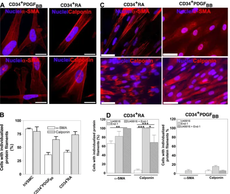
Co-culture of hESC-derived SMPCs with hVSMCs induces the assembly ofa-SMA and calponin into filaments
Next we sought to investigate whether hESC-derived SMPCs could be further matured into a SMC phenotype with an organized contractile filament network. To accomplish this goal, hESC-derived SMPCs were initially labeled with PKH67 fluorescent dye and co-cultured on top of hVSMCs for 5 days. After this time, the cells were sorted and characterized by immunocytochemistry. Remarkably, hESC-derived SMPCs showed significant improve-ment in the organization of the fibers after contact with hVSMCs: 36.3% and 41.2% of CD34+
PDGFBB and CD34+RA cells,
respectively, had organized a-SMA fibers, while 64.8% and 73.8% had organized calponin fibers (Figure 7A and Figure 7B). These results show that hESC-derived SMPCs are plastic cells and can be induced to differentiate into a more mature SMC phenotype displaying an organized contractile network.
Vasoactive agents U46619 and End-1 improve the organization of contractile protein fibers
Next, we identified molecules able to maturate the hESC-derived SMPCs into SMCs having an organized contractile network. We sought to evaluate the effect of the agonists U46619 and End-1 involved in the CaM/MLCK- and RhoA/Rho kinase-contraction pathways, respectively. In CD34+RA cells, each agonist independently improved dramatically the organization of a-SMA and calponin fibers, being End-1 the most effective. The addition of both agonists did not significantly improve the effect of End-1 (Figure 7C and Figure 7D). Importantly, the induction effect of the vasoactive agents was not observed on CD34+PDGF
BB (Figure 7D) or CD342PDGFBB cells (Figure S8) showing that the inductive effect is dependent on the differentiation history of the SMPCs. The mature SMCs (derived Figure 4. Contractility of hESC-derived cells.(A) Concentration of intracellular Ca2+
in FURA-2-loaded cultured hESC-derived cells in response to several agonists (100mM histamine, 1027M bradykinin, 1025M angiotensin II or 50 mM KCl). hVSMCs and HUVECs were used as positive and
negative controls, respectively. Traces are representative of 4 independent experiments for each condition. (B) Contractility of hESC-derived cells when exposed to the effects of carbachol (1025M in DMEM medium, for 30 min) or atropine (1024M, 1 h) plus carbachol (1025M for 30 min). hVSMCs were used as controls. Results are Mean6SEM (n= 3);#denotes statistical significance (P
from CD34+
RA) express SMC markers including a-SMA, SM-MHC and calponin but not the cardiac markera-actinin (Figure S9). Furthermore, the cells show no expression of troponin T, a protein found in skeletal and cardiac muscle but not in smooth muscle and vascular endothelial-cadherin, a protein typically expressed in endothelial cells (Figure S10).
Discussion
In this study, we demonstrate that CD34+
cells have higher SMC potential than CD342 cells. We further show that RA or
PDGFBBdrive the differentiation of CD34+cells into SMPCs We
have characterized the differentiated cells at gene and at protein level, their secretome, the ability to contract when incubated with several pharmacological agents, and contraction mechanism mediated by Ca2+. Envisioning the use of these cells for vascular engineering, they were encapsulated on 3D fibrin scaffolds and characterized at gene and functional levels. Finally, we identified End-1 as a key molecule to induce the polymerization of contractile proteins in SMPCs (CD34+RA cells).
We examined the capacity of three hESC populations (CD34+ , CD34+
KDR2, CD342) isolated from EBs at day 10, and cultured Figure 5. Evaluation of the role of Rho A/Rho kinase- and Ca2+/CaM/MLCK-dependent pathways in the contraction of SMPCs and
hVSMCs.CD34+PDGF or CD34+RA cells were initially treated for 3 days with serum-free medium containing the agonists endothelin-1 (End-1
(10 nM), A and B.1) or U46619 (1mM) (A and B.2) in the presence or absence of the Rho-kinase inhibitor Y27632 (13mM) or the calmodulin inhibitor
W-7 (12mg/mL). The cells were then encapsulated in fibrin gels and the diameter of the gel assessed at time 0 (1) and 14 h (2,3,4). Graph displays the
variation in gel diameter (percentage) encapsulating hVSMCs (2), CD34+
PDGFBBcells (3) or CD34+
RA cells (4). Data are shown as mean6SEM (n= 6), *P,0.001 relatively to samples inhibited with Y27632 or W-7, by Student’st-test.
as single cells on media supplemented with inductive signals including PDGFBB, RA, TGFb-1 and TGFb-1 plus PDGFBB to
differentiate into SMCs. Notably, TGFb-1 [5,21,38], PDGFBB
[5,12,19], and RA [9,17] have been reported to be very important inductive signals for SMC differentiation from different initial stem cell populations. We report that from all inductive signals tested in this study, RA and PDGFBBare the most effective in guiding the
differentiation of hESC-derived CD34+
cells into SMPCs, based on gene and protein analysis, response to the depolarization agents
and vasoactive peptides, contraction profile, secretion of cytokines, and behavior in a 3D scaffold. Although SMPCs express most of the SMC markers, they exhibit disorganized contractile proteins. We further show that CD34+cells have higher propensity to yield contractile SMPCs than CD342cells, when exposed to the same inductive signals. Interestingly, CD34+
KDR2 cells which have been reported to be of mesenchymal origin [18], can give rise to SMPCs but they respond less efficiently to vasoactive peptides and depolarization agents, have lower contractile properties than
Figure 7. Maturation of hESC-derived SMPCs.(A and B) Expression and organization of SMC proteins on hESC-derived SMPCs cultured on top of inactivated-hVSMCs for 5 days. hVSMCs were used as controls. Bar corresponds to 50mm. Results are Mean6SEM (n= 10). (C and D) Expression and organization of SMC proteins on hESC-derived SMPCs treated with vasoactive agents for 3 days. Bar corresponds to 50mm. Results are Mean6 SEM (n= 8); *, **, *** denote statistical significance (P,0.05,P,0.01,P,0.001, respectively).
doi:10.1371/journal.pone.0017771.g007
Figure 6. Characterization of hESC-derived SMPCs encapsulated in 3D fibrin gel scaffolds. SMPCs were characterized after being encapsulated in fibrin gels for 3 days. (A) SMC gene expression on CD34+RA (A.1) and CD34+PDGF (A.2) cells, as assessed by qRT-PCR. Gene
expression of hESC-derived SMPCs was normalized by gene expression of hVSMCs under the same culture conditions. Results are Mean6SEM (n= 4). (B) Comparison of extracellular matrix and adhesion molecules-related gene expression in CD34+
PDGFBB(B.1) or CD34+
CD34+
PDGF cells, and have a different cytokine secretion profile as compared to hVSMCs. In line with previous results [39], our results suggest that cells expressing KDR receptor are needed for an efficient SMC differentiation.
The development of mature contractile SMCs from stem cells occurs in multiple steps comprising (i) the commitment to the SMC lineage, (ii) the differentiation into early immature and (iii) the maturation into the mature contractile phenotype [40]. Previously, we have reported that CD34+
cells could give rise to SMLCs when cultured in medium supplemented with PDGFBB;
however, the differentiation of SMLCs was not complete since the assembly ofa-SMA or SM-MHC proteins into filaments was not observed [12]. Curiously, similar results have been obtained in this work for the cell populations tested and exposed to different inductive signals including RA and TGFb-1.
Our results indicate that the co-culture of SMPCs with fully differentiated hVSMCs induces the assembly of a-SMA and calponin proteins into individualized filaments. This indicates that the cells are able to maturate into a fully contractile phenotype. It is known that both assembly and disassembly of actin filaments is regulated by RhoA [41]. Consistent with this, our results show that End-1, an agonist of RhoA pathway, dramatically increases actin polymerization in CD34+
RA cells and the cells exhibit individu-alized a-SMA and calponin filaments. However, such inductive effect was not observed in CD34+PDGF
BBcells, and this could be
due to the inhibition of mature SMC marker expression by PDGFBB [42]. Strikingly, these cells are contractile (Figure 5)
despite presenting a small percentage of polymerized actin fibers (6%;Figure S3). Nevertheless, it is known that the total amount of actin that undergoes polymerization after induction of contraction is relatively small [43]. Further research is needed to study the molecular processes that regulate the assembly of actin filaments in smooth muscle tissues and the nature of the actin filaments network that are formed during contractile activation [43].
Contractile and synthetic SMCs, which represent the two ends of a spectrum of SMCs with intermediate phenotypes, clearly show different morphologies [22]. The SMPCs derived in this work seem to have the contractile phenotype, as they are spindle-shaped [44], express proteins involved in SMC contraction includinga -SMA, SM-MHC, calponin and SMa-22 [22,24], and contract when exposed to carbachol and vasoactive peptides being this effect reversed by the presence of the respective antagonists.
We show that SMPCs are able to contract to a plethora of vasoactive agents including carbachol, angiotensin II, histamine, thromboxane-mimetic U46619, endothelin-1, and bradykinin, as hVSMCs. Most of these agonists act through different receptors coupled to G-proteins activating membrane-bound phospholipase C, which leads to the formation of inositol-1,4,5-triphosphate (IP3)
and diacylglycerol (DAG) [26]. Through different mechanisms, both molecules induce the accumulation of Ca2+ in the cell cytoplasm [26]. The increase of intracellular Ca2+
activates the cell’s contraction machinery. Our results agree with previous ones showing that hESC-derived SMCs respond to bradykinin, histamine and carbachol by increasing at different levels the intracellular concentration of Ca2+
[14]. The accumulation of intracellular Ca2+
in hESC-derived SMCs exposed to carbachol is minimal, indicating that the contraction of the cell may involve the activation of other intracellular players (e.g. protein kinase C) than Ca2+, as described in other studies [45].
Our results show that hESC-derived SMPC contraction is mediated by the activation of CaM/MLCK- and Rho/Rho kinase-dependent pathways [7,26]. A similar mechanism has been reported recently for SMLCs obtained from human adipose
tissue-derived mesenchymal stem cells [7]. The results also indicate that the absence of fully organized protein filaments did not prevent the contraction of SMLCs. This is in line with results reported previously by us [12] and by others [13] despite the absence of maturity and organization in the contractile machinery of the cell.
SMCs have key biological functions in terms of contraction and secretion of soluble signaling molecules [46]. In the present study we report for the first time the secretion profile of hES-derived SMPCs. CD34+
cells that differentiate in medium supplemented with PDGFBB have similar secretome profile as hVSMCs. They
express high levels (.100 pg/mL) of IL-6 and IL-8, moderate levels (between 100 and 10 pg/mL) of IFN-c, and small levels of (between 10 and 1 pg/mL) of IL-7, G-CSF, MCP-1 and TNF-a. IL-6 is a cytokine with potent inflammatory properties and metabolic effects in SMCs. It has been shown that IL-6 secretion is inversely correlated to glucose consumption [47]. IL-8 is a cytokine that induces the proliferation and chemotaxis of smooth muscle cells and increases the activity of mitogen-activated protein kinase (MAPK) [48]. IFN-cacts on vascular smooth muscle cells to induce cellular proliferation [49].
Although several studies have reported the differentiation of different stem cells into SMCs, very few evaluated the impact of the 3D environment at the geno- and phenotype of the differentiated cells [5]. Gene studies have demonstrated that human aortic SMCs encapsulated in a 3D collagen matrix have significantly less focal adhesions, lower proliferation, and lowera -SMA expression than cells in 2D [50]. However, to our knowledge, no study has compared the gene expression of SMLCs from different origins with fully differentiated SMCs when cultured in 2D or 3D systems. Our results show that hESC-derived SMPCs encapsulated for 3 days in fibrin gels express similar gene levels of a-SMA, SM-MHC, and SMa22 as encapsulated hVSMCs. In addition, considering an array of 84 genes related to cell-cell and cell-matrix interactions, 3D-cultured-SMPCs had a more hVSMC-similar gene expression profile than 2D-cultured SMPCs. This shows that 3D scaffolds may induce further the differentiation of SMPCs into SMCs. Several factors might account for the differences found between 2D and 3D culture systems including (i) ECM stiffness and (ii) ECM 3D environment. It has been demonstrated that the stiffness of the ECM has a high impact in the cytoskeletal and focal adhesion assembly of SMCs [51]. Furthermore, SMCs cultured within 3D polyethylene glycol-fibrinogen [52] or collagen [50] gels had less proliferation, stress fibers and focal adhesion than on 2D culture systems. Future studies should evaluate the effect of both factors in the modulation of geno- and phenotype of the differentiated cells over the time and study the underlining mechanism. In this study we assessed the effect of the 3D matrix after 3 days of cell encapsulation. During this time, previous studies have shown that SMCs are able to migrate and remodel the ECM [53]; however, it would be interesting to extend these studies in time.
bioreactors. Second, our study identifies a methodology to induce the organization of the contractile protein filaments. Third, it demonstrates the importance of a 3D environment to modulate the activity of hESC-derived SMLCs.
Recent studies reported the derivation of pluripotent stem cells from human somatic cells by retroviral transduction [54]. These reprogrammed cells share many features with hESCs in terms of morphology, gene expression and differentiation potential, and open a new avenue for the potential derivation of autologous cells for regenerative medicine and drug screening. Indeed, the differentiation of iPSCs into SMCs has been recently reported [55]. It remains to be determined whether the methodology described in this work can be applied on iPSCs and this is an issue that will be evaluated in future work.
Supporting Information
Figure S1 SMC proteins are expressed on hESC-derived SMPCs. CD34+, CD34+KDR2and CD342 cells differentiated under different media conditions express a-SMA, calponin and SM-MHC, as evaluated by immunofluorescence. hVSMCs were used as a positive control and HUVECs as a negative control for the SMC markers. In all figures, bar corresponds to 50mm.
(TIFF)
Figure S2 Isotype controls for SMPC immunostainning.
Stained the isotype controls IgG2Aand IgG1for the SMC markers: a-SMA, SM-MHC and Calponin. Cell nuclei were stained with 49, 69-diamidino-2-phenylindole (DAPI). Cells were labeled with mouse anti–human IgG2Aand IgG1antibodies. Bar corresponds to 50mm.
(TIFF)
Figure S3 Organization and expression of contractile proteins.(A) Quantification of organizeda-SMA, SM-MHC and calponin filaments. (B) Expression of a-SMA in differentiated CD34+
, CD34+
KDR2and CD342cells. Cells were differentiated for 3 passages (approximately 18 days after cell seeding). hVSMCs and HUVECs were used as positive and negative controls, respectively. In all graphs, the percentages of positive cells were calculated based in the isotype controls (gray plot) and are shown in each histogram plot.
(TIFF)
Figure S4 Secretomic profile of hESC-derived cells.17 cytokines were measured simultaneously in the medium collected from CD34+KDR2PDGF
BB and CD34+KDR2EGM-2 cells. A
standard range of 0.2 to 3,200 pg/mL was used. Samples and controls were run in triplicate, standards and blanks in duplicate. (TIFF)
Figure S5 Contractility of hESC-derived cells.Cells were loaded with FURA-2/AM and their response to carbachol (1025M) was monitored by fluorescence. The response profile was compared to the one observed for hVSMCs and HUVECs, as positive and negative controls, respectively.
(TIFF)
Figure S6 Contractility of hESC-derived cells.Cells were loaded with FURA-2/AM and their response to vasoactive agonists
(bradykinin (1027M), angiotensin II (1025M) and histamine (100mM) and depolarization agents (KCl; 50 mM) was monitored by fluorescence. The response profile was compared to the one observed for hVSMCs and HUVECs, as positive and negative controls, respectively.
(TIFF)
Figure S7 Integrin gene expression. Gene expression on CD34+
PDGFBB and CD34+RA cells was normalized by gene
expression on hVSMCs, both cultured in 3D or 2D systems. Gene expression was obtained from the RT2ProfilerTMPCR array. (TIFF)
Figure S8 Expression and organization of SMC proteins on CD342PDGFBB cells treated with vasoactive agents
for 3 days.A) Quantification by immunocytochemistry analysis. Results are Mean6 SEM (n= 8). B) Expression of a-SMA and
calponin in CD342PDGFBBcells treated with End-1 (10 nM) for 3
days. Bar corresponds to 50mm. (TIFF)
Figure S9 Gene expression in CD34+PDGF
BB and
CD34+RA cells after treatment with vasoactive agents
for 3 days.Gene expression in CD34+PDGF
BBand CD34+RA
cells was normalized by gene expression in hVSMCs. Results are Mean6SEM (n= 4).
(TIFF)
Figure S10 Expression and organization of troponin T (TrpnT) and vascular endothelial-cadherin (VECad) on CD34+RA (A) and CD34+PDGF
BB (B) cells treated with
End-1 for 3 days. Human cardiac tissue (for TrpnT) and HUVECs (for VeCad) were used as positive controls. Bar corresponds to 50mm.
(TIFF)
Table S1 Primers used for Real Time PCR. PCR conditions: initial denaturation step at 94uC for 5 min; 40 cycles of denaturation at 94uC for 30 sec, annealing at 60uC for 33 sec and extension at 72uC for 30 sec. At the end was performed a final 7 minutes extension at 72uC. After amplification, the melting curve profile or agarose gel electrophoresis was used to determine the specificity of PCR products.
(TIFF)
Materials and Methods S1 (DOC)
Acknowledgments
The authors would like to thank Renata Gomes for help in the preparation of this manuscript.
Author Contributions
References
1. Matsubayashi K, Fedak PWM, Mickle DAG, Weisel RD, Ozawa T, et al. (2003) Improved left ventricular aneurysm repair with bioengineered vascular smooth muscle grafts. Circulation 108 Suppl 1: II219–225.
2. Niklason LE, Gao J, Abbott WM, Hirschi KK, Houser S, et al. (1999) Functional arteries grown in vitro. Science 284: 489–493.
3. Oberpenning F, Meng J, Yoo JJ, Atala A (1999) De novo reconstitution of a functional mammalian urinary bladder by tissue engineering. Nat Biotechnol 17: 149–155.
4. Kashiwakura Y, Katoh Y, Tamayose K, Konishi H, Takaya N, et al. (2003) Isolation of bone marrow stromal cell-derived smooth muscle cells by a human SM22alpha promoter: in vitro differentiation of putative smooth muscle progenitor cells of bone marrow. Circulation 107: 2078–2081.
5. Ross JJ, Hong Z, Willenbring B, Zeng L, Isenberg B, et al. (2006) Cytokine-induced differentiation of multipotent adult progenitor cells into functional smooth muscle cells. J Clin Invest 116: 3139–3149.
6. Rodrı´guez LV, Alfonso Z, Zhang R, Leung J, Wu B, et al. (2006) Clonogenic multipotent stem cells in human adipose tissue differentiate into functional smooth muscle cells. Proc Natl Acad Sci USA 103: 12167–12172.
7. Kim M, Jeon E, Kim Y, Lee J, Kim J (2008) Thromboxane A2 Induces Differentiation of Human Mesenchymal Stem Cells to Smooth Muscle-Like Cells. Stem Cells.
8. Le Ricousse-Roussanne S, Barateau V, Contreres JO, Boval B, Kraus-Berthier L, et al. (2004) Ex vivo differentiated endothelial and smooth muscle cells from human cord blood progenitors home to the angiogenic tumor vasculature. Cardiovascular Research 62: 176–184.
9. Huang H, Zhao X, Chen L, Xu C, Yao X, et al. (2006) Differentiation of human embryonic stem cells into smooth muscle cells in adherent monolayer culture. Biochemical and Biophysical Research Communications 351: 321–327. 10. Xie CQ, Zhang J, Villacorta L, Cui T, Huang H, et al. (2007) A highly efficient
method to differentiate smooth muscle cells from human embryonic stem cells. Arteriosclerosis, Thrombosis, and Vascular Biology 27: e311–312.
11. Gerecht-Nir S, Ziskind A, Cohen S, Itskovitz-Eldor J (2003) Human embryonic stem cells as an in vitro model for human vascular development and the induction of vascular differentiation. Lab Invest 83: 1811–1820.
12. Ferreira LS, Gerecht S, Shieh HF, Watson N, Rupnick MA, et al. (2007) Vascular progenitor cells isolated from human embryonic stem cells give rise to endothelial and smooth muscle like cells and form vascular networks in vivo. Circulation Research 101: 286–294.
13. Vo E, Hanjaya-Putra D, Zha Y, Kusuma S, Gerecht S (2010) Smooth-Muscle-Like Cells Derived from Human Embryonic Stem Cells Support and Augment Cord-Like Structures In Vitro. Stem Cell Rev and Rep.
14. Hill KL, Obrtlikova P, Alvarez DF, King JA, Keirstead SA, et al. (2010) Human embryonic stem cell-derived vascular progenitor cells capable of endothelial and smooth muscle cell function. Experimental Hematology 38: 246–257.e241. 15. Grynkiewicz G, Poenie M, Tsien RY (1985) A new generation of Ca2+
indicators with greatly improved fluorescence properties. J Biol Chem 260: 3440–3450.
16. Bernardino L, Agasse F, Silva B, Ferreira R, Grade S, et al. (2008) Tumor necrosis factor-alpha modulates survival, proliferation, and neuronal differen-tiation in neonatal subventricular zone cell cultures. Stem Cells 26: 2361–2371. 17. Drab M, Haller H, Bychkov R, Erdmann B, Lindschau C, et al. (1997) From totipotent embryonic stem cells to spontaneously contracting smooth muscle cells: a retinoic acid and db-cAMP in vitro differentiation model. FASEB J 11: 905–915.
18. Vodyanik MA, Thomson JA, Slukvin II (2006) Leukosialin (CD43) defines hematopoietic progenitors in human embryonic stem cell differentiation cultures. Blood 108: 2095–2105.
19. Simper D, Stalboerger PG, Panetta CJ, Wang S, Caplice NM (2002) Smooth muscle progenitor cells in human blood. Circulation 106: 1199–1204. 20. Sinha S, Hoofnagle MH, Owens GK (2009) Derivation of contractile smooth
muscle cells from embryonic stem cells. Methods Mol Biol 482: 345–367. 21. Sinha S, Hoofnagle M, Kingston P, McCanna M, Owens G (2004)
Transforming growth factor-b1 signaling contributes to development of smooth muscle cells from embryonic stem cells. American Journal of Physiology- Cell Physiology 287: 1560–1568.
22. Rensen SSM, Doevendans PAFM, van Eys GJJM (2007) Regulation and characteristics of vascular smooth muscle cell phenotypic diversity. Netherlands heart journal : monthly journal of the Netherlands Society of Cardiology and the Netherlands Heart Foundation 15: 100–108.
23. Frid M, Shekhonin B, Koteliansky V, Glukhova M (1992) Phenotypic changes of human smooth muscle cells during development: late expression of heavy caldesmon and calponin. Developmental biology(Print) 153: 185–193. 24. Owens GK, Kumar MS, Wamhoff BR (2004) Molecular regulation of vascular
smooth muscle cell differentiation in development and disease. Physiol Rev 84: 767–801.
25. Duband J, Gimona M, Scatena M, Sartore S, Small J (1993) Calponin and SM22 as differentiation markers of smooth muscle: spatiotemporal distribution during avian embryonic development. Differentiation 55: 1–11.
26. Hathaway DR, March KL, Lash JA, Adam LP, Wilensky RL (1991) Vascular smooth muscle. A review of the molecular basis of contractility. Circulation 83: 382–390.
27. Dora KA, Xia J, Duling BR (2003) Endothelial cell signaling during conducted vasomotor responses. Am J Physiol Heart Circ Physiol 285: H119–126. 28. Takeda T, Yamashita Y, Shimazaki S, Mitsui Y (1992) Histamine decreases the
permeability of an endothelial cell monolayer by stimulating cyclic AMP production through the H2-receptor. J Cell Sci 101(Pt 4): 745–750. 29. Graier WF, Simecek S, Bowles DK, Sturek M (1994) Heterogeneity of
caffeine-and bradykinin-sensitive Ca2+stores in vascular endothelial cells. Biochem J 300 (Pt 3): 637–641.
30. Montiel M, Ga´mez MI, Garcı´a-Vallejo JJ, Pe´rez De La Blanca E, Martı´n E, et al. (2003) Angiotensin II effect on calcium mobilization and mitogen-activated protein kinase activation in human umbilical vein endothelial cells. Signal Transduction 3: 201–208.
31. Kansui Y, Garland CJ, Dora KA (2008) Enhanced spontaneous Ca2+events in endothelial cells reflect signalling through myoendothelial gap junctions in pressurized mesenteric arteries. Cell Calcium 44: 135–146.
32. Frampton JE, Orchard CH (1992) The effect of a calmodulin inhibitor on intracellular [Ca2+] and contraction in isolated rat ventricular myocytes. J Physiol 453: 385–400.
33. Bouallegue A, Daou GB, Srivastava AK (2007) Endothelin-1-induced signaling pathways in vascular smooth muscle cells. Curr Vasc Pharmacol 5: 45–52. 34. Ross JJ, Tranquillo RT (2003) ECM gene expression correlates with in vitro
tissue growth and development in fibrin gel remodeled by neonatal smooth muscle cells. Matrix Biol 22: 477–490.
35. Majack RA, Cook SC, Bornstein P (1986) Control of smooth muscle cell growth by components of the extracellular matrix: autocrine role for thrombospondin. Proc Natl Acad Sci U S A 83: 9050–9054.
36. Deb A, Skelding KA, Wang S, Reeder M, Simper D, et al. (2004) Integrin profile and in vivo homing of human smooth muscle progenitor cells. Circulation 110: 2673–2677.
37. Wang H, Keiser JA (1998) Vascular endothelial growth factor upregulates the expression of matrix metalloproteinases in vascular smooth muscle cells: role of flt-1. Circ Res 83: 832–840.
38. Dickson M, Martin J, Cousins F, Kulkarni A, Karlsson S, et al. (1995) Defective haematopoiesis and vasculogenesis in transforming growth factor-beta 1 knock out mice. Development 121: 1845–1854.
39. Yamashita J, Itoh H, Hirashima M, Ogawa M, Nishikawa S, et al. (2000) Flk1-positive cells derived from embryonic stem cells serve as vascular progenitors. Nature 408: 92–96.
40. Owens GK (1995) Regulation of differentiation of vascular smooth muscle cells. Physiol Rev 75: 487–517.
41. Hellstrand P, Albinsson S (2005) Stretch-dependent growth and differentiation in vascular smooth muscle: role of the actin cytoskeleton. Can J Physiol Pharmacol 83: 869–875.
42. Dandre´ F, Owens GK (2004) Platelet-derived growth factor-BB and Ets-1 transcription factor negatively regulate transcription of multiple smooth muscle cell differentiation marker genes. Am J Physiol Heart Circ Physiol 286: H2042–2051.
43. Gunst SJ, Zhang W (2008) Actin cytoskeletal dynamics in smooth muscle: a new paradigm for the regulation of smooth muscle contraction. Am J Physiol, Cell Physiol 295: C576–587.
44. Hao H, Gabbiani G, Bochaton-Piallat ML (2003) Arterial smooth muscle cell heterogeneity: implications for atherosclerosis and restenosis development. Arterioscler Thromb Vasc Biol 23: 1510–1520.
45. Harnett KM, Biancani P (2003) Calcium-dependent and calcium-independent contractions in smooth muscles. Am J Med 115 Suppl 3A: 24S–30S. 46. Gerthoffer WT, Singer CA (2002) Secretory functions of smooth muscle:
cytokines and growth factors. Mol Interv 2: 447–456.
47. Mayr M, Zampetaki A, Sidibe A, Mayr U, Yin X, et al. (2008) Proteomic and metabolomic analysis of smooth muscle cells derived from the arterial media and adventitial progenitors of apolipoprotein E-deficient mice. Circ Res 102: 1046–1056. 48. Yue TL, Wang X, Sung CP, Olson B, McKenna PJ, et al. (1994) Interleukin-8. A mitogen and chemoattractant for vascular smooth muscle cells. Circ Res 75: 1–7. 49. Wang Y, Bai Y, Qin L, Zhang P, Yi T, et al. (2007) Interferon-gamma induces human vascular smooth muscle cell proliferation and intimal expansion by phosphatidylinositol 3-kinase dependent mammalian target of rapamycin raptor complex 1 activation. Circ Res 101: 560–569.
50. Li S, Lao J, Chen BPC, Li YS, Zhao Y, et al. (2003) Genomic analysis of smooth muscle cells in 3-dimensional collagen matrix. FASEB J 17: 97–99.
51. Peyton SR, Raub CB, Keschrumrus VP, Putnam AJ (2006) The use of poly(ethylene glycol) hydrogels to investigate the impact of ECM chemistry and mechanics on smooth muscle cells. Biomaterials 27: 4881–4893.
52. Peyton SR, Kim PD, Ghajar CM, Seliktar D, Putnam AJ (2008) The effects of matrix stiffness and RhoA on the phenotypic plasticity of smooth muscle cells in a 3-D biosynthetic hydrogel system. Biomaterials 29: 2597–2607.
53. Naito M, Nomura H, Iguchi A (1996) Migration of cultured vascular smooth muscle cells into non-crosslinked fibrin gels. Thromb Res 84: 129–136. 54. Takahashi K, Tanabe K, Ohnuki M, Narita M, Ichisaka T, et al. (2007)
Induction of pluripotent stem cells from adult human fibroblasts by defined factors. Cell 131: 861–872.