r
A
a
l
þ
a
t
n
ý
i
r
j
m
ir
a
O
O
h
r
c
i
r
g
a
in
e
a
s
e
R
l
Hatice Ceyda Deniz¹, Hüseyin Levent Keskin², Elçin Işlek Seçen², Gülin Feykan Yeğin Akçay², Işık Üstüner², Ayşe Filiz Avşar² ¹Department of Family Medicine, ²Department of Gynecology and Obstetrics, Atatürk Education and Research Hospital, Ankara, Turkey Tiroid Disfonksiyonunun Kemik Mineral Yoğunluğuna Etkisi / The Efect of Thyroid Disfunction on the Bone Mineral Density
Thyroid Dysfunction Does Not Afect the
Bone Mineral Density in Postmenopausal Women
Postmenapozal Kadınlarda Tiroid Disfonksiyonu
Kemik Mineral Yoğunluğunu Etkilememektedir
DOI: 10.4328/JCAM.1197 Received: 08.07.2012 Accepted: 05.08.2012 Printed: 01.01.2014 J Clin Anal Med 2014;5(1): 25-8
Corresponding Author: Gülin Feykan Yeğin Akçay, Yıldızevler Mah. 740/1 Sok. Havuzlubağ Sitesi D Blok No:7, Ankara, Turkey. T.: +90 3122912525/3839 F.: +90 3122912726 E-Mail: [email protected]
Özet
Giriş: Postmenapozal kadınlarda tiroid fonksiyon bozukluğu durumunda kemik mineral yoğunluğunun (KMY) etkilenip etkilenmediğini araştırmaktır. Gereç ve Yöntem: Çalışmaya randomize seçilmiş, en az 1 yıldır postmenapozal dönem-de olan 2006 - 2008 tarihleri arasında başvurmuş 261 olgu dahil edildi. Olgu-ların tiroid stimülan hormon (TSH), triiyodotironin (serbest T3), tiroksin (ser-best T4) ve tiroid otoantikorları ölçümleri yapıldı. Tiroid fonksiyon bozukluğu olanlar; hipertiroidi, hipotiroidi ve otoimmün tiroidit olarak gruplandırılırken, tiroid fonksiyon bozukluğu olmayanlar kontrol grubu olarak kabul edildi. Ke-mik Mineral Yoğunluğu (KMY) ; DEXA yöntemi kullanılarak Lomber 1-4 ver-tebra ve femur boynu bölgelerinde ölçüldü. Bulgular: Tiroid disfonksiyonu olan grup ve kontrol grupları karşılaştırıldığında; L1-L4 T-skoru hipotiroidi olan 56 (21.5%) olguda -1.26 ±1.25, hipertiroidi olan 42 (16.1%) olguda -1.46 ±1.36, otoimmun tiroidit saptanan 37 (14.2%) olguda ise -1.51 ±1.22 olarak saptan-dı. Kontrol grubunda olan 126 ( olguda 48.3%) L 1-4 T- skoru -1.28 ±1.20 idi. Femur boynu T-skoru hipotiroidi olan 56 olguda -0.31 ±1.15, hipertiroidi olan 42 olguda -0.80 ±1.41, otoimmun tiroidit saptanan 37 olguda ise -0.60 ±1.19 olarak saptandı. Kontrol grubunda olan 126 olguda ise femur boynu T sko-ru-0.55 ±1.08 olarak saptandı. Kontrol grubu ile hipotroidi, hipertroidi ve oto-immün troidit grupları karşılaştırıldığında ayrıca kontrol gurubu ile çalışma grupları kendi aralarında karşılaştırıldığında hem L 1-4 T-skoru hem de fe-mur boynu T-skoru arasında anlamlı bir fark izlenmedi. Tartışma: Bu çalışma-da postmenopozal kadınlarçalışma-da tiroid fonksiyon bozukluklarının tipine de bağ-lı olmaksızın KMY üzerinde etkisi olmadığı saptandı.Sonuçlar tiroid disfonksi-yonun postmenopozal kadınlarda osteoporoz gelişimine etkisinin olmadığını bunun muhtemel nedeninin de tiroid hormonlarının etkisini maskeleyen başka bir takım mekanizmalar olduğunu düşündürmektedir.
Anahtar Kelimeler
Tiroid Disfonksiyonu; Kemik Mineral Yoğunluğu; Postmenopozal
Abstract
Aim: To investigate the relationship between bone mineral density (BMD) and thyroid dysfunction in postmenopausal women. Material and Method: A total of 261 postmenopausal women, who were examined between 2006 and 2008, were included in this prospective cohort study. Levels of thyroid stimu-lating hormone (TSH), free T3 (triiodothyronine), free T4 (tiroxin), and thyroid antibodies (anti-thyroglobulin antibody -antiTG Ab; anti-thyroid peroxidase antibody - antiTPO Ab) were measured in all subjects. The subjects were classiied into four groups: hyperthyroidism, hypothyroidism, autoimmune thyroiditis, and euthyroid(control). Bone mineral densities (BMDs) from the lumbar 1–4 (L 1–4) vertebrae and the femoral neck regions of interest were measured using the dual energy x-ray absorptiometry (DEXA) method and used to yield T-score values which were compared between groups. Results: The mean L1 – 4 T-score was 1.26 ± 1.25 in 56 cases (21.5%) with hypothy-roidism ; -1.46 ± 1.36 in 42 (16.1%) cases with hyperthyhypothy-roidism and -1.51 ± 1.22 in 37 cases (14.2%) with autoimmune thyroiditis . The mean L1 – 4 T-score of the control group that consisted of 126 (48.3%) cases was -1.28 ± 1.20. The mean femoral neck T-score was -0.31 ±1.15 in hypothyroid group; -0.80 ±1.41 in hyperthyroid group and -0.60 ±1.19 in cases with autoimmune thyroiditis . The mean femoral neck T-score of the control group was -0.55 ±1.08. When the T-scores of the entire L1 – 4 region and those of the femoral neck were compared, the values were not signiicantly diferent between the four patient groups (p = 0.680 and p = 0.258, respectively). Discussion: The present study indicated that thyroid dysfunction does not signiicantly afect BMD in postmenopausal women with hyperthyroidism, hypothyroidism, or autoimmune thyroiditis. This result suggests that thyroid dysfunctions do not have a signiicant role in the development of osteoporosis during the postmenopausal period, perhaps because there may be other mechanisms at work that blunt or mask the efects of thyroid hormones.
Keywords
Thyroid Dysfunction; Bone Mineral Density; Postmenopausal
Tiroid Disfonksiyonunun Kemik Mineral Yoğunluğuna Etkisi / The Efect of Thyroid Disfunction on the Bone Mineral Density
2
Introduction
Osteoporosis a condition characterized by reduction in bone mass with deformation in the bone microarchitecture ,is one of the most common disease in the general population and one of the most common diseases of any kind observed in the ge-riatric population. It is frequently encountered in clinics along with other chronic disorders, such as cardiac disease, diabetes, and cancer [1]. It is clear that osteoporosis and related bone fractures will be a serious community health care problem in the coming years with the expected extended average lifespan. It is known that thyroid hormone is necessary for normal bone growth and maturation, but the relevant mechanism for thyroid hormone regulation of bone metabolism is not well understood [2]. The hypothalamic-pituitary-thyroid axis, which maintains normal euthyroid status, is a key homeostatic regulator of skel-etal development that may determine fracture risk [3,4]. However, studies examining whether there is a relationship be-tween thyroid function and osteoporosis and fracture risk have had conlicting results .
In this study, our aim was to investigate the relationship be-tween thyroid hormone dysfunction and bone mineral density (BMD) in postmenopausal patients. Because the preclinical pe-riod of osteoporosis is without bone fracture or any
other clinical symptoms, the gold standard method for osteoporosis detection is measuring BMD. Thus, we used this method to demonstrate osteoporosis in our study.
Material and Method
This prospective cohort study was conducted at an education and research hospital between the years 2008 – 2011. The study was approved by the Local Ethics Committee. A total of 261 women were en-rolled consecutively in this study. The women were postmenopausal for ≥ 1 year , had not received hor-mone replacement therapy, had not received any
treatments for osteoporosis/osteopenia ( e.g. antiresorptive agents, calcium or vitamin D). They also had not received any drugs that afect bone metabolism, such as corticosteroids, or had any chronic disorders or history of illegal drug use. The pa-tient’s age, age of menopause onset, duration of menopause, menopause type, weight, and height were recorded. Each pa-tient’s body mass index (BMI) was calculated using the kg/m2 formula. The BMD in lumbar 1–4 (L1–4) vertebrae and the fem-oral neck regions were measured with the Hologic QDRV4500W device using dual energy x-ray absorptiometry (DEXA) method and the T-scores values from L1 – 4 and total femur were re-corded.
The levels of thyroid stimulating hormone (TSH), free triiodo-thyronine (f-T3), free thyroxine (f-T4), and thyroid antibodies (tiroglobulin antibody, antiTG Ab; troidperoksidaz anti-body, antiTPO Ab) of all subjects were measured. TSH, f-T3, and f-T4 were evaluated using chemiluminescence method. AntiTG Ab and antiTPO Ab levels were determined with a commercially available radioimmunoassay kit ( Roche cobas e601 hormon analyzer ). The subjects were diagnosed with newly developed thyroid dysfunction in accordance with reference values ( TSH :0.27-4.2 , f-T3 :1.8-4.6 , f-T4 : 0.9-1.7 , antiTPO : 0-34 ,
an-tiTG : 0-115 ). The thyroid dysfunctions were classiied as hy-perthyroidism, hypothyroidism, or autoimmune thyroiditis. None of the patients had a previous thyroid dysfunction diagnosis or received prior therapy for thyroid dysfunction. Those without thyroid dysfunction were placed in the control group.
For statistical analysis, SPSS statistics package program (ver-sion 11.5) was used. The distribution of variables was tested using the Shapiro-Wilk normality test. The normally distributed variables were analyzed using one-way analyses of variance (ANOVAs). The statistical values for continuous variables are reported as means ± standard deviations (SDs). Statistical sig-niicance was set at p ≤ 0.05.
Results
The mean age of the all subjects was 56.0 ± 8.1 years (range, 44–71). The mean menopause onset age was 46.9 ± 5.8 years, and menopause duration was calculated as 9.2 ± 7.9 years. The mean L1–4 T-score was -1.34 ± 1.23 and the mean femoral neck T-score was -0.55 ± 1.17.
According to the subjects’ thyroid hormone values and antibody levels, 56 women (21.5%) were diagnosed with hypothyroid-ism, 42 (16.1%) were determined to have hyperthyroidhypothyroid-ism, and
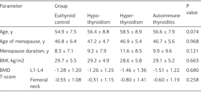
37 (14.2%) were diagnosed with autoimmune thyroiditis. The remaining 126 women (48.3%) were allocated to the control group (Table 1). The patients’ mean age, age of menopause on-set, duration of menopause, and BMI were similar between the four groups (p > 0.05) (Table 1). The BMD T-scores for the L1–4 region and the femoral neck did not difer signiicantly between the groups (p = 0.680 and p = 0.258, respectively) (Table 1).
Discussion
In the present study, postmenopausal subjects with hypothyroid-ism, hyperthyroidhypothyroid-ism, and autoimmune thyroiditis had BMDs in the L1-L4 and femoral neck regions that were similar to BMD values in euthyroid controls of similar age and menopause his-tory. These indings provide evidence indicating that these thy-roid dysfunction conditions do not disrupt bone metabolism in postmenopausal women.
Thyroid hormones increase bone resorption and bone forma-tion, resulting in an approximately 10% loss in bone mass per remodeling cycle as demonstrated by patients with thyrotoxi-cosis [5]. Therefore, hyperthyroidism a major cause of second-ary osteoporosis [6]. Hypothyroidism reduces bone turnover that afects both bone resorption and formation in a manner Table 1. Mean (±SD) demographic and BMD values were similar between the groups (p > 0.05).
Parameter Group P
value Euthyroid
control
Hypo-thyroidism Hyper-thyroidism
Autoimmune thyroiditis
Age, y 54.9 ± 7.5 56.4 ± 8.8 58.5 ± 8.9 56.6 ± 7.9 0.074
Age of menopause, y 46.8 ± 6.4 47.2 ± 4.7 46.9 ± 5.4 46.7 ± 5.6 0.968
Menopause duration, y 8.3 ± 7.1 9.2 ± 7.9 11.6 ± 8.5 9.9 ± 9.6 0.121
BMI, kg/m2 29.7 ± 5.5 29.2 ± 4.9 28.6 ± 5.8 29.1 ± 5.2 0.663
BMD
T-score L1-L4Femoral -1.28 ± 1.20 -1.26 ± 1.25 -1.46 ± 1.36 -1.51 ± 1.22 0.680
neck
-0.55 ± 1.08 -0.31 ± 1.15 -0.80 ± 1.41 -0.60 ± 1.19 0.258
| Journal of Clinical and Analytical Medicine 26
Tiroid Disfonksiyonunun Kemik Mineral Yoğunluğuna Etkisi / The Efect of Thyroid Disfunction on the Bone Mineral Density
3 that is similar to hyperthyroidism. The bone formation phase in
hypothyroidism is prolonged by the lengthened mineralization phase [7].
Subclinical hyperthyroidism (suppressed TSH in the presence of normal T3 and T4 levels) and TSH suppression by an overdose of T4 replacement are associated with reduced BMD and frac-ture [8–11]. Recent studies have reported that subclinical hyper-thyroidism can increase the risk of fracture, especially in the hip and the lumbar spine of postmenopausal women [8–12]. Meta-bolic changes related to thyroid hormones can afect BMD and mass [13]. It was reported that hypothyroidism and hyperthy-roidism reduced BMD and increased bone fractures [13]. Thy-rotoxicosis causes expedited growth, increased bone age, and reduced bone mass. On the other hand, hypothyroidism causes insuicient osteogenesis and growth failure [14].
Thyroid dysfunction is a common problem in perimenopausal and postmenopausal women [8,9,13]. Therefore, it may be im-portant to emphasize TSH measurement for postmenopausal women with low BMD, even if thyroid disease is not present [15]. Risk of osteoporosis has been reported to be increased in postmenopausal patients, and further increased in those with thyroid diseases [14] Yet the occurrence of vertebra fractures appears to be independent of age and BMD in postmenopausal women with low TSH values [14–16]. It was reported that the risk of new hip and vertebral fractures is increased in with low TSH levels, and that normalization of TSH levels as a result of thyroid hormone therapy can prevent that increase in risk [8]. A previous study investigating associations between bone sta-tus and levels of TSH and f-T4 in postmenopausal women ater long-term treatment for thyroid disorders suggested that TSH levels might support regulation of BMD [18]. Murphy et al. [15] analyzed the relationship between physiological variation in nor-mal thyroid status and BMD, as well as the relationship between thyroid status and non-vertebral fracture, in healthy postmeno-pausal women. They determined that higher levels of f-T3 and f-T4 were associated with increased bone loss and lower BMD in the hip. Indeed, BMD correlates positively with TSH in healthy postmenopausal women receiving T4 or drugs that afect bone metabolism and osteoporosis is more frequently observed in women with low to normal levels of TSH . However, BMD has been reported to be independent of T4 levels [17].
In a large population-based, cross-sectional study of women over 40 years of age, the prevalence of osteoporosis as deter-mined by ultra-distal forearm BMD measurements was found to be higher in women who self-reported hyperthyroidism (N = 944) than in women without self-reported thyroid disease (N = 5778) conirmed by serum TSH levels. Moreover, the women with the lowest TSH levels (<0.5 mIU/L) had lower forearm BMD measurements [20]. Yet the putative relationship between thy-roid hormone levels and bone loss or BMD remains controversial as other researchers could not ind any signiicant associations when they investigated these associations [23]. Interestingly, Grimnes et al. [21] found that abnormally high TSH levels (>4.6 mIU/L) were associated with femoral neck BMD above that ob-served in euthyroid women, while an association between TSH levels and BMD did not hold in women with normal TSH levels and BMD values.
Van der Deure et al. [22] performed a longitudinal investigation
of 151 men and women who were ≥55 years of age for a period 8.7 years. No associations were identiied between f-T4 or TSH levels with fracture risks in subjects with nonthyroidal illness. There were also no associations between patients who received drugs that afected bone metabolism. BMD correlated inversely with f-T4 levels and positively with TSH levels, although the correlation with f-T4 was stronger. Bauer et al. [8] determined that low thyrotropin levels were not associated with bone loss in postmenopausal women. This study group performed another prospective cohort study in 2001 that determined exogenous thyroid hormone therapy was not a risk factor for fracture in women with normal serum TSH concentrations. However, wom-en with low serum TSH levels were at an increased risk for new hip and vertebral fractures [24]. A study conducted by Pala et al. [23] showed there was no signiicant correlation between BMD and TSH values in healthy, postmenopausal women. This ob-servation indicated that TSH value was not a good marker for evaluating BMD.
In conclusion, the present study indicated that thyroid dys-function does not signiicantly afect BMD in postmenopausal women with hyperthyroidism, hypothyroidism, or autoimmune thyroiditis. This result suggests that thyroid dysfunctions do not have a signiicant role in the development of osteoporosis dur-ing the postmenopausal period, perhaps because there may be other mechanisms at work that blunt or mask the efects of thyroid hormones.
Competing interests
The authors declare that they have no competing interests.
References
1. Kanis JA. Diagnosis of osteoporosis. Osteoporos Int 1997;7(Suppl 3):108-16. 2. Galliford TM, Murphy E, Williams AJ, Bassett JH, Williams GR. Efects of thyroid status on bone metabolism: a primary role for thyroid stimulating hormone or thyroid hormone? Minerva Endocrinol 2005;30(4):237-46.
3. Andersen S, Bruun NH, Pedersen KM, Laurberg P.Biologic variation is important for interpretation of thyroid function tests.Thyroid 2003;13(11):1069-78. 4. Andersen S, Pedersen KM, Bruun NH, Laurberg P.Narrow indivicual variations in serum T(4) and in T(3) in normal subjects : a clue to the understanding of subclini-cal thyroid disease. J Clin Endocrinol Metab 2002;87(3):1068-72.
5. Mosekilde L, Eriksen EF, Charles P. Efects of thyroid hormones on bone and mineral metabolism. Endocrinol Metab Clin North Am 1990;19(1):35-63. 6. Akalin A, Colak O, Atalas O, Efe B .Bone remodeling markers and serum cyto-kines in patients with hyperthyroidism. Clin Endocrinol 2002;57(1):125-9. 7. Eriksen EF , Mosekilde L, Melsen F .Kinetics of trabecular bone resorption and formation in hypothyroidism:evidence for a positive balance per remodeling cycle. Bone 1986;7(2):101-8.
8. Bauer DC, Ettinger B,Nevitt MC, Stone KL . Risk for fracture in women with low serum levels of thyroid-stimulating hormone. Ann Intern Med 2001;134(7):561–8. 9. Cummings SR, Nevitt MC, Browner WS, Stone K, Fox KM, Ensrud KE et al.Risk factors for hip fracturein white women. Study of Osteoporotic Fractures Research Group. N Engl J Med 1995;332(12):767–73.
10. Heemstra KA, Hamdy NA, Romijn JA, Smit JW.The efects of thyrotropin-sup-pressive therapy on bone metabolism in patients with well-diferentiated thyroid carcinoma. Thyroid 2006;16(6):583–91.
11. GuoCY, Weetman AP, EastellR. Longitudinal changes of bone mineral density and bone turnover in postmenopausal women on thyroxine. Clin Endocrinol (Oxf) 1997;46(3):301–7.
12. Jamal SA, Leiter RE, Bayoumi AM, Bauer DC, Cummings SR . Clinical utility of laboratory testing in women with osteoporosis. Osteoporos Int 2005;16(5):534– 40.
13. Dhanwal DK. Thyroid disorders and bone mineral metabolism. Indian Journal of Endocrinology and Metabolism 2011;15(Suppl 2):107-11.
14. Pearce EN. Thyroid dysfunctionin perimenoupausal and postmenoupausal women. Menopause Int 2007;13(1):8-13.
15. Murphy E, Williams GR. The thyroid and the skeleton. Clin Endocrinol (Oxf) 2004;61(3):285–98.
16. Mazziotti G, Porcellia T, Patellia I, Vescovib PP, Giustinaa A. Serum TSH values and vertebra fructure risk in normal thyroid functioned postmenopausal women with low bone mineral density. Bone 2010;46(3): 94-100.
Journal of Clinical and Analytical Medicine | 27
Tiroid Disfonksiyonunun Kemik Mineral Yoğunluğuna Etkisi / The Efect of Thyroid Disfunction on the Bone Mineral Density
4
17. Morris MS . The association between serum thyroid-stimulating hormone in its reference range and bone status in postmenopausal American women. Bone 2007;40(4):1128–34.
18. Baqi L, Payer J, Killinger Z, Hruzikova P, Cierny D, Susienkova K et al. The level of TSH appeared favourable in maintaining bone mineral density in postmeno-pausal women. Endocr Regul 2010;44(2):57-63.
20. Svare A, Nilsen TI, Bjøro T, Forsmo S, Schei B, Langhammer A. Hyperthyroid levels of TSH correlate with low bone mineral density: the HUNT 2 study. Eur J Endocrinol 2009;161(5):779-86.
21. Grimnes G, Emaus N, Joakimsen RM, Figenschau Y, Jorde R. The relationship between serum TSH and bone mineral density in men and postmenopausal wom-en: the Tromsø study. Thyroid 2008;18(11) :1147-55.
22. Van der Deure WM, Uitterlinden AG, Hofman A, Rivadeneira F,Pols HA, Peeters RP et all.Efects of serum TSH and FT4 levels and the TSHR-Asp727Glu polymor-phism on bone: the Rotterdam Study. Clin Endocrinol (Oxf) 2008;68(2):175–81. 23. Pala HG, Acar B, Altunyurt S, Arık H. Bone Mineral Density in Postmenopausal Healthy Women – Thyroid Stimulant Hormon Relation. DEÜ Tıp Medical Faculty Journal 2008; 22(1) :1-7.
24. Bauer DC, Nevitt MC, Ettinger B, Stone K . Low thyrotropin levels are not associated with bone loss in older women: a prospective study. J Clin Endocrinol Metab 1997;82(9):2931–6.
| Journal of Clinical and Analytical Medicine 28