Infectious after the Emergence of Anatomically Modern
Humans
Aashish R. Jha1*, Douglas F. Nixon2, Michael G. Rosenberg3, Jeffrey N. Martin4, Steven G. Deeks4, Richard R. Hudson5, Keith E. Garrison6., Satish K. Pillai7
*.
1Department of Human Genetics, University of Chicago, Chicago, Illinois, United States of America,2Division of Experimental Medicine, University of California San Francisco, San Francisco, California, United States of America,3Albert Einstein College of Medicine, Bronx, New York, United States of America,4HIV/AIDS Division, Department of Medicine, San Francisco General Hospital, University of California San Francisco, San Francisco, California, United States of America,5Department of Evolution and Ecology, The University of Chicago, Chicago, Illinois, United States of America,6Department of Biology, Saint Mary’s College of California, Moraga, California, United States of America,7Division of Infectious Diseases, Department of Medicine, San Francisco Veterans Affairs Medical Center, University of California San Francisco, San Francisco, California, United States of America
Abstract
HERV-K113 and HERV-K115 have been considered to be among the youngest HERVs because they are the only known full-length proviruses that are insertionally polymorphic and maintain the open reading frames of their coding genes. However, recent data suggest that HERV-K113 is at least 800,000 years old, and HERV-K115 even older. A systematic study of HERV-K HML2 members to identify HERVs that may have infected the human genome in the more recent evolutionary past is lacking. Therefore, we sought to determine how recently HERVs were exogenous and infectious by examining sequence variation in the long terminal repeat (LTR) regions of all full-length HERV-K loci. We used the traditional method of inter-LTR comparison to analyze all full length HERV-Ks and determined that two insertions, HERV-K106 and HERV-K116 have no differences between their 59and 39LTR sequences, suggesting that these insertions were endogenized in the recent evolutionary past. Among these insertions with no sequence differences between their LTR regions, HERV-K106 had the most intact viral sequence structure. Coalescent analysis of HERV-K106 39LTR sequences representing 51 ethnically diverse individuals suggests that HERV-K106 integrated into the human germ line approximately 150,000 years ago, after the emergence of anatomically modern humans.
Citation:Jha AR, Nixon DF, Rosenberg MG, Martin JN, Deeks SG, et al. (2011) Human Endogenous Retrovirus K106 (HERV-K106) Was Infectious after the Emergence of Anatomically Modern Humans. PLoS ONE 6(5): e20234. doi:10.1371/journal.pone.0020234
Editor:Michael Hendricks, Harvard University, United States of America ReceivedJanuary 25, 2011;AcceptedApril 19, 2011;PublishedMay 25, 2011
Copyright:ß2011 Jha et al. This is an open-access article distributed under the terms of the Creative Commons Attribution License, which permits unrestricted use, distribution, and reproduction in any medium, provided the original author and source are credited.
Funding:This work was supported by National Institutes of Health grants AI076059, AI060379, AI084113, UCSF CFAR (P-30 AI27763), UCSF CTSI (UL1 RR024131), CNICS (R24 AI067039), NIAID (K24AI069994), and 1K01DA024654 grants. The funders had no role in study design, data collection and analysis, decision to publish, or preparation of the manuscript.
Competing Interests:The authors have declared that no competing interests exist. Dr. Douglas Nixon is a section editor for PLoS ONE. This manuscript was handled by a different section editor.
* E-mail: [email protected] (ARJ); [email protected] (SKP)
.These authors contributed equally to this work.
Introduction
Endogenous retroviruses (ERVs) are the fossilized, germ line-integrated remnants of exogenous retroviruses and comprise approximately 8% of the human genome [1,2]. Upon integration into germ cells, ERVs are transmitted in a Mendelian fashion, accumulating mutations over time that erode and ultimately eliminate their original viral sequence structure. Just as ERV genomes are subjected to modification within the host, host-ERV interactions may also induce changes in the host genome. For example, homologous recombination between distant HERV loci have likely induced chromosomal rearrangements in the human host, and coevolutionary conflicts with the exogenous precursors of HERVs may have resulted in sequence modifications in human antiretroviral genes such as APOBEC3H and TRIM5a [3,4,5].
Thus, identifying HERVs that integrated into the human genome in the recent evolutionary past may allow us to catalog proviruses that influenced the evolution of genes involved in antiviral
Results
The human genome consists of numerous HERV insertions that contain 59and 39LTRs and varying remnants of the viral coding genes in between. Many more insertions currently exist as solo-LTRs, which are the remnants of full-length insertions that have been truncated into a single LTR by virtue of inter-LTR recombination events. Inter-LTR comparison is the standard method used to estimate the age of full-length HERV insertions. At the time of insertion, the LTR sequences are presumed to be identical and the differences between the 59and 39 LTRs of the same provirus are believed to arise due to substitutions accumulating in a clocklike manner post-insertion [18]. To elucidate the insertion timeline of the human-specific endogenous retroviruses into the human genome, we obtained the complete genome sequences of all the human-specific full-length HERV-K insertion previously reported [15,16] using the UCSC genome browser (hg18).
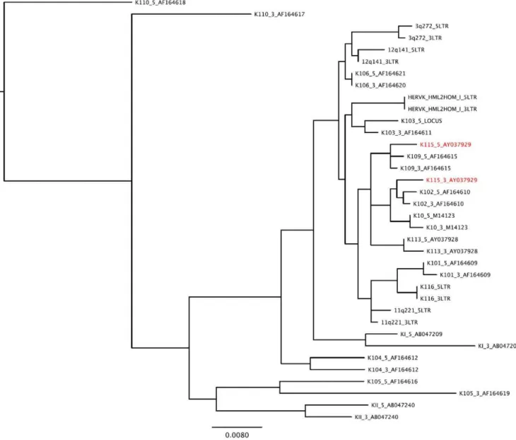
It has previously been hypothesized that gene conversion may be rampant amongst HERV loci [19]. Pervasive gene conversion would prevent us from applying a molecular clock to these data, since observed sequence variation between LTR regions would be attributed to recombination rather than the stepwise accumulation of mutations predicted by the neutral theory of evolution [20]. We reconstructed a maximum likelihood phylogeny of all HERV-K LTRs to determine the prevalence of gene conversion between insertions in the HERV-K family (Figure 1). The clustering of the 59and 39LTR sequences associated with each insertion locus in our phylogeny suggests that gene conversion is rare among HERV-Ks, with the single exception of HERV-K115. Evidence of a gene conversion event in HERV-K115 has been reported previously [17,19].
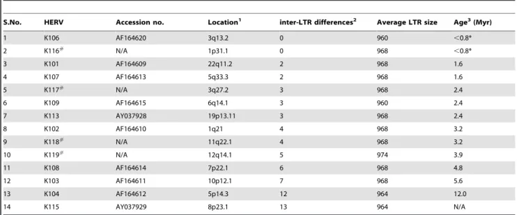
To estimate the insertion times of each human-specific endogenous retrovirus, we calculated the age of each human-specific full-length HERV-K insertion using the traditional inter-LTR comparison method [18]. Inter-inter-LTR divergence measure-ments of the fourteen insertions that did not undergo gene conversion in the LTR region were converted to insertion age estimates by applying an established HERV-K LTR-specific divergence rate of 0.13% per million years (Myr) to these data [21] (Table 1). Based on this method, HERV-K1p31.1 (referred to as HERV-K116 from here onwards) and HERV-K106 have the highest probability of being the youngest full-length endogenous retroviruses in the human genome. Due to the perfect identity between their 59 and 39 LTR sequences, an inter-LTR comparison is only informative to calculate a maximum, upper-bound age estimate. A single mutation is predicted to arise within an LTR every 0.8 Myr based on the divergence rate of 0.13%/ Myr. Having no sequence differences between their LTRs, inter-LTR analysis of HERV-K116 and HERV-K106 suggests that both insertions must be younger in age than the time required for one sequence polymorphism to arise between the two LTRs, or 0.8 Myr.
We used Multalin [22] to align HERV-K113, K115, K106, and K116 with experimentally reconstituted infectious HERVs KCON [23] and Phoenix [24] that are based on artificial consensus sequences of full-length human-specific HERV-K (HML-2) insertions. We observed that while all four HERV insertions (K113, K115, K106, and K116) exhibited similarities to the reconstituted viruses, all four contained mutations that were unique to each insertion (Figure S1). We observed that both HERV-K106 and HERV-K116 are members of the type I HERV-K family as evidenced by the presence of a 292 bp ‘deletion’ in env which is the signature of all type I HERV
members. The presence of this 292 bp env deletion in multiple HERV-K type I members suggests that this deletion may have been present in the infectious ancestral precursors of these viruses and probably does not render a HERV insertion dysfunctional on its own. However, HERV-K116 also has a 2846 bp deletion in its pol sequence [16]. In contrast, HERV-K106 exhibits relatively intact retroviral genome architecture (Figure 2A). Thus, these data suggest that HERV-K106 is the youngest endogenous retrovirus that survives largely intact in the human genome.
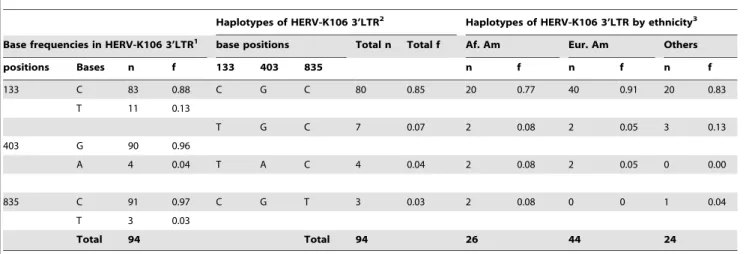
While inter-LTR comparison is a useful tool to estimate the age of HERV insertions, it becomes less informative with fewer mutations between the LTR regions and provides only an upper bound age estimate in the absence of sequence differences between the LTR regions. We recently developed an alternative dating method to infer insertion age when the inter-LTR method is inapplicable (to estimate the insertion dates of solo-LTRs and HERV loci such as K115 that show evidence of gene conversion) [25]. HERV insertions with identical 59and 39LTR sequences represent another scenario in which our alternative method is useful. We applied this method to the HERV-K106 insertion to derive a more precise estimate of its age. Our approach involves the application of coalescent inference to inter-host sequence variation in one of the proviral LTR sequences to estimate HERV insertion age. We generated complete HERV-K106 39LTR sequences from 51 individuals representing various ethnicities and three different geographical locations within the United States (Table 2). PCR amplification and sequencing of the HERV-K106 LTR revealed three single nucleotide polymorphic positions (SNPs): 133, 403, and 835 (numbered according to their position in the GenBank reference sequence AF164620) (Table 2). Four HERV-K106 haplotypes could be constructed based on the SNPs identified in the 39LTR region (Figure 2B).
The coalescent estimation of insertion age rests on the assumption that the insertion site is evolving neutrally. Therefore, we conducted tests on the HERV-K106 insertion site to determine whether the assumption of neutrality was maintained. We performed 10,000 coalescent simulations using MS software [26] and Schaffner’s calibrated model of human genome evolution [27] to calculate the probability of observing exactly 3 SNPs in a neutrally evolving, 960 bp stretch of human DNA. We assumed no recombination, and an inferred human mutation rate of 9.061029subsitutions per site per generation. These simulations yielded a Gaussian mutational probability distribution with a mean of 8 SNPs, and revealed that the presence of only 3 SNPs in a 960 bp neutrally evolving region of the human genome deviates significantly from expectations based on Schaffner’s model (p,0.05). These findings suggest one of two possibilities about HERV-K106. Either it is evolving under selection, or it is evolving neutrally but has not evolved in tandem with the human genome for a sufficient amount of time to conform to the predictions of Schaffner’s human genome-based model. We explored the selection hypothesis by performing standard tests of neutrality on the K106 locus, including Tajima’s D [28], and Fu and Li’s D* and H [29] (performed using DNASP [30]). In all cases, we could not reject the null hypothesis that HERV-K106 is evolving neutrally (p.0.10). HERV-K106 itself may have been evolving neutrally but could have been driven to fixation due to hitchhiking effects if the region flanking the insertion had been under positive selection. We used the HGDP Selection Browser [31] to test whether the genomic region containing the HERV-K106 insertion site is under selection by calculating the iHS and
XP-EHH statistics on genotypes in the Human Genome Diversity Panel. The iHS and XP-EHH statistics are haplotype homozygosity-based tests used to detect signatures of recent selection on variants that have not yet reached fixation [32] and can be applied to detect selective sweeps in alleles that have approached fixation in one population but are polymorphic in the overall human population [33]. Even though HERV-K106 is fixed in all humans, the genetic region flanking the K106 insertion may contain SNPs that could reveal if a selective sweep has occurred in this region. We found that HERV-K106 is incorporated into a genomic location with only a few genes nearby (Figure S2) and the region in chromosome 3 containing HERV-K106 exhibited no signatures of selection, as both iHS and XP-EHH did not yield extreme values (Figure S3). These data collectively support that K106 is evolving neutrally and has only shared its evolutionary history with the human genome for a relatively short period of time.
We constructed a maximum likelihood phylogeny of all 94 observed HERV-K106 39LTR haplotype sequences to estimate the age of the K106 insertion (Figure S4). According to coalescent theory, the genetic distance to the inferred most recent common ancestor (MRCA) should reflect the time that has elapsed since the establishment of the ancestral sequence, and in this particular case, the age of the proviral insertion itself. We used two previously reported evolutionary rates to translate genetic distance into coalescence time. The upper-bound for the coalescence-based age estimate was inferred using the HERV-K LTR specific mutation rate of 1.361029 mutations/site/year [21], and the inferred
mammalian genome mutation rate of 2.261029mutations/site/
year [34] was used to calculate a lower-bound estimate. Based on the two divergence rates, we estimate that HERV-K106 was integrated into the human genome between 91,000 and 154,000 years ago, after the emergence of anatomically modern humans [35,36].
Figure 1. Phylogenetic tree of full-length HERV-K (HML-2) LTR sequences.The clustering of the HERV-K 59and 39LTR sequences of each insertion suggests that gene conversion is rare in the HERV-K family. Each HERV is indicated by its name (K106 for HERV-K106). HERV taxa that may have undergone gene conversion are indicated in red. HERV-K115 59LTR clusters with HERV-K109 suggesting a gene conversion occurred between these two HERV-K members. HERV-K10, HERV-K HML2HOM, and HERV-K110 are sometimes referred to as HERV-K107, HERV-K108, and HERV-K18, respectively. HERV-K110 was used to root the phylogeny because it is present in both Humans and Gorillas [16].
Discussion
Our study suggests that HERV-K106 is a recent acquisition in the human genome and is possibly specific toHomo sapiens sapiens. K106 is fixed in humans, but K113 and HERV-K115 remain insertionally polymorphic [17], despite being inserted into the human genome several hundreds of thousands of years before K106 [25]. Features associated with the three regions of the human genome harboring these HERV insertions may help to explain the apparent discordance between insertion age and fixation. The fixation probability of an ERV may be inversely correlated with local chromosomal recombination rate and local gene density [37,38]. We employed the high resolution recombination map of Kong et al. [39] to examine local recombination rates in the genomic neighborhoods of HERV-K106, K113 and K115 insertions. Analysis of the map revealed that HERV-K106 lies in one of only nineteen recognized ‘‘recombinaton deserts’’ in the human genome (crossover rates less than 0.3 c/Mb), which may have accelerated its fixation. Conversely, K113 and K115 reside in regions that are considerably more recombinogenic (1.18 cM/Mb and 2.14 cM/ Mb, respectively) [39]. The low rate of recombination in the region surrounding K106 may have accelerated its fixation, or the high rates of recombination in the regions surrounding K113 and K115 may have decelerated the fixation of these older insertions. We next employed the NCBI Human Genome Map Viewer [40] to investigate the potential role of gene density in the neighborhood of these three HERV insertions, since a highly significant inverse correlation between local gene density and HERV fixation rate has been demonstrated [37]. The insertionally polymorphic HERV-K113 resides in chromosome 19, which has the highest gene density of all human chromosomes. We surveyed regions within a window of 1 Mb upstream to 1 Mb downstream of each HERV insertion for the presence of characterized genes to
obtain a more localized estimate of gene density. HERV-K106, K113 and K115 are flanked by 32, 64, and 115 known genes, respectively. Again, the low gene density in the region surrounding K106 may have accelerated its fixation, or the higher gene densities in the regions surrounding K113 and K115 may have hindered the fixation of these older insertions. Taken together, the patterns of local recombination rate and gene density support genomic context as a primary determinant of fixation that explains the discordance between insertion age and insertional polymor-phism in the human population observed within the HERV-K family.
There are many fixed and insertionally polymorphic HERV-K(HML-2) solo-LTRs in the human genome [16,41,42] and it is possible that some of these unfixed solo-LTRs may have been derived from proviral insertions that are younger than HERV-K106. However, the absence of a second LTR from these insertions prevents us from including them in our current investigation because we relied on the inter-LTR comparisons to identify HERVs that recently integrated in the human genome. A second caveat stems from the reliance of our age estimates on previously established mutation rates; changes in local mutation or recombination rate may influence the apparent age of this insertion. However, our analyses of the local recombination rates surrounding the K106 and K113 insertions suggest that this is not likely to be a confounding factor. Furthermore, we previously hervotyped a small subset of individuals in our samples (n = 16) to study haplotype diversity in HERV-K113 [25]. Haplotype diversity for K113 in this smaller subset of samples was higher than the haplotype diversity that we observed for HERV-K106 in the same subset, as would be expected when comparing an older to a younger insertion. The frequencies of the K113 minor haplotypes in individuals of both African and European descent were much higher than the frequencies of minor haplotypes that we observed for HERV-K106 (Figure 3).
Table 1.Human specific complete HERV-K (HML-2) proviruses within the human genome.
S.No. HERV Accession no. Location1 inter-LTR differences2 Average LTR size Age3(Myr)
1 K106 AF164620 3q13.2 0 960 ,0.8*
2 K116#
N/A 1p31.1 0 968 ,0.8*
3 K101 AF164609 22q11.2 2 968 1.6
4 K107 AF164613 5q33.3 2 968 1.6
5 K117#
N/A 3q27.2 3 968 2.4
6 K109 AF164615 6q14.1 3 960 2.4
7 K113 AY037928 19p13.11 3 968 2.4
8 K102 AF164610 1q21 4 968 3.2
9 K118# N/A 11q22.1 4 968 3.2
10 K119#
N/A 12q14.1 5 974 3.9
11 K108 AF164614 7p22.1 6 968 4.8
12 K103 AF164611 10p12.1 7 968 5.6
13 K104 AF164612 5p14.3 12 964 12.0
14 K115 AY037929 8p23.1 13 964 N/A
Table 1 Notes
1The identity and location of human specific complete HERV-K (HML-2) proviruses within the human genome were obtained from previous reports [15,16].#
HERV-K116, K117, K118 and K119 were previously referred to by their genomic locations.
2Three HERV-K members have as many differences as the insertionally polymorphic HERV-K113 and four HERV-K members have fewer differences between their LTR than HERV-K113 of which, HERV-K106 and HERV-K116 have identical LTR, however, the latter has a 2846 bp deletion in thepolregion.
3
All age estimates are based on inter-LTR comparisons and are in million years (Myr). *Age estimates for HERV-K106 and HERV-K116 are based on 1 SNP between their LTRs. The age of K115 is listed as ‘N/A’ because it cannot be determined by inter-LTR comparison method. The age of K115 was previously estimated to be at least 1.1 Myr using coalescent approach [25].
doi:10.1371/journal.pone.0020234.t001
To the best of our knowledge, this is the first rigorous evidence that the infectious progenitors of human endogenous retroviruses were active after the origin of anatomically modern man,Homo sapiens,in Africa [35,36].
Materials and Methods
Samples and DNA extraction
Peripheral blood samples were obtained with written consent from a total of 51 individuals drawn from the pediatric HIV cohort at Jacobi Medical Center (Bronx, NY), the SCOPE cohort of chronic HIV-1 infection (San Francisco, CA), and from blood donors to the Stanford Blood Bank (Palo Alto, CA). Twelve participants were perinatally HIV-1 infected children followed at Jacobi Medical Center, Bronx, NY. Twenty-five samples of HIV-1 infected adults were obtained from the SCOPE cohort in San Francisco, California and fourteen individuals from Stanford Blood Bank were anonymous, healthy HIV-1 negative individuals. The study was approved by the Institutional Review Boards at Jacobi Medical Center and the University of California at San Francisco (UCSF). All partici-pants self-identified their ethnicities. HERV-K106 hervotyping [25] and haplotype construction for this study was carried out
without prior knowledge of the ethnicity of the participants. Genomic DNA was isolated according to the manufacturer’s protocol using QIAmp DNA Blood Mini Kit (Qiagen, Valencia, California, USA).
PCR and gel electrophoresis
Insertion sites, solo-LTR, and full length HERV-K106 were detected using the HERV-K106 specific primers 59 -GGTGTTGCTGTGGAAGGTATTC-39and 59 -TCCATGGC-TATCCACGAGA-39and the two step PCR protocol previously described [15,16]. Neither insertional polymorphism nor solo-LTR formation was detected in our sample. We designed two primers HERV-K1063LTR1 59 -ATTTGGTGCCAGGAACT-GAG-39and HERV-K1063LTR2 59 -AAGAAAAGGGGGAAA-TGTGG-39 that were specific to the HERV-K106 39LTR for sequencing purposes. HERV-K1063LTR1 annealed to the host flanking DNA upstream of HERV-K106 insertion in chromo-some 3 and HERV-K1063LTR2 bound downstream of the 39LTR to theenvgene of HERV-K106. PCR was performed in a volume of 50 uL with 25 ng of genomic DNA in 1.0 uM of each primer, 200 uM of each dNTP, 25 mM of MgCl2, and 0.5 units
of Taq DNA polymerase obtained from Applied Biosystems, Foster City, California, USA. PCR was conducted with 10
Figure 2. Genome organization and haplotypes of HERV-K106.Figure 2A: Genomic characterization of HERV-K106 demonstrating the two LTRs, gag, pol, and env genes. The HERV-K106 genome was annotated with the aid of the HERV-K consensus sequence (HERV-KCON) [23], two HERV-K HML2 members that have somewhat functionalgag(HERV-K101 and HERV-K109) [43], and insertionally polymorphic HERV-K113 that has intact ORF [17]. Known functional elements within the HERV-K106 LTR are shown in colored boxes. All SNP positions in the coding genes are counted from the beginning ofgagORF. Five nonsynonymous SNPs shown ingagregion are HERV-K106 specific. These SNPs do not include I516M mutation which singlehandedly eliminates functionality of HERV-K113gag[43] warranting the future investigation of functionality of K106 gag. Although K106 lacks this vital SNP it harbors one base deletion at position 1977 which causes a frameshift near the end of thegaggene. Whether this frameshift causes gag to be dysfunctional is unknown. The HERV-K106 polymerase gene (pol) that includes reverse transcriptase is distinct from the other HERV-K family members used in our sequence comparison. In addition to the SNP shown, it harbors a 5 bp deletion from 4849–4853 bp that results in a frameshift mutation. The large 292 base deletion beginning at 5392 bp of pol extending into the env gene is the signature of type-I HERV-K [16]. In addition to the 292 bp deletion, HERV-K106envgene has a premature stop codon. ( ) indicate stop codons in HERV-K106 genome. Figure 2B: SNPs and surrounding bases in 39LTR of HERV-K106 demonstrating four haplotypes. Each haplotype is listed on the left, each SNP is represented in red and the position containing the SNP is highlighted in yellow.
minutes of initial denaturing at 95uC, 30 cycles of 10 seconds at 94uC, 30 seconds at annealing temperature (61.5uC), and 59 seconds of elongation at 72uC followed by a 10 min final extension at 72uC. Amplified PCR products were stored at 4uC. The amplified PCR products were detected as,1200 bp bands
in 2% agarose gel. A low-mass DNA ladder (Invitrogen, Carlsbad, California, USA) was used to verify the product size (Figure S5).
Sequencing and sequence analysis
Sequencing was performed at MCLAB, South San Francisco using an ABI 3730XL sequencer. To determine an individual’s hervotype (HERV genotype) for HERV-K106, sequences were aligned and single nucleotide polymorphisms (SNPs) were identified using Sequencher version 4.9 [25]. To rule out errors introduced during the PCR or sequencing process, PCR was performed twice on each genomic DNA sample and SNPs were
Table 2.Base frequencies and haplotypes of HERV-K106 3’LTR with haplotype frequencies in various ethnic groups within the United States.
Haplotypes of HERV-K106 3’LTR2 Haplotypes of HERV-K106 3’LTR by ethnicity3 Base frequencies in HERV-K106 3’LTR1 base positions Total n Total f Af. Am Eur. Am Others
positions Bases n f 133 403 835 n f n f n f
133 C 83 0.88 C G C 80 0.85 20 0.77 40 0.91 20 0.83
T 11 0.13
T G C 7 0.07 2 0.08 2 0.05 3 0.13
403 G 90 0.96
A 4 0.04 T A C 4 0.04 2 0.08 2 0.05 0 0.00
835 C 91 0.97 C G T 3 0.03 2 0.08 0 0 1 0.04
T 3 0.03
Total 94 Total 94 26 44 24
Table 2 Notes
1Alleles at all three SNP sites in HERV-K106 39LTR consisted of two alternate nucleotide bases with one being predominant than the other (133: C.T, 403: G.A, and 835: C.T). Using these SNP four haplotypes of HERV-K106 could be constructed.
2The most common haplotype was C-G-C (f = 0.90) followed by T-A-C (f = 0.05), C-G-T (f = 0.02) and T-G-C (f = 0.02).
377% of African American and 91% European Americans in our study had CGC haplotype of HERV-K106 LTR. CGC was also the most prevalent in a heterogeneous sample labeled as ‘others’ that consisted of a few (n,5 from each group) individuals of Hispanic, Asian, and East Indian origins. Haplotypes TGC and TAC were present in 8% and 5% of African Americans and European Americans respectively whereas haplotype CGT was present in 5% of African Americans but was absent in European Americans. African Americans, although in lower numbers in our study, demonstrated greater haplotype diversity than European Americans which is consistent with the ‘‘out of Africa’’ hypothesis that HERV-K106 originated in Africa and migrated out of Africa with human migration.
doi:10.1371/journal.pone.0020234.t002
Figure 3. Comparison of haplotype frequencies between HERV K106 and HERV-K113.Haplotypes and haplotype frequencies of HERV K113 and K106 from individuals in the same sample set are shown. Haplotypes and haplotype frequencies of K113 were obtained from our previous study [25]. Higher minor haplotype frequencies (MHFs) were observed for K113 (18%, 9%, 9%) compared to K106 (8%, 4%, 3%) even though the numbers of K113 LTRs sequenced was much smaller than that compared to K106. Higher K113 MHFs in a subset of our samples compared to that of K106 suggests that K106 integrated into the human genome much later than K106 such that MHFs of K106 have not had enough time to reach higher frequencies in global populations. This serves as an additional evidence for recent insertion integration time of HERV-K106.
doi:10.1371/journal.pone.0020234.g003
verified by bidirectional sequencing of the amplicons produced by the two independent PCR on the genomic DNA. There were ten individuals who were heterozygous at the SNP sites. Six were heterozygous at only one site and the haplotypes of these six individuals could be inferred. The remaining four individuals were heterozygous at more than one position and were excluded from the analysis since their haplotypes could not be recon-structed. After excluding the ambiguous heterozygotes, we analyzed the base frequencies at the three SNP sites of the HERV-K106 39LTR in 102 HERV-K106 sequences. The base frequencies and SNP positions are summarized in table 2. We also compared HERV-K106 diversity between various ethnicities in the USA represented in our sample. While African Americans (n = 13) and European Americans (n = 22) were abundant in our sample, other ethnic groups such as Asians, Hispanics, East Indians, and Native Americans were represented in very low numbers (n,5) in our sample. Thus, we compared HERV-K106 haplotype frequencies in African Americans and European Americans and the rest of the ethnicities were grouped together as ‘others’.
Coalescence and phylogenetic inference
Coalescence time was calculated as described previously [25]. The sequence of the most recent common ancestor (MRCA) of the HERV-K106 insertion was inferred using maximum likelihood inference and K110 (GenBank Accession number AF164617) as an outgroup taxon (Figure S4). Coalescence (insertion) times were approximated using the formula T~ð1=nPDiÞ=R where T is
the time (My) passed since the insertion event,nis the number of observed taxa,Dis the divergence associated with a descendant taxon (cumulative branch lengths between observed haplotype and inferred MRCA), andRrepresents one of two previously reported divergence (evolutionary) rates (2.261029 and 1.361029
muta-tions/site/year) used to generate upper and lower bound estimates of insertion times [21,34]. We used measurements of genetic divergence from inferred ancestral 5’ LTR sequences (Figure S4) to estimate the HERV-K106 insertion time.
Supporting Information
Figure S1 Comparison of HERV-K106 genome to K113, K115, K116, KCON and Phoenix.Multalin [22] was used to align K106 genome to that of two full length human specific HERV insertions (K113 and K115). We also included in the alignment two experimentally reconstituted HERVs KCON and Phoenix that are infectious and K116 that has a 2846 bp deletion in its pol gene. We observed that while all four HERV insertions (K113, K115, K106, and K116) exhibited similarities to the reconstituted viruses, all four contained mutations that were unique to each insertion. The identical regions in the genomes of all these HERV insertions are shown in blue. Genomic regions in red indicate regions that vary between at least one of these HERV insertions and ‘–’ indicates deleted regions.
(TIF)
Figure S2 Genomic location of HERV-K106. Top: High resolution genomic location of HERV-K106 showing insertion in
a gene desert. No genes can be seen within,1 kb upstream or
downstream of HERV-K106. Bottom: In a lower resolution genomic location map of the K106 insertion, the immune genes such as CD200R1 and CD200RL1 are visible far upstream of HERV-K106.
(TIF)
Figure S3 Lack of selective sweep on or near HERV-K106 region. The fixation of HERV-K106 in the human population could be due to drift, selection on HERV-K106, or a hitchhiking effect driven by other gene(s). If HERV-K106 reached fixation because it is under selection or due to a hitchhiking effect (i.e., if selection drove a nearby mutation to fixation and that mutation happened to be on a haplotype containing HERV-K106), then the XP-EHH statistic which measures complete selective sweeps should indicate selection acting on or nearby HERV-K106. We searched for signals of selection as measured by iHS and XP-EHH statistics in the samples from Human Genome Diversity Panels using tools on Prof. Johnathan Pritchard’s webpage (http://hgdp.uchicago.edu/). We were unable to detect any signals of selection on or near HERV-K106. We were able to detect signals of selection in regions 500 kb farther away from HERV-K106 insertion region, indicating that selection can be detected in that region but HERV-K106 is not under positive selection.
(TIF)
Figure S4 HERV-K106 39LTR haplotypes.ML tree of four haplotypesof HERV-K106 39LTR. The most prevalent haploty-pewas CGC (green) which is also the haplotypeof the HERV-K106 39LTR in Genbank(AF164620). Minor haplotypes CGT (red), TGC (blue), and TAC (orange) are also shown. All haplotypescluster together. The HERV-K110 39LTR was used as the outgroup.
(TIF)
Figure S5 HERV-K106 39LTR amplification. The two primers specific to HERV-K106 39LTR that we designed (HERV-K1063LTR1 59-ATTTGGTGCCAGGAACTGAG-39and HERV-K1063LTR2 59-AAGAAAAGGGGGAAATGTGG-39) were used to amplify the complete HERV-K106 39 LTR as detected by
,1200 bp bands in 2% agarosegel. Low DNA Mass ladder
(Invitrogen, Carlsbad, California, USA) was used to verify the product size.
(TIF)
Acknowledgments
We thank the academic editor Dr. Michael Hendricks and the anonymous reviewer for helpful comments.
Author Contributions
Conceived and designed the experiments: ARJ SKP KEG DFN. Performed the experiments: ARJ. Analyzed the data: ARJ SKP RRH. Contributed reagents/materials/analysis tools: RRH KEG DFN MGR JNM SGD. Wrote the paper: ARJ DFN MGR SGD KEG SKP.
References
1. Bannert N, Kurth R (2006) The evolutionary dynamics of human endogenous retroviral families. Annu Rev Genomics Hum Genet 7: 149–173.
2. Paces J, Pavlicek A, Paces V (2002) HERVd: database of human endogenous retroviruses. Nucleic Acids Res 30: 205–206.
3. Hughes JF, Coffin JM (2001) Evidence for genomic rearrangements mediated by human endogenous retroviruses during primate evolution. Nat Genet 29: 487–489.
4. OhAinle M, Kerns JA, Li MM, Malik HS, Emerman M (2008) Antiretroelement activity of APOBEC3H was lost twice in recent human evolution. Cell Host Microbe 4: 249–259.
5. Kaiser SM, Malik HS, Emerman M (2007) Restriction of an extinct retrovirus by the human TRIM5alpha antiviral protein. Science 316: 1756–1758. 6. Christensen T (2010) HERVs in Neuropathogenesis. J Neuroimmune
7. Moyes DL, Martin A, Sawcer S, Temperton N, Worthington J, et al. (2005) The distribution of the endogenous retroviruses HERV-K113 and HERV-K115 in health and disease. Genomics 86: 337–341.
8. Contreras-Galindo R, Kaplan MH, Leissner P, Verjat T, Ferlenghi I, et al. (2008) Human endogenous retrovirus K (HML-2) elements in the plasma of people with lymphoma and breast cancer. J Virol 82: 9329–9336.
9. Contreras-Galindo R, Lopez P, Velez R, Yamamura Y (2007) HIV-1 infection increases the expression of human endogenous retroviruses type K (HERV-K) in vitro. AIDS Res Hum Retroviruses 23: 116–122.
10. Garrison KE, Jones RB, Meiklejohn DA, Anwar N, Ndhlovu LC, et al. (2007) T cell responses to human endogenous retroviruses in HIV-1 infection. PLoS Pathog 3: e165.
11. Moyes D, Griffiths DJ, Venables PJ (2007) Insertional polymorphisms: a new lease of life for endogenous retroviruses in human disease. Trends Genet 23: 326–333.
12. Trono D, Van Lint C, Rouzioux C, Verdin E, Barre-Sinoussi F, et al. (2010) HIV persistence and the prospect of long-term drug-free remissions for HIV-infected individuals. Science 329: 174–180.
13. Tristem M (2000) Identification and characterization of novel human endogenous retrovirus families by phylogenetic screening of the human genome mapping project database. J Virol 74: 3715–3730.
14. Glazko GV, Nei M (2003) Estimation of divergence times for major lineages of primate species. Mol Biol Evol 20: 424–434.
15. Barbulescu M, Turner G, Seaman MI, Deinard AS, Kidd KK, et al. (1999) Many human endogenous retrovirus K (HERV-K) proviruses are unique to humans. Curr Biol 9: 861–868.
16. Macfarlane C, Simmonds P (2004) Allelic variation of HERV-K(HML-2) endogenous retroviral elements in human populations. J Mol Evol 59: 642–656. 17. Turner G, Barbulescu M, Su M, Jensen-Seaman MI, Kidd KK, et al. (2001) Insertional polymorphisms of full-length endogenous retroviruses in humans. Curr Biol 11: 1531–1535.
18. Dangel AW, Baker BJ, Mendoza AR, Yu CY (1995) Complement component C4 gene intron 9 as a phylogenetic marker for primates: long terminal repeats of the endogenous retrovirus ERV-K(C4) are a molecular clock of evolution. Immunogenetics 42: 41–52.
19. Belshaw R, Pereira V, Katzourakis A, Talbot G, Paces J, et al. (2004) Long-term reinfection of the human genome by endogenous retroviruses. Proc Natl Acad Sci U S A 101: 4894–4899.
20. Kimura M (1986) DNA and the neutral theory. Philos Trans R Soc Lond B Biol Sci 312: 343–354.
21. Lebedev YB, Belonovitch OS, Zybrova NV, Khil PP, Kurdyukov SG, et al. (2000) Differences in HERV-K LTR insertions in orthologous loci of humans and great apes. Gene 247: 265–277.
22. Corpet F (1988) Multiple sequence alignment with hierarchical clustering. Nucleic Acids Res 16: 10881–10890.
23. Lee YN, Bieniasz PD (2007) Reconstitution of an infectious human endogenous retrovirus. PLoS Pathog 3: e10.
24. Dewannieux M, Harper F, Richaud A, Letzelter C, Ribet D, et al. (2006) Identification of an infectious progenitor for the multiple-copy HERV-K human endogenous retroelements. Genome Res 16: 1548–1556.
25. Jha AR, Pillai SK, York VA, Sharp ER, Storm EC, et al. (2009) Cross-sectional dating of novel haplotypes of HERV-K 113 and HERV-K 115 indicate these proviruses originated in Africa before Homo sapiens. Mol Biol Evol 26: 2617–2626.
26. Hudson RR (2002) Generating samples under a Wright-Fisher neutral model of genetic variation. Bioinformatics 18: 337–338.
27. Schaffner SF, Foo C, Gabriel S, Reich D, Daly MJ, et al. (2005) Calibrating a coalescent simulation of human genome sequence variation. Genome Res 15: 1576–1583.
28. Tajima F (1989) Statistical method for testing the neutral mutation hypothesis by DNA polymorphism. Genetics 123: 585–595.
29. Fu YX, Li WH (1993) Statistical tests of neutrality of mutations. Genetics 133: 693–709.
30. Librado P, Rozas J (2009) DnaSP v5: a software for comprehensive analysis of DNA polymorphism data. Bioinformatics 25: 1451–1452.
31. Browser THS The HGDP Selection Browser.
32. Voight BF, Kudaravalli S, Wen X, Pritchard JK (2006) A map of recent positive selection in the human genome. PLoS Biol 4: e72.
33. Sabeti PC, Varilly P, Fry B, Lohmueller J, Hostetter E, et al. (2007) Genome-wide detection and characterization of positive selection in human populations. Nature 449: 913–918.
34. Kumar S, Subramanian S (2002) Mutation rates in mammalian genomes. Proc Natl Acad Sci U S A 99: 803–808.
35. McDougall I, Brown FH, Fleagle JG (2005) Stratigraphic placement and age of modern humans from Kibish, Ethiopia. Nature 433: 733–736.
36. White TD, Asfaw B, DeGusta D, Gilbert H, Richards GD, et al. (2003) Pleistocene Homo sapiens from Middle Awash, Ethiopia. Nature 423: 742–747. 37. Katzourakis A, Pereira V, Tristem M (2007) Effects of recombination rate on human endogenous retrovirus fixation and persistence. J Virol 81: 10712–10717. 38. Dolgin ES, Charlesworth B (2008) The effects of recombination rate on the distribution and abundance of transposable elements. Genetics 178: 2169–2177. 39. Kong A, Gudbjartsson DF, Sainz J, Jonsdottir GM, Gudjonsson SA, et al. (2002) A high-resolution recombination map of the human genome. Nat Genet 31: 241–247.
40. Viewer NHGM NCBI Human Genome Map Viewer.
41. Belshaw R, Dawson AL, Woolven-Allen J, Redding J, Burt A, et al. (2005) Genomewide screening reveals high levels of insertional polymorphism in the human endogenous retrovirus family HERV-K(HML2): implications for present-day activity. J Virol 79: 12507–12514.
42. Hughes JF, Coffin JM (2004) Human endogenous retrovirus K solo-LTR formation and insertional polymorphisms: implications for human and viral evolution. Proc Natl Acad Sci U S A 101: 1668–1672.
43. Heslin DJ, Murcia P, Arnaud F, Van Doorslaer K, Palmarini M, et al. (2009) A single amino acid substitution in a segment of the CA protein within Gag that has similarity to human immunodeficiency virus type 1 blocks infectivity of a human endogenous retrovirus K provirus in the human genome. J Virol 83: 1105–1114.