Skeletal Muscle in Zebrafish
Tomoaki Fujii1, Shin-ichiro Tsunesumi1, Kiyoshi Yamaguchi1, Sumiko Watanabe2, Yoichi Furukawa1*
1Division of Clinical Genome Research, Advanced Clinical Research Center, Institute of Medical Science, The University of Tokyo, Japan,2Division of Molecular Developmental Biology, Institute of Medical Science, The University of Tokyo, Tokyo, Japan
Abstract
Modifications of histone tails are involved in the regulation of a wide range of biological processes including cell cycle, cell survival, cell division, and cell differentiation. Among the modifications, histone methylation plays a critical role in cardiac and skeletal muscle differentiation. In our earlier studies, we found that SMYD3 has methyltransferase activity to histone H3 lysine 4, and that its up-regulation is involved in the tumorigenesis of human colon, liver, and breast. To clarify the role of Smyd3 in development, we have studied its expression patterns in zebrafish embryos and the effect of its suppression on development using Smyd3-specific antisense morpholino-oligonucleotides. We here show that transcripts ofsmyd3were expressed in zebrafish embryos at all developmental stages examined and that knockdown ofsmyd3in embryos resulted in pericardial edema and defects in the trunk structure. In addition, these phenotypes were associated with abnormal expression of three heart-chamber markers including cmlc2, amhc and vmhc, and abnormal expression of myogenic regulatory factors includingmyodandmyog. These data suggest that Smyd3 plays an important role in the development of heart and skeletal muscle.
Citation:Fujii T, Tsunesumi S-i, Yamaguchi K, Watanabe S, Furukawa Y (2011) Smyd3 Is Required for the Development of Cardiac and Skeletal Muscle in Zebrafish. PLoS ONE 6(8): e23491. doi:10.1371/journal.pone.0023491
Editor:Axel Imhof, Ludwig-Maximilians-Universita¨t Mu¨nchen, Germany
ReceivedDecember 1, 2010;AcceptedJuly 19, 2011;PublishedAugust 24, 2011
Copyright:ß2011 Fujii et al. This is an open-access article distributed under the terms of the Creative Commons Attribution License, which permits unrestricted use, distribution, and reproduction in any medium, provided the original author and source are credited.
Funding:This work was supported in part by Grant-in-Aid (#17015009) for Scientific Research from The Ministry of Education, Culture, Sports, Science and Technology Japan. The funders had no role in study design, data collection and analysis, decision to publish, or preparation of the manuscript.
Competing Interests:The authors have declared that no competing interests exist.
* E-mail: [email protected]
Introduction
The regulation of gene expression is achieved, in part, through epigenetic mechanisms that govern the association of transcription factors to DNA, and the nature of DNA packaging into chromatin [1]. The structure of chromatin containing nucleosome proteins and DNA is controlled dynamically through the modifications in histone tails, which include methylation, acetylation, phosphory-lation and ubiquitination [2]. Among the modifications, methyl-ation of H3K4, H3K36, and H3K79 is associated with transcriptional activation, while that of H3K9, H3K27, and H4K20 is associated with transcriptional repression. These methylations are catalyzed by histone methyltransferases contain-ing a SET domain, and reversed by demetylases containcontain-ing a jumonji domain. More than 60 SET domain-containing proteins have been identified so far, and among them, SET- and MYND-containing proteins termed SMYD proteins are evolutionally conserved from yeast to vertebrates. In human, there are five members of SMYD proteins; SMYD1, SMYD2, SMYD3, SMYD4, and SMYD5. Investigation on their catalytic activities disclosed that SMYD1, SMYD2 and SMYD3 have methyltrans-ferase activities to histone H3 lysine4 [3–5], and that SMYD2 additionally exerts methylation on histone H3 lysine36 and p53 [6,7].
We showed in our earlier reports that SMYD3 is up-regulated in colorectal, hepatocellular and breast cancer cells, and that its up-regulation plays a key role in the proliferation and survival of cancer cells. SMYD3 has a histone H3 lysine4 methyltransferase activity that is enhanced by HSP90a. Among adult tissues that we
examined, SMYD3 is abundantly expressed in the testis and skeletal muscle [5]. Another report showed that it was ubiquitously expressed in zebrafish [8]. However, the physiological role of SMYD3 in development remains unknown.
Here, we investigated the expression of two forms of zebrafish
smyd3transcripts during embryonic development and showed that Smyd3 plays a crucial role in the development of cardiac and skeletal muscle. These data may be useful for the understanding of diseases associated with cardiac abnormality or skeletal muscle defects.
Results
Identification of zebrafishsmyd3
Using the BLAST program, we searched the zebrafish smyd3
cDNA in the UCSC zebrafish database and obtained two sequences, ENSDART00000080847 and ENSDART00000105236, which shared 38% and 47% identity with human SMYD3 cDNA, respectively. Except for a 144-nucleotide region being deleted from the middle of the sequence, the sequence for EN-SDART00000080847 was identical to ENSDART00000105236, and both sequences were located on zebrafish chromosome 17. We termed the shorter ENSDART00000080847 transcript assmyd3_tv1
and the longer ENSDART00000105236 transcript as smyd3_tv2. Comparison of these sequences with the zebrafish genome revealed thatsmyd3contains 12 exons, and that the two forms are generated by alternative splicing. The 144 nucleotides lacking in smyd3_tv1
corresponds to a part of exon8 and the entire sequence of exon9.
a deduced 428-amino acid protein containing an extra 48-amino acid insertion at position 252–299. The Smyd3_tv1 and Smyd3_tv2 proteins contain two conserved domains, a MYND domain (codons 49–87) and a SET domain (codons 156–239), and share 42% and 46% identity with human SMYD3 protein, respectively. However, a post-SET domain (codons 253–266) is included in Smyd3_tv2, but not in Smyd3_tv1 (Figure S1).
Expression ofsmyd3in zebrafish development
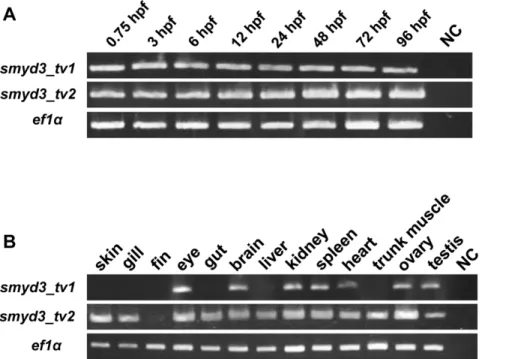
To determine the expression of zebrafishsmyd3in embryogen-esis, we carried out RT-PCR using RNA extracted from embryos at different developmental stages and variant-specific primer sets. The analysis revealed that both forms of transcripts were expressed at all developmental stages from as early as 0.75 hpf to 96 hpf (Figure 1A). In adult zebrafish, RT-PCR detected smyd3_tv1
transcripts in eye, brain, kidney, spleen, heart, ovary and testis, and smyd3_tv2 in skin, gill, eye, gut, brain, liver, kidney, spleen, heart, muscle of the trunk, ovary and testis, but not in fin (Figure 1B), These data indicate that smyd3_tv2 is specifically expressed in skin, gill, gut, liver, and muscle in the trunk.
Knockdown ofsmyd3 in developing embryos
To determine the role of Smyd3 in the development of zebrafish embryos, we injected morpholino-oligonucleotides (MOs) designed to suppress Smyd3 (Smyd3-MO) or mutant MOs containing a five-nucleotide-mismatched sequence against Smyd3-MO se-quence (Smyd3-mis-MO) into fertilized zebrafish eggs. We tested the effect of Smyd3-MO by co-injection with mRNA of smyd3
fused with EGFP in zebrafish eggs. Expectedly, we observed significant decrease of EGFP signals by Smyd3-MO but not by Smyd3-mis-MO at 10 hpf (Figure 2A, B, and C). To confirm the knock-down effect of Smyd3, we additionally prepared MOs that block normal splicing (Smyd3-SB-MO) and performed RT-PCR using asmyd3-specific primer set that amplifies both normal and
abnormal transcripts with exon-skipping. A band corresponding to normal splicing (465 bp) was detected in embryos injected with and without Smyd3-SB-MO, but a band corresponding to aberrant splicing (401 bp) was in embryos injected with Smyd3-SB-MO (Figure 2D). The abnormal transcripts ofsmyd3_tv1 and
smyd3_tv2 were deduced to result in the production of mutant proteins without its conserved region. These results suggested that Smyd3-MO and Smyd3-SB-MO effectively knocked down Smyd3. Interestingly, embryos injected with Smyd3-MO (termed Smyd3 morphants) exhibited pericardial edema and curved trunk (Figure 2E), which was not observed in embryos injected with Smyd3-mis-MO (Figure 2F). Of note, we could observe the normal morphology of the heart chambers (one atrium and one ventricle) and heartbeat in the morphants (Movie S1, Movie S2, and Movie S3).
We classified the severity of heart defect into three grades at 48 hpf when cardiac looping was completed [9]; Grade1: a mild looping defect alone (Figure 2H); Grade2: a moderate looping defect with mild pericardial edema (Figure 2I); Grade3: a severe looping defect with pericardial edema (Figure 2J). Approximately 12% of without injection embryos died spontaneously, indicating the infertility of embryos in our culture condition. Injection with 3 ng of Smyd3-MO led to Grade2 and Grade3 defect in approximately 34% and 26% of embryos, respectively, while injection with 1.5 ng led to Grade2 and Grade3 defect in approximately 14% and 5% of embryos, respectively, suggesting a significant increase of cardiac defect (p,0.001) in a dose-dependent fashion (Figure 3A). On the other hand, Grade2 and Grade3 defects were found in 0% and 2% respectively, of embryos injected with Smyd3-mis-MO, indicating that Grade2 and Grade3 heart defects are significantly increased (p,0.001) in the Smyd3 morphants. Regarding trunk defect, injection with 1.5 ng and 3 ng of Smyd3-MO induced the curved trunk in approximately 40% and 65% of embryos, respectively, but only 3% of embryos
Figure 1. Expression ofsmyd3_tv1andsmyd3_tv2in the developing zebrafish embryos and adult tissues (A) RT-PCR analysis was performed usingsmyd3_tv1andsmyd3_tv2-specific primer sets, with RNA extracted from zebrafish embryos at 0.75, 3, 6, 12, 24, 48, 72, and 96 hpf. NC: negative control (RNase free water).Expression ofef1aserved as an internal control. (B) Expression ofsmyd3_tv1and
smyd3_tv2in various adult tissues. doi:10.1371/journal.pone.0023491.g001
developed the abnormality with 3 ng of Smyd3-mis-MO, which also showed a significant increase of curved trunk (p,0.001) in the morphants (Figure 3B). To confirm these phenotypes, we injected zebrafish eggs with Smyd3-SB-MO that suppressed normal splicing. As a result, the embryos injected with Smyd3-SB-MO consistently showed cardiac and muscle defects as observed in those injected with Smyd3-MO, although their severities and frequencies were less than Smyd3-MO (Figure 3A and B). These data suggested that Smyd3 plays a crucial role in the development of the heart and trunk.
To clarify the importance of Smyd3_tv1 and/or Smyd3_tv2 in cardiac and trunk defects of Smyd3 morphants, we performed a rescue experiment usingsmyd3_tv1and_tv2mRNA. Consequent-ly, the cardiac defect and curved trunk in the Smyd3 morphants were significantly rescued by the injection withsmyd3_tv2mRNA (p,0.001), but not bysmyd3_tv1mRNA (Figure 3A and B). These
data suggested that smyd3_tv2 might play a major role in the development of the heart and trunk.
Expression of cardiac markers in Smyd3 morphants
To further disclose the mechanism(s) of heart defect in Smyd3 morphants, we studied the expression of seven markers; four anterior lateral plate mesoderm (ALPM) markers including GATA-binding protein 5 (gata5), stem cell leukemia protein (scl), NK2 transcription factor related 5 (nkx2.5), and heart and neural crest derivatives expressed transcript2 (hand2), and three cardiac chamber markers including ventricular myosin heavy chain (vmhc), atrial myosin heavy chain (amhc) and cardiac myosin light chain2 (cmlc2). The gata5, scl, nkx2.5 and hand2 are markers specific to ALPM, rostral ALPM, caudal ALPM and medial ALPM, respectively [10]. The three markers, vmhc, amhc and cmlc2 are specific to ventricle, atrium, and both chambers, respectively, and Figure 2. Effect of Smyd3 knockdown in zebrafish embryos by Smyd3-MO or Smyd3-SB-MO.(A, B, C, and D) Suppression ofsmyd3was examined at 10 hpf in embryos injected withsmyd3-EGFPmRNA alone (A),smyd3-EGFPmRNA and Smyd3-mis-MO (B), andsmyd3-EGFPmRNA and Smyd3-MO (C). Signals of EGFP were examined in the fluorescent macroscope (lower panel). The frequencies of EGFP-positive embryos were (A) 78.7%62.3, (B) 83.7%61.5, and (C) 17.7%63.4. Data are shown as means6SEM. (D) Effect of Smyd3-SB-MO onsmyd3transcripts. The wild-type transcripts were detected as a band at 465 bp and the aberrant transcripts at 401 bp. The lower panel shows the expression ofef1aas a control. (E and F) Phenotype of embryos injected with Smyd3-MO (E) or Smyd3-mis-MO (F) at 72 hpf. The pericardial edema (arrow head) and curved trunk (arrow) were observed in Smyd3 morphants (E). (G, H, I, and J) Morphological classification of heart defect. The degree of cardiac defect in the morphants was classified into three grades at 48 hpf. Grade1: Heart shows abnormality with mild looping defect and pericardial edema (H); Grade2: Heart shows abnormality with moderate looping and defect and pericardial edema (I); Grade3: Heart shows abnormality with string-like heart with severe pericardial edema (J). Normal: Normal heart (G). Embryos are shown in lateral view.
they are expressed in the heart tube of zebrafish embryos at 24 hpf [9]. In situ hybridization demonstrated that at 12 hpf, the expression ofgata5,scl,nkx2.5andhand2in Smyd3 morphants was similar to that in the control embryos injected with Smyd3-mis-MO or without injection (Figure 4A, B, C, and D), suggesting that Smyd3 is not involved in the early myocardial specification. At 24 hpf, the expression ofamhcandcmlc2was slightly shifted to the left side in the control embryos (Figure 4F and G), illustrating the normal elongation of the heart tube toward the left ventral side of the embryos [9]. On the other hand, their expression was localized at the midline of the Smyd3 morphants (Figure 4F and G). At 48 hpf,vmhcwas slightly expressed in the atrium of the morphants in addition to its abundant expression in the ventricle, but it was confined to the ventricle in the control embryos (Figure 4H). This abnormal expression ofvmhcwas observed in 9 of 13 morphants, but not in any of 11 controls. Furthermore, expression ofamhcand
cmlc2was enhanced in the atrium of morphants compared with the controls (Figure 4I and J). These findings indicate that cardiac defect in Smyd3 morphants may result from impaired maturation and/or delayed development of cardiomyocytes.
Expression of myogenic markers in Smyd3 morphants
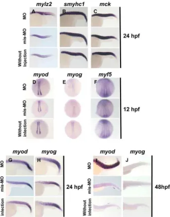
To clarify the mechanism(s) underlying curved trunk, we investigated the expression of six markers; three terminal differentiation makers for skeletal muscle including skeletal muscle myosin light polypeptide 2 (mylz2), slow myosin heavy chain 1 (smyhc1), and muscle creatine kinase (mck), and three myogenic regulatory factors including myogenic differentiation (myod),
myogenic factor 5 (myf5) and myogenin (myog).mylz2,smyhc1, and
mckare differentiation markers for first muscle, slow muscle, and both slow and first muscle, respectively [11]. myodand myf5are expressed in the two lines of adaxial cells flanking the notocord of somites, while myog is expressed in the two lines of cells and paraxial mesoderm at 12 hpf. In situ hybridization clarified that the expression patterns of mylz2, smyhc1, and mck in Smyd3 morphants were indistinguishable from control embryos injected with Smyd3-mis-MO or without injection at 24 hpf when skeletal muscle differentiation is completed (Figure 5A, B and C). Expression ofmyod,myog, and myf5was not different between the morphants and controls at 12 hpf (Figure 5D, E, and F). The morphants and the controls maintained high levels ofmyod and
myog expression in the trunks at 24 hpf (Figure 5G and H). Although the control embryos showed rapid decrease inmyodand
myogexpression at 48 hpf, the morphants sustained significantly high myod and myog expression levels (Figure 5I and J). This sustainedmyodandmyogexpression was observed in all morphants depicting curved trunk. These data suggest that the abnormal trunk morphogenesis in Smyd3 morphants is not caused by the perturbation of muscle differentiation, but possibly by the deregulated expression of myogenic regulatory factors such as
myodandmyog.
Discussion
Recent studies have unveiled that SMYD proteins are involved in the development of cardiac and skeletal muscle. For example, inactivation ofSmyd1, also known asBop, showed hypoplasia of the Figure 3. Frequencies of cardiac and trunk defects in Smyd3 morphants treated with different concentration of morpholinos, and with or without pre-injection withsmyd3_tv1orsmyd3_tv2mRNA.(A) Frequencies of heart defects in embryos injected with 1.5 or 3.0 ng of Smyd3-MO, 3.0 ng of Smyd3-mis-MO, 3.0 ng of Smyd3-SB-MO, 3.0 ng of Smyd3-MO in combination withsmyd3_tv1orsmyd3_tv2mRNA at 48 hpf. The histogram shows percentages of embryos with Normal (open box), Grade1 (light gray box), Grade2 (dark gray box), Grade3 (hatched box) or dead embryos (closed box). (B) Frequencies of curved trunk in embryos injected with 1.5 or 3.0 ng of MO, 3.0 ng of mis-MO, 3.0 ng of Smyd3-SB-MO, 3.0 ng of Smyd3-MO in combination withsmyd3_tv1orsmyd3_tv2mRNA at 48 hpf. The histogram shows percentages of embryos with Normal (open box), curved trunk (gray box), or dead embryos (closed box). Error bars represent the SEM. N is the total number of injected animals. doi:10.1371/journal.pone.0023491.g003
right ventricle in mice through disrupted maturation of ventricular cardiomyocytes [12,13], and defect of muscle contraction in zebrafish through impaired myofibril organization [3]. SMYD1 expression is controlled by MYOD, Myogenin, and MEF2, transcription factors related to myogenesis, and is essential for
Hand2expression that encodes a basic helix-loop-helix transcrip-tion factor expressed in cardiac muscle [12]. Smyd2 was abundantly expressed in skeletal muscle and the face region during embryogenesis in Xenopus laevis [14]. Both SMYD1 and
SMYD2 expression was gradually increased during porcine fetal muscle development [15]. In addition, muscle specific-depletion of
Drosophila Smyd4led to the failure of eclosion resulting in late pupal death [16]. Besides SMYD proteins, other methyltransferases have been revealed to play a crucial role in muscle development. EZH2, a polycomb protein containing a SET domain, controls skeletal muscle differentiation through transcriptional repression of SRF and MYOD [17]. PEDM1 or Blimp-1/u-boot induces slow-twitch fiber-specific muscle differentiation by suppression of fast muscle-specific gene expression [18,19]. The WDR5/ASH2L/MLL2 histone methyltransferase (HMT) complex activates MYOD, while SUV39H1 represses it [20,21]. In addition to these reports, we have shown here that Smyd3 plays an important role in the development of cardiac and skeletal muscle.
We have additionally revealed that two forms of smyd3 are expressed during zebrafish embryogenesis and in adult zebrafish. The two forms of transcripts encode proteins sharing most regions including the MYND and SET domains, but the short form (smyd3_tv1) lacks the post-SET domain. Since a post-SET was reported to enhance the methyltransferase activity coupled with another cystein in SET domain [22], the enzymatic activity of the long form (smyd3_tv2) may be higher than the short form. Consistent with this view, our rescue experiment showed that the long form (smyd3_tv2) seems to be more important than
smyd3_tv1for cardiogenesis and trunk formation. We also found that their expression was different in several adult tissues; the expression ofsmyd3_tv1was almost diminished in the gill, skin, gut, Figure 4. In situ hybridization analysis of ALPM and cardiac
chamber markers.(A, B, C, and D) Expressiongata5,scl,nkx2.5and hand2in Smyd3 morphants, control embryos injected with Smyd3-mis-MO and without injection at 12 hpf. (E, F, G, H, I, and J) Expression of vmhc,amhc,and cmlc2in the morphants, control embryos and without injection embryos at 24 hpf (E, F, and G) and 48 hpf (H, I, and J). Arrowhead indicates abnormal vmhc expression in the atrium (H). Embryos are shown in dorsal view, anterior toward the left (A, B, C, D, E, F, and G). Embryos are shown in frontal view, dorsal toward the left (H, I, and J).
doi:10.1371/journal.pone.0023491.g004
Figure 5. In situ hybridization analysis of terminal differenti-ation markers of skeletal muscle and myogenic regulatory factors.(A, B, and C) Expression ofmylz2,smyhc1andmckin Smyd3 morphants, control embryos injected with Smyd3-mis-MO and without injection at 24 hpf. (D, E, F, G, H, I, and J) Expression ofmyod,myog, and myf5in Smyd3 morphants, control embryos injected with Smyd3-mis-MO and without injection at 12 hpf (D, E, and F), 24 hpf (G and H) and 48 hpf (I and J). Embryos are shown in lateral view, anterior toward the left (A, B, C, G, H, I, and J). Embryos are shown in dorsal view, anterior toward the top (D, E, and F).
liver and trunk muscle although smyd3_tv2 was expressed ubiquitously in adult tissues. Therefore, the two forms of Smyd3 protein may have different roles in embryogenesis and adult tissues. Although the human ortholog SMYD3 protein contained a post-SET domain, a variant form termed SMYD3-NY lacking the N-terminal region was expressed in placenta, testis, ovary, kidney, spleen, and skeletal muscle [23].
In this study, we found that knockdown of zebrafish Smyd3 resulted in abnormal looping of heart tube accompanied by pericardial edema, which is similar to the Smyd1 morphants [3]. Heart development is governed by a complex gene regulatory network consisting of transcription factors, their co-factors, and downstream genes modulating cell fate specification, cell differen-tiation, cell proliferation, and cell migration. Among the network, transcription factors including Nkx2, GATA, Mef2, and Hand1/2 play a crucial role in early myocardial differentiation and morphogenesis [24,25]. In situ hybridization demonstrated that Smyd3 morphants did not show abnormal expression ofgata5,scl,
nkx2.5, and hand2, at early stages but showed deregulated expression of amhc, vmhc, and cmlc2. These data may imply that Smyd3 is not involved in early specification of cardiomyocytes. It is of note that SMYD3 up-regulates the expression ofNKX2.5in an embryonic kidney cell line HEK293 [5]. Unexpectedly, however, we found here that the expression ofnkx2.5was unchanged in the Smyd3 morphants compared to control embryos. Since Smyd3 is a histone H3K4 methyltransferase, other H3K4 methyltransfer-ase(s) such as Smyd1 may compensate the modification during heart development. Alternatively, nkx2.5 may be regulated by different histone modification enzymes and/or transcription factors between kidney and cardiac muscle.
In addition to the heart defects, we have shown that Smyd3 morphants developed curved trunk, which was associated with sustained expression ofmyodandmyogat a late developmental stage (48 hpf). Trunk skeletal muscle in vertebrates originates from a primary myotomal component of somites. Activation of myogenesis is regulated by a complex network comprising of the basic helix-loop-helix domain-containing myogenic regulatory factors (MRFs). Among the MRFs, Myod, the myogenic master transcription factor, is activated in adaxial cells adjacent to the notochord as early as 7– 7.5 hours in zebrafish embryogenesis [26]. The myod-expressing cells expand in an anterior-to-posterior wave by 14.5 hpf, and markedly drop the myod expression by 24 hpf. The sustained expression of myod in Smyd3 morphants might result from deregulation of its upstream regulator such as Pax3 [27], or an undetermined mechanism of Myod regulation. Notably,Smyd1/Bop
is transcriptionally regulated by MEF2C in the developing heart [13], and serum response factor and myogenin in myogenesis. From these data, it is tempting to speculate that Smyd3 is also regulated by MRFs and that inhibition of Smyd3 may activate Myod through a negative feedback loop. Since myod is known to enhance the expression ofmyog, the sustained expression ofmyogis likely due to the elevated expression of myod. Although additional studies are needed to clarify the mechanism(s) by which Smyd3 is implicated in muscle development, our findings should be a starting point for elucidating the roles of Smyd3 in myogenesis. Although Smyd3 morphants depicted cardiac defect and curved trunk, cardiac and skeletal myogenesis seem to be normally accomplished in early stages. Therefore, Smyd3 may not be involved in cell specification or differentiation, but involved in maturation or proliferation of differentiated myogenic cells.
In the present study, we have shown thatsmyd3plays a crucial role for cardiac and skeletal muscle development. These findings will be helpful for the understanding of molecular mechanisms underlying the development of heart and skeletal muscle.
Materials and Methods
Maintenance of zebrafish
Zebrafish (Danio rerio) were purchased from a local pet shop, and maintained under a 14-h day/10-h night cycle at 28.5uC. Fertilized eggs were obtained by mating adult fish from our outbred colonies soon after the light was turned on. Embryos were staged according to hours post-fertilization (hpf) and morpholog-ical criteria [28]. In our university, approval from the institutional committee for animal experiments is not necessary when using fish.
Reverse transcription-polymerase chain reaction (RT-PCR) analysis
Total RNA was extracted from embryos or adult tissues using TRIzol solution (Life Technologies, Carlsbad, CA). cDNA was generated using 0.5mg of total RNA with Surperscript II reverse transcriptase (Life Technologies) and oligo (dT)15 primers (Life
Technologies). PCR reaction was performed using the cDNA as template. Primers used for the amplification were as follows: 59 -CGTGGCCCGATCATAAGAGG-39and 59-ACAGCTCATCC-CAGTGCTGG-39forsmyd3_tv1, 59-GGAGCAATACCACTTC-CGGTGT-39, and 59-GCACTCGCTCAGTCTCCTCT-39 for
smyd3_tv2, 59-TCACCCTGGGAGTGAAACAGC-39 and 59 -ACTTGCAGGCGATGTGAGCAG-39 for ef1a, 59-CCGGAA-TTCTGAAATGATGGAGGCTGTG-39 and 59 -CGTCGTGC-AGAGATGCTTCA-39for the assessment of Smyd3-SB-MO.
Microinjection of morpholino-oligonucleotides (MOs)
using the Quick Change Site-Directed Mutagenesis kit II (Agilent Technologies, Santa Clara, CA). The primers used for the amplification were, 59 -ACAGACGTTCCCAGCACTGGGAT-GAGCTGTTGAAG-39 and 59-ACAGCTCATCCCAGTGC-TGGGAACGTCTGTCTTTA-39. Rescue experiments were performed by a pre-injection with 300 pg of capped smyd3_tv1
or_tv2mRNA and a subsequent injection with MOs as described earlier [30,31]. The capped mRNA was synthesized using a m7G(59)PPP(59) G (Roche, Mannheim, Germany) and T7 or SP6 RNA polymerase (Roche) with pc-Smyd3_tv1, pc-Smyd3_tv2 or pCS2-Smyd3-EGFP. Fisher’s exact test was employed for the analysis, andp,0.05 was considered statistically significant.
Whole mount in situ hybridization
For in situ hybridization, the following genes were used as cRNA probes:gata5, scl, hand2[25], cmlc2, vmhc [32],amhc [33],
mck, mylz2, smyhc1 [16], myod, myf5and myog [26]. cDNAs were amplified by RT-PCR and the products were cloned into pcDNA3.1 plasmids (Life Technologies). Digoxigenin (DIG)-labeled RNA probes were transcribed using RNA DIG labeling mix (Roche) and T7 RNA polymerase (Roche). Whole mount in situ hybridization was carried out essentially as described elsewhere [29].
Supporting Information
Figure S1 (A) Multiple alignment of human SMYD3, zebrafish
smyd3_tv1andtv2protein sequences using CLUSTAL W. MYND,
SET, and post-SET domain are indicated as a solid line above the sequence. Identical residues are indicated by asterisks, conserved substitutions by colons, and semi-conserved substitutions by periods.
(TIF)
Movie S1 Heartbeats of a control embryo without injection at 48 hpf.
(WMV)
Movie S2 Heartbeats of a control embryo injected with Smyd3-mis-MO at 48 hpf.
(WMV)
Movie S3 Heartbeats of a Smyd3 morphant at 48 hpf. (WMV)
Acknowledgments
We thank Atsumi Iida, Yoko Tabata, Keisuke Sasaki, and Nagahiro Kaneko for technical assistance, and Drs. Yasuo Ohuchi and Shinya Satoh for helpful discussions.
Author Contributions
Conceived and designed the experiments: SW YF. Performed the experiments: TF S-iT. Analyzed the data: KY. Wrote the paper: TF YF.
References
1. Sims RJ, Reinberg D (2004) From chromatin to cancer: a new histone lysine methyltransferase enters the mix. Nat Cell Biol 6: 685–687.
2. Sims RJ, Nishioka K, Reinberg D (2003) Histone lysine methylation: a signature for chromatin function. Trends Genet 19: 629–639.
3. Tan X, Rotllant J, Li H, DeDeyne P, Du SJ (2006) SmyD1, a histone methyltransferase, is required for myofibril organization and muscle contraction in zebrafish embryos. Proc Natl Acad Sci U S A 103: 2713–2718.
4. Abu-Farha M, Lambert JP, Al-Madhoun AS, Elisma F, Skerjanc IS, et al. (2007) The tale of two domains: proteomics and genomics analysis of SMYD2, a new histone methyltransferase. Mol Cell Proteomics 7: 560–572.
5. Hamamoto R, Furukawa Y, Morita M, Iimura Y, Silva FP, et al. (2004) SMYD3 encodes a histone methyltransferase involved in the proliferation of cancer cells. Nat Cell Biol 6: 731–740.
6. Brown MA, Sims RJ, Gottlieb PD, Tucker PW (2006) Identification and characterization of Smyd2: a split SET/MYND domain-containing histone H3 lysine 36-specific methyltransferase that interacts with the Sin3 histone deacetylase complex. Mol Cancer 5: 26.
7. Huang J, Perez-Burgos L, Placek BJ, Sengupta R, Richter M, et al. (2006) Repression of p53 activity by Smyd2-mediated methylation. Nature 444: 629–632.
8. Sun XJ, Xu PF, Zhou T, Hu M, Fu CT, et al. (2008) Genome-Wide Survey and Developmental Expression Mapping of Zebrafish SET Domain-Containing Genes. PLoS One 30: e1499.
9. Stainier DY (2001) Zebrafish genetics and vertebrate heart formation. Nat Rev Genet 2: 39–48.
10. Schoenebeck JJ, Keegan BR, Yelon D (2007) Vessel and blood specification override cardiac potential in anterior mesoderm. Dev Cell 13: 254–267. 11. Xu Y, He J, Wang X, Lim TM, Gong Z (2000) Asynchronous activation of 10
muscle-specific protein (MSP) genes during zebrafish somitogenesis. Dev Dyn 219: 201–215.
12. Gottlieb PD, Pierce SA, Sims RJ, Yamagishi H, Weihe EK, et al. (2002) Bop encodes a muscle-restricted protein containing MYND and SET domains and is essential for cardiac differentiation and morphogenesis. Nat Genet 31: 25–32. 13. Phan D, Rasmussen TL, Nakagawa O, McAnally J, Gottlieb PD, et al. (2005)
BOP, a regulator of right ventricular heart development, is a direct transcriptional target of MEF2C in the developing heart. Development 132: 2669–2678.
14. Kawamura S, Yoshigai E, Kuhara S, Tashiro K (2008) smyd1 and smyd2 are expressed in muscle tissue in Xenopus laevis. Cytotechnology 57: 161–168. 15. Peng YB, Yerle M, Liu B (2009) Mapping and expression analyses during
porcine foetal muscle development of 12 genes involved in histone modifications. Anim Genet 40: 242–246.
16. Thompson EC, Travers AA (2008) A Drosophila Smyd4 homologue is a muscle-specific transcriptional modulator involved in development. PLoS One 3: e3008.
17. Caretti G, Di Padova M, Micales B, Lyons GE, Sartorelli V (2004) The Polycomb Ezh2 methyltransferase regulates muscle gene expression and skeletal muscle differentiation. Genes Dev 18: 2627–2638.
18. Roy S, Ng T (2004) Blimp-1 specifies neural crest and sensory neuron progenitors in the zebrafish embryo. Curr Biol 14: 1771–1777.
19. Liew HP, Choksi SP, Wong KN, Roy S (2008) Specification of vertebrate slow-twitch muscle fiber fate by the transcriptional regulator Blimp1. Dev Biol 15: 226–235.
20. McKinnell IW, Ishibashi J, Le Grand F, Punch VG, Addicks GC, et al. (2008) Pax7 activates myogenic genes by recruitment of a histone methyltransferase complex. Nat Cell Biol 10: 77–84.
21. Mal AK (2006) Histone methyltransferase Suv39h1 represses MyoD-stimulated myogenic differentiation. EMBO J 25: 13611–13626.
22. Zhang X, Yang Z, Khan SI, Horton JR, Tamaru H, et al. (2003) Structural basis for the product specificity of histone lysine methyltransferases. Mol Cell 12: 177–185. 23. Zhou Z, Ren X, Huang X, Lu L, Xu M, et al. (2005) SMYD3-NY, a novel
SMYD3 mRNA transcript variant, may have a role in human spermatogenesis. Ann Clin Lab Sci 35: 270–277.
24. Olson EN (2006) Gene regulatory networks in the evolution and development of the heart. Science 313: 1922–1927.
25. Schoenebeck JJ, Yelon D (2007) Illuminating cardiac development: Advances in imaging add new dimensions to the utility of zebrafish genetics. Semin Cell Dev Biol 18: 27–35.
26. Weinberg ES, Allende ML, Kelly CS, Abdelhamid A, Murakami T, et al. (1996) Developmental regulation of zebrafish MyoD in wild-type, no tail and spadetail embryos. Development 122: 271–280.
27. Bryson-Richardson RJ, Currie PD (2008) The genetics of vertebrate myogenesis. Nat Rev Genet 9: 632–646.
28. Kimmel CB, Ballard WW, Kimmel SR, Ullmann B, Schilling TF (1995) Stages of embryonic development of the zebrafish. Dev Dyn 203: 253–310. 29. Kurita R, Sagara H, Aoki Y, Link BA, Arai K, et al. (2003) Suppression of lens
growth by alphaA-crystallin promoter-driven expression of diphtheria toxin results in disruption of retinal cell organization in zebrafish. Dev Biol 255: 113–127. 30. Zhao J, Cao Y, Zhao C, Postlethwait J, Meng A (2003) An SP1-like transcription
factor Spr2 acts downstream of Fgf signaling to mediate mesoderm induction. EMBO J 22: 6078–6088.
31. Cao Y, Zhao J, Sun Z, Zhao Z, Postlethwait J, et al. (2004)fgf17b, a novel member of Fgf family, helps patterning zebrafish embryos. Dev Biol 271: 130–143. 32. Yelon D, Horne SA, Stainier DY (1999) Restricted expression of cardiac myosin
genes reveals regulated aspects of heart tube assembly in zebrafish. Dev Biol 214: 23–37.