Jonathan Bramsiepe1, Katja Wester2¤a, Christina Weinl3¤b, Farshad Roodbarkelari3¤c, Remmy Kasili4¤d, John C. Larkin4, Martin Hu¨lskamp2, Arp Schnittger1,3*
1Institut de Biologie Mole´culaire des Plantes du CNRS, Universite´ de Strasbourg, Strasbourg, France,2Lehrstuhl fu¨r Botanik III, Universita¨t zu Ko¨ln, Ko¨ln, Germany,
3Unigruppe am Max-Planck-Institut fu¨r Pflanzenzu¨chtungsforschung, Lehrstuhl fu¨r Botanik III, Universita¨t zu Ko¨ln, Ko¨ln, Germany,4Department of Biological Sciences, Louisiana State University, Baton Rouge, Louisiana, United States of America
Abstract
Cell-fate specification is typically thought to precede and determine cell-cycle regulation during differentiation. Here we show that endoreplication, also known as endoreduplication, a specialized cell-cycle variant often associated with cell differentiation but also frequently occurring in malignant cells, plays a role in maintaining cell fate. For our study we have used Arabidopsis trichomes as a model system and have manipulated endoreplication levels via mutants of cell-cycle regulators and overexpression of cell-cycle inhibitors under a trichome-specific promoter. Strikingly, a reduction of endoreplication resulted in reduced trichome numbers and caused trichomes to lose their identity. Live observations of young Arabidopsis leaves revealed that dedifferentiating trichomes re-entered mitosis and were re-integrated into the epidermal pavement-cell layer, acquiring the typical characteristics of the surrounding epidermal cells. Conversely, when we promoted endoreplication in glabrous patterning mutants, trichome fate could be restored, demonstrating that endoreplication is an important determinant of cell identity. Our data lead to a new model of cell-fate control and tissue integrity during development by revealing a cell-fate quality control system at the tissue level.
Citation:Bramsiepe J, Wester K, Weinl C, Roodbarkelari F, Kasili R, et al. (2010) Endoreplication Controls Cell Fate Maintenance. PLoS Genet 6(6): e1000996. doi:10.1371/journal.pgen.1000996
Editor:Li-Jia Qu, Peking University, China
ReceivedMarch 1, 2010;AcceptedMay 19, 2010;PublishedJune 24, 2010
Copyright:ß2010 Bramsiepe et al. This is an open-access article distributed under the terms of the Creative Commons Attribution License, which permits unrestricted use, distribution, and reproduction in any medium, provided the original author and source are credited.
Funding:The work in the laboratory of MH is funded by the Deutsche Forschungsgemeinschaft (DFG) through the collaborative research program SFB 572. Research in the JCL group is supported by National Science Foundation grant IOS 0744566. JB receives an Allocation Pre´sidence grant from the Universite´ de Strasbourg. This work was supported by an Action The´matique et Incitative sur Programme AVENIR (ATIP-AVENIR) grant from the Centre National de la Recherche. Scientifique (CNRS) and an European research council (ERC) Starting Grant from the European Union to AS. The funders had no role in study design, data collection and analysis, decision to publish, or preparation of the manuscript.
Competing Interests:The authors have declared that no competing interests exist. * E-mail: [email protected]
¤a Current address: Lehrstuhl fu¨r Botanik II, Universita¨t zu Ko¨ln, Ko¨ln, Germany
¤b Current address: Division of Medical Biochemistry, Innsbruck Medical University, Innsbruck, Austria ¤c Current address: Max-Planck-Institut fu¨r Pflanzenzu¨chtungsforschung, Ko¨ln, Germany
¤d Current address: National Institute of Child Health and Human Development, Bethesda, Maryland, United States of America
Introduction
Many different cell cycle programs can be found in developing multicellular organisms [1]. Typically, embryonic cell cycles are short with a rapid sequence of the DNA-synthesis phase (S-phase) and mitosis (M-phase) rapidly generating cells or nuclei in a syncytium. At later stages of development, differentiating cells in many animal and plant species frequently enter an endoreplication cycle in which mitosis is skipped and DNA is re-replicated leading to polyploid cells [2–4]. However, very little is known about the biological importance of endoreplication and the resulting cellular polyploidy.
Progression of mitotic cell cycles is controlled by cyclin-dependent kinase (CDK)–cyclin heterodimeric complexes. Their action is in particular required for the entry into S-phase and M-phase [5]. The major determinant of CDK activity is the abundance of the cyclin co-factor and cyclin levels are controlled both at the level of transcription and by protein degradation [6]. In particular two multi-protein complexes, the Skip-F-box-Cullin (SCF) complex and the Anaphase-Promoting-Complex/Cyclo-some (APC/C) ligate ubiquitin moieties to cyclins marking them for subsequent degradation by the proteasome [7,8]. The
specificity of these ubiquitin ligases is brought about by adaptor proteins, i.e. F-box proteins for the SCF complex and Cdh1/ FZR/CCS52 and Cdc20/FZY for the APC/C [8–12].
In addition to cyclins, CDKs are also controlled by the binding of inhibitors, for instance p27Kip1 in mammalian cells [13].
Moreover, CDK activity is regulated by posttranslational modi-fications such that phosphorylation of a conserved threonin residue (position 161 in Arabidopsis) in the so-called T-loop is absolutely required for kinase activity [5,14,15]. Conversely, phosphorylation of two residues in the P-loop (typically T14 and Y15) can block kinase activity. All these control mechanisms appear to be globally conserved in eukaryotes and are present from yeast to plants [16–18].
substrate-depletion and lateral inhibition mechanism [21–23]. The trichome pattern is established in the basal part of the leaf where the positive regulators (activators of trichome fate) GLABRA1 (GL1), a R2R3 MYB transcription factor, GLABRA3 (GL3), a bHLH transcription factor, and TRANSPARENT TESTA GLABRA1 (TTG1), WD-40 protein, are initially ubiquitously expressed.
The current model postulates that due to stochastic fluctuations some cells express these regulators at a higher concentration and these differences in expression levels become greatly enhanced due to a positive feed back loop of the activator complex. In turn, the positive regulators induce the expression of inhibitors, small R3 single repeat MYB transcriptional regulators that are then released from cells that have high levels of activators and inhibit the formation of activator complexes in the surrounding cells [21,23]. So far six inhibitors have been identified: TRIPTYCHON (TRY), CAPRICE (CPC), ENHANCER OF TRY AND CPC (ETC) 1, 2, 3 (also called CPC-LIKE PROTEIN [CPL]), and TRICHOME-LESS (TCL) [24–28]. The current model suggests that once a cell has reached a certain threshold of activator complex, trichome fate is established and the incipient trichome cell starts to express downstream genes, such as the HD bZIP transcription factor GLABRA2 (GL2). Many downstream trichome-specific genes are then activated that regulate further outgrowth of the formation of typically three to four branches [29–31].
One of the earliest signs of trichome differentiation is the entry into an endoreplication cycle and along with further outgrowth trichomes undergo usually three to four rounds of DNA replication leading to a final DNA content of approximately 32C [19,20,32]. Endoreplication correlates well with trichome growth and typically mutants that have reduced endoreplication levels also display smaller trichomes with fewer branches while mutants with increased endoreplication levels have larger trichomes with more branches [33,34].
Two core cell-cycle regulators have so far been shown to be required for the trichome endoreplication cycle. The first one is CDKA;1, the major regulator of mitotic cycles and the
Arabidopsis homolog of the yeast Cdc2/CDC28 kinase. Either a substitution of the CDKA;1 amino acid T161 with D161 (further on abbreviated as D) or a mutant in which T14 has been substituted by D14 and Y15 by E15 (abbreviated asDE) resulted in reduced CDKA;1 kinase activity and both weak loss-of-function alleles displayed smaller nuclei in trichomes along with a reduction of trichome size [14,17].
The other cell-cycle regulator involved in trichome cell-cycle control is SIAMESE (SIM), a putative CDK inhibitor, and insim
mutants endoreplication is partially converted into a mitotic program resulting in multicellular trichomes [35,36]. One likely target of SIM is a CDK-cyclin D complex since a paralog of SIM in rice can inhibit cyclin D action when expressed in yeast [37]. In addition, the misexpression of CYCLIN D3;1 (CYCD3;1) in trichomes has resulted in the formation of multicellular trichomes [38]. A second target of SIM could be mitotic B-type cyclins since also the ectopic expression of a constitutive active B-type cyclin resulted in multicellular trichomes [39,40].
Remarkably, some of the trichome patterning genes appear to have a function in endoreplication control. Loss of GL3 function results in a reduction of endoreplication levels and conversely,try
mutants undergo one additional round of endoreplication [34]. In addition, GL1 might also control endoreplication although the situation is less clear since in one report the overexpression ofGL1
was found to result in increased endoreplication levels in trichomes while in another study no such effect in was observed [41,42]. Interestingly, it has been found thatSIMis a direct early target of GL3 and GL1 pinpointing to a tight interaction between patterning genes and the regulation of the endoreplication cycle [43].
Here we have dissected the relationship between pattern formation and cell-cycle control by using plants with reduced endoreplication levels in trichomes. We found that trichome fate is surprisingly plastic and trichome fate can be changed into an epidermal pavement cell fate even in advanced stages of trichome differentiation. Our data show that progression through an endoreplication cycle is an important aspect of cell fate acquisition and is crucial for cell fate maintenance.
Results
Reduction of endoreplication is correlated with a decrease in trichome numbers
Since several trichome patterning mutants are also affected in endoreplication control, we asked whether there is a functional connection between cell-cycle progression and pattern formation during very early trichome development. We therefore first revisited
simmutants in which trichomes undergo cell divisions leading to multicellular trichome [35,36]. Remarkably, we found that under our growth conditionssimmutants develop significantly fewer (T-test with p = 0.0001) trichome initiates sites (TIS) per leaf in comparison to wild type (Figure 1G, dark blue bars). A patterning defect of sim mutants became even more prominent when we compared the number of trichomes per epidermal cell number (light blue bars), heresim mutants had only approximately half of the trichomes found in wild type.
To test whether cell-cycle progression and trichome initiation are functionally linked, we sought for additional possibilities to promote cell proliferation in trichomes. In earlier experiments, we have used theGL2promotor to drive expression ofCYCD3;1or a N-terminally truncated B-type cyclin resulting in the formation of multicellular trichomes. Both the multicellular trichomes and the remaining single-celled trichomes, displayed a strong reduction in their ploidy level indicating that a mitotic cycle could inhibit and override an endoreplication cycle [38–40].
Author Summary
Differentiating cells often amplify their nuclear DNA content through a special cell-cycle variant, called endoreplication, in which cell division is skipped. Although this process is widespread from humans to plants, not much is currently known about the biological importance of endoreplication. Moreover, the control of cell-cycle activities has been thought to follow developmental decisions and the adoption of a specific cell fate. Here we have uncovered a previously unrecognized function of endoreplication in maintaining cell identity, presenting a striking example of how cell fate and cell-cycle progression are linked. Using leaf hairs on the reference plant Arabidopsis as a model, we show that compromising endoreplication leads to dedifferentiation of the newly forming leaf hair cell. Live observations of young Arabidopsis leaves revealed that dedifferentiating leaf hairs underwent repeated rounds of cell division and were re-integrated into the epidermal cell layer acquiring the typical characteristics of the surrounding epidermal cells. Conversely, promoting endoreplication in mutants that fail to develop hairs could at least partially restore their differentiation program. With this, our findings also pinpoint an important role of the social context of a cell, revealing a differentiation control system at the tissue level.
To manipulate cells during the initial patterning process we used here the promoters of theCPCandTRYgene that are active earlier than theGL2promoter to drive expression ofCYCD3;1[44]. Similar to the expression ofCYCD3;1from theGL2promoter, expression from the CPC and the TRY promoter resulted in multicellular trichomes. However, in addition, we obtained more than eight transgenic lines that were devoid of trichomes out of more than 20 primary transformants (Figure 1A and 1B).
We envisioned two scenarios that could explain these results. First, expression ofCDCD3;1and the loss of the CDK inhibitor SIM might interfere with trichome-fate establishment by promot-ing/allowing cell division of an incipient trichome cell and, thereby constantly diluting the presumptive activator levels. Alternatively, or in addition, endoreplication cycles may be instrumental for trichome patterning. To discriminate between these two possibilities, we analyzed plants that have reduced endoreplication levels in trichomes.
Previously, we had generated plants that express the CDK inhibitor ICK1/KRP1under the trichome-specific GL2 promoter, which led to strongly reduced endoreplication levels in trichomes [45]. This effect was even stronger when the N-terminally truncated ICK1/KRP1 variant ICK1/KRP1109–191, which lacks the first 108 amino acids was expressed from the GL2 promoter. The ICK1/ KRP1109–191form of the protein displays increased protein stability and interacts more strongly with CDKA;1 and CYCD3;1 than the full length ICK1/KRP1 protein in yeast two hybrid experiments [45–49]. Examination of trichome numbers on leaves of these
ICK1/KRP1-misexpressing plants revealed that trichome numbers were reduced (Figure 1G and Table S1). To complement this set of experiments, we analyzed two weakcdka;1mutants,DandDE, that were previously found to display reduced endoreplication levels in leaves [14,17]. None of these genotypes resulted in multicellular trichomes or would be expected to favor increased cell division, making dilution of cell fate transcriptional activators unlikely as an explanation of the observed reduction in trichome number.
Quantification of DAPI-stained trichomes revealed that both D and DE plants have decreased endoreplication levels in trichomes in comparison with wild-type plants (Figure 2I and 2M) and consistent with the above obtained results with ICK1/KRP1-misexpressing plants both weakcdka;1loss-of-function mutants also displayed fewer trichomes on leaves than control plants (Figure 1D, 1E, 1G, and Table S1). To reduce endoreplication levels further, we combined D plants with plants misexpressing ICK1/KRP1109–191. These plants displayed the severest trichome effect among the cell-cycle mutants studied and total trichome numbers dropped almost ten fold in comparison to wild type (Figure 1F and 1G and Table S1).
It was previously shown that ICK1/KRP1 can act non-cell-autonomously and thus, PROGL2:ICK1/KRP1-expressing plants
have typically fewer but larger epidermal cells that surround trichomes [46]. Plants with a reduction of CDK activity either due to a strong overexpression of CDK inhibitors or due to a compromised kinase, as inDandDE plants, have also generally larger epidermal cells [14,17,50,51]. This increase in cell size makes it difficult to discriminate between a reduction of trichome number due to reduced leaf size and fewer cells versus abona fide
patterning defect. To correct for cell-size and cell-number differences, we determined the ratio of trichomes per epidermal cells. This estimate revealed that there is indeed a true trichome patterning defect in bothICK1/KRP1overexpressing plants as well as inDandDEplants (Figure 1G, light blue bars; Table S1).
To substantiate that the reduction of trichome number is due to a true patterning defect rather than due to alterations of leaf growth, we analyzed plants expressing a cell-autonomous version of ICK1/KRP1 in trichomes; in this construct, the N-terminally
Figure 1. Reduced trichome numbers on plants with altered cell-cycle control in trichomes.Scanning electron micrographs of rosette leaf number 4 of Arabidopsis seedlings. (A) Columbia, (B)PROCPC:CYCD3;1,
(C) PROGL2: ICK1/KRP1im, (D) CDKA;1T161D, (E) CDKA;1T14D/Y15E and (F) PROGL2:KRP1109 - CDKA;1T161D plants. (G) Quantification of trichome
numbers of leaf 3 and 4 in comparison with Columbia. Col Columbia n = 31. sim siamese n = 18. ICK1/KRP1IM, PRO
GL2:GUS:YFP:KRP1109 n = 30. KRP1109PROGL2:KRP1
109
n = 32.D CDKA;1T161Dn = 32.DE CDKA;1T14D/Y15E
n = 32. D; ICK1/KRP1109 CDKA;1T161D; PRO
GL2:KRP1109 n = 26. Error bars:
truncated CDK inhibitor ICK1/KRP1109–191is fused to GUS and GFP [46,49]. We could also observe significantly reduced trichome numbers in plants expressing the immobile GUS:YF-P:ICK1/KRP1109–191 (hereafter referred to as ICK1/KRP1im) under theGL2promoter (T-test, p = 0.0001) confirming that local interference with endoreplication results in a patterning defect proper (Figure 1C and 1G and Table S1).
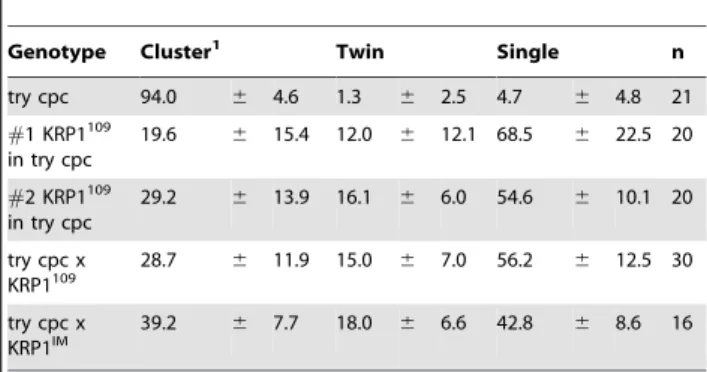
Genetic interactions between plants with reduced endoreplication levels and patterning mutants
To dissect the epistasis of the relationship between endoreplica-tion and pattern formaendoreplica-tion, we introgressed the weakcdka;1 loss-of-function mutantDandDEas well asICK1/KRP1-overexpressing plants into several trichome patterning mutants. First, we analyzed the effect of reduced endoreplication incpc-trydouble mutants that develop large trichome clusters due to reduced lateral inhibition [44] (Figure 3A). The combination with plants expressing the
PROGL2:ICK1/KRP1109–191construct orDandDEplants strongly
reduced cluster formation and cluster size (Figure 3C and 3E): 56 percent of trichome initiation sites now contain only one trichome and approximately 2/3 of the clusters form just two trichomes whereas incpc-trydouble mutant 95 percent of the sites contain more than one trichome with more than 93 percent clusters containing more than two trichomes. Similar, although somewhat weaker, effects were found when introducing the cell-autono-mously acting ICK1/KRP1imconstruct intocpc-tryplants with 39
percent clusters and 18 percent cluster with only two trichomes (Table 1). This shows that a reduction in endoreplication can override the effect of patterning mutants.
Next we analyzed crosses ofD,DE, andPROGL2:ICK1/KRP1im
with gl3 mutants, in which the trichome activator complex is compromised [22,43,52]. This combination resulted in a syner-gistic effect with a dramatic reduction of trichomes on leaf blades (Figure 3B, 3D, 3F, and 3G). Interestingly, we found on these plants a number of rudimentary, aborting trichomes that appeared to be arrested in their development and that we could never observe on wild-type plants (Figure 3D, 3F, and 3G). Taken together, these findings corroborate the importance of endorepli-cation, and place endoreplication control in an early phase of the trichome patterning process.
Aborting trichomes lose their fate and transdifferentiate The trichome pattern in Arabidopsis is established in the youngest part of a developing leaf. Therefore we analyzed the patterning zone of young leaves of plants with reduced endoreplication levels in trichomes. Consistent with previous studies [34,41,53], we found that in wild type emerging trichomes appeared with a minimal distance of approximately 3 cells (Figure 4A and 4B). As expected, epidermal cell size in the trichome patterning zone was much larger in D and DE
plants than in wild type (Figure 4C). None-the-less, trichomes were patterned with roughly the same distance as in wild type resulting in a similar number of trichomes per cells (Figure 4C and 4D and Table S2). Thus, while the trichome pattern on old leaves is significantly different between wild-type plants and plants with a compromised endoreplication cycle, the initial pattern of trichomes appears to be rather similar.
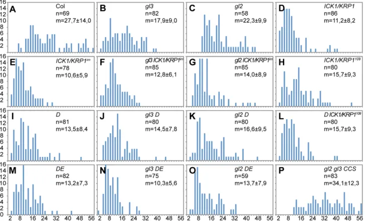
Figure 2. Analysis of DNA content in trichomes.(A–P) Distribution of the trichome DNA contents in relative fluorescence units (RFU). Two RFUs roughly represent 2C, calibrated with wild-type andgl3trichome nuclei. (A) Wild type. (B)glabra3(gl3). (C)glabra2(gl2). (D)PROGL2:ICK1/KRP1. (E) PROGL2:GUS:YFP:ICK1/KRP1109-191(ICK1/KRP1im). (F)gl3 - ICK1/KRP1im. (G) gl2 - ICK1/KRP1im. (H)PROGL2:ICK1/KRP1109–191. (I)CDKA;1T161D(D). (J)gl3 - D. (K) gl2 - D. (L)D-PROGL2:ICK1/KRP1
109–191
. (M)CDKA;1T14D/Y15E(DE). (N)gl3-DE. (O)gl2-DE. (P)gl2-gl3-PROGL2:CCS52A1.
doi:10.1371/journal.pgen.1000996.g002
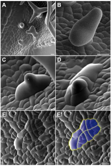
We therefore analyzed next how the trichome pattern becomes different over time in plants with reduced endoreplication levels and first examined in detail young leaves of these plants by scanning electron microscopy. We found that outside of the trichome patterning zone of leaves of ICK1/KRP1-mixexpressing
plants as well as our other mutant lines several trichomes were arrested in their development, i.e. large cells with an outgrowth cone typical of developing trichomes, but with a much wider base (Figure 5C); these trichomes were never found on wild-type leaves (Figure 5A and 5B). Strikingly, we found a few cases where such an aborted trichome showed several constrictions suggesting recent cell divisions (Figure 5D). Finally, we found unusual patches of cells that displayed common division planes (Figure 5E). Typically, cell divisions in the wild-type leaf epidermis are not coordinated but mosaic whereas in the above identified cell patches division planes were aligned over more than eight cells (Figure 5E). One explanation for this common orientation could be that a large precursor cell would have undergone many successive divisions. However, the cell size of this precursor must have been very large, much larger than the typical epidermal pavement cell.
To understand the origin of these cell patches and to test whether they could be derived from aborting trichomes, we followedin vivo
the fate of trichome initials on very young leaves. We first monitored wild-type trichomes labeled by GFP expressed from the GL2
promoter. We could track trichome initials developing into mature trichome cells during the time course of two days with pictures being taken every 24 hours; during the entire period theGL2reporter gave a strong fluorescence signal (Figure 6A and 6B). Since aborting trichomes are difficult to find, especially on young leaves that have a reduced number of epidermal cells inD,DEorKRP-misexpressing plants, we decided to follow trichome development in combinations of ICK1/KRP1-misexpressing plants withgl3mutants since these plants show one of the largest discrepancies between young and old leaves in terms of trichome numbers (compare Figure 3F and 3G with Figure 4D). In addition, we used for KRP expression anICK1/ KRP1-YFPfusion construct driven from theGL2promoter to mark trichomes and monitor at the same time the accumulation of the KRP protein.
Several leaves were observed over two to three days with pictures being taken every 24 hours. On many leaves we could observe that trichome initials were formed displaying strong YFP fluorescence similar to a fluorescent signal found in wild-type plants that express GFP under the control of theGL2promoter. In contrast to wild type, we could detect in a number of cases in which trichomes did not grow out further but underwent cell division. Figure 6 shows an example of a leaf in which three trichome initials divide, two initials undergoing one cell division giving rise to two cells and one initial dividing even twice leaving a patch of four enlarged cells that resembled the cluster that we had previously seen by SEM.
Figure 3. Genetic combinations of trichome patterning mutants with plants that have a reduced endoreplication levels in trichomes.Scanning electron micrographs of rosette leaf number 4. (A)
cpc try. (B)gl3. (C)PROGL2:KRP1109
incpc try. (D)PROGL2:KRP1109
ingl3. (E)
CDKA;1T161Dincpc try(F)CDKA;1T161Dingl3. (G) Quantification of trichome numbers of leaf 3 and 4 in comparison with wild type (Columbia). Error bars: standard deviation. Scale bars: (A–D) 50mm; (E,F) 200mm. doi:10.1371/journal.pgen.1000996.g003
Table 1.Trichome initiation sites containing multiple trichomes ontry-cpc-PROGL2:ICK1/KRP1plants.
Genotype Cluster1 Twin Single n
try cpc 94.0 6 4.6 1.3 6 2.5 4.7 6 4.8 21
#1 KRP1109 in try cpc
19.6 6 15.4 12.0 6 12.1 68.5 6 22.5 20
#2 KRP1109 in try cpc
29.2 6 13.9 16.1 6 6.0 54.6 6 10.1 20
try cpc x KRP1109
28.7 6 11.9 15.0 6 7.0 56.2 6 12.5 30
try cpc x KRP1IM
39.2 6 7.7 18.0 6 6.6 42.8 6 8.6 16
1A cluster is defined here as a trichome initiation site with more than 2 trichomes.
Moreover, in the aborting trichomes the YFP fluorescence rapidly diminished and was finally absent after two days. This could indicate that the KRP fusion protein would be rapidly degraded in this genetic background. However, non-aborting and outgrowing trichomes, as occasionally found on gl3–PROGL2: ICK1/KRP1-YFP plants, showed a strong YFP fluorescence (Figure 6C). Thus, we conclude that in the aborting and dividing trichomes theGL2promoter is switched off indicating that along with the entry into a mitotic cell cycle these cells have lost their trichome identity.
To corroborate the trichome cell fate loss, we introgressed four trichome markers into D and DE plants, comprising of the 59
regulatory region ofGL2,NOK, the putative kinaseAt2g36090and the F-box protein F9C22.2 encoding gene AT2G36090, respec-tively [29,54]. The GL2 reporter was found to be expressed in young wild-type and aborting trichomes but, while its expression continued in wild-type trichomes, the activity of the reporter ceased in aborting trichomes (Figure S1). In contrast, the reporter construct for NOK, At2g36090 and F9C22.2were only active in mature, three-branched trichomes and were not found to be expressed in aborting trichomes of mutants with reduced endoreplication levels (Figure S2, data not shown).
Finally, we measured the DNA content of these aborting trichomes. The aborting trichomes were found to have large nuclei, sometime even larger than wild-type trichomes at comparable stages (see also Figure 6D9 and 6E9). However, nuclear size is well known to not inevitably reflect DNA content and indeed the fluorescence intensity of DAPI-stained trichome
nuclei was much weaker of aborting trichomes than of wild type (Figure 7A–7C). Quantification of the exact DNA content of these aborting trichomes was difficult due to strong background fluorescent in young leaf parts and the often not clear distinction between a large epidermal cell and an aborting trichome during early developmental stages. None-the-less, our measurements always gave a similar trend with aborting trichomes having a DNA content between 2 and 4C, i.e. being not endoreplicated, in comparison with trichomes on wild-type plants at comparable stages that were found to have DNA levels between 4 and 8C (Figure 7D).
Taken together, our findings represent a case of trans-differentiation. Cells programmed to become trichomes and already expressing trichome identity genes changed their fate to an epidermal pavement cell program and were incorporated into the developing leaf epidermis.
Promotion of endoreplication partially rescues aborting trichomes
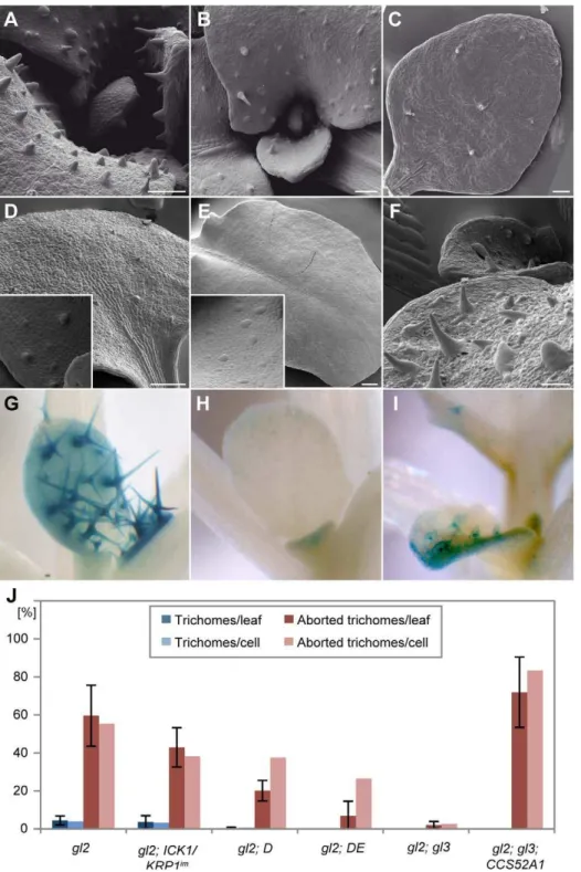
Rudimentary trichomes also occur on leaves ofgl2mutants and genetic combinations ofgl2withgl3have a synergistic effect and were previously reported to display leaves completely devoid of trichomes [34,55]. In the light of our above findings, we hypothesized that a decrease of ploidy levels in gl2–gl3 double mutants might be the main factor responsible for the lack of trichomes. Therefore we introduced our set of lines with reduced endoreplication levels in trichomes intogl2mutants. These genetic
Figure 4. Analysis of early trichome initation.Light micrographs of young rosette leaves. (A) Overview of a young leaf 4 in wild type. The trichome initiation zone was defined as the leaf region where trichomes emerge (indicated in blue) and only unbranched trichomes were counted. (B) Trichome initiation zone in wild type (Columbia). (C) Trichome initiation zone inCDKA;1T161D
leaves. (D) Quantification of the number of trichome initiation sites (TIS) compared to Columbia. Error bars: standard deviation n$8. Scale bars: (A) 100mm; (B,C) 25mm.
doi:10.1371/journal.pgen.1000996.g004
combinations with gl2 displayed a strong decline in trichome number and in the most severe cases (with the gl2 DE double mutant) all out-growing trichomes were eliminated (Figure 8A–8C and 8J and Table S1).
This raised the question whether trichomes were initiated in
gl2–gl3 double mutants but lose their fate similarly to the above made observations. Indeed, analysis by SEM revealed that large and distinct cells reminiscent of initial trichomes can be seen on young leaves but later on no traces of these cells are left (Figure 8E). We hypothesized that stimulation of a mitotic cycle ingl2mutants might further enhance trichome cell-fate loss. To test this we generated agl2-simdouble mutant and indeed the resulting double homozygous mutants resembledgl2–gl3mutants with no trichomes left on the leaf epidermis (Figure 8D). Again, we could find out-bulging cells on younger leaf parts indicating that trichomes were initially specified but lost their fate.
GL2 and GL3 are transcription factors that play a major role during trichome development and to test how important endocycle control versus a failure of regulating other target genes is, we sought for a way to specifically stimulate an endoreplication
cycle and/or block mitosis ingl2andgl2–gl3mutants. Since it was recently shown thatCCS52overexpression can induce endorepli-cation while blocking mitosis [56,57], we generated a
PROGL2:CCS52A1 construct and first introduced this into
wild-type plants. Similar to the misexpression ofCCS52from the35S
promoter, its overexpressing under the GL2 promoter control resulted in overbranched trichomes with an increase in trichome endoreplication levels but no obvious alteration of the trichome pattern was observed [56] (data not shown).
Next, we transformed thePROGL2:CCS52construct intogl2and gl2–g3mutants. Indeed, the number of trichomes and trichome-like structures in gl2 mutants that expressed PROGL2:CCS52
construct was higher than ingl2mutants (Figure S3A and S3B). The most striking effect was found in gl2–gl3 double mutants expressingCCS52: While old leaves ofgl2–gl3double mutants are devoid of trichomes, the CCS52 overexpression lines displayed distinct cells with an outgrowth cone that resembled trichomes on
gl2 mutants (Figure 8F and 8J). These trichome-like structures never branched or developed papillae typical for mature trichomes. However, given that GL2 and GL3 are two major regulators of trichome development, a complete restoration to wild-type like trichomes was not expected.
To test the differentiation status of these cells, we introduced the above-used NOK, At2g36090and F-box protein F9C22.2 promoter GUS reporter line into gl2, gl3, and into gl2-gl3 mutants that expressedCCS52under theGL2promoter control. In all genotypes analyzed, theAt2g36090andF9C22.2reporter were only active in outgrowing and neither in aborting trichomes nor in the trichome-like structures found ingl2orgl2–gl3mutants expressing CCS52 (Figure S2, data not shown). In contrast, the NOK reporter was expressed in outgrowinggl2mutant trichomes but was neither active in aborting trichomes ingl2nor ingl2–gl3mutants. However, ingl2– gl3mutants expressingPROGL2:CCS52we observed that almost all
rudimentary trichomes displayed a strong and enduring expression of this marker line (Figure 8G–8I). Similarly,gl2mutants expressing
PROGL2:CCS52construct showed activity of the NOK reporter in
the partially restored trichomes that developed in the center of the leaves (Figure S3C and S3D). Thus, the block of the re-initiation of a mitotic program and promotion of endoreplication ingl2and ingl2– gl3double mutants is sufficient to at least partially maintain and promote trichome fate.
Discussion
Traditionally, trichome development has been divided into three separate phases, pattern formation from a field of initially equivalent epidermal cells. Next,morphogenesiswith outgrowth and branch formation of the incipient trichome cell, accompanied by endoreplication. Finally,maturationwith expansion growth and the formation of papillae on the surface [34]. Here we show that at least the two earlier phases have a substantial overlap with a new role for endoreplication and the repression of mitosis in maintaining and/or reinforcing the patterning process (Figure 9). Thus, in addition to a feedback loop of the trichome activator complex, including GL3, we postulate a second partially overlapping module with another feedback loop containingGL2
acting during very early trichome pattern formation. This model is supported by the similarity of thegl2–gl3with thegl2-simdouble mutant. In both cases trichomes abort shortly after their initial formation but–as demonstrated for gl2–gl3–can be rescued by promoting endoreplication and the inhibition of cell division. Moreover, the need for such second feedback loop during pattern formation gives a glimpse at the dynamics and complexity of tissue organization in living organisms.
Figure 5. Morphology of aborted trichomes. Scanning electron micrographs of rosette leaves of wild-type plants (A,B) and plants expressing PROGL2:ICK1/KRP1 (C–E). (A) Trichome development on a
young wild-type leaf. (B) Emerging wild-type trichome. (C) Aborting trichome on a young leaf ofPROGL2:ICK1/KRP1plant. (D) Putative aborted
Endoreplication in trichome patterning
The data presented here show that endoreplication and the inhibition of mitosis are required for stabilization or maintenance of trichome cell fate, suggesting that there are developmental constraints on the patterning system. The switch from initial patterning to maintenance might be a sensitive phase in cell fate commitment, involving the transition from one major transcrip-tional program to another major transcriptranscrip-tional program, i.e. from a generic pattern module, also used for root hair patterning, to a
cell type-specific readout, i.e. trichome morphology and physiol-ogy. Consistent with this view, genome-wide transcript profiling and the analysis of promoter reporter constructs have revealed that the expression of many patterning genes is strongly reduced in maturing trichomes [29] (and reference list there in). At the same time, these profiling studies have shown thatGL2andSIMwere among the most strongly expressed genes in mature trichomes in comparison to leaves without trichomes (99.4 and 28.0 fold, respectively). Since GL2 was identified as a direct target of a
Figure 6. Live-imaging of aborting trichomes.Confocal-scanning micrographs of young rosette leaves; (A-C and D0-F0) overlay of propidium iodine channel and YFP/GFP channel; (D–F) Propidium iodine channel; (D9–F9) YFP/GFP channel. (A,B) Leaves ofPROGL2:GFPexpressing plants in
wild-type background after 24 h (A) and 48 h (B). (C) Leaf ofgl3-PROGL2:GUS:YFP:ICK1/KRP1109-191with a putative trichome precursor cell (arrow head) after
cell division without YFP fluorescence while an outgrowing, i.e. non-aborting, trichome (square) displays a strong YFP signal. (D) Young leaf ofgl3
-PROGL2:GUS:YFP:ICK1/KRP1109-191; young trichome cells can be identified by their increased size in comparison to the surrounding epidermal pavement
cells and a bright fluorescent signal in the nucleus. Please note that some surrounding epidermal cells also display YFP fluorescence due to the low activity of theGL2promoter in groups of epidermal cells encompassing the future trichome. (E) The same leaf as in (D) after 24 h and after 48 h (F). Three trichome initials are marked by a plus, an asterisks, a number sign (D0–F0). Please note yellow background fluorescence in E9/E0(cell walls are marked) and F9/F0(some cell walls and stomata) as a consequence of the high sensitivity in the detection procedure.
doi:10.1371/journal.pgen.1000996.g006
patterning gene complex [58–60] (see also Figure 9), one needs to postulate a second transcriptional input after/or in parallel to the initial pattern formation. Similarly,SIMexpression was found to positively correlate withGL3expression and chromatin immuno-precipitation experiments revealed that GL3 directly binds to the
SIM promoter region [29,35,43]. It seems possible that endor-eplication might be important to bridge these different transcrip-tional programs, for instance by increasing the intracellular concentrations of mRNAs for the proteins of the trichome activator complex (Figure 9).
Additionally, endoreplication is known to influence chromatin dynamics [61], and recent findings have even presented a molecular link between chromatin organization, pattern formation and cell-cycle control in particular during Arabidopsis root development. The Arabidopsis root epidermis is composed of alternating files of cells that all develop into hair cells (trichoblasts) or non-root hair cells (atrichoblasts). Root hair patterning and trichome patterning share many of the same regulators, such as
GL2 [21,62]. Evidence that the chromatin state of GL2 is connected to the specific cell fate in the root epidermis comes from the demonstration that the genomic region ofGL2was only accessible to FISH hybridization in atrichoblasts whereGL2is also expressed, and not in trichoblasts where theGL2promoter is not active [63]. Remarkably, the chromatin state was found to be very dynamic and could be established during a single cell cycle, likely between M-phase and G1 phase. It is plausible that endoreplica-tion might influence chromatin accessibility by fixing a certain chromatin state in trichomes.
GL2 expression was recently found to be also controlled by protein called GEM for GL2-EXPRESSION MODULATOR and GEM misexpression resulted in reduced levels of GL2
whereas gemmutants displayed higher amounts of GL2mRNA [64]. Consistently, gem mutants produced more trichomes and
GEMmisexpression lines produced fewer trichomes. GEM was found to influence histone modifications, i.e. acetylation and methylation, around genes involved in trichome patterning, i.e.
GL2 and CPC, presenting one possibility for the expression control ofGL2. GEM was found to interact with CDT1, a central component of the DNA replication machinery. CDT1 misexpres-sion in turn enhances endoreplication levels in trichomes and trichome branch numbers [65]. In addition, in these CDT1
misexpression lines,GL2is also upregulated offering a second link between DNA replication and GL2 expression [64]. Thus, chromatin organization and DNA replication might be more intrinsically linked to pattern formation, and could represent the second feedback loop that we have postulated to function during early trichome patterning (Figure 9).
Dedifferentiation and tissue patterning
The observation that trichomes can lose their fate and can be completely reintegrated into the pavement layer may shed light on general principles in pattern formation and tissue organization. A key question is which constraint underlies the necessity of a second feed back loop after pattern formation. Two major possibilities might explain this: The first scenario (cell-autonomous scenario) is based on the consideration that all epidermal cells, including trichome initials, are formed from an epidermal ground state. In this scenario, pavement cell fate would be the default state that must be overwritten during commitment of cells to the trichome fate. A failure to stabilize this program would reveal the default fate and accordingly, aborting trichomes would return to a pavement cell stage.
Figure 7. DNA content of young aborting trichomes.Light micrographs of young rosette leaves stained with DAPI. (A) Columbia wild-type plants. (B)ICK1/KRP1im. (C)gl3 - ICK1/KRP1im. (D) Quantification of DNA contents of trichome nuclei in relative fluorescence units (RFU). The RFU are calibrated by dividing epidermal cells nuclei so that 2 RFU roughly represents 2C.
In this scenario, chromatin regulation might play a key role since it is known from Drosophila that cell fate is fixed and epigenetically inherited over many cell divisions by establishing repressive or activating chromatin states. Of key importance here are the Polycomb repressive complex one and two (PRC1 and PRC2) and the trithorax complex. The PRC2 mediates the
tri-methylation of lysine 27 of Histone H3 leading to the recruitment of PRC1 complexes and the stable inactivation of the respective chromatin segment [66–68]. Homologs of the animal PRC2 complex have been identified in plants and among other target genes,GL2was found to be lysine K27 trimethylated in a PRC2-dependent manner [69] (D. Bouyer and A.S. unpublished data).
Figure 8. Partial rescue of trichome development ofgl2 gl3byPROGL2:CCS52A1expression.Scanning electron micrographs of rosette leaves (A–F) and light micrographs of GUS-stained leaves (G–I). (A)gl2. (B)gl2 - ICK1/KRP1im
. (C)gl2 - DE. (D)gl2 - sim. (E) Mature leaves ofgl2–gl3
double mutant are completely devoid of trichomes. (F)PROGL2:CCS52A1expression promotes endoreplication in trichome precursor cells and causes
the formation of trichome-like cells ingl2–gl3double mutants. (G–I) Expression of the trichome markerPRONOK:GUS. (G) Wild type. (H) Ingl2–gl3
mutants only a weak PRONOK:GUS activity can be detected in leaf margins. (I) The trichome-like cells in gl2–gl3-PROGL2:CCS52A1plants show PRONOK:GUSactivity. (J) Quantification of trichome number of leaf 3 and 4 in comparison with wild type.
doi:10.1371/journal.pgen.1000996.g008
Another not mutually exclusively possibility is that the fate of an aborting trichome could be influenced by its neighboring cells (non-cell-autonomous scenario). Support for non-cell-autonomous influences on maintenance of cell fate come from grafting or ablation experiments where it was found that a cell or its progeny, when invading from one developmental context to another, adapts its fate according to its new position [70–73]. The existence of locally acting tissue-specific supervision mechanisms are presum-ably very important to organize and maintain body architecture by correcting incorrectly oriented cell divisions. However, mutants that affect the organization of tissue layers are very rare, likely due to the fundamental nature of this process and the probable pleiotropic mutant phenotype. One example might be mutants in the receptor-like kinase CR4 from maize or its Arabidopsis homolog ACR4 that have been found to be crucial for epidermis development. Both are expressed in the epidermal cell layer but may receive signals from underlying cell layers, thus also coordinating inter-tissue organization [74–78].
Trichome cell fate seems to be determined by a very robust developmental program since trichomes can be initiated and differentiate in subepidmermal layers in try mutants plants that ectopically expressGL1[79]. It seems possible that endoreplication is one of the mechanisms that strongly stabilizes trichome fate and protects it from otherwise observed fate conversions induced by the neighboring cells. Interestingly, introgressing the patterning and endoreplication mutantgl3intotry-PRO35S:GL1plants dramatically
reduced the formation of subepidermal trichomes (A.S. and M.H., unpublished data). The emerging new tools to precisely study the development of single cell type for instance by laser dissection microscopy and a new round of mutant screens [31] will help to go in future one level deeper in the understanding of endoreplication during pattern formation and tissue integrity and will help to answer long standing questions in developmental biology.
Materials and Methods
Plant material and growth conditions
Arabidopsis (Arabidopsis thaliana) plants were grown on soil under long-day conditions (16 h of light, 8 h of darkness) between 18uC and 25uC at standard greenhouse conditions. To avoid the possibility of accession-specific variability in trichome develop-ment, only plants of the accession Columbia-0 (Col-0) were used with the exception of the cpc-try double mutant, which is a combination of the accessions Landsbergerecta(Ler) and Wasilews-kaja-0(WS-0) [44]. Thegl2,gl3andsimmutants inCol-0have been described previously[29,35,80]. For acdka;1mutant the previously characterized SALK T-DNA insertion allele was used [81]. The
CDKA; 1T161D (D) and CDKA; 1T14D/Y15E (DE) rescue lines of
cdka;12/2were generated by Dissmeyer et al. (2007, 2009). The PROGL2:ICK1/KRP1, PROGL2:ICK1/KRP1
109–191
and PROGL2: GUS:YFP:KRP1109–191 (ICK/KRP1im) misexpression lines are characterized in Weinl et al. and Jakoby et al. [46,49]. The PROMOTER-GUS reporter lines for MYB106 At3g01140 and
At2g36090 are described in Jakoby et al. [51]. Genotypes were confirmed by PCR, antibiotic selection and/or segregation analysis of the following generation.
Transgenic lines generated in this study
ThePROGL2:ICK1/KRP1109–191construct was transformed into cpc-try double mutants. For construction of PROCPC:CYCD3;1, PROTRY:CYCD3;1, and PROGL2:CCS52A1 the Gateway cloning
system was used. The destination vectors pAMPAT-PROCPC and pAMPAT-PROTRY are a kind gift of Martin Pesch, University of
Cologne [44], the pAMPAT-PROGL2 vector has been previously
described [46]. Molecular manipulations were performed accord-ing to standard procedures and plants were transformed by a modified version of the floral dip method according to Clough and
Figure 9. New model of trichome development.Trichome development appears to rely on two positive feed back loops. The first loop centers around the transcriptional activator GL3. The loop achieves the initial trichome pattern. Among other targets, GL3 directly activatesGL2andSIMthat are involved in a second feed back loop that is important for trichome cell fate maintenance and entry into an endoreplication cycle. Please note that in both feedback loops, GL3 and GL2 are operating together with many more regulators, indicated here as other factors.
Bent [82]. At least 20 transgenic plants were generated for all expression constructs. A number of representative reference lines displaying a typical phenotype were chosen for further analysis. Genotypes were confirmed by PCR, antibiotic selection and/or segregation analysis of the following generation.
Accession numbers
Sequence data for material used in this work can be found at TAIR (www.arabidopsis.org) and NCBI (www.ncbi.nlm.nih.gov) under the following accession numbers. For TAIR: At2g36090, At3g48750 (At CDKA;1), At2g46410 (At CPC), At4g34160 (At CYCD3;1), At2g36090 (At F9C22.2), At4g22910 (At FZR2/CCS52A1), At1g79840 (At GL2), At5g41315 (At GL3), At2g23430 (At ICK1/ KRP1), At3g01140 (At NOK), At5g04470 (At SIM), At5g53200 (At TRY). Germplasm information for deposited T-DNA-lines: cdka;1
(SALK_106809/Germplasm: 4824368), cpc (CS6399/Germplasm: 1007963690), gl3–3 (GK 545D05/Germplasm:3510637538),sim-1
(Germplasm:5529955621),try-EM1(Germplasm:3510701804).
Microscopy and image processing
Light microscopy was performed with an Axiophot microscope (Zeiss) and confocal laser scanning microscopy with a TCS SP2 AOBS CLSM system (Leica Microsystems). Scanning electron microscopy was done using a SUPRA 40VP (Zeiss) equipped with a K1250X Cryogenic SEM Preparation System (EMITECH). For image processing Leica Confocal Software Lite 2.05, Zeiss AxioVision 4.7, Adobe Photoshop CS2 and Adobe Illustrator CS2 were used. Image analysis was performed with Image J 1.43l (http://rsb.info.nih.gov/ij/).
DNA measurements
For DNA quantification of trichome nuclei, rosette leaf 4 was vacuum infiltrated for 30 min in formaldehyde solution (3.7% formaldehyde in PBS, 0.1% Tween [PBST]) followed by incubation at 4uC overnight. Samples were washed two times for 15 min in PBST. Afterwards, leaves were vacuum infiltrated in DAPI solution (0.25 mg/mL, 5% DMSO in PBST) for 15 min and incubated overnight in DAPI solution at 4uC; thereafter, leaves were washed twice in PBST. The DAPI intensity was quantified and the background fluorescence was subtracted using the ImageJ software (rsbweb.nih.gov/ij/). The median value of
Columbiaandgl3-3trichomes was set as 32C and 16C respectively. From this value, the corresponding C values of the trichome nuclei were estimated. For a comparison of the DNA content of young trichomes and aborting trichomes in the initiation zone, Z-sacks of the initiation zone were taken with an ApoTome (Zeiss). The exact number and intensity of each pixel of the trichome nuclei were measured using the Z-axis profile plot function in ImageJ (rsbweb.nih.gov/ij/). The relative fluorescent units (RFU) were scaled by comparing to the RFU of trichome nuclei with nuclei of surrounding dividing epidermal cells. The smallest epidermal nuclei were set to 2C.
Histology
The expression of the GUS protein was visualized as previously described [46]. For cell wall staining, leaves were directly mounted in a saturated solution of propidium iodide (100mg/ml) in water
and incubated for 5 min.
Trichome counting
Trichomes were counted on leaf 3 and 4 at a leaf length of 4 mm length. Agarose (2% in Water) prints were taken of each leaf allowing an accurate measurement of the total leaf size as well as
cell numbers and cell sizes by light microscopy. Total cell numbers and cell sizes per leaf were estimated by counting the number of cells in a square of 10000mm2located atJandLof the distance
between tip and base of a leaf, halfway between midrib and leaf margin. All measurements were repeated three times on separately grown plants. For analyzing early pattering processes, trichome initiation sites (TIS) were counted in the trichome initiation zone of leaf 4 before it reached a length of 800mm. The trichome
initiation zone was defined at the most basal region of a leaf restricted at its distal end by appearance of branched trichomes [41] Total cell numbers and cell sizes in a trichome initiation zone were estimated by counting all pavement cells per square of 961mm2. All measurements were repeated three times on
separately grown plants.
Trichome tracking
Plants were germinated and grown for 10 days on soil under long day conditions. Single leaves including the petioles and an upper part of the hypocotyls were cut off and put into a block of 1% MS agar so that the hypocotyls were embedded but the leaf blade was not in contact with the agar. These blocks were placed into Petri dishes and stored in a plant growth chamber under long day conditions for up to 72 hours. Each time before fluorescent images were obtained, leaves were stained in 200mM propidium
iodine for 5 min and washed with water. After image taking, the leaves were embedded to a new agar block and placed back into a Petri dish. Every 24 hours fluorescent images were taken by confocal laser-scanning microscopy using a 40x water-immersion objective without a cover slip.
Supporting Information
Figure S1 Activity of PROGL2:GUS in DE rosette leaves.
Expression of PROGL2:GUSin young (A) and mature trichomes
(B). (C) GUS staining of an early aborting trichome (marked by an arrowhead). (D) Cell patch putatively derived from an aborted trichome with no GUS expression (marked by an arrow). Note the GUS positive surrounding young trichomes.
Found at: doi:10.1371/journal.pgen.1000996.s001 (0.91 MB TIF)
Figure S2 Expression of PRONOK:GUS and PRO At2g36090:
GUS. (A,C,E,G,I) PRO At2g36090:GUS activity in rosette leaves.
(B,D,F,H,J) PRONOK:GUS. (A,B) Wild-type Columbia. (C,D)gl3.
E,Fgl2. G,Hgl2-gl3. I,Jgl2-gl3-PROGL2:CCS52A1.
Found at: doi:10.1371/journal.pgen.1000996.s002 (4.11 MB TIF)
Figure S3 Stereo-micrographs of rosette leaves (A,B) and light micrographs of GUS-stained rosette leaves (C,D). (A)gl2mutants predominantly form under-branched and small trichomes at the margin of leaves. (B) gl2 - PROGL2:CCS52A1 plants develop
trichome-like structures on central leaf areas; one is indicated by an arrow. (C) PRONOK:GUS activity can only be detected in gl2
mutants in the outgrowing trichomes near leaf margins. (D)
PRONOK:GUS marks trichome like structures on gl2 - PROGL2: CCS52A1plants. Scale bars: (A,B) 500mm; (C,D) 100mm.
Found at: doi:10.1371/journal.pgen.1000996.s003 (1.68 MB TIF)
Table S1 Trichome numbers and epidermal cell sizes of mature rosette leaves.
Found at: doi:10.1371/journal.pgen.1000996.s004 (0.02 MB XLS)
Table S2 Numbers and cell sizes of incipient trichomes and surrounding epidermal cells on young rosette leaves.
Found at: doi:10.1371/journal.pgen.1000996.s005 (0.02 MB XLS)
Acknowledgments
We thank Christian Fleck and Florian Geier for a fruitful and stimulating discussion on trichome pattern formation. The authors acknowledge Rolf-Dieter Hirtz for his excellent assistance in the Scanning electron microscope work. We thank Martina Pesch for providing cloning and expression vectors used in this study.
Author Contributions
Conceived and designed the experiments: JB AS. Performed the experiments: JB KW CW FR RK. Analyzed the data: JB KW CW FR JCL MH AS. Contributed reagents/materials/analysis tools: RK JCL MH. Wrote the paper: JB JCL MH AS.
References
1. Jakoby M, Schnittger A (2004) Cell cycle and differentiation. Curr Opin Plant Biol 7: 661–669.
2. Edgar BA, Orr-Weaver TL (2001) Endoreplication cell cycles: more for less. Cell 105: 297–306.
3. Kondorosi E, Roudier F, Gendreau E (2000) Plant cell-size control: growing by ploidy? Curr Opin Plant Biol 3: 488–492.
4. Sugimoto-Shirasu K, Roberts K (2003) ‘‘Big it up’’: endoreduplication and cell-size control in plants. Curr Opin Plant Biol 6: 544–553.
5. Morgan DO (1997) Cyclin-dependent kinases: engines, clocks, and micropro-cessors. Annu Rev Cell Dev Biol 13: 261–291.
6. Pines J (1999) Four-dimensional control of the cell cycle. Nat Cell Biol 1: E73–9. 7. Peters JM (2006) The anaphase promoting complex/cyclosome: a machine
designed to destroy. Nat Rev Mol Cell Biol 7: 644–656.
8. Nakayama KI, Nakayama K (2005) Regulation of the cell cycle by SCF-type ubiquitin ligases. Semin Cell Dev Biol 16: 323–333.
9. Capron A, Okresz L, Genschik P (2003) First glance at the plant APC/C, a highly conserved ubiquitin-protein ligase. Trends Plant Sci 8: 83–89. 10. Lechner E, Achard P, Vansiri A, Potuschak T, Genschik P (2006) F-box proteins
everywhere. Curr Opin Plant Biol 9: 631–638.
11. Fu¨lo¨p K, Tarayre S, Kelemen Z, Horva´th G, Kevei Z, et al. (2005) Arabidopsis anaphase-promoting complexes: multiple activators and wide range of substrates might keep APC perpetually busy. Cell Cycle 4: 1084–1092.
12. Pesin JA, Orr-Weaver TL (2008) Regulation of APC/C activators in mitosis and meiosis. Annu Rev Cell Dev Biol 24: 475–499.
13. De Clercq A, Inze D (2006) Cyclin-dependent kinase inhibitors in yeast, animals, and plants: a functional comparison. Crit Rev Biochem Mol Biol 41: 293–313. 14. Dissmeyer N, Nowack MK, Pusch S, Stals H, Inze D, et al. (2007) T-Loop Phosphorylation of Arabidopsis CDKA;1 Is Required for Its Function and Can Be Partially Substituted by an Aspartate Residue. Plant Cell 19: 972–985. 15. Harashima H, Shinmyo A, Sekine M (2007) Phosphorylation of threonine 161 in
plant cyclin-dependent kinase A is required for cell division by activation of its associated kinase. Plant J 52: 435–448.
16. De Veylder L, Beeckman T, Inze D (2007) The ins and outs of the plant cell cycle. Nat Rev Mol Cell Biol 8: 655–665.
17. Dissmeyer N, Weimer AK, Pusch S, De Schutter K, Kamei CL, et al. (2009) Control of cell proliferation, organ growth, and DNA damage response operate independently of dephosphorylation of the Arabidopsis Cdk1 homolog CDKA;1. The Plant Cell 21: 3641–3654.
18. Gutierrez C (2008) The Arabidopsis Cell Division Cycle. In: Last R, Chang C, Jander G, Kliebenstein D, McClung R, Millar H, eds. The Arabidopsis Book The American Society of Plant Biologists. pp 1–19.
19. Marks MD (1997) Molecular Genetic Analysis of Trichome Development in Arabidopsis. Annu Rev Plant Physiol Plant Mol Biol 48: 137–163.
20. Hulskamp M, Schnittger A, Folkers U (1999) Pattern formation and cell differentiation: trichomes in Arabidopsis as a genetic model system. Int Rev Cytol 186: 147–178.
21. Ishida T, Kurata T, Okada K, Wada T (2008) A genetic regulatory network in the development of trichomes and root hairs. Annu Rev Plant Biol 59: 365–386. 22. Bouyer D, Geier F, Kragler F, Schnittger A, Pesch M, et al. (2008) Two-dimensional patterning by a trapping/depletion mechanism: the role of TTG1 and GL3 in Arabidopsis trichome formation. PLoS Biol 6: e141. doi:10.1371/ journal.pbio.0060141.
23. Pesch M, Hu¨lskamp M (2009) One, two, three.models for trichome patterning in Arabidopsis? Current Opinion in Plant Biology 12: 587–592.
24. Tominaga R, Iwata M, Sano R, Inoue K, Okada K (2008) Arabidopsis CAPRICE-LIKE MYB 3 (CPL3) controls endoreduplication and flowering development in addition to trichome and root hair formation. Development 135: 1335–1345.
25. Kirik V, Simon M, Huelskamp M, Schiefelbein J (2004) The ENHANCER OF TRY AND CPC1 gene acts redundantly with TRIPTYCHON and CAPRICE in trichome and root hair cell patterning in Arabidopsis. Dev Biol 268: 506–513. 26. Kirik V, Simon M, Wester K, Schiefelbein J, Hulskamp M (2004) ENHANCER of TRY and CPC 2 (ETC2) reveals redundancy in the region-specific control of trichome development of Arabidopsis. Plant Mol Biol 55: 389–398. 27. Wang S, Kwak SH, Zeng Q, Ellis BE, Chen XY, et al. (2007)
TRICHOME-LESS1 regulates trichome patterning by suppressing GLABRA1 in Arabidopsis. Development 134: 3873–3882.
28. Wester K, Digiuni S, Geier F, Timmer J, Fleck C, Hulskamp M (2009) Functional diversity of R3 single-repeat genes in trichome development. Development 136: 1487–1496.
29. Jakoby MJ, Falkenhan D, Mader MT, Brininstool G, Wischnitzki E, et al. (2008) Transcriptional profiling of mature Arabidopsis trichomes reveals that NOECK
encodes the MIXTA-like transcriptional regulator MYB106. Plant Physiol 148: 1583–1602.
30. Lieckfeldt E, Simon-Rosin U, Kose F, Zoeller D, Schliep M, Fisahn J (2008) Gene expression profiling of single epidermal, basal and trichome cells of Arabidopsis thaliana. Journal of Plant Physiology 165: 1530–1544.
31. Marks MD, Wenger JP, Gilding E, Jilk R, Dixon RA (2009) Transcriptome analysis of Arabidopsis wild-type and gl3-sst sim trichomes identifies four additional genes required for trichome development. Molecular Plant 2: 803–822.
32. Larkin JC, Brown ML, Schiefelbein J (2003) HOW DO CELLS KNOW WHAT THEY WANT TO BE WHEN THEY GROW UP? Lessons from Epidermal Patterning in Arabidopsis. Annual Review of Plant Biology 54: 403–430.
33. Folkers U, Berger J, Hulskamp M (1997) Cell morphogenesis of trichomes in Arabidopsis: differential control of primary and secondary branching by branch initiation regulators and cell growth. Development 124: 3779–3786. 34. Hulskamp M, Misra S, Jurgens G (1994) Genetic dissection of trichome cell
development in Arabidopsis. Cell 76: 555–566.
35. Churchman ML, Brown ML, Kato N, Kirik V, Hulskamp M, et al. (2006) SIAMESE, a Plant-Specific Cell Cycle Regulator, Controls Endoreplication Onset in Arabidopsis thaliana. Plant Cell 18: 3145–3157.
36. Walker JD, Oppenheimer DG, Concienne J, Larkin JC (2000) SIAMESE, a gene controlling the endoreduplication cell cycle in Arabidopsis thaliana trichomes. Development 127: 3931–3940.
37. Peres A, Churchman ML, Hariharan S, Himanen K, Verkest A, et al. (2007) Novel plant-specific cyclin-dependent kinase inhibitors induced by biotic and abiotic stresses. J Biol Chem 282: 25588–25596.
38. Schnittger A, Schobinger U, Bouyer D, Weinl C, Stierhof YD, Hulskamp M (2002) Ectopic D-type cyclin expression induces not only DNA replication but also cell division in Arabidopsis trichomes. Proc Natl Acad Sci U S A 99: 6410–6415.
39. Schnittger A, Schobinger U, Stierhof Y, Hulskamp M (2005) Erratum: Ectopic B-Type Cyclin Expression Induces Mitotic Cycles in Endoreduplicating Arabidopsis Trichomes. Curr Biol 15: 980.
40. Schnittger A, Schobinger U, Stierhof YD, Hulskamp M (2002) Ectopic B-type cyclin expression induces mitotic cycles in endoreduplicating Arabidopsis trichomes. Curr Biol 12: 415–420.
41. Schnittger A, Folkers U, Schwab B, Jurgens G, Hulskamp M (1999) Generation of a spacing pattern: the role of triptychon in trichome patterning in Arabidopsis. Plant Cell 11: 1105–1116.
42. Szymanski DB, Marks MD (1998)GLABROUS1Overexpression and TRIPTY-CHONAlter the Cell Cycle and Trichome Cell Fate in Arabidopsis. Plant Cell 10: 2047–2062.
43. Morohashi K, Grotewold E (2009) A systems approach reveals regulatory circuitry for Arabidopsis trichome initiation by the GL3 and GL1 selectors. PLoS Genet 5: e1000396. doi:10.1371/journal.pgen.1000396.
44. Schellmann S, Schnittger A, Kirik V, Wada T, Okada K, et al. (2002) TRIPTYCHON and CAPRICE mediate lateral inhibition during trichome and root hair patterning in Arabidopsis. Embo J 21: 5036–5046.
45. Schnittger A, Weinl C, Bouyer D, Schobinger U, Hulskamp M (2003) Misexpression of the cyclin-dependent kinase inhibitor ICK1/KRP1 in single-celled Arabidopsis trichomes reduces endoreduplication and cell size and induces cell death. Plant Cell 15: 303–315.
46. Weinl C, Marquardt S, Kuijt SJ, Nowack MK, Jakoby MJ, et al. (2005) Novel Functions of Plant Cyclin-Dependent Kinase Inhibitors, ICK1/KRP1, Can Act Non-Cell-Autonomously and Inhibit Entry into Mitosis. Plant Cell 17: 1704–1722.
47. Wang H, Qi Q, Schorr P, Cutler AJ, Crosby WL, Fowke LC (1998) ICK1, a cyclin-dependent protein kinase inhibitor from Arabidopsis thaliana interacts with both Cdc2a and CycD3, and its expression is induced by abscisic acid. Plant J 15: 501–510.
48. Zhou Y, Li G, Brandizzi F, Fowke LC, Wang H (2003) The plant cyclin-dependent kinase inhibitor ICK1 has distinct functional domains for in vivo kinase inhibition, protein instability and nuclear localization. Plant J 35: 476–489.
49. Jakoby MJ, Weinl C, Pusch S, Kuijt SJ, Merkle T, et al. (2006) Analysis of the subcellular localization, function and proteolytic control of the Arabidopsis CDK inhibitor ICK1/KRP1. Plant Physiol 141: 1293–1305.
51. Wang H, Zhou Y, Fowke LC (2006) The emerging importance of cyclin-dependent kinase inhibitors in the regulation of the plant cell cycle and related processes. Canadian Journal of Botany 84: 640–650.
52. Payne CT, Zhang F, Lloyd AM (2000)GL3encodes a bHLH protein that regulates trichome development in Arabidopsis through interaction withGL1
andTTG1. Genetics 156: 1349–1362.
53. Larkin JC, Young N, Prigge M, Marks MD (1996) The control of trichome spacing and number in Arabidopsis. Development 122: 997–1005.
54. Szymanski DB, Jilk RA, Pollock SM, Marks MD (1998) Control of GL2 expression in Arabidopsis leaves and trichomes. Development 125: 1161–1171. 55. Ohashi Y, Oka A, Ruberti I, Morelli G, Aoyama T (2002) Entopically additive expression of GLABRA2 alters the frequency and spacing of trichome initiation. Plant J 29: 359–369.
56. Larson-Rabin Z, Li Z, Masson PH, Day CD (2009) FZR2/CCS52A1 expression is a determinant of endoreduplication and cell expansion in Arabidopsis. PLANT PHYSIOLOGY 149: 874–884.
57. Kasili R, Walker JD, Simmons LA, Zhou J, De Veylder L, Larkin JC. SIAMESE cooperates with a CDH1-like protein to establish endoreplication. Genetics, (in press).
58. Wang S, Chen JG (2008) Arabidopsis transient expression analysis reveals that activation of GLABRA2 may require concurrent binding of GLABRA1 and GLABRA3 to the promoter of GLABRA2. Plant Cell Physiol 49: 1792–1804. 59. Zhao M, Morohashi K, Hatlestad G, Grotewold E, Lloyd A (2008) The
TTG1-bHLH-MYB complex controls trichome cell fate and patterning through direct targeting of regulatory loci. Development 135: 1991–1999.
60. Morohashi K, Zhao M, Yang M, Read B, Lloyd A, et al. (2007) Participation of the Arabidopsis bHLH factor GL3 in trichome initiation regulatory events. PLANT PHYSIOLOGY 145: 736–746.
61. Kato N, Lam E (2003) Chromatin of endoreduplicated pavement cells has greater range of movement than that of diploid guard cells in Arabidopsis thaliana. J Cell Sci 116: 2195–2201.
62. Schiefelbein J, Kwak SH, Wieckowski Y, Barron C, Bruex A (2009) The gene regulatory network for root epidermal cell-type pattern formation in Arabi-dopsis. J Exp Bot 60: 1515–1521.
63. Costa S, Shaw P (2006) Chromatin organization and cell fate switch respond to positional information in Arabidopsis. Nature 439: 493–496.
64. Caro E, Castellano MM, Gutierrez C (2007) A chromatin link that couples cell division to root epidermis patterning in Arabidopsis. Nature 447: 213–217. 65. del Mar Castellano M, Boniotti MB, Caro E, Schnittger A, Gutierrez C (2004)
DNA replication licensing affects cell proliferation or endoreplication in a cell type-specific manner. Plant Cell 16: 2380–2393.
66. Schuettengruber B, Chourrout D, Vervoort M, Leblanc B, Cavalli G (2007) Genome regulation by polycomb and trithorax proteins. Cell 128: 735–745. 67. Schwartz YB, Pirrotta V (2008) Polycomb complexes and epigenetic states.
Current Opinion in Cell Biology 20: 266–273.
68. Simon JA, Kingston RE (2009) Mechanisms of polycomb gene silencing: knowns and unknowns. Nat Rev Mol Cell Biol 10: 697–708.
69. Zhang X, Clarenz O, Cokus S, Bernatavichute Y, Pellegrini M, et al. (2007) Whole-genome analysis of histone H3 lysine 27 trimethylation in Arabidopsis. PLoS Biol 5: e129. doi:10.1371/journal.pbio.0050129.
70. van den Berg C, Willemsen V, Hage W, Weisbeek P, Scheres B (1995) Cell fate in the Arabidopsis root meristem determined by directional signalling. Nature 378: 62–65.
71. Kidner C, Sundaresan V, Roberts K, Dolan L (2000) Clonal analysis of the Arabidopsis root confirms that position, not lineage, determines cell fate. Planta 211: 191–199.
72. Stewart RN, Burk L (1970) Independence of tissues derived from apical layers in ontogeny of the tobacco leaf and ovary. American Journal of Botany 58: 1010–1016.
73. Dermen H, Stewart R (1973) Ontogenetic study of floral organs of peach (Prunus persica) utilizing cytochimeral plants. American Journal of Botany 60: 283–291.
74. Becraft PW, Stinard PS, McCarty DR (1996) CRINKLY4: A TNFR-like receptor kinase involved in maize epidermal differentiation. Science 273: 1406–1409.
75. Tanaka H, Watanabe M, Watanabe D, Tanaka T, Machida C, Machida Y (2002) ACR4, a putative receptor kinase gene of Arabidopsis thaliana, that is expressed in the outer cell layers of embryos and plants, is involved in proper embryogenesis. Plant Cell Physiol 43: 419–428.
76. Becraft PW, Kang SH, Suh SG (2001) The maize CRINKLY4 receptor kinase controls a cell-autonomous differentiation response. PLANT PHYSIOLOGY 127: 486–496.
77. Gifford ML, Dean S, Ingram GC (2003) The Arabidopsis ACR4 gene plays a role in cell layer organisation during ovule integument and sepal margin development. Development 130: 4249–4258.
78. Watanabe M, Tanaka H, Watanabe D, Machida C, Machida Y (2004) The ACR4 receptor-like kinase is required for surface formation of epidermis-related tissues in Arabidopsis thaliana. Plant J 39: 298–308.
79. Schnittger A, Jurgens G, Hulskamp M (1998) Tissue layer and organ specificity of trichome formation are regulated by GLABRA1 and TRIPTYCHON in Arabidopsis. Development 125: 2283–2289.
80. Kirik V, Lee MM, Wester K, Herrmann U, Zheng Z, et al. (2005) Functional diversification of MYB23 and GL1 genes in trichome morphogenesis and initiation. Development 132: 1477–1485.
81. Nowack MK, Grini PE, Jakoby MJ, Lafos M, Koncz C, Schnittger A (2006) A positive signal from the fertilization of the egg cell sets off endosperm proliferation in angiosperm embryogenesis. Nat Genet 38: 63–67.
82. Clough SJ, Bent AF (1998) Floral dip: a simplified method for Agrobacterium-mediated transformation ofArabidopsis thaliana. Plant J 16: 735–743.