Catarina Gonçalves Pimpão
Licenciada em Bioquímica
Screening and Validation of Aquaporin Inhibitors for
Cancer Therapeutics
Dissertação para obtenção do Grau de Mestre em
Bioquímica para a Saúde
Orientadora: Prof. Drª Graça Soveral, Professora Associada com
Agregação, Faculdade de Farmácia, Universidade de Lisboa
Júri:
Presidente: Prof. Drª Maria Teresa Nunes Mangas Catarino Arguente: Prof. Drª Maria Alice Santos Pereira
Vogal: Prof. Drª Graça Soveral
Faculdade de Ciências e Tecnologia da Universidade Nova de Lisboa
S cr ee n in g a n d V al id at ion of A q u a p or in I n h ib it or s for C an ce r T h er ap eu ti cs C at ar in a Pi m p ão 2019
Screening and Validation of Aquaporin Inhibitors for Cancer Therapeutics
Os direitos de autor pertencem a Catarina Gonçalves Pimpão, à Faculdade de Ciências e Tecnologia da Universidade Nova de Lisboa e à Universidade Nova de Lisboa.
A Faculdade de Ciências e Tecnologia da Universidade Nova de Lisboa tem o direito, perpétuo e sem limites geográficos de arquivar e publicar esta dissertação através de exemplares impressos reproduzidos em papel ou de forma digital, ou por qualquer outro meio conhecido ou que venha a ser inventado, e de a divulgar através de repositórios científicos e de admitir a sua cópia e distribuição com objetivos educacionais ou de investigação, não comerciais, desde que seja dado crédito ao autor e editor.
Copyright belongs to Catarina Gonçalves Pimpão and Faculdade de Ciências e Tecnologia da Universidade Nova de Lisboa and Universidade Nova de Lisboa.
Faculdade de Ciências e Tecnologia da Universidade Nova de Lisboa has the perpetual and geographically unlimited right of archiving and publishing this thesis through printed or digital copies, or by any other means known or to be invented, and to divulgate its contents through scientific repositories and to admit its copy and distribution with educational or research, non-commercial goals, as long as its author and editor are properly credit.
Part of the results obtained during this work were included in three conference abstracts and in a published scientific article:
C. Pimpão, A.S. Coxixo, M. Aureliano, A. Rompel, G. Soveral, “Screening Polyoxometalates as
Aquaporin Inhibitors for Cancer Therapeutics” in FEBS Congress 2019: From Molecules To
Living Systems, 6-11 July 2019, Krakow, Poland.
C. Pimpão, A.S. Coxixo, M. Aureliano, A. Rompel, G. Soveral, “Polyoxometalates as Aquaporin
Inhibitors with Potential Anticancer Properties” in 11th iMed.ULisboa Postgraduate Students
Meeting & 4rd i3DU Meeting, 15 July 2019, Lisbon, Portugal.
A. Serrano, C. Pimpão, A.S. Coxixo, G. Fraqueza, A. Rompel, G. Soveral, M. Aureliano, “Polyoxotungstates with anticancer activity: Are membrane proteins attractive targets?” in SINAL
2019: 10th Meeting in Signal Transduction, 18-19 October 2019, Olhão, Algarve, Portugal.
C. Rodrigues, C. Pimpão et al., “Human Aquaporin-5 Facilitates Hydrogen Peroxide Permeation and Cancer Cell Migration,” Cancers (Basel)., vol. 11, 2019.
Acknowledgments
Chegou ao fim mais uma etapa da minha vida. Esta fase que tanto esforço e dedicação pediu e que é mais uma prova da determinação e persistência que me caracteriza, além de uma enorme superação pessoal. Foi um ano de muitas aprendizagens, cheio de boas experiências a fazer aquilo que mais gosto. Claro que este caminho não teria sido feito sem o apoio e suporte das pessoas certas que contribuíram para o meu sucesso e bem-estar a quem quero expressar o meu agradecimento.
Quero primeiro agradecer à Professora Graça Soveral por me ter acolhido no seu laboratório, por estar sempre lá quando alguma coisa corria menos bem e por me ter dado esta oportunidade de fazer aquilo que gosto. Além disso, agradeço imenso ter tido a oportunidade de ir ao Congresso da FEBS em Cracóvia para apresentar os meus resultados, foi com certeza muito enriquecedor tal como ter trabalhado com Brech Aikman e Simon Krabbe em que me pôs em contacto com outras formas de trabalhar.
À Andreia, Claúdia e Inês tenho muito a agradecer. Obrigado pela paciência, pelo apoio e ajuda no que precisasse. Em especial à Claúdia, um obrigado pela música de fundo no laboratório e em geral, pela animação diária naquele laboratório. Muito orgulhosa da tua conquista, Doutora! Também um obrigado especial à Inês pela companhia nos congressos e pelos cappuccinos do Sr. Joaquim.
À Ana Coxixo, que no início era a minha colega de tese e que rapidamente se revelou num apoio e companhia diária naquele laboratório. A ti, um obrigado pela amizade, pelas conversas, por me fazeres rir mesmo quando estávamos em desespero, pela companhia nos dias compridos, por teres estado ao meu lado no Hospital Santa Maria durante aquelas longas oito horas e claro, a surpresa no dia da minha partida. Apesar de muitas dúvidas pelo meio, conseguimos amiga, tese feita!
Às minhas amigas Andreia, Sara, Inês, Kiki, Teresa, Painho e Carina, um especial obrigado pelo apoio neste último ano, foram cruciais para o meu sucesso no mestrado! Em especial, Andreia, Sara e Kiki, obrigada pelos lanchinhos, jantares e afins que me faziam descontrair do dia-a-dia. Andreia e Sara, obrigada por alinharem comigo em escrever a tese no caleidoscópio, que foi sempre uma boa desculpa para nos encontrarmos e apoiarmos mutuamente nesta fase.
Ao João, que chegou mais tarde, mas que sem dúvida foi das pessoas que esteve presente em todos os meus sucessos e nos momentos menos bons. Obrigada por nunca vacilares, pelos jantares e fins de semana que me aliviam do stress e por seres o companheiro de todas as horas, sempre a dar-me força e confiança para atingir os meus objectivos. À família do João, a minha segunda família que sempre me acolheu e compreendeu.
Quero também agradecer à Professora Teresa Catarino, sempre disponível para ajudar e esclarecer dúvidas e tão bem acolheu a nossa turma de mestrado. Também um obrigado aos meus colegas de mestrado e à FCT/UNL, ITQB e NMS por me terem acolhido e por todas as vivências. Um obrigado também aos escuteiros, que preencheram e animaram os meus fins-de-semana. É e sempre será uma parte essencial na minha vida e que moldou os meus princípios e valores, permitindo-me chegar onde cheguei. Aos caminheiros, à Rute e a todos os meus chefes e principalmente aos meus miúdos, o meu grande obrigado.
Por último, agradecer à minha família que teve sempre uma palavra de força, principalmente durante todo aquele mês de Agosto. Um grande obrigado por me apoiarem incondicionalmente e confiarem em mim e nas minhas capacidades.
Resumo
As aquaporinas (AQPs) são proteínas transmembranares que facilitam a difusão da água e glicerol através das membranas celulares, cruciais para a homeostase da água e metabolismo. As AQPs encontram-se sobre-expressas em diversas células e tecidos cancerígenos, estando envolvidas na proliferação, migração celular e tumorigénese, revelando o seu potencial como alvos terapêuticos. A identificação de novos inibidores de aquaporinas é essencial. Neste trabalho, foram avaliados compostos metálicos (polioxometalatos, compostos de vanádio, cobre, zinco e ouro) como inibidores de aquaporinas, utilizando como modelos celulares eritrócitos humanos (RBCs) e leveduras transformadas com aquaporinas humanas. O efeito inibidor das AQPs foi avaliado pela determinação da permeabilidade membranar à água e ao glicerol, utilizando a técnica de stopped-flow. O polioxotungstato-A (POT-A) revelou ser um inibidor da AQP3, com um IC50 de (0.71 ± 0.04) µM em RBCs. Em leveduras, POT-A inibiu AQP3 a 100%, confirmando
os resultados obtidos para os RBCs. O composto de vanádio P103 inibiu AQP1 com um IC50 de
(9.11 ± 0.03) µM em RBCs. O composto de ouro RBA29, mostrou inibir a AQP3 em RBCs com um IC50 de (2.29 ± 0.03) µM. Ainda, os compostos RBA29, RBA31 e STAM013 mostraram inibir
a AQP9 em leveduras, com um IC50 de (6.64 ± 0.09) µM para RBA29. Neste trabalho, foi também
avaliado o efeito de mutações na permeabilidade da AQP5 através da técnica de stopped-flow. Deste modo, foi descrito um novo mecanismo de regulação em que His73 localizada no filtro de seletividade interage com Ser183 fosforilada, levando à constrição do poro. Foi também avaliada a função de aquaporinas de diversos organismos, em que a aquaporina de tardígrado revelou maior permeabilidade e seletividade para a água do que as outras aquaporinas testadas. Os resultados obtidos contribuem para a descoberta e desenho de inibidores seletivos com aplicação terapêutica.
Abstract
Aquaporins (AQPs) are transmembrane proteins that facilitate the diffusion of water and glycerol across cell membranes, crucial for water and energy homeostasis. These proteins were found overexpressed in different cancer cells and tissues, being involved in tumor formation, cell proliferation and migration, suggesting their great potential as drug targets for cancer treatment. Identification of novel aquaporin modulators to be used in cancer therapeutics is of utmost importance. In this study, the inhibitory effect of polyoxometalates, vanadium, copper, zinc and gold-based compounds was screened by the stopped-flow technique in human red blood cells (RBCs) and then validated in aquaporin-expressing yeast cells. From the set of polyoxometalates, polyoxotungstate-A (POT-A) revealed as the most promising AQP3 inhibitor with an IC50 of (0.71
± 0.04) µM. Using yeast cells individually expressing human aquaporins, POT-A showed to selectively inhibit AQP3 with 100% inhibition, corroborating the results of RBCs assays. The vanadium compound P103, showed highly inhibition of AQP1 with an IC50 of (9.11 ± 0.03) µM in
RBCs. The gold-based compound RBA29 revealed as a promising AQP3 inhibitor with an IC50 of
(2.29 ± 0.03) µM in RBCs. Moreover, compounds RBA29, RBA31 and STAM013 showed an inhibitory effect in AQP9-tranformed yeasts, with an IC50 of (6.64 ± 0.09) µM for RBA29. In
addition, investigation of the channel residues important for AQP5 permeability revealed a new gating mechanism where His73 located in the selectivity filter interacts with phosphorylated Ser183, conducting to the pore constriction. Furthermore, the activity of aquaporins from diverse organisms was evaluated, wherein aquaporin from tardigrade revealed to be water selective and exhibited higher water permeability than the other aquaporins tested. Overall, these data contribute to the discovery and design of selective inhibitors with potential therapeutic application.
Table of Contents
Acknowledgments ...ix
Resumo...xi
Abstract ... xiii
Index of Figures ... xvii
Index of Tables ... xxv
List of Abbreviations ... xxvii
1. Introduction ... 1
1.1. Importance of water in living cells and discovery of aquaporins ... 1
1.2. Aquaporins ... 1
1.2.1. Aquaporins structure and function ... 2
1.2.2. Localization, biological functions of aquaporins and associated pathologies ... 7
1.2.3. Aquaporin inhibitors ... 14
2. Thesis aims ... 19
3. Materials and Methods ... 21
3.1 Compounds preparation ... 21
3.1.1. Polyoxometalates ... 21
3.1.2. Vanadium, copper and zinc compounds ... 22
3.1.3. Gold compounds ... 23
3.2. RBCs assays ... 23
3.2.1. Ethics statement ... 23
3.2.2. Buffers and solutions preparation ... 24
3.2.3. Erythrocyte sampling, preparation and compound incubation ... 24
3.2.4. Reversibility assay ... 24
3.3. Yeast assays ... 25
3.3.1. Strains ... 25
3.3.2. Buffers, culture media and amino acids preparation ... 26
3.3.3. Growth and maintenance conditions ... 27
3.3.4. Storage of the yeast cells ... 28
3.3.5. Yeast cells preparation and compound incubation ... 28
3.3.6. Detection of aquaporin localization through fluorescence microscopy ... 29
3.4. Aquaporins functional assays using the stopped-flow spectroscopy ... 29
3.4.1. Functional assays for RBCs ... 31
3.4.2. Functional assays for yeast cells ... 32
3.5. Statistical analysis ... 34
4. Results and Discussion ... 35
4.1. Screening of metallodrugs as aquaporin inhibitors... 35
4.1.1. Inhibition studies of water and glycerol permeability in aquaporins by polyoxometalates ... 37
xvi 4.1.2. Inhibition studies of water and glycerol permeability in aquaporins by vanadium,
copper and zinc compounds ... 44
4.1.2.1. Inhibitory effect of vanadium, copper and zinc compounds in RBCs permeability 44 4.1.3. Inhibition studies of water and glycerol permeability in aquaporins by gold compounds... 49
4.2. Functional assays of hAQP5 and important residues for permeability ... 55
4.2.1. hAQP5 localization and function in the yeast plasma membrane ... 55
4.2.2. Role of hAQP5 residues in gating ... 56
4.3. Functional assays of aquaporins from diverse organisms ... 59
5. Conclusions ... 65
Index of Figures
Figure Caption Page
Figure 1.1 Top view of the extracellular face of an aquaporin-1 (APQ1) homotetramer, with monomer labelled from 1 to 4, based on the X-ray structure of bovine AQP1. (Adapted from [19]).
3
Figure 1.2 Topology and three-dimensional structure of an aquaporin
protein within the membrane.
A – Topology structure of an aquaporin within the
membrane, representing the six transmembrane helices from I to VI and the five loops from A to E. Loops B and E contains the conserved NPA motifs. C represents the carboxyl terminus and N represents the amino terminus. B – “Hourglass” three-dimensional structure of an aquaporin monomer (a ribbon model of NtAQP1, a PIP1 protein from tobacco) within the membrane. The arrows represent short α-helices, that were formed by loop B and E, entering the membrane from the extracellular and intracellular surfaces; the two NPA boxes are indicated in green; the six transmembrane helices from I to VI are also shown. (Adapted from [9]).
4
Figure 1.3 AQP1 selectivity filter water molecules and residues forming the hydrophilic face of the channel pore. Cut-away side view of the channel with secondary structure shown in ribbon format. Side chains critical to establishing the hydrophilic path across the length of the selectivity filter are shown. The four water molecules located within the selectivity filter are represented as green spheres. Of these four water molecules only the middle two are close enough to form a hydrogen bond between water molecules. The constriction region and NPA motifs are indicated by light blue and black arrows, respectively. (Adapted from [18]).
5
Figure 1.4 Representation of the mechanism for water permeation and blockage of proton transport of AQP1. A – Representation of how the partial charges from the short helices’ dipoles reorient the water molecules passing through the constriction of the pore. B and C – Representation of the hydrogen bonding of the oxygen atom of the water molecule to the amide groups of Asn 76 and Asn 192. (Adapted from [23]).
6
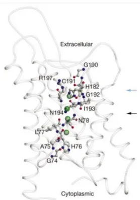
10
Figure 1.5 Proposed mechanism of AQP-dependent cell migration, showing water influx through AQP at the tip of a lamellipodium. (Adapted from [30]).
Figure 1.6 Overexpression of aquaporins in various types of cancer: brain, lung, liver, colorectal and breast. (Adapted from [63]).
xviii
Figure 1.7 Gold(III) coordination compounds as inhibitors of aquaglyceroporins. (Adapted from [25]).
17
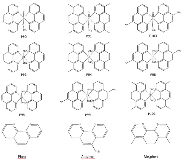
Figure 3.1 Structures of the metallodrugs provided by the Instituto Superior Técnico.
22
Figure 3.2 Structures of the gold compounds provided by Cardiff University.
23
Figure 3.3 Circular map of pUG35 vector with restriction site to insert the aquaporin gene, regulated by MET25 promoter and in line with yEGFP3, the GFP gene.
26
Figure 3.4 Scheme of the stopped-flow technique. (Adapted from [103]).
30
Figure 3.5 Cell shrinkage due to water efflux after an imposed hyperosmotic gradient with a non-permeable solute. (Adapted from [2]).
31
Figure 3.6 Cell volume change after imposing a hyperosmotic glycerol solution. First, the cells shrink rapidly due to water efflux and then glycerol enters the cell followed by water influx, causing the reswelling of the. (Adapted from [2]).
32
Figure 4.1 Traces of water and glycerol transport through cell membrane. A – Stopped-flow representative signal for water transport after imposing a hyperosmotic sucrose solution to the cells, in control RBCs and in the presence of inhibitor. B – Stopped-flow representative signal for glycerol transport after imposing a hyperosmotic glycerol solution to the cells, in control RBCs and in the presence of an inhibitor.
35
Figure 4.2 Inhibition of water and glycerol transport in RBCs by eleven polyxoxometalates at a concentration of 100 µM after 30 minutes incubation at RT. Data are shown as media ± SEM for one or two experiments. Significance levels: **** P < 0.0001, *** P < 0.001, ** P < 0.01, * P < 0.05, ns – nonsignificant, compared to the control by student t-test.
37
Figure 4.3 Inhibitory effect of POT-1 on RBCs glycerol permeability.
A - Concentration dependent inhibition of glycerol
permeability in RBCs by POT-1 for concentrations between 0.1-100 µM (incubated for 30 minutes at RT) with an IC50
value of (2.78 ± 0.09) µM (n=2) B – Stopped-flow signal for glycerol transport after imposing a hyperosmotic glycerol solution on the cells, for control RBCs and in the presence of 100 µM of POT-1.
Figure 4.4 Inhibitory effect of POT-3 on RBCs glycerol permeability.
A - Concentration dependent inhibition of glycerol
permeability in RBCs by POT-3 for concentrations between 0.1-100 µM (incubated for 30 minutes at RT) with an IC50
> 50 µM (n=1) B – Stopped-flow signal for glycerol transport after imposing a hyperosmotic glycerol solution on the cells, for control RBCs and in the presence of 100 µM of POT-3.
39
Figure 4.5 Inhibitory effect of POT-A on RBCs glycerol permeability.
A - Concentration dependent inhibition of glycerol
permeability in RBCs by POT-A for concentrations between 0.1-100 µM (incubated for 30 minutes at RT) with an IC50 value of (0.71 ± 0.04) µM (n=2) B – Stopped-flow
signal for glycerol transport after imposing a hyperosmotic glycerol solution on the cells, for control RBCs and in the presence of 100 µM of POT-A.
39
Figure 4.6 Inhibitory effect of POT-B and POT-C on RBCs glycerol permeability. A - Concentration dependent inhibition of
glycerol permeability in RBCs by POT-B for
concentrations between 0.1-100 µM (incubated for 30 minutes at RT) with an IC50 > 50 µM (n=1) B –
Concentration dependent inhibition of glycerol
permeability in RBCs by POT-C for concentrations between 0.1-100 µM (incubated for 30 minutes at RT) with an IC50 > 100 µM (n=1).
40
Figure 4.7 Inhibitory effect of POT-F on RBCs glycerol permeability.
A - Concentration dependent inhibition of glycerol
permeability in RBCs by POT-F for concentrations between 0.1-100 µM (incubated for 30 minutes at RT) with an IC50 value of (3.10 ± 0.05) µM (n=2) B – Stopped-flow
signal for glycerol transport after imposing a hyperosmotic glycerol solution on the cells, for control RBCs and in the presence of 100 µM of POT-F.
41
Figure 4.8 Inhibition of glycerol permeability of RBCs after treatment with 5 µM of POT-A (incubated for 30 minutes at RT) and assessment of reversibility by washing with PBS or by incubation with 1 mM of 2-mercaptoethanol (for 30 minutes at RT). Data are shown as media ± SEM for one experiment. Significance levels: *** P < 0.001, compared to the control by student t-test.
42
Figure 4.9 Inhibitory effect of POT-A on glycerol permeability in pUG35, hAQP3, hAQP7 and hAQP9. A - Effect of POT-A on glycerol permeability in pUG35, hAQP3, hAQP7 and hAQP9 (incubation for 30 minutes at RT at a concentration of 100 µM). Data are shown as media ± SEM for one experiment. B – Time course of the relative cell volume changes after a hyperosmotic glycerol solution, for yeast cells transformed with plasmid encoding hAQP3 in the absence of compound and in the presence of 100 µM of
xx POT-A. Significance levels: *** P < 0.001, * P < 0.05, ns
– nonsignificant, compared to the control by student t-test.
Figure 4.10 Inhibition of water and glycerol transport in RBCs by metallodrugs at a concentration of 100 µM after 30 minutes incubation at RT. For P103, the maximum concentration tested was 20 µM considering that above this concentration, the cells decrease their volume with the effect of the compound. Data are shown as media ± SEM from one to three experiments. Significance levels: **** P < 0.0001, *** P < 0.001, * P < 0.05, ns – nonsignificant, compared to the control by student t-test.
44
Figure 4.11 Inhibitory effect of vanadium compounds (P90, P91 and P103) in RBCs permeability. A - Concentration dependent inhibition of water permeability in RBCs by P90 and P91 for concentrations between 0.1-100 µM (incubated for 30 minutes at RT) with an IC50 > 50 µM (n=2 for P90 and n=3
for P91). B – Concentration dependent inhibition of water permeability in RBCs by P103 for concentrations between 0.1-100 µM (incubated for 30 minutes at RT) with an IC50
value of (9.11 ± 0.03) µM (n=2). C - Concentration dependent inhibition of glycerol permeability in RBCs by P90 and P91 for concentrations between 0.1-100 µM (incubated for 30 minutes at RT) with an IC50 > 50 µM (n=2
for P90 and n=1 for P91). D - Concentration dependent inhibition of glycerol permeability in RBCs by P103 for concentrations between 0.1-100 µM (incubated for 30 minutes at RT) with an IC50 value of (21.17 ± 0.07) µM
(n=1).
45
Figure 4.12 Stopped-flow signal for water transport after imposing a hyperosmotic sucrose solution on the cells, for control RBCs and in the presence of 20 µM of P103.
46
Figure 4.13 Inhibitory effect of copper compounds (P93, P94 and P98) in RBCs permeability. A - Concentration dependent inhibition of water permeability in RBCs by copper compounds for concentrations between 0.1-100 µM (incubated for 30 minutes at RT) with an IC50 > 50 µM (n=1
for all compounds). B – Concentration dependent inhibition of glycerol permeability in RBCs by copper compounds for concentrations between 0.1-100 µM (incubated for 30 minutes at RT) with an IC50 > 45 µM (n=1 for all
compounds).
47
Figure 4.14 Inhibitory effect of zinc compounds (P96, P99 and P102) in RBCs permeability. A - Concentration dependent inhibition of water permeability in RBCs by zinc compounds for concentrations between 0.1-100 µM (incubated for 30 minutes at RT) with an IC50 > 50 µM (n=1 for all
compounds). B – Concentration dependent inhibition of glycerol permeability in RBCs by zinc compounds for concentrations between 0.1-100 µM (incubated for 30
minutes at RT) with an IC50 > 50 µM (n=1 for all
compounds).
Figure 4.15 Inhibition of water and glycerol transport in RBCs by metallodrugs at a concentration of 10 µM after 30 minutes incubation at RT. Data are shown as media ± SEM from one to three experiments. Significance levels: **** P < 0.0001, *** P < 0.001, ** P < 0.01, * P < 0.05, ns – nonsignificant, compared to the control by student t-test.
48
Figure 4.16 Protonated and deprotonated state of the gold compound RBA29 provided by Cardiff University.
50
Figure 4.17 Inhibition of water and glycerol permeability in RBCs by gold compounds at a concentration of 100 and 10 µM after 30 minutes incubation at RT. Data are shown as media ± SEM for one or three experiments. Significance levels: *** P < 0.001, ns – nonsignificant, compared to the control by student t-test.
50
Figure 4.18 Structures of Auphen, RBA29, RBA31 and STAM013 provided by Cardiff University.
51
Figure 4.19 Inhibitory effect of RBA31 and STAM013 in RBCs glycerol permeability. A - Concentration dependent inhibition of glycerol permeability in RBCs by RBA31 for concentrations between 0.1-100 µM (incubated for 30 minutes at RT) with an IC50 > 50 µM (n=1). B –
Concentration dependent inhibition of glycerol
permeability in RBCs by STAM013 for concentrations between 0.1-100 µM (incubated for 30 minutes at RT) with an IC50 > 50 µM (n=1).
52
Figure 4.20 Inhibitory effect of RBA29 on RBCs glycerol permeability.
A – Concentration dependent inhibition of glycerol
permeability in RBCs by RBA29 for concentrations between 0.1-100 µM (incubated for 30 minutes at RT) with an IC50 value of (2.29 ± 0.03) µM (n=3). B – Stopped-flow
signal for glycerol transport after imposing a hyperosmotic glycerol solution on the cells, for control RBCs and in the presence of 10 µM of RBA29 (concentration at which the compound already shows maximum inhibition).
52
Figure 4.21 Inhibitory effect of RBA29, STAM013 and RBA31 on glycerol permeability in hAQP9 at a concentration of 10 µM (incubation for 30 minutes at RT). Data are shown as media ± SEM for one experiment. Significance levels: *** P < 0.001, ** P < 0.01, compared to the control by student t-test.
53
Figure 4.22 Inhibitory effect of RBA29 on hAQP9 glycerol permeability. A - Concentration dependent inhibition of hAQP9 glycerol permeability by RBA29 for concentrations between 0.1-50 µM (incubated for 30 minutes at RT) with
xxii an IC50 value of (6.64 ± 0.09) µM (n=1). B – Time course
of the relative cell volume changes after being exposed to a hyperosmotic glycerol solution, for cells transformed with plasmid encoding hAQP9 in the absence of compound and in the presence of 10 µM of RBA29.
Figure 4.23 Localization, expression and function of hAQP5-expressing yeast cells. A – Phase contrast (left) and epifluorescence (right) microscopy images of yeast aqy-null cells transformed with plasmid encoding GFP-tagged human AQP5 (100× objective). B – Representative time course of relative cell volume (V/V0) changes after
exposure to a hyperosmotic shock. C – Water permeability coefficients of control and hAQP5-expressing yeast cells before and after incubation with HgCl2 for 5 minutes at RT
at pH 5.1. Data are shown as mean ± SEM of ten measurements. D – Activation energy for water permeability of control and hAQP5 cells. Data are shown as mean ± SEM of three independent experiments. Significance levels: *** P < 0.001, ns – nonsignificant, compared to the control by student t-test.
55
Figure 4.24 Top view of human AQP5 monomer structure with phosphorylation consensus sites Ser156 localized in intracellular loop D and Ser183 and His173 both localized in the selectivity filter. As proposed, when His67 side chain switches its orientation, it opens the channel, allowing the water passage. In such cases, if the proximity of His173 to Ser183 is higher than 7 Å and lower than 10 Å, the hAQP5 monomer shows a wide conformation. Structures were generated with Chimera (http://www.cgl.ucsf.edu/chimera) and are based on the AQP5 X-ray structure (PDB databank code 3D9S).
57
Figure 4.25 Residues involved in water permeability through hAQP5.
A – Water permeability values for control cells and for
yeast transformed with plasmid encoding AQP5 WT and mutants, at 23ºC for pH 5.1 and pH 7.4. Data are shown as mean ± SEM of 10 measurements. B – Relative membrane expression of AQP5 WT and mutants for pH 5.1 and pH 7.4, calculated from fluorescence intensity profiles (10 cells in each experimental condition, 4 profiles for each cell, from at least 3 independent experiments). Significance levels: *** P < 0.001, ns – nonsignificant, compared to the control by student t-test.
58
Figure 4.26 Water and glycerol permeability coefficients for pH 5.0 and pH 7.4 for the eight aquaporins under study. A – Water permeability values for control cells and yeast transformed with plasmid encoding the aquaporins of interest, at 23ºC for pH 5.0 and pH 7.4. B – Glycerol permeability values for control cells and yeast transformed with plasmid encoding the aquaporins of interest, at 23ºC for pH 7.4. Data are shown as media ± SEM for two measurements. Significance
levels: **** P < 0.0001, *** P < 0.001, ** P < 0.01, * P < 0.05, ns – nonsignificant, compared to the control by student t-test.
Figure 4.27 Arrenhius plot for water permeability assays at pH 5.0 and pH 7.4 for control cells and M. thermautotrophicus and tardigrades aquaporins-expressing yeast cells. A – Arrhenius plot for water permeability assays at pH 5.0 for yeast transformed with the empty vector and transformed with plasmid encoding M. thermautotrophicus and tardigrade aquaporins, for a range of temperatures from 9ºC to 34ºC. B - Arrhenius plot for water permeability assays at pH 7.4 for yeast transformed with the empty vector and transformed with plasmid encoding M. thermautotrophicus and tardigrade aquaporins, for a range of temperatures from 9ºC to 34ºC.
Index of Tables
Table Caption Page
Table 3.1 Compounds and respective anions formula and the location where the polyoxometalates were synthetized.
21
Table 3.2 Composition of the phosphate buffered saline solution.
24
Table 3.3 Composition of the potassium-citrate buffer.
27
Table 3.4 Composition of the yeast nitrogen base (YNB) culture media for Saccharomyces
cerevisiae.
27
Table 4.1 Activation energy values calculated from the slope determined from the experimental points using simple linear regression.
List of Abbreviations
AQP(s) – Aquaporin(s) AQP1 – Aquaporin-1 AQP2 – Aquaporin-2 AQP3 – Aquaporin-3 AQP4 – Aquaporin-4 AQP5 – Aquaporin-5 AQP7 – Aquaporin-7 AQP9 – Aquaporin-9 Arg197 – Arginine 197 Asn76 – Asparagine 76 Asn192 – Asparagine 192 [Au(phen)Cl2]Cl - AuphencAMP – Cyclic adenosine monophosphate CFDA - 5(6)-carboxyfluorescein diacetate CNS – Central nervous system
CPE – Choroid plexus epithelium CSF – Cerebrospinal fluid Cys40 – Cysteine 40 Cys189 – Cysteine 189 Cys191 – Cysteine 191
Ea – Activation energy (J.mol-1)
FO – Forward osmosis
GFP – Green fluorescent protein
GlpF – Glycerol facilitator from Escherichia coli hAQP(s) – Human aquaporin(s)
H173A – Histidine replaced by alanine H173W – Histidine replaced by tryptophan His67 – Histidine 67
His173 – Histidine 173 His182 – Histidine 182 Met47 – Methionine 47
NDI - Nephrogenic diabetes insipidus NMO - Neuromyelities optica
NPA – Asparagine-proline-alanine OD – Optical density
xxviii
PBS - Phosphate buffered saline Pf - Water permeability coefficient
Pgly - Glycerol permeability coefficient
Phe58 – Phenylalanine 58 Phen - 1,10 – phenanthroline
PIP(s) - Plasma membrane intrinsic protein(s) PKA – Protein kinase A
POMs – Polyxoxometalates POTs – Polyoxotungstates POVs – Polyoxovanadates RBCs – Red blood cells RO – Reverse osmosis
ROS – Reactive oxygen species RT – Room temperature
S156A – Serine replaced by alanine S156E – Serine replaced by glutamic acid S183A – Serine replaced by alanine S183E – Serine replaced by glutamic acid SEM – Standard error of the mean Ser156 – Serine 156
Ser183 – Serine 183
V
rel–
Relative cell volume1. Introduction
1.1. Importance of water in living cells and discovery of aquaporins
Water is an important and abundant component for all living cells, where the maintenance of water homeostasis is essential to the organisms’ physiological processes [1]–[3]. Over the last decades, water exchanges between the inside and outside of the cells have been investigated and major advances have been achieved. At first, when lipid bilayer was discovered, investigators proposed that water permeability would occur only by simple diffusion through the lipid plasma membrane. However, studies in various cell models, as red blood cells (RBCs) [4] suggested that water permeability by simple diffusion through the hydrophobic bilayer could not explain the high water permeability observed. Therefore, it was proposed the existence of specialized water-selective channels in the cells’ plasma membranes [3]. Currently, it is well known that water crosses the cell membranes by two distinct pathways: partition/diffusion of water molecules across the hydrophobic bilayer and single water diffusion through specialized protein channels known as aquaporins [5]. Considering that the activation energy (Ea) measures the energy barrier
to water movement across a membrane, water transport mediated by aquaporins present lower values of Ea compared with diffusion through lipid bilayer [5], [6].
The first water channel was discovered in erythrocytes’ membrane and was named CHIP28; when cloned in oocytes from Xenopus laevis, it induced increased osmotic water permeability. When RBCs were incubated with HgCl2, a knownpotent inhibitor of water channels,
water permeability decreased and could be restored by incubation with reducing agents [7]. Prior to these experiments with CHIP28, the discovery of the effect of these two reagents in RBCs’ water permeability was reported by Robert Macey and colleagues that concluded that the water channel protein should contain a free sulfhydryl in the pore [3]. Moreover, considering that both reagents did not affect water transport across the lipid bilayer, Agre and co-workers concluded that CHIP28 protein, presently called as aquaporin-1 (AQP1), was a functional unit of membrane water channels [7]. Later, the developed work by Peter Agre and colleagues in this field, was recognized with the Chemistry Nobel Prize in 2003.
1.2. Aquaporins
Aquaporins (AQPs) belong to a highly conserved and diversified group of membrane proteins called membrane intrinsic proteins (MIPs) that facilitate the transport of water, glycerol and other small solutes as urea, ammonia, hydrogen peroxide and carbon dioxide across cell
2 membranes. This family of ubiquitous channel proteins comprise more than 1700 integral membrane proteins found in all-living organisms [8]. Therefore, aquaporins have representatives in all kingdoms: archaea, eubacteria, fungi, plants and animals [9]. In archaea, it was identified an aquaporin-like sequence – AqpM - that showed to be a moderate water channel and a very poor glycerol transporter with high thermostability [10]. Various eubacteria possess only one orthodox aquaporin and one aquaglyceroporin. For example, Escherichia coli has AQPZ as an aquaporin strictly selective for water and one AQP-like sequence, the glycerol facilitator (GlpF) [11], [12]. In the case of yeast Saccharomyces cerevisiae, it was identified two orthodox aquaporins (ScAqy1 and ScAqy2) and two aquaglyceroporins (YFL054Cp and ScFps1) [13]. Also, plant aquaporins show a high multiplicity of isoforms and are divided into four subgroups: plasma membrane intrinsic proteins (PIPs), tonoplast intrinsic proteins (TIPs), nodulin-26-like intrinsic membrane proteins (NIPs) and small basic intrinsic proteins (SIPs) [14]. Finally, in mammalian cells, in particular in human cells, there are 13 aquaporin isoforms (AQP0-12) found differentially expressed in organs and tissues that can be divided into three subfamilies:
(1) classical or orthodox aquaporins (AQP0, AQP1, AQP2, AQP4, AQP5, AQP6, AQP8), considered strictly selective to water. However, AQP6 also transports anions as nitrate and AQP8 transports free radicals as hydrogen peroxide (H2O2);
(2) aquaglyceroporins (AQP3, AQP7, AQP9, AQP10) that besides water, are also permeable to small uncharged molecules, in particular glycerol, and other small solutes;
(3) S-aquaporins (AQP11, AQP12), also called subcellular aquaporins, that show deviations from AQP signature sequence (asparagine-proline-alanine (NPA) boxes that will be described below) [15], [16]. AQP11 was recently reported to transport both water and glycerol [17] however, AQP12 function is still not clear.
1.2.1. Aquaporins structure and function
In 2001, the first AQP1 structure from bovine RBCs was determined by X-ray crystallography at a 2.2 Å resolution [18] (Figure 1.1).
Figure 1.1: Top view of the extracellular face of an aquaporin-1 (APQ1) homotetramer, with monomers labelled from 1 to 4, based on the X-ray structure of bovine AQP1. (Adapted from [19]).
As shown in Figure 1.1, AQP1 is organized as tetramers in membranes where each monomer functions as an independent water pore, granting higher selectivity and higher water permeation [20]. The arrangement of the 4 monomers creates a central pore that may act as a gas (CO2, NO) or a cation channel [21]. Nowadays, more high-resolution structures of AQPs are
available, allowing to understand the atomic-level mechanisms and enabling their application for molecular dynamics simulations and virtual screening [18], [20].
Earlier to the identification of AQP1 structure by X-ray crystallography, the studies on AQP structure began with electron crystallographic analyses of 2D crystals purified from red blood cells [22], with low-resolution, that allowed the comprehension of the general topology of AQPs [19]. In 2000, these analyses reached a final density map with a resolution of 3.8 Å that was sufficient to build an atomic model for AQP1, revealing for the first time the architecture of an AQP water pore [23] (Figure 1.2).
4 Figure 1.2: Topology and three-dimensional structure of an aquaporin protein within the membrane. A – Topology structure of an aquaporin within the membrane, representing the six transmembrane helices from I to VI and the five loops from A to E. Loops B and E contains the conserved NPA motifs. C represents the carboxyl terminus and N represents the amino terminus. B – “Hourglass” three-dimensional structure of an aquaporin monomer (a ribbon model of NtAQP1, a PIP1 protein from tobacco) within the membrane. The arrows represent short α-helices, that were formed by loop B and E, entering the membrane from the extracellular and intracellular surfaces; the two NPA boxes are indicated in green; the six transmembrane helices from I to VI are also shown. (Adapted from [9]).
AQPS monomeric units are ≈ 30 kDa [19] and consist of six transmembrane helices (from I to VI) and five connecting loops (from A to E), with N- and C-terminal domains located in the cytoplasm. Loops A, C and E are extracellular and loops B and D are intracellular. The protein is composed of two internal tandem repeats, comprising the amino- and carboxy terminal halves of the protein. Each one of the repeats includes three transmembrane helices and a highly conserved loop following the second transmembrane helix – loop B and loop E – that include NPA sequences, a conserved signature motif. Loops B and E form short α-helices (HB and HE) that folds back into the membrane, with loop B entering the membrane from the cytoplasmic side and loop E from the extracellular side. The NPA boxes are orientated in 180 degrees to each other, creating an aqueous pathway through the aquaporin pore as proposed in the hourglass model [9] (Figure 1.2B).
This hourglass-shaped structure of AQPs has an extracellular and a cytoplasmic vestibule where water molecules are presented to be permeated through a central amphipathic pore region of 20-25 Å, that connects the two vestibules. In AQPs amphipathic pore, two-stage filters responsible for high selectivity to water and glycerol permeation are found: one corresponding to the NPA motifs interaction at the membrane centre, that generates an electrostatic barrier for proton exclusion, and a selectivity filter located approximately 10 Å away from these motifs [24]. This selectivity filter is also known as the aromatic/arginine (ar/R) constriction, because it is composed by conserved aromatic and arginine residues [18]. It is located at the narrowest point
of the channels, being formed by three (in aquaglyceroporins) or four (in orthodox aquaporins) residues that determine the size of molecules allowed to be permeated, distinguishing features that identify the aquaporins subfamilies [23], [25], [26].
Figure 1.3 – AQP1 selectivity filter, important amino acid residues and water molecules forming the hydrophilic face of the channel pore. Cut-away side view of the channel with secondary structure shown in ribbon format. Side chains critical to establishing the hydrophilic path across the length of the selectivity filter are shown. The four water molecules located within the selectivity filter are represented as green spheres. Of these four water molecules only the middle two are close enough to form a hydrogen bond between water molecules. The constriction region and NPA motifs are indicated by light blue and black arrows, respectively. (Adapted from [18]).
AQPs strictly prevent the conduction of protons through the pore, being highly selective to water or glycerol transport. The mechanism for proton exclusion was proposed by Murata and co-workers from the AQP1 structure obtained by electron crystallography [23]. Loops B and E are held together by Van der Waals interactions between the proline residues of the NPA motifs, close to the centre of the pore. On the other hand, the two asparagine residues of the NPA motifs (Asn76 and Asn192) are held tightly in position and extend their polar side chain residues into the pore. Due to the positive electrostatic field generated by the dipole moments of the pore helices, a water molecule approaches the pore centre and orients itself to the side of NPA motifs to allow the oxygen atom form hydrogen bonds with the amine groups of the two asparagine residues [22], [23] (Figure 1.4A e 1.4B). Consequently, the two hydrogen atoms from the water molecule are oriented perpendicular to the pore axis, preventing the formation of hydrogen bonds with the adjacent water molecules (Figure 1.4C). Therefore, the water molecule can only form hydrogen bonds to the NPA motifs by the oxygen atom while the hydrogen atoms can only establish hydrogen bonds outwards the centre of the pore and towards the extracellular and
6 cytoplasmic pore entrance. Thus, NPA motifs are essential to compensate the energy cost of dehydration by replacing the interactions from the solvent, and to establish a pathway for coordinating water transport (or glycerol in aquaglyceroporins), guaranteeing the blockage of the protons permeation [18], [19], [23], [24].
Figure 1.4: Representation of the mechanism for water permeation and blockage of proton transport of AQP1. A – Representation of how the partial charges from the short helices’ dipoles reorient the water molecules passing through the constriction of the pore. B and C – Representation of the hydrogen bonding of the oxygen atom of the water molecule to the amide groups of Asn76 and Asn192. (Adapted from [23]). The ar/R constriction region in AQP1 is located at the beginning of 20 Å long selectivity filter. At the constriction region, residues His182 and Arg197, along with the accessible carbonyl oxygen of residue Cys189, compose the hydrophilic face of the pore. The imidazole ring of His182 is extended into the pore while Arg197 is pointed upwards, parallel to the pore axis [18]. Concerning Cys189, it is the site for blockage of AQP1 by HgCl2. Its side chain extends to the
pore from the extracellular side of the constriction region, suggesting that inhibition by this mercury compound results in physical blockage of the water transport. Opposite to the hydrophilic face, there is the hydrophobic face where Phe58 is located. Therefore, ar/R constriction region in AQP1 is formed by Arg197, His182, Phe58 and Cys189 being the first three residues highly conserved across water-specific aquaporins. This region corresponds to the narrowest point of the pore with a 3 Å diameter that limits the size of substrates allowed to permeate the pore, which is
A
only slightly larger than the 2.8 Å diameter of a water molecule [22]. Therefore, the pore constriction prevents permeation of all molecules bigger than water, including hydrated ions. For aquaglyceroporins, in the case of GlpF, His182 is replaced by glycine that provides additional room to Cys191 be replaced by phenylalanine. These two substitutions increase the size and hydrophobicity of the GlpF constriction region, allowing simultaneously the transport of glycerol and the low permeability to water [24].
1.2.2. Localization, biological functions of aquaporins and associated
pathologies
AQPs are widely expressed in the body, especially in tissues that involve fluid transport since their function is to facilitate the water and glycerol transport through the plasma membrane. Therefore, these channels are involved in many physiological processes in epithelia, central nervous system, epidermis, adipocytes, collecting duct of kidney, among others [9], [19], [27]. Studies of phenotypes of AQP-knockout mice have provided information about the AQP physiology and, consequently, of the pathologies associated, being also relevant for the discovery of potential AQP modulators. Below, data from AQP-knockout mice studies are discussed for the main aquaporins’ physiological functions, especially detailed for the aquaporins under study in this work: AQP1, AQP3, AQP5, AQP7 and AQP9.
1.2.2.1.
Transepithelial water transport
AQPs facilitate transepithelial water transport in the kidneys, in response to osmotic gradients created by transport of active ions and neutral solutes. AQP1 is expressed in the apical and basolateral membrane of the renal proximal tubule and the thin descending limb of Henle, as well as in the endothelial cells of descending vasa recta. Studies with mice deficient in AQP1 revealed the reduced water permeability leading to the impairment of the water reabsorption in the renal proximal tubule and of the renal count-current multiplication in descending limb of Henle and vasa recta. Therefore, AQP1-knockout mice are unable to concentrate urine leading to water diuresis, enhancing the important role of this aquaporin in the renal urinary concentrating mechanism [19], [28]. Moreover, this defects in urine concentration when AQP1 is not being expressed are unlikely to affect NaCl and urea permeabilities since AQP1 does not permeate these solutes, suggesting that the water diuresis is just a result of water permeability depletion [29].
Aquaporin-2 (AQP2), aquaporin-3 (AQP3) and aquaporin-4 (AQP4) are located in the kidney collecting duct epithelial cells: AQP2 is expressed in the apical membrane and intracellular vesicles and AQP3 and AQP4 are expressed in the basolateral membrane [30]. For an effective water reabsorption, kidney cells from the collecting duct require a high transepithelial permeability to water that is attained by the constitutively active AQP3 and AQP4 in basolateral cell membrane and also by the vasopressin-induced trafficking of AQP2 from intracellular
8 vesicles to the apical cell membrane [19]. In mice deficient of AQP2, AQP3 and AQP4, the urine concentration is impaired in the collecting duct, being expected that potential modulators of these AQPs would present a similar effect, such as the vasopressin V2 receptor antagonists that can
regulate AQP2 expression [31]. Mutations in AQP2 that interfere with its folding and cellular processing and/or its intrinsic water function, are often associated with nephrogenic diabetes insipidus (NDI), one of the known aquaporinopathies, which demonstrates the essential role of this aquaporin in the urine concentrating mechanism [32]. NDI, a hereditary disease, is characterized by polydipsia and polyuria, being the latter the main cause for the death of mutant neonatal mice since it induces renal failure [33].
Aquaporin-5 (AQP5) is expressed at the luminal membrane of serous epithelial cells in submucosal glands in nasopharynx and upper airways. Data from knockout-mice lacking AQP5 studies suggest that this aquaporin facilitates fluid secretion in submucosal glands meaning that its modulation could alter the gland secretions in cystic fibrosis or in other airway diseases [34]. Moreover, AQP1 is expressed in the apical membrane of the choroid plexus epithelium (CPE), being important to facilitate the water transport across the CPE membrane during the secretion of cerebrospinal fluid (CSF). Mice lacking AQP1 present a lower osmotic permeability that produce a significant reduction in CPE permeability and in CSF production. Consequently, the intercranial pressure decreases, suggesting that discovery of novel AQP1 inhibitors would be important to treat elevated intercranial pressure [35]. Furthermore, this aquaporin is also expressed in ciliary epithelium in the eye and data from AQP1-knockout mice revealed the decrease in aqueous fluid production and in intraocular pressure, suggesting that AQP1 inhibitors would be important to treat elevated intraocular pressure in diseases as glaucoma [36].
1.2.2.2.
Central nervous systems – brain swelling, epilepsy and NMO
Aquaporin-4 (AQP4) is most abundant in astrocytes and ependymal-cerebrospinal fluid barrier, with its highest expression on the astrocytes end-feet that surround blood vessels in the central nervous system (CNS), being responsible for the control of the bidirectional fluid exchange [37], [38]. Studies with AQP4-null mice revealed the role of AQP4 in regulation of the water flux across the blood-brain barrier being involved in: water uptake into brain tissue in cytotoxic edema [39], [40] (water moves into the brain through an intact blood–brain barrier); water clearance after vasogenic edema [41] (water moves into the brain by bulk fluid flow through a leaky blood-brain barrier [27]); absorption of cerebrospinal fluid in hydrocephalus [42] and a novel therapeutic target for control of seizures, in particular, for epilepsy, since AQP4 knockout mice increase the seizure duration with increment in extracellular space volume [43]. Besides the pathophysiological functions already mentioned, this orthodox aquaporin is also implicated in neuroinflammation. Neuromyelities optica (NMO) is an autoimmune inflammatory demyelinating disease of CNS that affects optic nerves and spinal cord, associated with
autoantibodies against AQP4, the target antigen [27], [37], [44]. Also, about 50% of patients lose vision in at least one eye or are unable to walk independently [45]. The binding of AQP4-IgG to AQP4 in astrocyte end-feet activates the complement, leading to the primary astrocyte injury. After this lesion, inflammatory cells are recruited which further cause the blood-brain barrier disruption. The astrocyte loss and inflammation lead to demyelination of oligodendrocytes and neuron loss. Current NMO treatments include general immunosuppression, immunomodulation and plasma exchange [46].
1.2.2.3.
Tumor – cell migration, proliferation and angiogenesis
Considering that the aim of this work is focused on the role of AQPs overexpression in cancer, their implication in this disease will be described in more detail in this chapter and especially focused for the aquaporins under study: AQP1, AQP3, AQP5, AQP7 and AQP9.
Cancer is known to be the second leading cause of mortality and is estimated to account for 9.6 million death in 2018. It is characterized by the growth of abnormal cells that have the ability to invade surrounding tissue and spread throughout the body [47]. As a result of the major progress for the comprehension of this disease, treatment has evolved for the past decades, improving cancer survival. Still, much more advances are needed in this field in terms of efficacy and improving life quality, since the therapies available are very toxic and lack effectiveness [48]. Therefore, it is essential to find new possible therapeutic targets. Considering that aquaporins are overexpressed in several types of cancer tissues, they could be potential drug targets to discover new cancer therapies.
AQP1 is highly expressed in vascular endothelial cells [49] in several different tumor cell types (as brain, breast colorectal, lung, ovarian, among others) [50] where it increases the membrane water permeability. Saadoun et al. reported that mice lacking AQP1 have reduced angiogenesis in vivo, leading to reduced tumor growth and extensive tumor necrosis [51]. Additionally, it was found evidences that AQP1 is involved in cell migration, revealing that AQP1-expressing tumors increase their invasiveness and their ability to metastasize by crossing microvascular endothelial barriers [51], [52]. It has been proposed a mechanism for AQPs in cell migration that involves a series of cellular events: first, AQPs are polarized on the front edge of cell protrusions (lamellipodium) in migrating cells, that combined with actin cleavage and the ion uptake at the tip of lamellipodium, creates osmotic gradients leading to the influx of water across the cell membrane. This causes the increase of the local hydrostatic pressure, inducing the expansion of the lamellipodium, which can create space for actin polymerization and aquaporin-dependent cell migration (Figure 1.5). Therefore, this suggests that AQPs mediate the rapid changes in cell shape when migrating cells squeeze through the extracellular matrix, since these cell changes demand rapid water fluxes that can only be performed in the presence of aquaporins. This explains the need of overexpression of AQPs in tumor cells, increasing the invasion and
10 metastasis to local tissue. Accordingly, several in vitro and in vivo assays have confirmed a decreased ability of cells to migrate when AQP1 is not being expressed [50]–[53].
Figure 1.5 - Proposed mechanism of AQP-dependent cell migration, showing water influx through AQP at the tip of a lamellipodium. (Adapted from [30]).
AQP5 is expressed in the epithelial cells of secretory tissues [54], being upregulated in various tumors such as lung, colon and breast tumors, among others [50]. In the case of lung adenocarcinomas, AQP5 is highly expressed at the apical membrane of the alveolar type I cells, strongly increasing water permeability, with potential for local invasiveness and metastasis ( normally at lymph nodes) [55], [56]. When AQP5 gene is silenced, migration and invasion are decreased, indicating that AQP5 plays an important role in the development of lung adenocarcinoma. AQP5 is also overexpressed in the ductal epithelial cells of human breast cancer tissues, normally associated with invasive carcinoma with lymph node metastasis. The highly expression of AQP5 in these cells, propose its essential role in the tumorigenesis, cancer proliferation and migration. These processes are strongly reduced when the AQP5 expression is decreased. Furthermore, Jung et al. suggested that the alterations in osmolarity conditions of the tumor microenvironment could be related to the progression of breast cancer through cell proliferation and migration [57]. Additionally, AQP5 is upregulated in the membrane of gastric carcinoma cells, being also overexpressed in lymph node metastasis. This upregulation leads to an increased water permeability that conducts to enhanced metastatic potential and increment of cell proliferation and migration, as in the other tumors mentioned [54].
Aquaporin-3 (AQP3) is an aquaglyceroporin expressed at the basolateral membrane of epithelial cells in kidney collecting duct, airways, intestine, urinary bladder, conjunctiva, cornea, among others. AQP3, in mammalian skin, is expressed in keratinocytes residing in the plasma membranes of the basal epidermal cell layer, being overexpressed in skin tumors [50], [58]. Hara-Chikuma and Verkman reported that AQP3-null mice are resistant to the formation of skin tumors, probably as a result of the impairment of glycerol uptake by tumor cells, with absence of papillomas. Furthermore, this deletion and reduced glycerol content leads to the decrease of intracellular ATP, suggesting that glycerol is the key regulator of ATP. For this reason,
AQP3-facilitated glycerol transport is essential for the growth and survival of tumor cells, being glycerol the main energy source for its deregulated metabolism [50], [58], [59].
Aquaporin-7 (AQP7) is expressed in the plasma membranes of normal ovarian epithelium and of benign tumor cells and nuclear membrane of borderline and malignant tumor cells whereas AQP9 is expressed in the basolateral membranes of ovarian benign and borderline tumor cells and throughout the plasma membranes of malignant cells. Yang and co-workers showed that both AQP7 and AQP9 expression is higher in malignant tumors, indicating their role in energy metabolism in ovarian epithelial tumors and in ovarian carcinogenesis [60].
Aquaporin-9 (AQP9) is uniformly distributed in the surface of the astrocytes, being localized at the border of the cerebrospinal fluid spaces and responsible for brain water homeostasis. During ischemia and in malignant gliomas, the lactate permeability of AQP9 increases as a response to lactate acidosis, resulting in glycerol and lactate clearance from the extracellular space [61]. Moreover, unlike the other cases described, AQP9 was reported to be down regulated in hepatocellular carcinoma, being localized in the basolateral membrane of hepatocytes in the liver. This aquaglyceroporin when overexpressed in these tissues, conducts to the decrease in invasion and migration of the tumor cells, preventing the tumor growth and metastasis [62].
Figure 1.6 – Overexpression of aquaporins in various types of cancer: brain, lung, liver, colorectal and breast. (Adapted from [63]).
1.2.2.4.
Aquaglyceroporins functions
Besides their role in the energy metabolism and progression of the tumor, there are other functions associated to aquaglyceroporins. Due to their broad pathophysiological functions already reported, the aquaglyceroporins under study (AQP3, AQP7 and AQP9) will be specifically discussed in this chapter.
12 As already mentioned, AQP3 is expressed in keratinocytes localized in the basolateral layer of the epidermis. By acting as a water-retaining “humectant”, glycerol is essential to preserve the skin moisture. Studies with mice lacking AQP3 showed reduction of the glycerol content, leading to a decrease in stratum corneum (SC) hydration and, consequently, a decrease in skin elasticity. With glycerol replacement, the skin hydration and elasticity were restored, as well as barrier properties, evidencing the involvement of AQP3 in skin physiology [64], [65]. Moreover, AQP3-null mice have shown defective wound healing, that involves the impairment of cell proliferation and migration. As a result of AQP3-facilitated water transport, cell migration is accelerated while glycerol transport via AQP3 is required in cell proliferation [66].
Moreover, AQP3 is strongly expressed at the basolateral membrane of colonic epithelial cells. When mice have colitis and are not expressing AQP3, the colonic epithelial cells proliferation is impaired and is restored after glycerol supplementation, improving the survival and reducing the severity of colitis [67]. AQP3 is also localized in peritoneal macrophages being involved in the water and glycerol transport through the membrane in macrophage phagocytosis and migration [68].
AQP7 is expressed in adipocytes and AQP9 is expressed in hepatocytes, being both involved in the adipose metabolism. Hara-Chikuma and co-workers reported that AQP7-null mice had reduced adipocyte glycerol permeability, resulting in the intracellularly increase of glycerol and consequently, triglycerides accumulation and adipocyte hypertrophy [69]. Thus, it is proposed that AQP7 is responsible for the secretion and uptake of glycerol from the adipocyte. When AQP7 is downregulated, triglycerides start to accumulate, resulting in the development of obesity and could also result in higher concentrations of insulin due to the increase of intracellular glycerol [70]. About AQP9, it is responsible for hepatic uptake of glycerol and may be associated with the development of diabetes in terms of glycerol and glucose metabolism. In AQP9-defficient mice, levels of glycerol and triglycerides are increased and since AQP9 is not being expressed, glycerol cannot enter the cell and consequently, hepatic gluconeogenesis is impaired. This implies that glucose is not being produced and so, with glycerol oral administration, the levels of glycerol and triglycerides return to normal, enabling the production of glucose [71].
1.2.2.5.
Peroxiporins and their role in cancer
Recent evidences showed that some AQPs can transport H2O2, called peroxiporins. H2O2
is a major redox metabolite, present in all aerobic organism, that intervenes in redox sensing, signalling and redox regulation [72]. It belongs to the reactive oxygen species (ROS) and when their production is imbalanced, it results in oxidative stress in the cells, causing cell damage, namely in lipids, proteins DNA, and even cell death, playing a key role in cancer development [73]–[75]. In particular, the permanent modification of DNA by oxidative damage is a critical step in mutagenesis and consequently, leads to carcinogenesis. On the other hand, H2O2 is an
important intracellular signalling molecule. Recent studies have reported that redox-dependent signalling is also essential to multiple cell processes such as differentiation and growth and can act as second messengers in immune cells, being involved in autophagy and mitophagy [75] and in cancer cells apoptosis. In addition to the participation in many cell processes, ROS are responsible for triggering cellular defense mechanisms that end up as a long-term protective shield [76]. Therefore, H2O2 has a dual effect wherein at intracellular concentrations below toxic levels,
it functions as a second messenger in homeostatic redox signalling, designated as oxidative eustress. At higher concentrations, hydrogen peroxide leads to adaptive stress responses being involved in biomolecules’ damage and designated as oxidative distress [72]. The regulation of these processes is maintained by multiple intracellular redox-regulating molecules such as thioredoxins that have cysteine residues for which H2O2 is highly reactive [73], [74]. For this
redox regulation, aquaporins also play an important role since they control the passage of H2O2
through the membrane and thus, control H2O2 intracellular concentration. Currently, the
aquaporins that are known to permeate hydrogen peroxide are: AQP3, AQP5, AQP8, AQP9 and probably also AQP11. Recent studies suggested that some aquaporins are able to transport NOX-derived H2O2, affecting redox signalling pathways mediated by hydrogen peroxide, being
associated with leukaemia cell proliferation [77].
In our lab, it was recently studied the transport of hydrogen peroxide for AQP5 that plays a crucial role in cancer. Accordingly, AQP5 has been reported to be overexpressed in cancer cells and tumor tissues, suggesting its implication in tumor formation, cell migration, proliferation and adhesion to human cancer [78]. Likewise, it is hypothesized that this upregulation is due to the ability of AQP5 to permeate H2O2 besides water, that are essential for the cancer progression [73],
[79]. In addition, AQP5 is found preferentially phosphorylated in tumor cells by cAMP-dependent phosphorylation through PKA, that regulate the expression and trafficking of this aquaporin. When there is a short exposure to cAMP, it results in AQP5 internalization from the plasma membrane while in a long exposure to this molecule, AQP5 is translocated to the plasma membrane, being abundantly expressed [78], [80]. Furthermore, Woo et. al showed that phosphorylation of the PKA consensus site – Ser156 – on AQP5, activates the Ras signalling pathway increasing cell proliferation [81], [82]. Moreover, AQP5 can change between open and closed state by a tap-like mechanism caused by the translation of His67 in the cytoplasmic end. Also, when in the open state, the selectivity filter regulates the passage of water molecules by two conformations: wide and narrow. When His173 is close to Ser183, the channel is in the narrow conformation, restricting the water passage. In the wide state, the two residues are far from each other, allowing the passage of water molecules. Moreover, by rAQP5 studies in our lab, it was showed that AQP5 phosphorylation along with deprotonation of His183 residue at pH 7.4, leads to the widening of the pore. Therefore, AQP5 might be regulated by pH dependent on phosphorylation and might also be involved in cell oxidative stress response [79]. In this work,


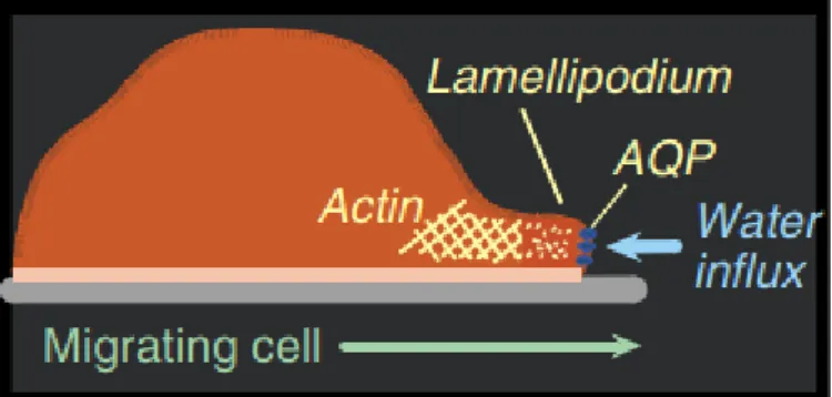

![Figure 1.7: Gold(III) coordination compounds as inhibitors of aquaglyceroporins. (Adapted from [25])](https://thumb-eu.123doks.com/thumbv2/123dok_br/15732080.1071599/45.892.181.714.211.403/figure-gold-iii-coordination-compounds-inhibitors-aquaglyceroporins-adapted.webp)