The effect of wine on the survival and invasiveness ability of
Listeria monocytogenes
THESIS
submitted in partial fulfilment of the requirements for the degree
EUROPEAN M.Sc
in
Food Science, Technology and Nutrition
by
Andrea Zuleta Pérez
Supervision: José António Couto
Co-supervision: Vânia Ferreira and Tim Hogg
To my mother, father and brother we might be scattered around the
world yet so close
Resumo
Listeria monocytogenes é um patogénio de origem alimentar conhecido pela gravidade da sua infeção, a listeriose, e pela sua capacidade de crescer em condições adversas, como temperaturas de refrigeração, ampla gama de valores de pH, atividade de água baixa e elevada salinidade. L. monocytogenes pode induzir a sua própria fagocitose por células epiteliais do intestino do hospedeiro, seguido de replicação e de transferência direta para outras células. O vinho é uma solução de composição complexa e tem atividade antimicrobiana comprovada devido, principalmente, ao seu conteúdo de etanol, ácidos orgânicos e compostos fenólicos. Este trabalho centrou-se em (i) comparar a suscetibilidade de estirpes de L. monocytogenes de origem alimentar e clínica à ação antimicrobiana do vinho, e (ii) na avaliação da influência do vinho sobre a capacidade de invasão de L. monocytogenes utilizando a linhagem de células Caco- 2 (células epiteliais intestinais humanas).
Foram utilizados 39 isolados de L. monocytogenes, 22 de origem alimentar e 17 de origem clínica pertencentes a diferentes serogrupos. Para medir o efeito de inativação do vinho tinto, cada isolado foi submetido a uma diluição de 1:10 de vinho, durante 120 segundos, a 25 ºC. Verificou-se que o vinho exerceu um forte efeito antilisterial, porém observou-se uma alta variabilidade fenotípica entre os isolados – os isolados clínicos mostraram ser significativamente mais resistentes ao vinho do que os isolados alimentares. Células viáveis de uma estirpe clínica (isolada a partir de um surto de listeriose que ocorreu em Portugal) recolhidas após a exposição ao vinho durante 15 seg evidenciaram uma maior capacidade de invadir a linha de células humanas Caco-2 em comparação com o controlo correspondente não exposto ao vinho. Este efeito não foi observado quando o teste de invasão foi realizado em dois outros isolados mais resistentes ao vinho. Tanto quanto é do nosso conhecimento, este é o primeiro estudo que utiliza um número substancial de isolados de L. monocytogenes de diferentes origens e com diversidade fenotípica e genética para estudar a sua suscetibilidade ao vinho. O trabalho também é inovador na avaliação da influência do vinho sobre as características de virulência de um organismo patogénio. Os resultados deste estudo evidenciam que o vinho tinto pode atuar como uma barreira ao crescimento e sobrevivência de L. monocytogenes, quando em contacto com alimentos contaminados.
Abstract
Listeria monocytogenes is a foodborne pathogen known for the severity of its infection, listeriosis, and for its capability of growing at harsh conditions such as refrigeration temperatures, wide pH range, low water activity and high salinity. L. monocytogenes can induce its own phagocytosis by the host’s intestinal epithelial cells, followed by replication and direct transfer to other cells. Wine is a complex solution with proved antimicrobial activity due to its content of ethanol, organic acids and phenolics. This work focused on (i) comparing the susceptibility of food and clinical strains of L. monocytogenes towards wine, and (ii) on the evaluation of the influence of wine on the invasiveness ability of L. monocytogenes using the human intestinal epithelial cells Caco-2 cell line.
Thirty-nine isolates of L. monocytogenes were used in this study, 22 of food and 17 of clinical origin belonging to different serogroups. To measure the inactivation effect of red wine, each isolate was subjected to a 1:10 dilution of wine, during 120 seconds, at 25 ºC. Wine was found to exert an antilisterial effect, though a high phenotypic variability was observed among isolates - clinical isolates were found to be significantly more resistant to red wine than food isolates. Viable cells of one clinical strain (isolated from a listeriosis outbreak occurred in Portugal), collected after exposure to wine for 15 sec showed enhanced ability to invade the human intestinal Caco-2 cell line when compared to the corresponding unexposed control. This effect was not observed when the invasion test was done on two other isolates more resistant to wine. This is, to the best of our knowledge, the first study using a substantial number of L. monocytogenes strains from different origins and diverse phenotypic and genetic characteristics to study the susceptibility to wine. It is also innovative on assessing the influence of red wine on virulence traits of a foodborne pathogen. The results of this study give evidence that red wine can be seen as a significant barrier to the growth and survival of L. monocytogenes when in contact with contaminated food (food consumption scenario, for
Acknowledgements
To the Erasmus Mundus Programme of the European Commission for granting me the scholarship to study this master’s programme; and to the partner universities KU Leuven, Dublin Institute of Technology and Universidade Católica Portuguesa for all the support during the development of my studies.
I acknowledge UCP-ESB for accepting me for this master’s thesis project and for providing all the conditions to conduct the research work in the person of my supervisor Professor José Antonio Couto, and co-supervisors Vânia Ferreira and Professor Tim Hogg.
To Professor Paula Teixeira and José Antonio Couto for always having the door open and the valuable observations and guidance.
To all the people in the microbiology and cell culture laboratory for making me feel welcome and their willingness to help if I needed, especially thanks to Claudia and Norton.
Thank you Rui for helping, particularly, at the beginning diluting (God knows how many Eppendorfs) and plating, and for the cheers during the writing process.
Special thanks to Vânia for all the knowledge, patience, support and enthusiasm during this project. I cannot express how grateful I am, not only for all you taught me in this short time but for the friendship, caring and for making this possible going the extra mile, even when that meant taking me to the doctor or staying up until 4 a.m.
To my beloved friends back in Mexico and the ones I made along the way for the complete confidence you had in me, even in the moments I doubted myself; the warmest thank you, I wouldn’t have made it without your support.
To Yair and Silvia for inspiring me to pursue and international degree and the guidance during the application process and for been there when I needed.
To Catalina for welcoming me in Ireland and showing how beautiful that country is. For all the good and for the bad moments turned into nice coffee chats, you were a piece of home abroad.
To Ulises for joining this adventure, filling it with magic making it, even more, unforgettable.
And last but not least to my, family, you have been in every step of the way, encouraging and supporting me. I love you.
Table of Contents
Resumo ... 5 Abstract ... 7 Acknowledgements ... 9 1 Introduction ... 13 1.1 Listeria ... 13 1.1.1 Listeria monocytogenes ... 13 1.1.2 Growth conditions ... 14 1.1.3 Listeriosis ... 14 1.1.4 Virulence factors ... 151.1.5 Human cases and outbreaks of listeriosis ... 17
1.2 Wine ... 18
1.2.1 Antimicrobial properties of wine ... 19
1.2.2 Wine compounds contributing to its antimicrobial properties. ... 23
1.3 Objective ... 26
2 Materials and methods ... 27
2.1 L. monocytogenes isolates ... 27
2.2 Storage conditions ... 28
2.3 L. monocytogenes inoculum preparation ... 28
2.4 Wine sterilization ... 29
2.5 The inactivation effect of wine on L. monocytogenes ... 29
2.6 Bacterial enumeration ... 29
2.7 Caco-2 Invasion assays ... 30
2.7.1 Growth of L. monocytogenes for Caco-2 invasion assays. ... 30
2.7.2 Tissue culture invasion assays. ... 30
2.8 Statistical Analysis ... 31
3 Results and discussion ... 32
3.1 Effect of wine on the survival of Listeria monocytogenes ... 32
3.2 Impact of wine on the invasion capacity of Listeria monocytogenes into the human intestinal epithelial Caco-2 cells ... 37
4 Conclusions ... 40
5 Future work ... 41
1 Introduction
1.1 Listeria
Listeria was named after the surgeon and pioneer of antisepsis Lord Lister in the 1860’s (Ledermann, 2007). Listeria species are gram-positive, anaerobic facultative, nonsporulating, catalase positive, and oxidase-negative, rod-shaped bacteria of 0.4-0.5 x 1-2 µm with parallel sides and blunt ends. They are widely distributed in the environment: soil, water, vegetation, fresh and frozen poultry, animal feed, slaughterhouse wastes effluents, and feces from healthy animals and humans (Ludwig et al., 2001). They are capable of growing at pH 6-9, high salt concentrations (10% (w/v)), and temperatures ranging from 4 to 45 ºC but optimal growth occurs at 30 – 37 ºC; Listeria do not survive heating at 60 ºC for 30 minutes. The motility of this bacteria depends on its growth temperature, producing peritrichous flagella below 30 ºC and repressed at 37 ºC, the latter needed to achieve full virulence (Ludwig et al., 2001).
Listeria genus comprises 19 species: L. aquatic, L. booriae, L. cornellensis, L. denitrificans, L. fleischmannii, L. floridensis, L. grandensis, L. grayi, L. innocua, L. ivanovii, L. marthii, L. monocytogenes, L. murrayi, L. newyorkensis, L. riparia, L. rocourtiae, L. seeligeri, L. weihenstephanensis, L. welshimeri (Euzéby, 2015); however, only L. monocytogenes and L. ivanovii are pathogenic affecting more than 50 animal species; furthermore, humans are only infected by L. monocytogenes (Zorn & Suárez, 2009; Bennett, 2015).
1.1.1 Listeria monocytogenes
L. monocytogenes was first described in 1926 by Murray et al. under the name of Bacterium monocytogenes because it caused fever and monocytes in their laboratory rabbits and guinea pigs. A year later, Pirie renamed it Listerella hepatolytica for the liver damage he found in gerbils and finally in 1940 he named it as this foodborne pathogen is currently known: Listeria monocytogenes (Farber & Peterkin, 1991; Ledermann, 2007).
Thirteen serotypes have been described for L. monocytogenes: 1/2a, 1/2b, 1/2c, 3a, 3b, 3c, 4a, 4ab, 4b, 4c, 4d, 4e and 7, based on serological reactions between somatic (O factor) and flagella (H factor) antigens and their corresponding antibodies (Farber & Peterkin, 1991); however serotypes 1/2a, 1/2b, and 4b comprise the majority of the strains associated with human listeriosis cases and outbreaks (Orsi et al., 2011). This species is divided in four major
evolutionary subdivisions: lineage I (1/2b, 3b, 3c, 4b); lineage II (1/2a, 3a, 1/2c); lineage III (4a, 4b, 4c); and lineage IV (4a, 4b, 4c). Serotypes 1/2a, 1/2c, 1/2b, and 4b (lineages I and II) are associated with the majority human listeriosis cases (> 98%), while serotypes 4a and 4c (lineage III) are rarely associated with outbreaks despite their frequent isolation from a variety of food and environmental specimens (Orsi et al., 2011).
1.1.2 Growth conditions
L. monocytogenes differentiates from other bacterium for its capability of growing at hard conditions such as refrigeration temperatures (-0.5 – 9.3 ºC) (Walker et al., 1990); wide pH range (4.2 – 9.5) (Bover & Garriga, 2014) surviving lower values (pH 3.3 – 3.5) (Phan-Thanh & Montagne, 1998); low water activity (0.90 – 0.93); and high salinity 12 – 16% (w/v) NaCl (Bover & Garriga, 2014). These conditions make L. monocytogenes a serious hazard for the food industry, because it is widespread in the environment and is able to survive and grow in the food processing environment and ready-to-eat foods; furthermore, it can contaminate a wide range of products at different stages of production e.g. after pasteurization, due to its capacity to attach to abiotic surfaces and form biofilms (Alessandria et al., 2010).
1.1.3 Listeriosis
Listeriosis is the foodborne infection caused by L. monocytogenes. For the general population is a rare disease, when presented in healthy individuals is in the form of flu-like symptoms or as self-limited gastroenteritis; conversely, for the risk population (pregnant women and their fetus, newborns, the elderly and immunocompromised people) can be lethal with a fatality rate of 16 – 3 0% (Vázquez-Boland et al., 2001; Bortolussi, 2008; Bennett et al., 2015).
The infective dose and incubation period of L. monocytogenes is unknown but is estimated to be 10 – 100 million colony forming units (CFU) in healthy hosts, whereas for the risk groups it is of 0.1 – 10 million CFU (Bortolussi, 2008). The symptoms can develop any time from 2 to 90 days after consumption with a mean of 30 days (Bennett, 2015; Bortolussi, 2008).
Listeriosis is presented as non-invasive and invasive disease. As mentioned before, in healthy individuals the non-invasive disease presents as gastroenteritis accompanied by fever, watery diarrhea, nausea, headache, and pains in joints and muscles, the onset of the disease
usually occurs in a range of 6 hours to 10 days and usually lasts 1 to 3 days up to 7; in contrast, the invasive form comprises bacteremia, endocarditis and central nervous system (CNS) affections such as meningitis, encephalitis, rhombencephalitis, brain abscess and spinal cord infection (Bennett, 2015). Pregnant women are prone to develop bacteremia manifested as acute febrile illness, often accompanied by myalgias, arthralgias, headache, and backache; furthermore, the mother can transmit the disease to the fetus through the placenta resulting in stillbirth, spontaneous abortion or neonatal death, however, early antimicrobial treatment can result in the birth of a healthy infant (Bennett, 2015). For treatment, ampicillin is the preferred agent; when CNS infection or endocarditis are present the addition of gentamicin and trimethoprim-sulfamethoxazole are used. For people allergic to penicillin, trimethoprim- sulfamethoxazole is used (Allerberger & Wagner, 2010).
1.1.3.1 Invasion and spread
Listeriosis can be such a severe illness because L. monocytogenes can induce its own phagocytosis by host cells (nonphagocytic cells), followed by replication within those cells and direct transfer to another cell; then it can spread through the body protected from antibodies and complement. The immunity to L. monocytogenes is T cell mediated (Doyle 2001; Swaminathan et al., 2007). The bacterium starts by infecting the intestinal epithelial cells. From the intestine bacteria disseminate via the blood or lymph to the liver and spleen where most are killed by neutrophils acting with Küpffer cells. In people with inadequate T-cell mediated immune response, listeriae multiply in the hepatocytes and macrophages and is transported to infect other organs, particularly the brain and uterus (Doyle, 2001).
1.1.4 Virulence factors
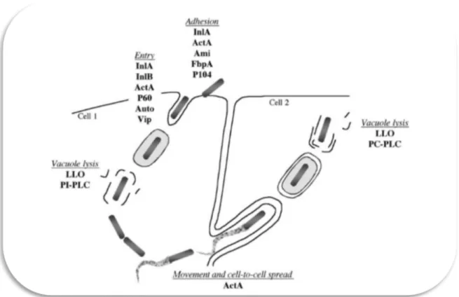
Figure 1.1 Representation of the of infectious process of L. monocytogenes and the factors implicated in each step (in Swaminathan et al., 2007).
Internalins A and B (InlA and InlB): are listeria surface proteins involved in the entry to host cells. IntlA binds to E-cadherin on the surface of host epithelial cells which stimulates the phagocytosis of L. monocytogenes. Similarly, InlB binds to Met receptor for the invasion of hepatocytes in the liver (Swaminathan et al., 2007).
Listeriolysin O (LLO): is a bacterial preforming toxin than enables the scape of L. monocytogenes from the vacuoles into the cytoplasm of the cell (Doyle, 2001).
Proteins P104 and P60: P104 is a surface protein involved in the adhesion to intestinal cells (Doyle, 2001). P60 is important in the immune response against listeriosis, because specific antibodies in immunocompetent individuals can prevent systemic infections (Swaminathan et al., 2007).
ActA protein: is a surface protein implicated in the attachment to cells and responsible for the actin-based motility of Listeria. It induces the polymerization of globular actin molecules to
form filaments along which the Listeria moves to adjacent cells without exposure to antibodies or other immunoactive molecules (Doyle, 2001).
Phospholipases: have a membrane-damaging activity and are involved in the escape from phagosomes (Swaminathan et al., 2007). Two are produced by L. monocytogenes phosphatidylinositol-specific phospholipase C (PI-PLC) and a broad-range or phosphatidylcholine-specific phospholipase C (PC- PLC). PI-PLC aids in escape from the primary vacuole while PC-PLC is active during cell-to-cell spread of bacteria and it can substitute LLO (Doyle, 2001).
Metalloprotease: PC-PLC is produced as an inactive precursor, to activate it a bacterial zinc-dependent metalloprotease and a host cell cysteine protease are required (Doyle, 2001).
Vip: is a virulence gene that encodes the LPXTG surface protein required for entering mammalian cells at intestinal level and later stages of the infection (Swaminathan et al., 2007).
Clp proteases (caseinolytic proteins) and ATPases: ClpC ATPase is a general stress protein that assists in the disruption of the vacuolar membrane. ClpC also modulates expression of the ActA protein and the internalins. ClpP serine protease is required for growth under stress affecting the activity of listeriolysin O, also ClpE, is involved in listerial pathogenesis (Doyle, 2001).
1.1.5 Human cases and outbreaks of listeriosis
According to the European Food Safety Authority (EFSA) latest European Union (EU) summary report on zoonoses, zoonotic agents and foodborne outbreaks for 2014, 27 member states confirmed a total of 2,161 cases of human listeriosis with a notification rate of 0.52 cases per 100,000 population representing an increase of 30% compared with 2013. For the period 2008 – 2014 the increasing trend of listeriosis was statistically significant. Of all the zoonotic diseases under EU surveillance, listeriosis caused the most severe human disease with 98.9% hospitalizations and 210 deaths, representing a case-fatality rate of 15% (out of the 1,401 confirmed cases with known outcome). The highest number of deceased was reported by France: 51 cases. Regarding outbreaks of listeriosis, there were several small but
Denmark reported a large outbreak comprising 41 cases; on the other hand, Sweden who in 2013 had an outbreak involving 50 cases, in 2014 only 27 cases were reported (EFSA, 2015).
1.2 Wine
Wine is a complex solution of a vast number of chemicals e.g. 160 esters have been identified, despite that individually they are found in concentrations below human detection (10-4
- 10-9
g/L) together they are important for the organoleptic characteristics (Jackson, 2014). The aromatic compounds are found in 0.8 – 1.2 g/L mainly as fusel alcohols (50% of all volatile substances), volatile acids and fatty acid esters. Carbonyls, phenols, lactones, terpenes, acetals, hydrocarbons, and sulfur and nitrogen compounds are present in much smaller concentrations but their importance lays in the varietal and sensory features conferred to the wine’s fragrance (Jackson, 2014).
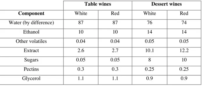
The main components of wine are water and ethanol (approximately 98%); followed by trace components (vitamins, sugars, nitrogenous components, cations and anions), volatiles (fusel alcohols, esters, ketones, C13 norisoprenoids, fatty acids, phenols, amides, other) and acids (tartaric and malic), representing 1%, 0.5% and 0.5% respectively (Jackson, 2014). Wines can be classified in two major groups: table wines whose alcohol content is below 14% and dessert wines produced from grapes high in sugar and low in acid content (Friedman, 2014). The general composition of red and white wines for both categories is shown in table 1.1 where the content of phenols between red and white wines is noted as the major difference in both table and dessert wines.
Table 1.1 Estimates of typical gross composition (% weight) of wines (in Soleas et al. 1997)
Table wines Dessert wines
Component White Red White Red
Water (by difference) 87 87 76 74
Ethanol 10 10 14 14 Other volatiles 0.04 0.04 0.05 0.05 Extract 2.6 2.7 10.1 12.2 Sugars 0.05 0.05 8 10 Pectins 0.3 0.3 0.25 0.25 Glycerol 1.1 1.1 0.9 0.9
Acids 0.7 0.6 0.5 0.05 Ash 0.2 0.2 0.2 0.2 Phenols 0.01 0.2 0.01 0.1 Amino acids 0.25 0.25 0.2 0.2 Fats, terpenoids 0.01 0.02 0.01 0.02 Vitamins, etc. 0.01 0.01 0.01 0.01 Total 100 100 100 100
1.2.1 Antimicrobial properties of wine
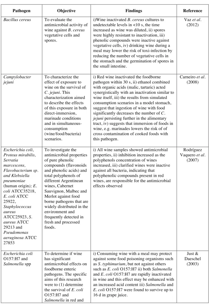
Wine is active against bacteria (human and plants), fungus, protozoans and several viruses, including herpes simplex virus, poliovirus, hepatitis A, as well as in common cold viruses (rhinoviruses and coronaviruses) (Jackson, 2014; Muñoz-González, et al. 2014; Friedman, 2014; Cueva et al., 2010; Király-Véghely et al., 2009). Takkouche et al. (2002) found an inverse association between moderate wine drinkers and the incidence of common cold. As stated before wine is a complex solution with numerous components showing antimicrobial properties, the mechanisms behind this are not well understood: low pH (3.0 to 4.0), high organic acid content (titratable acidity ≥6.0 g/L tartaric acid), relatively high ethanol (10% to 15%), and potentially high total sulfur dioxide (0 to 300 ppm) are contributors (Waite & Daeschel, 2007).
The concentrations of alcohol found in wine are not high enough to be fully responsible for the antimicrobial action, rather the interaction of different constituents, for example, the modification of anthocyanins during fermentation increases their toxicity to viruses, protozoans, and bacteria (Gram positive and negative) (Jackson, 2014); as Boban et al., (2010) found the antimicrobial activity cannot be attributed to the phenolic or nonphenolic constituents of wine, nor based on its components predict the antimicrobial activity of a wine. Furthermore, Møretrø & Daeschel (2006) evaluated wine components individually and combined concluding that the synergistic effect of organic acids, ethanol, and low pH seems to be responsible for a major part of the antibacterial effect. Table 1.2 shows selected studies on the effects of wine against foodborne pathogens. All the studies used red wine with the exception of Møretrø & Daeschel (2006) who reported that red wine was more effective than white for the tested bacteria, and Liu et al. (2006) who found no significant differences between red and white wines in the inactivation of Vibrio parahaemolyticus; furthermore no significant differences were found between those wines with and without added sulfite.
Table 1.2 Studies on the antimicrobial effect of wine against foodborne pathogens.
Pathogen Objective Findings Reference
Bacillus cereus To evaluate the
antimicrobial activity of wine against B. cereus vegetative cells and spores.
i)Wine inactivated B. cereus cultures to undetectable levels in <10 s, the time increased as wine was diluted, ii) spores were highly resistant to inactivation, iii) phenolic compounds were inactive against vegetative cells, iv) drinking wine during a meal may lower the risk of toxi-infection by reducing the number of vegetative cells in the stomach and the germination of spores in the small intestine.
Vaz et al. (2012) Campylobacter jejuni To characterize the effect of exposure to wine on the survival of
C. jejuni. This
characterization aimed to describe the effects of this exposure in both direct-immersion, marinade conditions and in simultaneous-consumption (wine/food/bacteria) scenarios.
i) Red wine inactivated the foodborne pathogen within 30 s, ii) ethanol combined with organic acids (malic, tartaric) acted synergistically with an inactivation similar to wine itself, iii) the results from simulated consumption scenarios in a model stomach, suggest that ingestion of wine with food significantly decreases the number of C.
jejuni persisting further in the alimentary
tract, iv) suggests that immersion of foods in wine, e.g. marinades lowers the risk of of cross contamination of cooked foods with this pathogen. Carneiro et al. (2008) Escherichia coli, Proteus mirabilis, Serratia marcescens, Flavobacterium sp. and Klebsiella pneumoniae (human origin); E. coli ATCC35218, E. coli ATCC 25922, Staphylococcus aureus ATCC25923, S. aureus ATCC 29213 and Pseudomonas aeruginosa ATCC 27853 To investigate the antimicrobial properties of pure phenolic compounds (flavonoids and phenolic acids) and total polyphenols of different Argentinean wines, Cabernet Sauvignon, Malbec and Merlot against food borne pathogens that are widely distributed in the environment and frequently detected in fresh and processed foods.
i) All wine samples showed antimicrobial properties, ii) inhibition increased as the polyphenols concentration of wines increased, iii) clarified wines were inactive against all bacteria, indicating that
polyphenolic compounds present in red wines, are responsible for the antimicrobial effects observed Rodríguez Vaquero et al. (2007) Escherichia coli O157:H7 and Salmonella spp To determine if wine has significant antimicrobial effects on foodborne enteric pathogens. The specific aims of this research were to (1) determine the survival of E. coli O157:H7 and
Salmonella in red and
i) Consuming wine with a meal may protect against some food poisoning organisms such as S. typhimurium, but not against others such as E. coli O157:H7 ii) both Salmonella and E. coli O157:H7 are rapidly inactivated in wine and this effect may be enhanced with an increased acid content iii) Salmonella and
E. coli O157:H7 were found to survive up to
16 d in grape juice.
Just & Daeschel
white wine and grape juice and (2) determine the effect of wine on E.
coli O157:H7 and Salmonella survival in a
model stomach system
Salmonella enteritidis, Escherichia coli O157:H7, and Vibrio parahaemolyticus Examine the antibacterial activity against the three entero-pathogenic bacteria in
vitro and in vivo. To
identify which fraction of wine had
antibacterial activity, and examine the ability of wine and the fraction to protect against infection in vivo.
i) Red and white wines have antibacterial activity against the 3 entero-pahtogens killing them within 30 minutes, ii) the evaporated fraction contained in wine seemed responsible for this and not
polyphenols, iii) when tested in mice, neither red or white wine showed prevention properties against S. enteritidis wether given “one shot” treatment or continoues
administration, iv) the antibacterial property seems to be limited to in vitro circumstances.
Sugita-Konishi et al.
(2001)
Listeria innocua To study the the
bactericidal effect of wine on L. innocua (surrogate for L.
monocytogenes) in a
food matrix, under simulated gastric conditions (stomach model), and to evaluate specific influence of some wine components on this effect.
i) Red wine volumes, equivalent to the ingestion of one glass and half a bottle, induced a 3−4 log CFU reduction of the initial pathogen count in <2 h, ii) the combination of ethanol and malic and tartaric acids was less effective than wine, iii) the kinetics of the bactericidal effect of red and white wine, differed demonstrating that the antimicrobial effects of wines depends on composition. Fernandes et al. (2007) L. monocytogenes, Escherichia coli O157:H7, Salmonella typhimurium, and Staphylococcus aureus
To test the antibacterial effect of wine against wild-type strains and sigma mutants of the pathogens E. coli
O157:H7, L. monocytogenes, S. typhimurium, and Staphy lococcus aureus.
The antibacterial effects of selected components of wines were also tested individually and in combination. Moreover, the protection of the bacteria against wine by stress dependent protection systems was evaluated.
i) Red and white had bactericidal activity against all strains, red wine was more effective than white, ii) of the wild-type strains, S. Typhimurium was the most sensitive to wine, followed by E. coli O157:H7, L. monocytogenes, and S. aureus, iii) the alternative sigma factors seemed to be involved in protection of the bacteria against wine, mutants (having this factor disrupted) were generally more sensitive to wine than their wild-type counterparts, iv) preincubation of E. coli O157:H7 and S.
aureus (wild-type) in sublethal
concentrations of wine and ethanol and pH 4.5 did not increase their tolerance against wine or against the mixture of organic acids and ethanol, v) the synergistic effect of organic acids, ethanol, and low pH seems to be responsible for a major part of the antibacterial effect of wine (the composition of 0.15% malic acid, 0.6% tartaric acid, 15% ethanol, and pH 3.0 had the strongest bactericidal effect). Møretrø & Daeschel (2006) Salmonella enterica serovar Enteritidis and Escherichia To examine antibacterial activity of the intact red wine and
i) Antibacterial activity of the samples was: intact wine > phenols-stripped wine > dealcoholized wine > combination of ethanol
Boban et al. (2010)
coli its derivatives under the same experimental conditions against 2 common foodborne pathogens, S. enterica serovar Enteritidis and
E. coli. To separate the
role of wine phenolics, ethanol, and pH from other wine constituents, the antimicrobial effects of intact wine were compared to that of phenols-stripped wine, dealcoholized wine, ethanol, and low pH applied separately and in combination.
and low pH > low pH > ethanol, ii) low pH and ethanol had synergistic effect, whereas individually their antibacterial activity is negligible, iii) antibacterial activity of the samples could not be related to their total phenolics and resveratrol content,
antioxidant capacity, ethanol content, or pH, iii) antimicrobial activity of complex solutions such as intact wine cannot be exclusively attributed to its phenolic or nonphenolic constituents, nor can the antimicrobial activity of wine be predicted on the basis of its particular components.
S. aureus and E. coli O157:H7.
To look at 4 wine parameters, pH, titratable acidity, sulfur dioxide concentration, and ethanol concentration, in various combinations within a wine background to evaluate antimicrobial activity against the food-borne pathogens S. aureus and
E. coli O157:H7.
i) pH was found to be the most critical factor in predicting inactivation of both S. aureus and E. coli O157:H7, ii) Molecular sulfur dioxide, titratable acidity, and ethanol concentration also contributed to the inactivation of S. aureus. Ethanol
concentration was also found to contribute the efficacy of wine treatments on E. coli O157:H7. iii)Total sulfur dioxide and free sulfur dioxide were not predictive of wine efficacy against either pathogen tested. These findings indicate the importance of each parameter in wine to be used for potential disinfection purposes.
Waite & Daeschel (2007) Vibrio parahaemolyticus To investigate the antibacterial activities of both red and white wines against V.
parahaemolyticus in
laboratory-contaminated oysters and compared the bactericidal effects of wines with and without added sulfites on inactivating V.
parahaemolyticus.
i) The populations in wine-treated whole oysters decreased by >1.7 and >1.9 log MPN/g after 24 h at 7 and 25 ºC,
respectively; no significant differences were found between red and white wines or between wines with and without added sulfite, ii) both red and white wines were more effective in inactivating V.
parahaemolyticus in oyster meat
homogenate populations decreased rapidly (a 3.89- log MPN/g reduction) to nondetectable levels after 30 min at 25 ºC, iii) These results suggest that chewing oysters before
swallowing when eating raw oysters may result in greater inactivation of V.
parahaemolyticus if wine is consumed
Liu et al. (2006)
1.2.2 Wine compounds contributing to its antimicrobial properties.
Several studies, evaluating wine as a whole or specific groups of compounds, have identified the major constituents with antimicrobial effects, which are described below.
1.2.2.1 Ethanol
The efficacy of ethanol as antiseptic is increased in the presence of water and its antimicrobial activity is optimum in the range of 60 – 90%, causing membrane damage by solubilizing lipids and denaturating proteins with subsequent interference with metabolism and cell lysis (McDonnell & Russell, 1999; Barker & Park, 2001), thus the concentrations of ethanol present in wine are not high enough to be fully responsible for the antimicrobial action of wine since the effects of wine are greater than the same concentration of diluted ethanol (Møretrø & Daeschel, 2006). This is consistent with Boban et al. (2010) findings who reported that low pH and ethanol had a synergistic effect, whereas individually their antibacterial activity is unimportant.
1.2.2.2 Organic Acids
Acidity in wine is divided into volatile and fixed, meaning the acids that can be readily removed by steam distillation and those that are poorly volatile, respectively; both make up the total acidity. Fixed acidity in wines can vary from less 2 g/L to over 5 g/L (Jackson, 2014). The main organic acids of wine are tartaric and malic, for the ones undergoing malolactic fermentation malic acid is metabolized to lactic acid. Fixed acidity is responsible for the antimicrobial effect of organic acids because it confers low pH to the wine in which most bacteria do not grow and the fatty acids remain undissociated. In a study evaluating the antimicrobial activity of wine against Bacillus cereus vegetative cells and spores Vaz et al. (2012) found that organic acids contribute to the antimicrobial effect of wine; additionally, they strengthen the action of phenolics and ethanol. Similarly, Møretrø & Daeschel (2006) found a higher reduction in viable cells of S. aureus, L. monocytogenes, S. typhimurium and E. coli in a mixture of ethanol, malic and tartaric acid (pH 3) than when each was tested individually. Carneiro et al., (2008) used a the mixture of tartaric, acetic, lactic and citric acids combined with ethanol in concentrations found in wine and their combination showed higher bactericidal effect than the mixture of acids and ethanol separately. The effectiveness
of organic acids is related to pH which determines the degree of dissociation of the acid. The undissociated organic acids cross the cell membrane lipid bilayer more easily. Once inside the cell, the acids dissociate because of the higher intracellular pH (near neutrality), releasing protons that acidify the cytoplasm which suppress cell enzymes affecting its metabolism. In order to restore the optimal intracellular pH, protons need to be pump out by the H+-ATPase demanding considerable metabolic energy in the form of adenosine triphosphate (ATP) that could lead to depletion of cellular ATP with the eventual cell death due to energy depletion; furthermore, the remaining anions in the cytoplasm can inhibit the synthesis of macromolecules, enzyme activity, nutrient transport systems within the cytoplasm resulting in cell death (Swaminathan et al., 2007; Ng & Koh, 2016).
1.2.2.3 Phenolic compounds
Phenolics or phenols are a complex group comprising a huge amount of compounds important for the quality (appearance, taste, mouth-feel, fragrance) of wine and its antimicrobial properties. The main source is the grape (skin, seed and stems) followed by smaller amounts that may be extracted from oak barrels, finally yeasts produce trace amounts during fermentation (Jackson, 2014). Chemically, phenols are cyclic benzene compounds with one or more hydroxyl groups associated directly with the ring structure (Soleas et al., 1997). Concentration of phenols is expressed as gallic acid equivalents (GAE) based on their chemical reducing capacity relative to the equivalent reducing capacity of that component. For red wines concentration ranges from 1800 to 4059 GAE mg/L (average 2567 GAE mg/L), and 165 to 331 GAE mg/L (average 239 GAE mg/L) for white (Frankel et al., 1995).
The main phenolics found in wine are: flavonoids (two phenyl groups) whose polymers are tannins, and non-flavonoids (containing one phenyl group).
Flavonoids
Flavonoids characterize red wines more than any other feature, accounting for more than 85% of their phenolic content (≥1000 mg/L), conversely in white wines they constitute less than 20% of the total phenolic content (≤50 mg/L) (Jackson, 2014). The rest consists mainly of the non-flavonoid, caffeic acid. The amount of flavonoids in wine is influenced by a number of factors starting from the grape production (cultivar, vintage, climatic conditions of the region) to the vinification process (temperature, length of skin contact, mixing, type of fermentation/aging). This family of compounds share a common C6-C3-C6 skeleton
consisting of two phenolic rings (named A and B) linked together by a heterocyclic pyran ring (C-ring) (Terrier et. al., 2009).
The most common flavonoids in wine are flavonols, catechins (flavan-3-ols) and anthocyanins (in red wines), all of them are either free or polymerized with other flavonoids, non-flavonoids or both. There are also small amounts of free leucoanthocyanins (Soleas et al., 1997).
Flavanol oligomers and polymers are also called condensed tannins or proanthocyanidins for their capacity to precipitate proteins and to release anthocyanidin when heated in acidic conditions (Terrier et. al., 2009). Anthocyanidins are the sugar-free counterparts of anthocyanins and five are identified in wine: delphinidin, cyanidin, petunidin, peonidin and malvidin (Monagas & Bartolomé, 2009).
Non-Flavonoids
Non-flavonoid phenolic constituents in wine are divided into hydroxybenzoic acids (HBA), hydroxycinnamic acids (HCA), volatile phenols, stilbenes and miscellaneous compounds (e.g. lignans and coumarins). They stabilize the color of red wines despite they are colorless, contribute to flavor and some of them exhibit potent biological activities (Rentzsch et al., 2009).
The most common derivatives from HBA found in wine are gallic acid, gentisic acid, p-hydroxybenzoic acid, protocatechuic acid, syringic acid, salicylic acid, and vanillic acid, and from HCA are caffeic acid, p-coumaric acid, ferulic acid, and sinapic acid in cis- and trans- forms, the latter is more prevalent due to its stability (Rentzsch et al., 2009).
The nature of non-flavonoids is influenced by the material the wine is aged in; for wines not aged in oak the derivatives of HCA are higher than those of HBA; whereas in wines aged in oak, levels of HBA derivatives (especially ellagic acid) are higher (Soleas et al., 1997).
Stilbenes are synthesized in the grape as response to stress and are known for their antioxidative, anticarcinogenic and antimutagenic potency; one of the most comprehensively studied is resveratrol. Resveratrol exists in the cis- and trans- isomers, as in the ß-glucoconjugated form. The 3-O-ß-D-glucosides of cis- and resveratrol as cis-and trans-configured resveratrol are called piceids. The oligomeric and polymeric forms of stilbenes are called viniferins (Rentzsch et al., 2009).
In wine ε-viniferin, δ-viniferin, pallidol (resveratrol dimer), α-viniferin, (resveratrol trimer) and hopeaphenol (resveratrol tetramer) have been identified. Free trans- and
cis-resveratrols are present in a concentration range of 0.2–13 mg/L in red wines and 0.1–0.8 mg/L in white wines; for resveratrol-3-O-glucoside, concentrations range from 0.3–9 mg/L in red and 0.1–2.2 mg/L in white. In comparison to wine, grapes contain mainly trans-resveratrol glucoside in concentrations ranging from 1.5 to 7.3 µg/g ( Rentzsch et al., 2009).
Phenolic compounds have been extensively studied for the health benefits aforementioned. Rodríguez Vaquero et al. (2007) studied the antimicrobial properties of four phenolic acids (gallic, vanillic, protocatechuic and caffeic acid) and three flavonoids (rutin, catechin and quercetin) of different wines and found that as their concentration increased so did the bactericidal effect of wine, furthermore the same wines after clarification (controls) showed no inactivation, relating directly the antimicrobial effects to the polyphenolic compounds; in contrast Vaz et al. (2012) found that resveratrol, ferulic acid, p-coumaric acid, kaempferol and quercetin were inactive against B. cereus vegetative cells. Boban et al. (2010) found that after wine, their phenols-stripped counterparts where most effective thus the antimicrobial activity of wine cannot be exclusively attributed to its phenolic or nonphenolic constituents; likewise, red wine with the highest resveratrol concentration had the highest inhibitory effect on Helicobacter pylori urease activity (virulence factor) however, resveratrol from wine required lower concentrations than the pure compound to produce the same results, probably due to synergic reactions with the other wine constituents (Paulo et al., 2011).
1.3 Objective
The bactericidal effect of wine on L. innocua (surrogate of L. monocytogenes) has already been shown in model stomach systems. This work further characterized the activity of wine against L. monocytogenes comparing the sensitivity of food strains with clinical strains. Another major aim of this work was to evaluate the effect of sub-lethal levels of wine on the capacity of L. monocytogenes to invade host cells which is the first step of the infective cycle. Human intestinal epithelial cells (Caco-2 cell line) were used to assess the influence of wine on the invasiveness ability of L. monocytogenes.
2
Materials and methods
2.1 L. monocytogenes isolates
Thirty-nine isolates of L. monocytogenes were used in this study (table 2.1). All the isolates belong to the Listeria Research Center of Escola Superior de Biotecnologia (LRCESB) (Porto, Portugal) and were selected to include two different origins: clinical and food. These isolates have been previously characterized by serogroup multiplex-PCR, pulsed-field gel electrophoresis (PFGE), and antibiotic resistance (Komora et al., 2016).
Table 2.1 List of food and clinical isolates of L. monocytogenes selected for this study.
Origin Isolate code Serogroup a PFGE typeb Antibiotic Susceptibilityc Food
Lm 654 IVb 263 ERYSNITSCIPS
Lm 864/4 IVb 133 ERYRNITSCIPS Lm 830/1 IVb 133 ERYRNITRCIPS Lm 841/2 IVb 133 ERYRNITRCIPS
Lm 1162 IVb 17 ERYS NITR CIPS Lm 1604/2 IVb 83 ERYS NITS CIPS Lm 1728 IVb 79 ERYS NITR CIPS Lm 1940/1 IVb ND ERYS NITS CIPR Lm 949 IIb 232 ERYS NITS CIPR Lm 969/3 IIb 151 ERYS NITR CIPS Lm 971 IIb 246 ERYS NITS CIPR Lm 1043 IIb 16 ERYS NITR CIPS Lm 800/2 IIb 145 ERYR NITR CIPS Lm 1216 IIb 278 ERYS NITS CIPS Lm 1382/1 IIb 147 ERYS NITR CIPS Lm 1486/1 IIb 10 ERYS NITR CIPS Lm 1535 IIb 156 ERYS NITR CIPS Lm 925/1 IIb 43 ERYS NITS CIPS Lm 1852/3 IIb ND ERYS NITR CIPS Lm 1846/1 IIb ND ERYS NITR CIPS Lm 863/1 IIc 129 ERYS NITS CIPS Lm 1305 IIc 206 ERYS NITS CIPS
Clinical Lm 2104 IVb 86 ERYS NITR CIPS Lm 2264 IVb 53 ERYS NITS CIPR Lm 2265 IVb 53 ERYS NITS CIPR Lm 2542 IVb 70 ERYS NITS CIPS Lm 2571 IVb 53 ERYS NITS CIPR Lm 3390 IVb 393 ERYS NITR CIPR Lm 1543 IVb 54 ERYS NITS CIPS Scott A 4b ND ND Lm 2103 IIa 9 ERYS NITS CIPR Lm 2388 IIa 96 ERYS NITS CIPR 3391 IIa 332 ERYS NITR CIPR 2086 IIa 36 ERYS NITS CIPS EGDe 1/2a ND ND 2065 IIb 37 ERYS NITS CIPS CLIP 21369 1/2b ND ERYS NITS CIPR 2658 IIb 87 ERYS NITR CIPR 1062 IIb 42 ERYS NITR CIPS
a Determined by Multiplex-PCR that differentiates major serogroups IVb (includes serotypes 4b, 4d, and 4e),
serogroup IIa (includes serotypes 1/2a and 3a), and serogroup IIb (serotypes 1/2b, 3b, and 7); except for reference strains Scott A, EGDe, and CLIP 21369 with known serotypes.
b Characterization by pulsed-field gel electrophoresis (PFGE) enzimes AscI and ApaI
c ERY - erythromycin, minimum inhibitory concentration (MIC) ≥ 4 μg/mL; NIT - nitrofurantoin, MIC ≥ 128
μg/mL;CIP - ciprofloxacin, MIC ≥ 8 μg/mL; R resistant; S susceptible ND – not determined
2.2 Storage conditions
Stock cultures of Listeria strains were kept in tryptic soya broth with yeast extract 0.6% w/v (TSBYE, Lab M, Heywood, Lanchashire, UK) supplemented with 30% (v/v) of glycerol at -80 °C.
2.3 L. monocytogenes inoculum preparation
Before use, frozen stocks were streaked onto brain heart infusion (BHI, Biokar Diagnostics, Beauvais, France) agar plates and incubated at 37 °C overnight. One single colony of each isolate was transferred into 5 mL of BHI (Biokar) and incubated at 37 ºC for 24 h. The cultures were then sub-cultured in 11 mL of BHI (0.1% v/v) and incubated at 37 ºC for 18 h to reach the stationary phase. The cells were washed by centrifugation (10,000 x g, 5
min, 4 ºC; Rotina 35R, Hettich, Germany) in sterile phosphate-buffered saline (PBS; pH = 7.4) and cell pellet was suspended in the same volume of PBS to obtain an inoculum concentration of approximately 109
colony forming units (CFU)/mL. This procedure was done immediately before the experiments and 100 µL of each sample were collected to obtain the initial cell count.
2.4 Wine sterilization
Alandra red wine (2014, Alentejo region, Portugal) with 13% of ethanol (v/v) was used. The wine was filter sterilized using 0.45 µm - 25 mm cellulose acetate membranes (Frilabo sterile syringe filters, USA) and kept at 4 °C in sterile Scott Duran flasks of 100 mL until used.
2.5 The inactivation effect of wine on L. monocytogenes
For each isolate, inoculum aliquots of 2.5 mL were added to 22.5 mL of diluted wine (1:10 in sterile deionized water) pre-warmed at 25 ºC during 30 min in a thermostated water bath (Julabo SW22, Seelbach, Germany) before inoculation to allow temperature equilibration. At defined time points (15, 30, 60 and 120 seconds after inoculation) 100 µL of sample were collected, and vortexed for homogenization. As a control, for each isolate, 2.5 mL of inoculum were added to 22.5 mL of sterile PBS that was kept at 25 ºC and 100 µL of sample were collected at 2 time points (0 and 120 seconds). The experiments and corresponding controls were conducted in duplicate.
2.6 Bacterial enumeration
Samples were serially diluted in sterile PBS, and the dilutions subsequently plated on BHI agar plates in duplicate by the drop count technique (Miles & Misra, 1938). After incubation at 37 ºC for 24 h the colonies were counted, and the CFU/mL calculated. Microbial counts were transformed to logarithmic reduction using the equation: log (N/N0), where N is the microbial cell density at a particular sampling time and N0 is the initial cell density.
2.7 Caco-2 Invasion assays
Two food and two clinical strains were selected for this assay; selection was preformed to include from each origin the isolate showing higher resistance to wine and the strain more sensitive to wine.
2.7.1 Growth of L. monocytogenes for Caco-2 invasion assays.
Resistant L. monocytogenes strains (1852/3 and 2658) were grown and exposed to diluted wine as described above. At 15 sec exposure, 25 mL of sterile PBS was added and cells were immediately centrifuged (7000 x g, 5 min, 4 ºC), washed once with PBS and resuspended in 4 mL of PBS. Susceptible strains (969/3 and 2542) were grown and exposed to diluted wine as described above, however due to the considerable decrease in cell numbers after 15 seconds (3.7 log reduction for 969/3 and 2.4 log reduction for 2542), and to guarantee enough cell numbers for the invasion assay, the experiment to the wine exposure was performed using 10 times more volume of diluted wine (i.e. 250 mL) and inoculum (i.e. 25 mL). After 15 sec, 250 mL of sterile PBS was added, and each suspension was immediately centrifuged (7000 x g, 15 min, 4 ºC), and washed once with PBS. Cell-free supernatant was discarded and the pellet was resuspended in 2.5 ml of fresh PBS. As a control, for each invasion assay, the four L. monocytogenes strains were grown and treated in the same exact conditions, however, diluted wine was replaced by PBS. Furthermore, for sensitive strains controls, in the last step, the pellet was resuspended in the same initial volume of PBS instead of 2.5 mL (otherwise the initial cell numbers for the invasion assay would be exceedingly high).
2.7.2 Tissue culture invasion assays.
Caco-2 invasion assays were performed as previously described by Nightingale et al. (2005) with minor adjustments. The tumor-derived human colorectal epithelial cell line Caco-2 was grown in T75 flasks using Eagle’s minimal essential medium (EMEM) (Lonza, Verviers, Belgium) containing 20% fetal bovine serum (FBS) (Lonza), 1% sodium pyruvate (Lonza) and 1% non-essential amino acids (Lonza), and incubated at 37 ºC with 7% (v/v) CO2 atmosphere. For invasion assays, 5.0x10⁴
Caco-2 cells were seeded into 24-well plates (Costar, Corning, NY, USA) in EMEM and incubated for 48 h at 37 °C. Caco-2 monolayers were subsequently inoculated with 10 µL of the L. monocytogenes suspension treated as
detailed above and incubated at 37 °C for 30 min. The inoculums were immediately serial diluted and plated on BHI agar plates. Each well was then washed three times with 1 ml of sterile PBS to remove any unattached, extracellular L. monocytogenes. Subsequently, infected cells were incubated with 1 ml of pre-warmed fresh EMEM and at 45 min post-inoculation, the medium was replaced with fresh Caco-2 medium containing 150 µg/ml gentamicin (Gibco BRL, Gaithersburg, MD) in order to kill remaining extracellular bacteria. At 90 min post-infection, the medium was aspirated and the wells were washed three times with sterile PBS. Caco-2 cells were lysed by the addition of 500 µL of ice-cold sterile ultra-pure water and vigorous pipetting. Lysed Caco-2 cell suspensions were collected, serial diluted in sterile PBS and plated by the spread plating method on BHI agar plates, which were incubated at 37 °C for 24 h for determination of bacterial counts. The invasion efficiency was reported as the percentage of the inoculum recovered by the enumeration of intracellular bacteria. An uninoculated BHI broth were included as controls in each invasion assay. Three independent invasion assays were performed for each strain.
2.8 Statistical Analysis
One-way analysis of variances (ANOVA) was carried out to assess statistically significant differences among isolates. All tests were performed to a 5% significance level using IBM SPSS® Statistics® 20 for Windows® (SPSS Inc., Chicago, USA).
3 Results and discussion
3.1 Effect of wine on the survival of Listeria monocytogenes
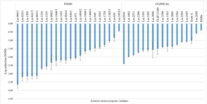
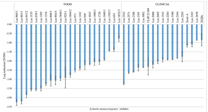
In this study we tested the resistance of 17 clinical and 22 food L. monocytogenes isolates to red wine (1:10 dilution) during 120 sec, at 25 ºC. These experimental conditions (i.e. wine dilution factor, temperature and time) were selected based on preliminary experiments where non diluted wine and dilutions 1:2, 1:4 and 1:8 dilutions at 37 ºC exerted a very strong lethal effect, dropping viable cell counts to no detectable levels in less than 15 sec (data not shown). Immediately after 15 sec exposure a high variability among isolates was observed (figure 3.1), with log reduction values ranging from -0.4 to -2.4 for clinical isolates and from -0.5 and -3.7 for food isolates. Within the time of the experiment a substantial decrease in cell viability occurred (figures 3.2 – 3.3); after 120 sec exposure time, 11 food (50%) and 4 clinical (23.5%) isolates suffered reductions higher than 4.5 log cycles (data not shown), i.e. cell counts below or at the detection limit of the enumeration technique (<500 CFU/mL).
Figure 3.1 Log reduction of food and clinical isolates of L. monocytogenes strains after 15 sec exposure to 1:10 dilution of red wine, rank-ordered according to their susceptibility. Data represent mean of duplicate assays and error bars represent the standard deviation of the mean. -4.50 -4.00 -3.50 -3.00 -2.50 -2.00 -1.50 -1.00 -0.50 0.00 L m 969/ 3 L m 925/ 1 L m 1305 L m 841/ 2 L m 863/ 1 L m 1216 L m 1535 L m 800/ 2 L m 1846/ 1 L m 1 162 L m 864/ 4 L m 830/ 1 L m 971 L m 1604/ 2 L m 1940/ 1 L m 1486/ 1 L m 949 L m 1043 L m 1728 L m 1382/ 1 L m 654 L m 1852/ 3 L m 2542 L m 1062 L m 2571 L m 2388 L m 3391 L m 2265 L m 2086 C L IP 21369 L m 3390 L m 1543 L m 2065 L m 2264 L m 2104 L m 2103 S cot t A L m 2658 EG D e FOOD CLINICAL L og re duc ti on (N /N 0)
Figure 3.2 Log reduction of food and clinical isolates of L. monocytogenes strains after 30 sec exposure to 1:10 dilution of red wine, rank-ordered according to their susceptibility. Data represent mean of duplicate assays and error bars represent the standard deviation of the mean.
Figure 3.3 Log reduction of food and clinical isolates of L. monocytogenes strains after 60 sec exposure to 1:10 dilution of red wine, rank-ordered according to their susceptibility. Data represent mean of duplicate assays and error bars represent the standard deviation of the mean. -4.50 -4.00 -3.50 -3.00 -2.50 -2.00 -1.50 -1.00 -0.50 0.00 L m 969/ 3 L m 863/ 1 L m 841/ 2 L m 1535 L m 830/ 1 L m 1305 L m 1216 L m 800/ 2 L m 864/ 4 L m 1846/ 1 L m 925/ 1 L m 1604/ 2 L m 971 L m 1 162 L m 949 L m 1043 L m 1486/ 1 L m 1728 L m 1940/ 1 L m 1382/ 1 L m 654 L m 1852/ 3 L m 2542 L m 2571 L m 2388 L m 3391 L m 1062 C L IP 21369 L m 2086 L m 2065 L m 1543 L m 2264 L m 3390 L m 2104 L m 2265 S cot t A L m 2103 L m 2658 EG D e FOOD CLINICAL L og re duc ti on (N /N 0)
Listeria monocytogenes isolates
-6.00 -5.50 -5.00 -4.50 -4.00 -3.50 -3.00 -2.50 -2.00 -1.50 -1.00 -0.50 0.00 L m 1535 L m 830/ 1 L m 969/ 3 L m 841/ 2 L m 1382/ 1 L m 1305 L m 654 L m 864/ 4 L m 800/ 2 L m 863/ 1 L m 1846/ 1 L m 1216 L m 925/ 1 L m 1728 L m 971 L m 1486/ 1 L m 1 162 L m 1043 L m 949 L m 1604/ 2 L m 1940/ 1 L m 1852/ 3 L m 2542 L m 2388 L m 2571 C L IP 21369 L m 1062 L m 2086 L m 2065 L m 2104 L m 3391 L m 1543 L m 3390 L m 2265 L m 2264 S cot t A L m 2103 EG D e L m 2658 FOOD CLINICAL L og re duc ti on (N /N 0)
Five isolates (all from food origin) presented a reduction of > 3 log cycles immediately after 15 sec of exposure time, while only one clinical isolate showed the same degree of decline, but at 60 sec of exposure time. The food isolate Lm 1852 (serogroup IIb) was the most resistant exhibiting a log reduction of only -1.5 at the end of the experimental period, followed by the clinical isolates Lm 2658 (serogroup IIb), Lm 2103 (serogroup IIa), and reference strain Scott A (serotype 4b) with log reduction values of -1.7, -2.0, and -2.2, respectively. The remain isolates presented reduction values of > 3.0 log cycles at the end of the experimental period.
In the selection of isolates for this study, we have included three clinical (Lm 2264, Lm 2265, and Lm 2671) and three food (Lm 830/1, Lm 841/2, and Lm 864/4) isolates that are grouped into two genotypes (table 1). The clinical isolates that share the same PFGE type 53 were isolated from three different patients in July 2008 and May 2010, in Portuguese hospitals located in the Centre and in the Lisboa e Vale do Tejo regions. The food isolates that share the PFGE type 133 were isolated from ready-to-eat foods produced in the same processing plant in August and October 2003 (Magalhães et al., 2015). Isolates of the same PFGE type performed similarly, not presenting significant differences in their susceptibility to the wine (p < 0.05).
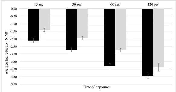
An overall ANOVA analysis indicated that mean values of log reduction of clinical and food isolates were statistically different (p < 0.05) at all sampling times (figure 3.4); food isolates were found to be more susceptible to wine, presenting higher log reduction means (more than one log-cycle reduction) than the clinical isolates. No statistical differences were found (p > 0.05) among serogroups IVb, IIb, and IIa, while serogroup IIc isolates were significantly more susceptible to wine (p < 0.05); however only two isolates of the latter were analyzed, hence further studies with more IIc isolates should be performed to validate this result.
Figure 3.4 Mean log reduction of L. monocytogenes isolates of food and clinical origin exposed to red wine (1:10 dilution) at each sampling time Data represent mean of duplicate assays and error bars represent the standard deviation of the mean. (<) Food isolates; (<) Clinical Isolates.
Antimicrobial effects of certain beverages such as coffee, tea, beer and wine have been widely reported in the literature (L’Anthoën & Ingledew, 1996; Almeida et al., 2006; Medina et al., 2007; Carneiro et al., 2008). Among them, wine is well known for its antibacterial properties (Møretrø & Daeschel, 2006), and despite its activity against foodborne pathogens like Campylobacter spp., Escherichia coli, Salmonella spp., or Staphylococcus aureus (Sheth et al., 1988; Just & Daeschel, 2003; Rodríguez Vaquero et al., 2007; Carneiro et al., 2008) has been extensively investigated, few studies have explored the effect of wine on the survival of L. monocytogenes. To measure the effects of wine-related stress exposure on L. monocytogenes, we compared the survival response of 39 food and clinical isolates. Our results indicate that, under the tested conditions, red wine had a strong antilisterial activity, and that there is intra-strain variability in the resistance of this pathogen to wine, the human clinical isolates being significantly more resistant than the isolates obtained from food products. Komora et al. (2016), using the same isolates of the present study, also reported a higher resistance of clinical isolates to lactic acid and to osmotic stress. As these strains have been characterized in terms of D-value at 58 ºC, antibiotic resistance, and resistance to lactic acid, we evaluated a possible association between resistance to different stresses, however no correlation was found (data not shown).
-5.00 -4.50 -4.00 -3.50 -3.00 -2.50 -2.00 -1.50 -1.00 -0.50 0.00
15 sec 30 sec 60 sec 120 sec
A ve ra ge l og re duc ti on( N /N 0) Time of exposure
Fontoura (2012), in a study with eight clinical and eight food L. monocytogenes isolates, also found variability on the behavior of this pathogen to diluted red wine, and that clinical isolates were significantly more resistant when submitted to a 1:100 dilution of red wine. However, contradictory to our results, when the three more resistant strains from each origin were challenged in red wine diluted to 1:10 (at 20 ºC), the differences in bacterial cell inactivation between the origins was not significant. Other studies have reported higher resistance of clinical isolates to different environmental stress conditions when compared to food isolates. Variation on isolates of L. monocytogenes response to environmental stresses such as temperature, salt, pH, or sanitizers has also been reported (Aryani et al., 2015; Magalhães et al., 2015; Cunha et al., 2016)
Although several studies have explored the major wine components that may play a critical role in bacterial inactivation (e.g. pH, ethanol, organic acids, phenols, etc.) a consensus as not been achieved most likely because wine is a complex solution that incorporates multiple elements; hence its antimicrobial activity is rather due to the synergistic interaction of several parts than due to a single factor (Boban et al., 2010). Nevertheless, some wine elements have been reported to hold a strong antilisterial activity, such as ethanol (Corral et al., 1990) or ethanol in combination with organic acids (Fernandes et al., 2007). Also, phenolic compounds such as caffeic acid, rutin, and quercetin have a strong activity (Rodríguez Vaquero et al., 2007). Rhodes and co-authors (2006) showed antilisterial activity in red grape juice without ethanol, and demonstrated its association with different polymeric phenolics of grape skin, seeds and juice. Red wine has also been proved to inactivate L. monocytogenes cells in 30 min, and this effect was exacerbated when different marinades prepared with wine, oregano leaves,garlic juice, andoregano oil were applied; in this case the inactivation was instantaneous (Friedman et al., 2007).
Wine is an acidic environment, primarily due to the presence of tartaric, malic, and lactic acids. The wine low-pH has been pointed as a key impact factor in bacterial inactivation (Waite & Daeschel, 2007). However, in a study by Boban and co-workers (2010) that evaluated the effect of different elements of the wine against S. enterica and E. coli found that, used in separate, pH and ethanol presented only a minor antibacterial activity, while in combination with other components a synergistic effect was observed; intact wine was the most effective against these pathogens. Just and Daeschel (2003) when comparing wine and grape juice demonstrated a higher antibacterial effect of wine against the same pathogens, even when both beverages presented the same level of acidity. L. monocytogenes is able to tolerate low-pH environments, a feature that is crucial for its survival either in food-associated
environments, as in the infection process during passage through the stomach and within the macrophage phagosome. Dykes and Moorhead (2000) found that clinical isolates were less susceptible to acidic stress (pH 2.5) comparing to isolates from meat. Ramalheira et al. (2010) and Barbosa et al. (2012) concluded that clinical isolates were more resistant than food isolates recovered from various food products during passage through simulated gastro intestinal tract. Oppositely, Cunha et al. (2016) evaluated the ability of 33 isolates from food (18) and clinical (15) origin to survive the gastrointestinal conditions and extreme pH values (1.5 – 12) and found no differences in survival among isolates of different origins.
Our understanding of inter-strain variation in L. monocytogenes phenotypic response to different stress conditions is still limited, whether the observed differences between isolates reflect specificities related to their ecology (e.g. adaptation to human, animal, food-associated, and natural environments), or entirely to specific genetic qualities, such as lineage or serotype, is still unclear. For example, a number of studies point out that lineage I isolates (predominantly serotype 4b), are overrepresented among isolates from human listeriosis cases, even though some outbreaks have been caused by lineage I serotype 1/2b and lineage II serotype 1/2a isolates, whereas lineage II isolates appear to be overrepresented among food isolates and may be better adapted to a saprotrophic and environmental life style (Nightingale et al., 2005; Orsi et al., 2011). Comparative studies on the phenotypic behavior of L. monocytogenes isolates representative of different origins, and genetic characteristics, are therefore valuable to gather more data to uncover additional diversity and contribute to our understanding on this pathogen ecology. Our results also underline the importance of using a high number of isolates in this type of studies, as it has become clear that using a low number of isolates or reference strains, may provide biased results or that do not fully reflect the entire spectrum of its features.
3.2 Impact of wine on the invasion capacity of Listeria monocytogenes into the human intestinal epithelial Caco-2 cells
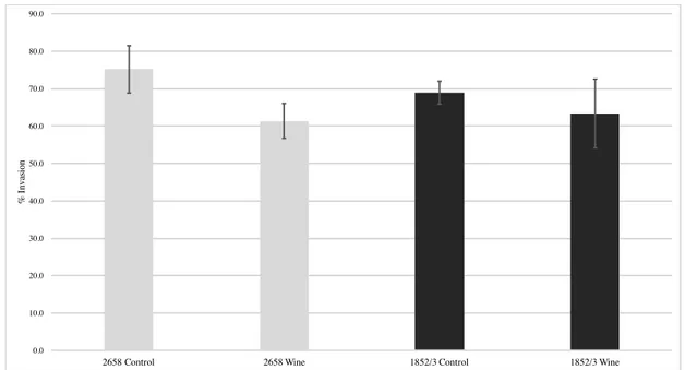
To evaluate the possible effect of wine on L. monocytogenes virulence, we investigated the ability of four selected isolates to invade the human epithelial Caco-2 cells after exposure to wine. From each origin one resistant and one susceptible isolate were selected, namely: clinical isolates Lm 2658 (IIb, resistant) and Lm 2542 (IVb, susceptible); and food isolates Lm 1852/3 (IIb, resistant) and Lm 969/3 (IIb, susceptible). The invasion
efficiency of resistant and susceptible L. monocytogenes exposed to diluted wine (1:10) during 15 sec is plotted in figures 3.5 and 3.6, respectively.
The invasion efficiency of the resistant isolates Lm 2658 and Lm 1852/3 after wine exposure was not statistically different of that observed for their respective controls (p > 0.05). Oppositely, the clinical isolate Lm 2542 demonstrated enhanced ability to invade the Caco-2 cells after exposed to the wine in comparison to the unexposed control (p < 0.05). This strain was isolated from a listeriosis outbreak in Portugal related with contaminated cheese (Magalhães et al., 2015). The food isolate Lm 969/3 unexposed to wine showed a low invasion efficiency. It has been demonstrated that attenuated invasion phenotypes in Caco-2 cells are frequently associated with premature stop codons (PMSC) in inlA, which encodes the surface protein InlA, that, as detailed previously in the Introduction section, is a key element for the initial bacterium attachment and invasion of intestinal epithelial cells through interaction with the cell host receptor E-cadherin. Strains with PMSC in inlA produce a truncated form of InlA that is secreted rather than anchored to the bacterial cell wall (Van Stelten & Nightingale, 2008). Other factors, such as reduced motility and nonsense mutations in prfA gene, that regulates the expression of a set of virulence genes have also been previously associated with impairment in invasion ability in Caco-2 cells (Roche et al., 2005; Handa-Miya et al., 2007; Roberts et al., 2009; Ferreira et al., 2011). As this is a poor invasive strain no comparison can be made between exposed and unexposed cells, because the initial bacterial numbers used to inoculate the Caco-2 cells monolayers were already low, in the end of the invasion assay the samples collected for enumeration were below the detection limit of the enumeration technique.
Exposure to stress conditions and food-associated environments may affect virulence-related characteristics in L. monocytogenes. For example, Garner et al. (2006) found that L. monocytogenes became more invasive when subjected to high pH, organic acids, and salt.
The effects of wine on the virulence of L. monocytogenes are almost certainly complex and might be related to the expression of virulence genes when this pathogen is under stress. For instance, the alternative sigma factor σB
(encoded by sigB) have been identified as regulating L. monocytogenes response to several environmental stresses response and virulence gene expression (Kim et al., 2004), and also plays a role in infection of human intestinal Caco-2 cells by regulating transcription of InlA (Garner et al., 2006). Further studies including more isolates are needed to confirm these results, and to evaluate the expression of virulence genes relevant for this outcome.