University of Trás-os-Montes and Alto Douro
Titanium-45: development and optimization of the
production process in low energy cyclotrons
PhD Thesis in Physical Sciences
PEDRO SILVA COSTA
Supervisors:
Prof. Doutor Marco Paulo Duarte Naia
Prof. Doutor Francisco José Cerqueira Alves
University of Trás-os-Montes and Alto Douro
Titanium-45: development and optimization of the
production process in low energy cyclotrons
PhD Thesis in Physical Sciences
PEDRO SILVA COSTA
Supervisors:
Prof. Doutor Marco Paulo Duarte Naia
Prof. Doutor Francisco José Cerqueira Alves
Jury:
Prof. Doutor José Boaventura Ribeiro da Cunha (President)
Prof. Doutor Luís Filipe dos Santos Garcia Peralta
Prof. Doutor Francisco José Cerqueira Alves
Prof. Doutor Armando da Assunção Soares
Prof. Doutor Norberto Jorge Alves Parente Gonçalves
Obstacles can’t stop you. Problems can’t stop you. Most important of all, other people can’t stop you. Only you can stop you.
I
Agradecimentos / Acknowledgments
Porque este trabalho não seria possível sem a intervenção (direta ou indireta) de várias pessoas, não poderia deixar de lhes expressar o meu profundo agradecimento:
Ao meu Orientador, Prof. Doutor Marco Naia; por me receber e aceitar como doutorando na UTAD, por ter encarado esse papel com a máxima dedicação e profissionalismo, por me ter mostrado o mundo da Física no seu estado puro e sem condicionar a minha própria visão pessoal, pelo desenho do meu plano de formação no Curso de Doutoramento, por todos os ensinamentos, por todos os momentos de discussão e reflexão, por todo o rigor, por toda a tranquilidade e por ter tornado este importante passo numa etapa incrivelmente proveitosa e agradável;
Ao meu Co-Orientador, Prof. Doutor Francisco Alves, por me aceitar como doutorando no ICNAS, por me abrir as portas do “seu” ciclotrão para toda a componente experimental deste trabalho, por toda a diversão e jeito peculiar de encarar cada noite de trabalho ou cada momento de discussão técnico-científica, por ter sido um importante contributo para a implementação deste trabalho;
Ao Prof. Luís F. Metello, meu “boss” na ESTSP.IPP e mentor deste trabalho; pelo desafio lançado, pela forma como encontrou sempre modo de financiar o material das experiências, pela insatisfação constante, pela exigência, pela pressão, mas também pela oportunidade de aprender, de crescer, de ser eu a implementar esta ideia que ele tinha em mente, pela sua rede de contactos que não hesitou em partilhar comigo, pelas viagens (nacionais e internacionais) para congressos e outros eventos científicos recheadas de discussão e aprendizagem, pela companhia nas noites de experiências, pela amizade e pelo incentivo;
Ao Doutor Sérgio do Carmo, Físico do ICNAS; pelo apoio prático inestimável, por operar o ciclotrão, pelas horas a fio de medições de espectros com o detetor HPGe, pelos contributos positivos na discussão de resultados e pela acessibilidade e diversão;
For all international collaborations, namely R.R. Johnson (BEST), W. Gelbart (ASD) and C. Artner (IASON), that made possible experimental work with beneficial technical inputs;
Aos meus colegas de gabinete na ESTSP.IPP (Diana, Domingos, Joana, Lídia, Mariana e Sara); pela
amizade e companheirismo, por demonstrarem que a opção de trabalhar em simultâneo com a realização de um doutoramento foi a mais louca, mas também a mais acertada, pelo ambiente de trabalho que sempre ajudou a transformar os piores momentos em momentos de diversão, por manterem sempre a “orquestra a tocar” e por estarem lá sempre que necessário – razão pela qual vários de vocês são já bons amigos;
Aos vários Professores do Departamento de Física da UTAD; pela acessibilidade, pelos ensinamentos nas unidades curriculares do 1º Ano do Curso de Doutoramento, pelo apoio, pela exigência em todas as
II
apresentações de trabalhos e por também terem contribuído, ao seu jeito e como lhes foi possível, para a concretização deste trajeto;
Aos meus amigos; por constantemente serem uma boa desculpa para atrasar o trabalho e me ir divertir, por incentivarem este trabalho e pela amizade que tanto ajuda nos momentos de maior tensão;
A toda a minha família; por saber da importância deste passo, por se preocupar, por incentivar e desejar o maior sucesso para mim;
Em especial, aos meus pais; por terem feito de mim aquilo que eu hoje sou, com as virtudes e defeitos que tenho, pela educação e valores que me transmitiram, por terem feito de mim uma pessoa feliz e grata, por saberem do peso deste trabalho para a minha vida e carreira, por estarem sempre lá independentemente do que a vida lhes possa ter trazido e dos rumos que possam ter tomado, por me amarem, por me darem o espaço e tempo necessário para me dedicar a este trabalho, por me mostrarem que seria sempre eu a beneficiar (ou sair prejudicado) por todas as minhas opções e atitudes… por tudo o que são;
À (muito especial) Joana; por ter transformado este projeto num projeto comum e numa luta a dois, pelo amor, pela amizade, pelo constante incentivo, motivação e preocupação, por tolerar as minhas falhas e ausências, por ter ouvido pacientemente os assuntos que nada lhe interessavam, por ser corresponsável pela exequibilidade deste trabalho em vários aspetos que nunca esquecerei…por perceber que isto é apenas um início e por aceitar que este tipo de projetos venha a fazer parte das nossas vidas;
…entre outros; porque todos aqueles que algum dia se cruzaram na minha vida são responsáveis pela pessoa e profissional em que me tornei e, para o bem ou para o mal, tiveram um determinado impacto neste trabalho;
Resta-me ainda agradecer de uma forma global a vida que eu tenho, os obstáculos e dificuldades que surgiram, os desafios e oportunidades aos quais estou grato, e tudo aquilo que me faz sonhar ou ambicionar mais e mais, e faz de mim aquilo que eu sou e que tento refletir em tudo o que faço.
III
Termo de responsabilidade
Declara-se que o autor desta Tese participou activamente na concepção e na execução de todo o trabalho experimental que esteve na origem dos resultados aqui apresentados, bem como em todo o trabalho teórico prévio, interpretação e discussão dos dados obtidos. Por opção, os capítulos textuais da Tese serão apresentados com recurso à Língua Inglesa.
V
Preface
In my personal point of view, a Doctoral Thesis is a restrict synthesis of a long route of learning, personal and professional development, and scientific achievements. There is much more than a hundred of written pages! Between literature reviews, hundreds of papers read, presentation of oral communications and posters in conferences, preparation and submission of papers…a PhD is really a hard task, a long way, a long journey to find the (hopefully) positive end.
The Thesis that will be presented here is the reflex of four years of work, always in part-time dedication, but always with a deep motivation and enthusiasm. And, of course, it is the reflex of my own soul, of the way I see physical phenomena, Nuclear Medicine or even the world itself.
Just to introduce the birth of this work, following a series of fruitful conversations and discussions, and always considering my background, expectations, profile and interests, my institutional boss (Prof. Luís F. Metello, ESTSP.IPP) gave me a preliminary idea that until that date was only on his mind; the challenge of studying the viability of produce an unconventional radionuclide (Titanium-45) in low energy cyclotrons to be used in PET imaging. After some study and investigation, the challenge was accepted and this PhD project started to be conceptualized, designed and implemented. And the rest of this story is written in the scientific work called Thesis and that will be presented in the next pages.
I would also like to state that this Thesis has also a challenging message: Nuclear Physics research is a field that could be occupied by a non-physicist researcher; and I humbly hope that this Thesis could prove that with the quality desirable and required for this level.
Finally, it is also important to add that this Thesis, but also all the research project involved on it, is the study to find a solution with the application of theory already known from fundamental research and using methodologies and approaches that came from applied research to design science research methods, but passing also through experimental science, culminating in the viability study of an industry-tailored process.
Read, analyze, criticize…and enjoy!
This Thesis is my first formal contribution for a better society!
VII
Publications List
The work performed under the scope of this Thesis resulted in the publication of the following scientific contributions:
Posters:
1. P. Costa, F. Alves, M. Duarte Naia, L.F. Metello. “New methods using low energy cyclotrons in radioisotope production for Nuclear Medicine”, in E3 Forum - Education, Employment & Enterpreneurship Conference, Lisboa (2013);
2. P. Costa, L.F. Metello, F. Alves, M. Duarte Naia. “Application of Monte Carlo simulation codes to plan an activation experiment in Scandium-45 targets” in Annual Congress of the European Association of Nuclear Medicine, Goteborg (2014);
3. P. Costa, L.F. Metello, F. Alves, M. Duarte Naia. “The use of radiolabeled nanoparticles for biomedical imaging” in Low Dose PT, Lisboa (2015);
4. P. Costa, L.F. Metello, L. Cunha, R.R. Johnson, L. Mattei, W. Gelbart, J. Obermair, B. Dietl, R. Nauschnig, C. Artner, P. Lass, G. Currie, S. Carmo, F. Alves, M. Duarte Naia. “Cyclotron produced 45Ti-Titanium: why & how…so Why Not?” in 5th Symposium on Medical Radionuclides, Brussels (2015);
5. P. Costa, L.F. Metello, L. Cunha, R.R. Johnson, L. Mattei, W. Gelbart, J. Obermair, B. Dietl, R. Nauschnig, C. Artner, P. Lass, G. Currie, S. Carmo, F. Alves, M. Duarte Naia. “Excitation function determination for 45Ti production using 45Sc(p,n) 45Ti reaction using low energy cyclotrons” in Annual Congress of the European Association of Nuclear Medicine, Hamburg (2015)
Oral Communications:
1. P. Costa, M. Naia. “Gamma Spectroscopy: Code for Analysis and Simulation” in Física 2012, Aveiro (2012);
2. P. Costa, L. Cunha, R.R. Johnson, W. Gelbart, C. Artner, F. Alves, M. Duarte Naia, L.F. Metello. “Using Monte Carlo to approach 45Ti direct production on low energy – medical – cyclotrons” in Cycleur 2014 – Cyclotron European Network Workshop, Ispra (2014);
3. P. Costa, L.F. Metello, L. Cunha, R.R. Johnson, W. Gelbart, J. Obermair, C. Artner, S. Carmo, F. Alves, M. Duarte Naia. “Experimental results on excitation functions for 45Ti direct production on low energy – medical – cyclotrons” in 11th Cycleur – Cyclotron European Network Workshop, Monastir (2015); 4. P. Costa, L.F. Metello, L. Cunha, P. Lass, G. Currie, R.R. Johnson, W. Gelbart, J. Obermair, C. Artner, F.
Alves, M. Duarte Naia. “45Ti-Titanium: from cyclotron production to potential applications evaluation” in Low Dose PT, Lisboa (2015);
VIII
5. P. Costa, L. Cunha, L.F. Metello. “The role of the Nuclear Medicine Technologist in cyclotron related Research & Development” in Annual Congress of the European Association of Nuclear Medicine, Hamburg (2015);
6. P. Costa, M. Naia, F. Alves, L.F. Metello. “Development of 45Ti for radiopharmaceutical purposes: results obtained and future perspectives” in 12th Cycleur – Cyclotron Cyclotron European Network Workshop – and 2nd Bern Cyclotron Symposium, Bern (2016);
Published abstracts:
1. P. Costa, L.F. Metello, F. Alves, M. Duarte Naia. PET imaging using Titanium-45: Could it be useful? Nuclear Medicine and Biology. 2014; 41(7). DOI: 10.1016/j.nucmedbio.2014.05.129
Peer-review journal articles:
1. P. Costa, L.F. Metello, F. Alves, M. Duarte Naia. Nanoparticle-based radiopharmaceuticals: Status quo
and future developments.
Under submission in: Molecular Diagnosis and Therapy (Impact Factor 2014: 2.891)
2. P. Costa, L.F. Metello, F. Alves, M. Duarte Naia. Study of 45Ti production by the experimental
determination of the excitation function of 45Sc(p,n) 45Ti nuclear reaction.
IX
Abstract
Introduction: Modern practice of Medicine include the use of technological innovations such as Medical Imaging. In vivo imaging techniques can be used to evaluate biological structures and functions non-invasively in almost all
living subjects.
In this Thesis it will be highlighted Nuclear Medicine. As a simple definition, Nuclear Medicine is a medical imaging modality based on the measurement of an internal source from an internal tracer, reason why it uses radiolabeled compounds to study in vivo physiologic processes. So, Nuclear Medicine relies on the supply of radionuclides. In the specific context of Positron Emission Tomography (PET) imaging, positron emitters are used to label several different compounds, allowing the study of almost all the major biological systems. Although there are several radionuclides to be potentially applied in PET imaging, routine clinical applications are still based on a small group constituted by 18F, 11C, 13N and, more recently, 68Ga. However, recent literature indicates that this trend is changing. Among others, 45Ti is being proposed as a potential candidate for PET imaging, since it presents some interesting properties: abundant positron emission, reduced positron energy, physical half-life of 3.09h, and relevant chemical properties, that enable radiolabelling with bifunctional chelates, ligands or even to radiolabel titanium dioxide nanoparticles. Given this, several issues should be solved before the real possibility to implement 45Ti clinical applications.
Aim: Considering that Radionuclide production is the first crucial technical step involved in PET, and that production
of 45Ti is yet very poorly explored in literature, this Project was designed and implemented with the aim to study the viability of the production of 45Ti in low energy cyclotrons, expecting the characterization of the excitation functions of the appropriate nuclear reaction, yield determination and development of a critical analysis to select the best methodology.
Materials and Methods: To evaluate nuclear reactions activation studies should be implemented. Nevertheless,
the execution of nuclear activation studies deserve special attention and a careful integration of all the available information already collected. In this sense, the first step of this activation experiment was the adequate planning using Monte Carlo simulation codes in the way to obtain several results that were totally integrated in the design of experimental studies.
Still before the experimental activation study, some preliminary experimental studies with gamma-spectroscopy were performed with calibrated radionuclide sources as demonstration of the ability of the technique to illustrate physical phenomena such as radioactive decay and radiation interaction with matter, and in the way to refine the analysis of the main experiment.
Then, the stacked foil technique was implemented in a 18 MeV cyclotron to study the 45Sc(p,n)45Ti nuclear reaction and its feasibility to effectively produce 45Ti. Activation was measured using HPGe gamma-spectroscopy.
Theoretical insight about the potential applications of 45Ti for PET imaging were also reviewed, analyzed, proposed and discussed.
X
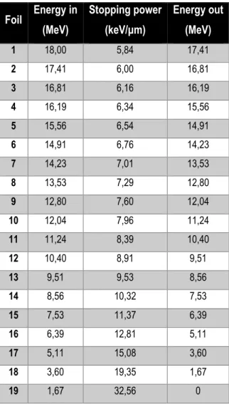
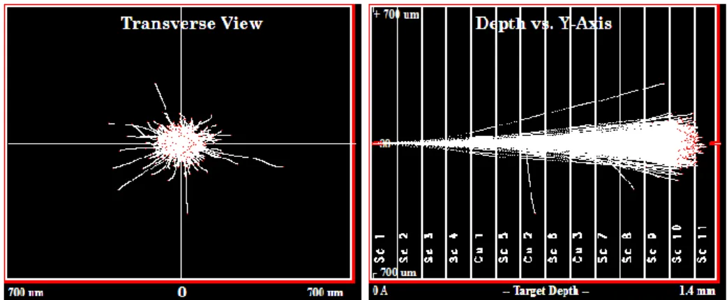
Results: According to TALYS code, and also due to other practical considerations, 45Sc(p,n)45Ti nuclear reaction was selected as the one with much more potential for industrial implementation in the way to obtain significant quantities of 45Ti. SRIM code simulations were used to understand the beam energy degradation along the stacked foil designed for the activation study, while SSSM sub-routine was used to study the implantation of the beam in the successive targets.
Results on the main excitation function under study were collected, with the addition of information regarding concurrent reactions leading to 44mSc, 44Sc and 44Ti. Experimental results showed that 45Sc(p,n)45Ti nuclear reaction seems to be feasible in low energy cyclotrons, with cross-section values presenting a peak for proton beam energies in the range between 10 and 14 MeV, while energies higher than 17 MeV should be avoided due to the increased production of contaminants such as 44Ti, 44Sc and 44mSc. Thick target yield for a saturation condition was experimentally determined as 433.64 MBq.µA-1sat.
Theoretical evidences collected demonstrate that 45Ti could provide good PET image quality, with some preclinical applications already tested in studies related to a new class of anticancer drugs based on titanium complexes. Other possible applications already cited include 45Ti-ligands for theranostics and personalized medicine. An innovative proposal on the use of 45Ti to radiolabel of titanium dioxide nanoparticles will also be presented and discussed.
Conclusion: Thus, there is the possibility to effectively obtain significant quantities of 45Ti allowing its possible commercial and industrial production, distribution and use. Given this, 45Ti could then provide good PET images and be used for labeling of different compounds already tested or to be incorporated in the development of nanoparticle-based radiopharmaceuticals.
XI
Resumo
Introdução: A Medicina praticada atualmente inclui o uso de inovações tecnológicas, tais como a Imagiologia
Médica. As técnicas de imagiologia in vivo permitem avaliar estruturas e funções de forma não-invasiva, em quase todos os sistemas biológicos. O tema deste trabalho está centrado na Medicina Nuclear, que é uma modalidade de imagiologia médica com base na medição de uma fonte utilizando um marcador interno. Este método utiliza compostos marcados radioactivamente para estudar processos fisiológicos in vivo, pelo que a Medicina Nuclear está baseada no fornecimento de radionuclídeos.
No contexto específico da imagiologia por Tomografia de Emissão de Positrões (PET), os emissores de positrões são utilizados para radiomarcar vários compostos diferentes que permitem o estudo de praticamente todos os principais sistemas biológicos. Embora existam vários radionuclídeos para ser potencialmente aplicados em imagens de PET, as aplicações clínicas de rotina utilizam apenas um pequeno grupo de nuclídeos, constituído por 18F, 11C, 13N e, mais recentemente, o 68Ga. No entanto, a literatura recente indica que esta tendência está a mudar. Entre outros, 45Ti tem sido proposto como um candidato potencial para geração de imagens PET, uma vez que apresenta algumas propriedades interessantes: emissão eficiente de positrões, reduzida energia dos positrões, semi-vida física de 3,09 h, bem como as suas propriedades químicas relevantes, que permitem a marcação radioativa com quelatos bifuncionais, ligandos diversos ou mesmo para marcar radioactivamente nanopartículas de dióxido de titânio Porém, várias questões devem ser resolvidas antes da real possibilidade de implementar aplicações clínicas de 45Ti.
Objetivo: A produção do radionuclídeo é o primeiro passo técnico fundamental envolvido em PET, contudo a
produção de 45Ti é ainda muito pouco explorada na literatura. O projeto relatado neste trabalho foi delineado e implementado de forma a estudar a viabilidade da produção de 45Ti em ciclotrões de baixa energia. Com este objetivo principal desenvolveu-se um programa de pesquisa completo para obter uma caracterização completa das funções de excitação da reação nuclear mais apropriada, determinação dos respetivos rendimentos e desenvolvimento de uma análise crítica para selecionar a metodologia mais apropriada.
Material e Métodos: Para avaliar as reações nucleares foram implementados estudos de ativação nuclear. No
entanto, a execução desses estudos merece atenção especial e uma integração cuidadosa de toda a informação disponível já recolhida e publicada. Neste sentido, o primeiro passo desta experiência de ativação foi o planeamento adequado utilizando códigos de simulação Monte Carlo para prever resultados e selecionar os cenários mais prováveis. Os dados calculados foram totalmente integrados na conceção dos estudos experimentais. Ainda antes do estudo experimental de ativação, foram realizados ensaios experimentais preliminares com espectroscopia-gama com fontes calibradas de radionuclídeos, para avaliar a capacidade da técnica para ilustrar fenómenos físicos, tais como o decaimento radioativo e a interação de radiação com a matéria, e refinar a análise da experiência principal.
XII
Finalmente, utilizou-se a técnica da pilha de lâminas num ciclotrão de 18 MeV para estudar a reação nuclear 45Sc(p,n)45Ti e a sua viabilidade para produzir efetivamente 45Ti. A ativação foi medida utilizando espectroscopia-gama com recurso a um detetor HPGe.
Resultados: De acordo com o código TALYS, e também devido a outras considerações práticas, a reação nuclear
45Sc(p, n)45Ti foi selecionada como aquela com mais potencial para obter quantidades significativas de 45Ti para aplicação industrial.
O cálculo com os códigos SRIM permitiu compreender a degradação da energia do feixe ao longo da pilha de lâminas concebida para o estudo da ativação induzida, enquanto que a sub-rotina SSSM foi usada para estudar a implantação do feixe do ciclotrão nos alvos sucessivos na pilha.
Obtiveram-se os dados relativos à função de excitação principal, bem como informações adicionais sobre reações concorrentes que levam à produção de 44mSc, 44Sc e 44Ti. Os resultados experimentais mostraram que a reação nuclear 45Sc(p,n)45Ti parece ser exequível em ciclotrões de baixa energia, com valores de secção eficaz que apresentam um pico para energias do feixe de protões compreendidas entre os 10 e os 14 MeV, enquanto que as energias superiores a 17 MeV devem ser evitadas, devido ao aumento da produção de contaminantes como o 44Ti, o 44Sc e o 44mSc. O rendimento de alvo espesso para uma condição de saturação foi determinado experimentalmente como 433,64 MBq.μA-1sat.
As evidências teóricas recolhidas demonstram que o 45Ti pode proporcionar uma boa qualidade de imagem PET, com algumas aplicações pré-clínicas já testadas em estudos relacionados com uma nova classe de fármacos anticancerígenos à base de complexos de titânio. Outras possíveis aplicações do 45Ti já citadas incluem a radiomarcação de ligandos para teragnóstico e medicina personalizada. Apresenta-se também uma proposta inovadora de uso de 45Ti para a marcação radioativa de nanopartículas de dióxido de titânio.
Conclusão: Existe a possibilidade de obter de forma eficaz quantidades significativas de 45Ti permitindo a sua possível produção comercial, distribuição industrial e utilização clínica. Como o 45Ti tem potencial de proporcionar boas imagens PET, e ser utilizado para a radiomarcação de diferentes compostos, deve ser testado de forma a ser incorporado no desenvolvimento de radiofármacos para imagiologia PET ou na marcação de nanopartículas para estudos aplicados de farmacologia e diferentes aplicações biomédicas.
XIII
Index
1.
Introduction ... 1
1.1 Nuclear Medicine basics ... 2
1.1.1. Nuclear (in)stability ... 4
1.1.2. Radioactive decay ... 7
1.1.3. Positron and positronium physics ... 10
1.1.4. Nuclear Medicine procedures ... 11
1.2 Radionuclides in Nuclear Medicine ... 12
1.2.1 Common radionuclides in Nuclear Medicine ... 12
1.2.2. Unconventional radionuclides in Positron Emission Tomography ... 13
1.3 The potential interest of Titanium-45 ... 15
1.4 Aim and outline of the Thesis ... 16
2.
Production of Radionuclides for Nuclear Medicine ... 19
2.1 Introduction to radionuclide production ... 19
2.2 Nuclear reactions physics ... 21
2.3 Cyclotron principles ... 26
2.3.1 Cyclotron physics and operation ... 27
2.3.2 Cyclotron targetry and target chemistry... 33
2.4 Study of radionuclide production processes ... 34
2.4.1 Industrial radionuclide production issues ... 35
2.4.2 Excitation functions and production process optimization ... 36
3.
Monte Carlo simulation codes to plan an activation experiment ... 39
3.1 Basic concepts on Monte Carlo simulation codes ... 39
3.2 Methodology of application of Monte Carlo simulation codes ... 41
3.2.1 Preliminary study of nuclear reaction excitation functions ... 41
3.2.2 Study of the proton beam energy degradation ... 41
XIV
3.3 Results and discussion on the application of Monte Carlo simulation codes ... 44
3.3.1 Preliminary study of nuclear reaction excitation functions ... 44
3.3.2 Study of the proton beam energy degradation ... 45
3.3.3 Study of the proton beam range and dispersion... 47
3.4 Final considerations on the application of Monte Carlo simulation codes ... 49
4.
Radiation detection and quantification using Gamma-ray spectroscopy ... 51
4.1 Gamma radiation interaction with matter ... 51
4.2 Introduction to radiation detection and quantification ... 55
4.3 Gamma-ray spectra: basic technical considerations on detection and analysis ... 57
4.4 Gamma-ray spectroscopy: calibration and sources of error ... 63
4.5 Final considerations on gamma-ray spectroscopy ... 67
5.
Experimental determination of the excitation function of
45Sc(p,n)
45Ti nuclear
reaction ... 69
5.1 Introducing Titanium-45: what is it and how to obtain it? ... 69
5.2 Materials and methods ... 70
5.2.1 General aspects ... 71
5.2.2 HPGe gamma spectroscopy ... 72
5.2.3 Cross-section determination ... 73
5.2.4 Preliminary beam energy calibration ... 74
5.2.5 Foil preparation ... 75
5.2.6 Qualitative study of 45Sc(p,n)45Ti energy threshold ... 76
5.2.7 Stacked foil irradiation ... 76
5.2.8 Data analysis ... 77
5.3 Results and discussion ... 77
5.3.1 HPGe calibration ... 77
5.3.2 Beam energy calibration ... 78
5.3.3 Cross-section determination: correction factors ... 88
XV
5.3.5 Determination of the excitation function for the 45Sc(p,n)45Ti nuclear reaction ... 89
5.4 Final considerations on the excitation function of 45Sc(p,n)45Ti nuclear reaction ... 99
6.
Potential applications of
45Ti ... 101
6.1 Nanoparticle-based radiopharmaceuticals: status quo and future developments ... 101
6.1.1 From Nanotechnology to Nanomedicine ... 101
6.1.2 Metodological aspects of the review ... 104
6.1.3 Nanoparticle-based radiopharmaceuticals: reviews and robust data ... 104
6.1.4 Nanoparticle-based radiopharmaceuticals: recent experiments and developments ... 107
6.1.5 Final remarks and future perspectives ... 115
6.2 Nanoparticle-based radiopharmaceuticals: is there a future to 45TiO2 nanoparticles? ... 116
6.3 45Ti-labeled compounds for use in PET imaging ... 118
6.4 Final considerations on potential applications of 45Ti ... 121
7.
Final remarks ... 123
7.1 Research summary and general discussion ... 123
7.2 Conclusion and future perspectives ... 125
XVII
Figures index
Chapter 1Figure 1.1 – Spectrum of macroscopic medical imaging modalities. ... 2 Figure 1.2 – Schematic representation of multidisciplinarity in Nuclear Medicine. ... 3 Figure 1.3 – Total nucleon-nucleon force. ... 5 Figure 1.4 – Binding energy per nucleon versus the mass number of the isotope. ... 6 Figure 1.5 – Neutron (N) vs. Atomic (A) number in nuclides found in nature. ... 7 Figure 1.6 – Beta decay energy spectrum. ... 9 Figure 1.7 – First known photo of the path of a positron. ... 10 Figure 1.8 – Relationship between Nuclear Medicine procedures and the decay mode of applied radionuclides. 12 Figure 1.9 – Technical steps inherent to a PET examination. ... 16 Chapter 2
Figure 2.1 – Schematic representation of cyclotron components. ... 28 Figure 2.2 – Cyclotron beam extraction systems: electrostatic deflectors (lefs) and stripper foils (right). ... 30 Figure 2.3 – Worlwide distribution of cyclotron maximum proton energies. ... 32 Figure 2.4 – Description of some common commercial cyclotrons used for radionuclide production. ... 33 Figure 2.5 – Experimental data considered for the definition of excitation function of 18O(p,n)18F nuclear reaction. ... 36
Figure 2.6 – Yield of 18F calculated from the recommended cross-section data. ... 37
Figure 2.7 – Excitation functions of proton induced reactions on 124Te... 38
Chapter 3
Figure 3.1 – Relationship between Theory and Monte Carlo approaches. ... 40 Figure 3.2 – Comparison between analytic methods and Monte Carlo methods. ... 40 Figure 3.3 – Schematic representation of the main phases of an activation experiment planning. ... 41 Figure 3.4 – Emittance diagram illustrating Twiss parameters, total geometrical beam emittance, maximum beam
extent, radius and maximum beam divergence. ... 43
Figure 3.5 – Graphical representation of TALYS-based excitation function of the nuclear reaction 45Sc(p,n)45Ti. 44
Figure 3.6 – Graphical representation of TALYS-based excitation functions of proton induced nuclear reactions for
production of 45Ti and some competitor nuclear reactions. ... 45
Figure 3.7 – SRIM/TRIM graphical outputs: transversal view of ions in the last foil of the stack (left); and longitudinal
XVIII
Chapter 4
Figure 4.1 – Schematic representation of the photoelectric effect. ... 52 Figure 4.2 – Schematic representation of the Compton scattering effect. ... 54 Figure 4.3 – Schematic representation of the pair production interaction phenomenon. ... 54 Figure 4.4 – Schematic representation of the relative predominance of the three main processes of photon
interaction with matter. ... 55
Figure 4.5 – Schematic representation of precision and accuracy of measurements. ... 56 Figure 4.6 – Gamma spectrum of a 60Co standard source obtained using a NaI(Tl) detector at Dept Física - UTAD. ... 58
Figure 4.7 – Calibrated gamma spectrum of the 60Co standard source obtained at Dept Física - UTAD. ... 58
Figure 4.8 – Typical gamma spectrum of a source with a photopeak greater than 1022 keV, obtained in an
intermediary size detector. ... 59
Figure 4.9 – Calibrated gamma spectrum of a 22Na standard source obtained at Dept Física - UTAD. ... 59
Figure 4.10 – Typical background spectrum using a semiconductor (HPGe) detector. ... 61 Figure 4.11 – Logic flowchart describing the overall functioning of the GSA software. ... 62 Figure 4.12 – GSA interface. ... 63 Figure 4.13 – Example of an energy calibration curve for HPGe-based spectroscopy. ... 64 Figure 4.14 – Example of an efficiency calibration curve obtained in an HPGe system. ... 66 Figure 4.15 – Efficiency calibration curves of an HPGe detector and its relation with source-detector distance and
sample configuration. ... 67
Chapter 5
Figure 5.1 – Real photograph of the IBA Cyclone® 18/9 HC cyclotron installed at ICNAS. ... 71 Figure 5.2 – Some real photographs of the in house stacked foil holder. ... 72 Figure 5.3 – Real photograph of the dismantled target holder and copper foils used for beam energy
calibration. ... 74
Figure 5.4 – Real photograph of some copper and scandium foils used in the experiments. ... 75 Figure 5.5 – Example of a gamma-spectrum of the 152Eu source used for HPGe detector calibration (linear scale). ... 77
Figure 5.6 – Example of a gamma-spectrum of the 152Eu source used for HPGe detector calibration (log
scale). ... 78
Figure 5.7 – Comparison of the 63Zn activation in two 25 μm copper foils interspaced by an 875 μm aluminum degrader given incident proton energies of 15.0 and 15.5 MeV. ... 79
Figure 5.8 – Representation of the stack of copper foils, including 100 μm copper monitor foils (grey), interspaced
XIX
Figure 5.9 – Example of a gamma-spectrum of a Copper foil irradiated presenting production of 62Zn, 63Zn and 65Zn. ... 81
Figure 5.10 – Graphical representation of the relation between nominal and calibrated energies. ... 82 Figure 5.11 – Experimental and tabulated excitation function for the natCu(p,x)63Zn reaction. ... 84
Figure 5.12 – Experimental and tabulated excitation function for the natCu(p,x)65Zn reaction. ... 84
Figure 5.13 – Beam energy calibration curves obtained using the independent, the linear and the quadratic model.
... 87
Figure 5.14 – Tabulated and experimental excitation functions for the natCu(p,x)63Zn and natCu(p,x)65Zn nuclear reactions . ... 91
Figure 5.15 – Example of a gamma-spectrum of a Scandium foil irradiated with a mean energy higher than 17 MeV
presenting production of 44Ti, 45Ti, 44Sc and 44mSc. ... 94
Figure 5.16 – Experimental excitation function of the 45Sc(p,n)45Ti nuclear reaction. ... 95
Figure 5.17 – Experimental excitation function of the 45Sc(p,n)45Ti nuclear reaction fitted with two different mathematical models. ... 96
Figure 5.18 – Thick target yield of the 45Sc(p,n)45Ti nuclear reaction calculated for a saturation condition for proton energies between 16 and 8 MeV. ... 97
Figure 5.19 – Comparison between TALYS simulated and experimental excitation functions for the 45Sc(p,n)45Ti nuclear reaction, with respective lines indicating mathematical fitting with Bi-Hill models and grey shadowed area indicating interval of experimental values considering experimental uncertainty. ... 98
Figure 5.20 – Graphical comparison of experimental data on the excitation function of the 45Sc(p,n)45Ti nuclear reaction obtained in this work with other published results, with grey shadowed area indicating interval of experimental values considering experimental uncertainty. ... 99
Chapter 6
Figure 6.1 – Techno-scientific implications in nanoparticle creation and development. ... 102 Figure 6.2 – Frequency of Nuclear Medicine procedures in experimental papers analyzed. ... 110 Figure 6.3 – Study design and population used in experimental papers analyzed. ... 111 Figure 6.4 – Relative frequency of application for each radionuclide in experimental papers analyzed. ... 102 Figure 6.5 – Relative frequency of articles analyzed per year of publication. ... 115 Figure 6.6 – Whole body images of 45Ti-phytate and 45Ti-DTPA acquired 10 mins and 60 mins after injection in rats. ... 118
Figure 6.7 – Comparison of image quality of 18F and 45Ti using a Derenzo phantom. ... 119
XXI
Tables index
Chapter 1Table 1.1 - Physical properties of the radionuclides most commonly applied in Nuclear Medicine. ... 13 Table 1.2 – Physical half-life of some of the unconventional radionuclides with most potential in PET imaging. .. 14 Chapter 2
Table 2.1 – Main production methods for the most common radionuclides in Nuclear Medicine. ... 20 Table 2.2 – Overview of the most common nuclear reaction types. ... 24 Table 2.3 – Classification of cyclotrons according to particle type and energy. ... 31 Table 2.4 – Alternative classification of cyclotrons according to particle type and energy. ... 31 Table 2.5 – Examples of radionuclide production routes and list of common targets used. ... 34 Chapter 3
Table 3.1 – Input parameters for beam generation in SSSM/SRIM. ... 43 Table 3.2 – SRIM simulation of beam energy degradation in a scandium stack irradiated with 18 MeV protons. . 46 Table 3.3 - SRIM simulation of beam energy degradation in a scandium-copper stack irradiated with 18 MeV
protons. ... 47
Table 3.4 – Range and dispersion of 18 MeV protons in a scandium-copper stack simulated with SRIM/TRIM. .. 48 Table 3.5 – Comparison of simulated range and dispersion of 18 MeV protons (SRIM/TRIM) and 18 MeV real beam
(S3M) implanted in a scandium-copper stack. ... 49
Chapter 4
Table 4.1 – Nuclear decay data of 152Eu. ... 65
Chapter 5
Table 5.1 – Nuclear data relevant for gamma spectroscopy calculations. ... 73 Table 5.2 – SRIM simulation of beam energy degradation in a copper stack of foils irradiated with 18 MeV protons.
... 75
Table 5.3 – SRIM simulation of beam energy degradation in a scandium-copper stack of foils irradiated with 18
MeV protons with the indication of mean energy per foil. ... 76
Table 5.4 – Activitites of 62Zn, 63Zn and 65Zn produced in the copper stack used for cyclotron beam energy calibration. ... 80
Table 5.5 – Fitting parameters for the adjustment of cyclotron beam energy calibration model. ... 81 Table 5.6 – Cyclotron beam energy calibration results following 63Zn activity experimental ratios. ... 82
XXII
Table 5.7 – Properties of targets used in the copper stacked foil. ... 83 Table 5.8 – Experimental cross-section values for the 63Cu(p,n)63Zn and 65Cu(p,n)65Zn. ... 83
Table 5.9 – Gaussian fitting parameters obtained using ORIGIN 2016®. ... 85 Table 5.10 – Linear cyclotron beam energy calibration results according to the Gaussian fiting of the excitation
functions of the monitor reactions on copper foils. ... 86
Table 5.11 – Quadratic cyclotron beam energy calibration results according to the Gaussian fiting of the excitation
functions of the monitor reactions on copper foils. ... 87
Table 5.12 – Activities of 45Ti produced on the stacked foils used for the experimental determination of the excitation function of the 45Sc(p,n)45Ti nuclear reaction, quantified using HPGe detector and corrected with factor F. ... 90
Table 5.13 – Activities of 62Zn, 63Zn and 65Zn produced on the monitor foils inserted in the stacked foil, quantified using HPGe detector and corrected with factor F. ... 90
Table 5.14 – Cyclotron beam energy calibration results for the stacked foil constituted by Scandium foils. ... 91 Table 5.15 – Irradiation parameters implemented in the two independent experiments using stacked foil technique
for the determination of the excitation function of the 45Sc(p,n)45Ti nuclear reaction. ... 92
Table 5.16 – Properties of targets used in the scandium-copper stacked foils applied in the main experiments. .. 92 Table 5.17 – Summarized results of experimental cross-section values for the 45Sc(p,n)45Ti nuclear reaction. .... 93
Chapter 6
Table 6.1 – Analysis of 20 review papers on nanoparticle-based radiopharmaceuticals found using
EBSCOhost. ... 105
XXIII
Abbreviations, Acronyms and Symbols List
A – number of mass Am – Amaricium Ba – Barium C – Carbon Co – Cobalt Cs – Cesium CT – Computed Tomography Cu – Copper Da – Dalton e- - electron E – energy EC – electron capture EOB – end of bombardment Eu – EuropiumeV – electron-volt F – Fluorine
FDA – Food and Drugs Administration Ga – Gallium
GSA – Gamma Spectra Analyzer GUI – Graphical User Interface h - hour
HPGe – High purity germanium I – Iodine
IAEA - International Atomic Energy Agency
ICNAS – Instituto das Ciências Nucleares Aplicadas à Saúde In – Indium
IT – isomeric transition K – Potassium keV – kiloelectron-volt LET – Linear Energy Transfer MeV – Megaelectron-volt MR – Magnetic Resonance n - neutron
XXIV Na – Sodium
NaI – Sodium iodide
NaI(Tl) – Sodium iodide doped with Tallium NM – Nuclear Medicine
O – Oxygen p - proton Pb – Lead
PET – Positron Emission Tomography Ra – Radium
Rb – Rubidium Re – Renium RF - radiofrequency
SATP – Standard Ambient Temperature and Pressure Sc - Scandium
Se – Selenium Sm – Samarium
SPECT – Single Photon Emission Computed Tomography SRIM – Stopping and Range of Ions in Matter
SSSM – SRIM Supporting Software Modules Tc – Technetium
TENDL – Talys Evaluated Nuclear Data Library Ti – Titanium
TiO2 – titanium dioxide Tl – Tallium
TRIM – Transport and Range of Ions in Matter US – Ultrasonography
WHO – World Health Organization Y – Yttrium Z – atomic number Zn – Zinc α – alpha particle β- - electron β+ - positron
1
1.
Introduction
In modern practice of Medicine the use and incorporation of technological developments into the clinical routine is becoming more and more common. In such an environment, one can specify the case of Medical Physics methods and technologies. In fact, it is very difficult to define the range and scope of Medical Physics, but for the purpose of this work it is sufficient to refer that it includes the use of radiation for diagnostic or therapeutic purposes, including specifically the field of Medical Imaging.
The application of imaging modalities and technologies has improved the quality of medical care and procedures available in nowadays’ practice of medicine. With the implementation of the current paradigms of Evidence Based Medicine and Science Based Medicine, medical imaging is occupying a primary position in clinical decision algorithms.
Noninvasive imaging modalities could allow accurate diagnosis, increase the precision in treatment choice and planning and give opportunity to follow the evolution of patients’ clinical status [1].
Medical or biomedical imaging modalities are actually the main pillars of medical care with many advantages including real time monitoring, accessibility without tissue destruction, minimal or no invasiveness. These modalities could also act over wide ranges of time and size scales involved in biological and pathological processes [2].
In vivo imaging techniques can be used to evaluate biological structures and functions non-invasively in almost all
living subjects. Multiple imaging techniques are available, including X-ray imaging, Computerized Tomography (CT), Magnetic Resonance (MR) imaging, Ultrasonography (US), Optical Imaging using fluorescent molecules and Nuclear Medicine (NM) using radioisotopes (planar imaging, Single Photon Emission Computed Tomography - SPECT - and Positron Emission Tomography - PET) [3].
Generally, imaging modalities can be divided according to different classifications; one option could be based on two main groups: those that primarily provide structural information (for example, CT or MR) and those that primarily provide functional and molecular information (for example, SPECT and PET) [4].
Another kind of classification of medical imaging modalities could be observed in Figure 1.1, showing the division based on the source of the measured signal and on its physical basis. Different imaging modalities or techniques are based on the detection of signals from different sources meaning different biological properties, as it is the example of CT and MRI where a contrast is produced by detecting differences in tissue density and water content, respectively, or in radionuclide imaging where contrast is conferred by detection of a clearly identified molecule labeled with a radioactive isotope [4].
2
This thesis will focus especially on Nuclear Medicine, more specifically in the PET field. The next topics will be dedicated to the definition and exploitation of Nuclear Medicine and the classification and description of the most important medically useful radionuclides.
1.1 Nuclear Medicine basics
Nuclear Medicine is an independent medical specialty defined by World Health Organization (WHO) since 1972, that “encompasses applications of radioactive materials in diagnosis, treatment or in medical research, with the exception of the use of sealed radiation sources in radiotherapy” [5].
Following the classification and differentiation present in Figure 1.1, Nuclear Medicine comprehends the medical imaging modality based on the measurement of an internal source from an internal tracer. It is considered to be a part of functional imaging field that is more uncommon in clinical practice than morphological imaging, even considering that it is gaining importance and frequency of application. This kind of methodology allows to access very important data, so increasing the efficiency of clinical diagnosis, essentially because of sooner detection of pathological processes. In fact almost all diseases start with functional/biochemical changes prior the morphological expression.
Figure 1.1 - Spectrum of macroscopic medical imaging modalities.
Source: H. Zaidi and B. H. Hasegawa, "Overview of Nuclear Medical Imaging: Physics and Instrumentation," in Quantitative Analysis
3
Nowadays it is quite common to present Nuclear Medicine imaging at the leading edge of Molecular Imaging, even considering that it is a hard task to define the real range of the Molecular Imaging field. Nevertheless, Nuclear Medicine is for sure one of the main parts of it, as it allows the in vivo measurement and follow-up of biological processes while enables the determination of different changes in cellular or molecular physiology [6].
This medical specialty implies a multidisciplinary environment, since its appearing, either as fundamental knowledge or as application to the specific context. This characteristic can be illustrated through the analysis of primary inputs such as: discovery of radioactivity (Henri Becquerel, 1896), definition of radioactivity (Marie Curie, 1897), the principle of radioactive tracer (George de Hevesy, 1924); followed by the application of these and other principles, for instance with the invention of the cyclotron (Ernest Lawrence, 1930), the gamma camera (Anger Hall, 1958) or upon obtaining the first image of cerebral neurotransmission (Henry Wagner, 1983) [7]. In summary, this field of science conjugates the knowledge and the experience from different scientific fields leading to the development of solid and valid solutions and methodologies (Figure 1.2).
From Figure 1.2 it is very clear the need for a continuous and complementary interaction between many different scientific fields in the Nuclear Medicine practice and application.
Moreover, Nuclear Medicine presents a tendency of growth over the past years. For instance, in the United States the number of procedures per year grown from 14 to 20 million between 1999 and 2005 [8]. Another study (Dose DATAMED II), designed to collect available data on the patient doses from the radiodiagnostic procedures (X-ray and Nuclear Medicine) in the European Union, shown that the frequency of Nuclear Medicine procedures over European Countries varies from 527 (Romania) to 35468 (Luxembourg) per year per million of inhabitants [9]. Together with other conclusions of that study, this data illustrate the high variability in frequency of examinations
4
between countries, but also identify a trend to homogenization with the increase of the annual number of procedures in several countries (even in the developing countries).
This situation of continuous and sustainable growth is related with clinical importance of the use of radiotracers that allows exceptional target specificity at the molecular level that cannot be accomplished with any other imaging technique [10]. Another advantage of radionuclide imaging is the ability to perform real time imaging studies, in order to increase understanding of physiological mechanisms underlying pathological processes or revealing the effects of drug administration [11].
Diagnostic Nuclear Medicine contributes to the clinical care, with all the advantages mentioned before, using radiopharmaceuticals (that combine a radionuclide with a specific molecule/probe), being possible to find specific radiopharmaceuticals for each organ, tissue or process. In overview, diagnostic Nuclear Medicine is playing an important clinical role in Oncology, Cardiology and Neurology, but also in Nephrology and investigations related with Infection or Inflammation processes, while the therapeutic branch of Nuclear Medicine is mainly concentrated in the Oncology field [12].
It is also known that Nuclear Medicine relies on the application of photons from the radioactive decay scheme of several elements, that can be detected from outside a patient or a simple organ or tissue sample [13]. On other hand, the possible usefulness of other decay particles for therapeutic purposes should also be considered. Considering that Nuclear Medicine rely on the use of radiopharmaceuticals it is very important to state a clear definition of these compounds. Radiopharmaceuticals are substances that result from the combination of a radionuclide and a molecular probe. The first is responsible for the signal to be read (or the energy to destroy the cells in therapeutic applications) after radioactive decay, and the second is the biological vector that guides the radionuclide during its biological distribution. Indeed, there are many possible combinations using a number of radionuclides and several molecules that have the ability to target cells, tissues or biochemical processes, demonstrating, and quite often quantifying, their respective metabolic/catabolic properties, creating real time information without interfering in the processes under study.
1.1.1. Nuclear (in)stability
Considering that radiopharmaceuticals are constituted by useful radionuclides, thus resulting in the dependence of Nuclear Medicine on radionuclides, it is important to point out physical basis inherent to the instability of nuclides. To understand and discuss the concept of nuclear stability, it is important to realize that atomic nuclei are quantum bound states of particles called nucleons (classified as hadrons - aggregates of quarks) of which there are two types; the positively charged (proton) and the uncharged (neutron).
One of the major aims of Nuclear and Particle Physics is to understand the properties of atomic nuclei mainly in terms of the interaction between pairs of nucleons. However, actually, with the onset of Quantum Chromodynamics, it became clear that the interaction between pairs of nucleons is not fundamental. According to this complex theory the strong nuclear force determines the interaction between quarks, and not exactly between aggregates of quarks
5
(such as nucleons) directly [14]. This consideration changes the paradigm of analysis from a two-body problem (nucleon-nucleon) to a six-body problem (three quarks for each nucleon). Even so, in the simplest approach towards a nuclear structure problem, it is common to assume nucleons to be elementary particles that interact by the strong nuclear force. The next theoretical background provided here will follow this simpler approach.
Only hadrons (including nucleons) can interact via the strong interaction, being scattered from other hadrons, producing changes in their motion and/or producing other particles, via this mechanism of interaction [15]. This strong interaction is charge independent (it is the same for neutrons as for protons), spin dependent and of short range (in the order of 10-15 m). Strong nuclear force is sufficiently strong to bind low energy hadrons together, and could be divided in three components: i) short-range; ii) intermediate/medium-range; and iii) long-range. Figure 1.3 illustrates the total nucleon-nucleon force according to the range of interaction.
Thus, strong force is responsible, for example, for the binding of neutrons and protons in the nucleus, and for the nuclei shape [16, 17]. Therefore, it is necessary to provide energy to separate its components. Inversely, a certain amount of energy is released during the formation of the stable structures from its components. Binding energy of the nucleus B(A,Z) is deducted from the energy difference between the nucleus and its stable free components [Z protons and N (=A-Z) neutrons]. From the relativistic conservation of mass-energy, the total mass of the stable nucleus must be inferior than the sum of the masses of its components (Mnucleus < Σ mi) and this is verified in this situation [18, 19].
Binding energy per nucleon is an important parameter that allows the understanding of stability in different nuclides. In stable isotopes binding energy per nucleon is comprehended between 7 and 9 MeV, and this binding energy
Figure 1.3 – Total nucleon-nucleon force.
6
between nuclei components depends on several factors. Overall binding energy is proportional to the number of nucleons (number of mass - A).
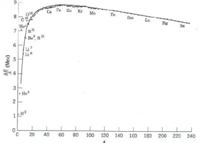
The binding energy per nucleon as a function of the number of mass is shown in Figure 1.4. As it can be observed, binding energy increases with the mass number in light nuclei until reaching a maximum at mass number of around 55-60 (in the iron-nickel region), with the nuclei 56Fe normally indicated as the one that has greater binding energy per nucleon. Beyond, it starts to decrease slowly in function of the rise of mass number. Larger binding energy and lower mass mean more stable nuclei. Of course, the more nucleons mean the greater the total binding energy. The combination of this trend show us that energy can be released by the fusion of light nuclei into heavier ones or by the fission of heavy nuclei into lighter ones [19].
However, other corrections should be considered in the determination of the binding energy per nucleon, including the contribution of the electrostatic repulsion between protons, which in turn is dependent on the square of its charge (Z2), leading to the importance of the relationship between A and Z2 in the study of nuclear stability [18-20]. These two considerations (dependence on A and on Z2) are mainly based on the liquid drop model for the nuclear structure.
On a first approximation, the later factor mentioned, Coulomb contribution to the binding energy, seems to indicate that it would be favorable to have less protons than neutrons in a nucleus. However, this is not the case and it is important to use some considerations from other nuclear structure model (shell model) to explain the fact that it is common to have roughly the same number of neutrons and protons in stable nuclei. The notion underlying this phenomenon is called symmetry and takes into account quantum mechanics in nuclei (quantum states of nucleons) [19]. Since isotopes have different mass numbers they will possess different levels of binding energy, which leads
Figure 1.4 - Binding energy per nucleon versus the mass number of the isotope.
7
to the appearance of isotopes of the same element with greater or lesser stability. A way to assess the stability of a nuclide is to evaluate the ratio of the number of neutrons and protons (N/Z) – Figure 1.5.
Indeed, the analysis of this N/Z ratio will also give an important data to predict the type of nuclear decay of each radionuclide, as it will be explained later.
Finally, the last factor contributing to the binding energy, and consequently to nuclear stability, is the physical evidence that nucleons tend to pair off. This means that binding energy is greater in nuclei where all the neutrons and protons are paired-off. Inversely, an odd-odd nucleus (odd number of protons and neutrons) tend to present a lower binding energy [18, 19].
1.1.2. Radioactive decay
Actually more than 2700 isotopes from elements of the Periodic Table are known. All the unstable nucleus tend to transform themselves into more stable ones, through one of several allowed processes: division into smaller fragments, particle emissions and/or emission of energy in the form of electromagnetic waves [21]. This is what is meant by radioactive decay and nuclides undergoing such transformations are called radionuclides.
The most common decay processes are based on the spontaneous emission of photons (gamma or X-rays) or particles (α, β- or β+). A possible method of division and classification is based on the fundamental force underlying to the decay process, i.e.: strong nuclear force or weak nuclear force. Other interactions due to some quantum nuclear structure rearrangements could also be added to this division.
There are radionuclides that decay to a nuclear state with increased energy compared to the ground state (excited state) with relatively long physical half-life. Radionuclides could have these excited states with duration longer than the nanosecond scale (meaning 100 to 1000 times longer than the other excited states) being designated by
Figure 1.5 - Neutron (N) vs. Atomic (A) number in nuclides found in nature.
8
metastable states [22, 23]. In these cases, the radioactive decay occurs by releasing the excess of energy by emitting gamma photons, in a process called isomeric transition.
Another possibility of decay in excited states is the internal conversion, namely the release of energy by a gamma photon that is absorbed internally by an orbital electron (typically from the layers K or L). By absorbing this energy, the electron is ejected from the electron cloud and remains with the difference between the photon energy and the binding energy as kinetic energy. Subsequently, occurs the occupation of the gap created by the electron in the cloud by an electron from an higher orbital and the consequent release of energy in the form of characteristic X-rays or Auger electrons [22].
Together, isomeric transition and internal conversion are examples of radiative processes, involving fundamental electromagnetic interactions, and resulting in the conservation of atomic and mass number [19].
In what concerns to strong force mediated decays, an alpha (α) particle could be emitted from the nucleus, in a process relatively common in very heavy elements.
Generally, individual nucleons cannot escape from the nucleus. However, a bound group of nucleons can sometimes escape because its binding energy increases the total energy available for the process. The most significant demonstration of this possibility is α-decay process. This aggregate of nucleons is very strongly bound and has this ability to escape from the nucleus. The potential energy of an α-particle is dependent on its distance from the center of the nucleus (due to the strong nuclear interaction) [18].
Alpha particles are helium nuclei (A = 4 ; Z = 2), and have the ability to deposit large amounts of energy (4 ∼ 8 MeV) within a very short range travelled in matter, due to their charge and mass - reason why they own high Linear Energy Transfer (LET). Equation
energy
A Z A Z
Y
α
X
42 , Eq. 1.1represents this decay in which the resulting nucleus will have a decrease in the atomic number (Z - 2) and in the mass number (A - 4).
One of the many facets of the weak interaction is the Beta-decay (β decay). Being slower by several orders of magnitude than electromagnetic and strong interactions, these weak processes cannot be observed if there are these competing interactions [24]. Weak interactions transform protons in neutrons or vice versa, changing as well the number of charged leptons (positive or negative electrons) and number of neutrinos [19].
β-decay processes include β-minus decay (β-), β-plus decay (β+) and electron capture. These processes occurs always in a strict dependence on the existence of isobars with smaller masses [18].
Given this, β- emission is energetically possible whenever the mass of the daughter atom is smaller than its isobaric neighbor in Z. In such cases, β- particles (negatively charged and very high speed electrons) are emitted simultaneously with an anti-neutrino, while a neutron is converted into a proton, resulting in a nucleus with an increase in the atomic number and maintenance of mass number according to
9
energy
A Z A Z
Y
β
X
1 . Eq. 1.2β- particles are normally emitted from isobars that have an excess number of neutrons. The emitted β- particles present a continuous kinetic energy spectrum, ranging from 0 to the energy endpoint (Emax) - correspondent to each radionuclide, with an average energy of β− emitted particles that is often approximately equal to 1/3 Emáx (Figure 1.6). The cited energy spectrum applies also to β+ (positrons) - to be described in detail in an appropriate topic.
In isobars that have an “excess” of protons, stability could be found by emitting a positron (positive electron). In this decay a proton is transformed in a neutron with the release of an antimatter particle named positron (or beta plus particle - β+) and a neutrino:
energy
υ
e
n
p
. Eq. 1.3This decay results in a nucleus with a decrease of one unit in the atomic number and maintenance of the mass number, as it is represented in equation
energy
A Z A Z
Y
e
υ
X
1 01 . Eq. 1.4If the nuclear masses of neighboring isobars in Z differ by less than the electron mass, stability could be reached by capturing an orbital electron that combines with a proton to obtain a neutron - electron capture decay. In this case, the resulting nuclide is an isotope of a different element, maintaining the mass number as it could be concluded by the decay equation
energy
Z A Z A
υ
Y
e
X
1 . Eq. 1.5Figure 1.6 - Beta decay energy spectrum.
10
The layer of the electron which is captured is filled by another electron, which in turn is more energetic and leads to the release of X-rays.
Except for a small difference in the energies involved, electron capture process has the same selection rules applied to β+ decay and is usually in competition with it. The probability of electron capture increases with Z3, due to the increased strength of the nuclear Coulomb field and decreased radii of electronic orbits [23].
1.1.3. Positron and positronium physics
Due to the main theme of this work, positron should be covered with a special attention and a detailed description here.
Back to its discovery, on 2nd of August of 1932 the scientist Carl D. Anderson was photographing cosmic-rays track using a Wilson chamber (based on a magnetic field of 1.5 T) and registered an interesting finding (Figure 1.7) [25]. At that time, properties of the discovered particle were carefully described although some limitations of the experiment: “(…) It is possible with the present experimental data only to assign rather wide limits to the magnitude
of the charge and mass of particle (…) It is concluded, therefore, that the magnitude of the charge of the positive electron which we shall henceforth contract to positron is very probably equal to that of a free negative electron (…) The magnitude of the proper mass cannot as yet be given further than to fix an upper limit to it about twenty times that of the electron mass” [25].
In fact, this insights were true and the photo presented below is yet known as the first photo of a positron path. Moreover, in Anderson’s paper it was already suggested that positrons must be secondary particles, ejected from unstable atomic nucleus [25].
Figure 1.7 - First known photo of the path of a positron.
11
In physical terms, positron has the same quantity of charge as the electron, 1.6 × 10⁻¹⁹ C, but positively charged, the very same rest mass of an electron, 9.10 × 10⁻³¹ kg, and an intrinsic spin of ½ , being thus a fermion. In matter it eventually annihilates with an electron after a short lifetime, even considering that current theories of particle physics emphasize that, in vacuum, positron is a stable particle [26]. The time frame from positron emission until the formation of positron-electron pair is inversely proportional to the local electron density. Its interaction with electrons in soft matter can result in the formation of positronium that is an unstable electron-positron bonded state. The positronium atom can be formed when the energy of the incident positron exceeds the difference between the ionization energy of the target atom and the binding energy [26]. However the probability of occurrence in condensed matter is very reduced, mainly due to the inexistence of free space in matter to its formation.
Positronium is electrically neutral, and its centre of mass is midway between the constituent electron and positron. When the positron-electron pair is in a singlet spin state it could totally annihilate with the consequent emission of two gamma-photons. The triplet spin state annihilates with the emission of three photons, having very reduced rates of occurrence, cause of pickoff [27]. Due to the long lifetime of triple state it become more probable that the positron annihilates with another electron via two photons.
In normal circumstances, the positron remains free in space and its annihilation with the electrons of matter result in two photons of around 511 keV - called annihilation photons [22]. This physical process is the basis of the signal measured in Positron Emission Tomography (PET) used in Nuclear Medicine – with special interest for the process reported in this work.
1.1.4. Nuclear Medicine procedures
As it was already focused, the information obtained in Nuclear Medicine depends on the detection of radioactive emissions from compounds known as radiopharmaceuticals, which are the combination of a drug with a pre-determined biological distribution with a radioactive isotope that decays to provide the signal for detection. In this sense, the selection of the radionuclide (source) must guarantee that its decay mode is adapted to the desired purpose. Generally, for diagnostic purposes, it uses primarily two types of sources; gamma radiation emitting sources (radioactive isotopes that decay by isomeric transition or electron capture - that will not be highlighted here) for Conventional Nuclear Medicine or beta plus emitting sources for Positron Emission Tomography - that will be discussed along this work. On the other hand, Nuclear Medicine includes also some therapeutic procedures, where the main goal of the radionuclide is to destroy specific cells or tissues. Figure 1.8 illustrates the relation between the type of procedure and the nuclear decay mode of the radionuclide.