FACULDADE DE FARMÁCIA
REGULATION OF NEURAL STEM CELL
PROLIFERATION AND DIFFERENTIATION BY
APOPTOSIS-RELEVANT FACTORS
Daniela Vanessa Moutinho dos Santos
DOUTORAMENTO EM FARMÁCIA
BIOQUÍMICA
FACULDADE DE FARMÁCIA
REGULATION OF NEURAL STEM CELL
PROLIFERATION AND DIFFERENTIATION BY
APOPTOSIS-RELEVANT FACTORS
Daniela Vanessa Moutinho dos Santos
Tese de Doutoramento em Farmácia (Bioquímica), apresentada à
Universidade de Lisboa através da Faculdade de Farmácia
Research supervisors:
Professor Cecília M. P. Rodrigues
Professor Susana Solá
Lisboa
2012
The studies presented in this thesis were performed at the Research Institute for Medicines and Pharmaceutical Sciences (iMed.UL) and at the Department of Biochemistry and Human Biology, Faculty of Phamacy, University of Lisbon, under the supervision of Professor Cecília M. P. Rodrigues and Professor Susana Solá.
Daniela Vanessa Moutinho dos Santos was the recipient of a Ph.D. fellowship (SFRH/BD/42008/2007) from Fundação para a Ciência e Tecnologia (FCT), Lisbon, Portugal. This work was supported by grants FCF/67912/2006, PTDC/SAU-NMC/117877/2010 and Pest-OE/SAU/UI4013/2011 from FCT and FEDER.
De acordo com o disposto no ponto 1 do artigo nº 41 do Regulamento de Estudos Pós-Graduados da Universidade de Lisboa, deliberação nº 93/2006, publicada em Diário da República – II Série nº 153 – 5 de Julho de 2003, o Autor desta dissertação declara que participou na conceção e execução do trabalho experimental, interpretação dos resultados obtidos e redação dos manuscritos.
Aos meus pais, que me ensinaram que é bom ter dúvidas e questionar
Resumo
As células estaminais são células indiferenciadas que se conseguem replicar e originar células especializadas e funcionais, através de processos de diferenciação celular. Existem vários tipos de células estaminais, originárias de tecidos embrionários e adultos, caracterizadas por possuirem potenciais de diferenciação diferentes. As células estaminais neurais (NSC) têm a capacidade de originar os principais tipos de células neurais, incluindo neurónios e células da glia (astrócitos e oligodendrócitos). Em cada divisão celular, as NSC podem seguir destinos celulares diferentes, nomeadamente continuar o processo proliferativo, ou iniciar o processo de diferenciação. Esta tomada de decisão é regulada por vários fatores intrínsecos e extrínsecos. Além disso, os mecanismos regulatórios envolvem também sinais de sobrevivência e de morte celular, sendo que o destino de proliferação, diferenciação ou morte depende do balanço e da conjugação dos múltiplos fatores.
Estudos recentes sugerem que fatores associados ao processo apoptótico podem também modular a proliferação e a diferenciação das NSC. De facto, o nosso grupo demonstrou já que as caspases, proteases de cisteína que desempenham um papel central no processo apoptótico, bem como a proteína p53, supressora de tumores, aceleram a diferenciação neural. Por outro lado, também demonstrámos o envolvimento de vários miRNAs associados à apoptose na diferenciação neural e clarificámos o papel do miRNA-34a durante este processo. Curiosamente, as variações na expressão e atividade de proteínas e miRNAs relacionados com a apoptose não se encontram associadas a um aumento de morte celular, o que sugere que estes fatores desempenham novas funções na regulação da diferenciação das NSC, independentemente do seu papel proapoptotico. Assim, propusémo-nos identificar e caracterizar novos fatores, associados à apoptose, com capacidade de regular o potencial de proliferação e diferenciação das NSC e, também, modular estes processos recorrendo a novos reguladores de vias apoptóticas.
Avaliámos, inicialmente, a capacidade de dois derivados de naftoquinona sintéticos regularem a apoptose em diferentes modelos celulares. De facto, compostos com uma estrutura de quinona são comuns na natureza e apresentam actividades anticancerígenas, antibacterianas, antimaláricas e fungicidas. Dois compostos com o potencial de inibir proteases de cisteína foram concebidos e sintetizados:
nafto[2,3-d]isoxazole-4,9-diona-3-carboxilatos 1a e 1b. O potencial antiapoptótico dos compostos
1a e 1b foi avaliado e comparado com o da naftoquinona 4, um composto estruturalmente
mais simples. Culturas primárias de hepatócitos de rato foram incubadas com os derivados de naftoquinona sintetizados e, posteriormente, tratadas com camptotecina, um
estímulo tóxico proapoptótico que induz danos no DNA. Os nossos resultados demonstraram que os derivados de naftoquinona 1a e 1b conferem uma proteção anti-apoptótica eficaz nestas condições, ao contrário da naftoquinona 4. Tanto o composto 1a, como o composto 1b, aumentaram significativamente a viabilidade celular e reduziram a fragmentação nuclear, a ativação da caspase-3, -8 e -9 e a libertação de citocromo c, provocadas pelo tratamento com camptotecina. Além disso, ambos os compostos aumentaram a expressão da Bcl-XL, uma proteína pró-apoptótica da família da Bcl-2 que
modula a via mitocondrial da apoptose. Avaliámos a extensão das propriedades anti-apoptóticas destes compostos a outros modelos, por recurso a linhas celulares e estímulos apoptóticos diversos. Obtivémos efeitos protetores semelhantes em hepatócitos primários de rato e em células HuH-7 de hepatoma humano expostas a TGF-β1, uma citocina pró-apoptótica. Verificámos ainda que as propriedades antiapoptóticas dos derivados de naftoquinona 1a e 1b não se restringem a células do fígado, uma vez que ambos os compostos se revelaram eficazes na proteção de células de feocromocitoma de rato (PC12) tratadas com rotenona, um inibidor da cadeia respiratória mitocondrial. Estes resultados sugeriram que os nafto[2,3-d]isoxazole-4,9-diona-3-carboxilatos 1a e 1b são agentes citoprotetores eficazes e potenciais moduladores de vias apoptóticas.
De seguida, investigámos a função de proteínas associadas à apoptose na regulação do potencial de proliferação e diferenciação das NSC. Uma vez que tínhamos confirmado anteriormente o efeito das caspases na diferenciação neural, investigámos agora outra família de proteases de cisteína, as calpaínas. As calpaínas dependem de cálcio para a sua ativação e já foram associadas a vários processos celulares, incluindo a apoptose, a migração e o ciclo celular. Também já foram implicadas em processos de diferenciação em diversos modelos celulares, mas o seu impacto na diferenciação neural é ainda pouco conhecido. Assim, começámos por inibir estas proteases em NSC de ratinho. Tratámos as NSC com calpeptina, um inibidor químico de calpaínas, ou, em alternativa, sobrexpressámos a calpastatina, um inibidor endógeno específico para as calpaínas. Verificámos que esta inibição resultou em redução de proliferação e indução de diferenciação das NSC. Este efeito foi acompanhado por alterações significativas na expressão de proteínas do ciclo celular. Experiências com inibidores de canais de cálcio presentes no retículo endoplasmático sugeriram que as calpaínas são ativadas por fluxos de cálcio nas NSC. De seguida, focámos o nosso estudo nas calpaínas mais estudadas e, também, mais abundantes no cérebro, as calpaínas 1 e 2. Curiosamente, observámos que estas duas isoformas são reguladas de modo díspar durante a diferenciação das NSC. A
análise da expressão das duas proteínas indicou que a calpaína 1 é mais expressa nas NSC, decrescendo os seus níveis de expressão durante a diferenciação. Por outro lado, os níveis de calpaína 2 aumentaram ao longo da diferenciação celular. De facto, a redução da expressão de cada uma destas proteínas, recorrendo à tecnologia de silenciamento com siRNAs, resultou em efeitos radicalmente diferentes. O silenciamento da calpaína 1 acelerou o processo de diferenciação neural, marcado pelo aumento de expressão de marcadores neuronais e gliais, respectivamente β-III tubulina e GFAP. O silenciamento da calpaína 2, contudo, traduziu-se numa diminuição de expressão da proteína GFAP. Estes resultados sugerem que a calpaína 1 poderá ter um papel na manutenção da proliferação das NSC, reprimindo a diferenciação, e que a calpaína 2 poderá estar mais envolvida na diferenciação de células da glia.
Por fim, avaliámos o efeito dos derivados de naftoquinona, previamente descritos, na regulação do destino celular das NSC. De facto, os compostos 1a e 1b apresentavam características promissoras, uma vez que foram sintetizados enquanto inibidores de proteases de cisteína, para além de que os resultados anteriores mostraram que as caspases e as calpaínas são potenciais moduladores do destino celular. Experiências preliminares sugeriram que, mais uma vez, os compostos 1a e 1b apresentavam propriedades semelhantes, pelo que apenas um dos compostos foi utilizado neste estudo. Verificámos que o tratamento com 1a resultou numa alteração do potencial de diferenciação das NSC, favorecendo a gliogénese em detrimento da neurogénese. Contudo, a modulação da atividade de caspases e calpaínas, através da adição de inibidores químicos, não reproduziu este efeito, o que indica que o papel do 1a nas NSC é independente da modulação destas proteases. Uma vez que as naftoquinonas são moléculas electrofílicas e pro-oxidantes, avaliámos também a produção de espécies reactivas de oxigénio (ROS) nas NSC após tratamento com 1a. Os resultados demonstraram que o derivado de naftoquinona promove um aumento de ROS nestas células, associado a um aumento de expressão e à translocação nuclear de proteínas de resposta antioxidante, como Nrf2 e Sirt1. A alteração do estado redox das NSC poderá ser um dos principais mecanismos pelos quais o composto 1a modula a diferenciação das NSC. De facto, a adição de antioxidantes reverteu, ainda que parcialmente, a alteração no potencial de diferenciação das NSC induzida pelo derivado de naftoquinona, reduzindo a geração de ROS e a translocação nuclear das proteínas Nrf2 e Sirt1. Assim, estes estudos revelaram uma nova função do derivado de naftoquinona 1a na modulação do destino celular das NSC, sublinhando a importância do ambiente redox neste processo.
Em conclusão, estes estudos contribuem para uma visão integrada dos processos de proliferação, diferenciação e apoptose, bem como do seu papel no destino das células, que poderá revelar-se útil no desenvolvimento de estratégias terapêuticas com células estaminais neurais.
Palavras-chave: Apoptose – Calpaínas – Células estaminais neurais – Derivados de
Abstract
Neural stem cell (NSC) fate decision is controlled by both intrinsic and extrinsic factors. Mounting evidence supports the hypothesis that apoptosis-associated factors modulate NSC proliferation and differentiation. Therefore, we set to identify and characterize new apoptosis-associated factors regulating NSC fate decision, and to modulate related pathways using novel regulators of apoptosis.
We started by evaluating the ability of synthetic naphthoquinone derivatives naphtho[2,3-d]isoxazole-4,9-dione-3-carboxylates 1a and 1b to prevent cell death in different cellular models. Notably, under apoptogen stimulation, both 1a and 1b increased cell viability, promoted up-regulation of Bcl-XL, and reduced nuclear
fragmentation, caspase-3, -8 and -9 activation, and cytochrome c release. These results indicate that 1a and 1b may act as efficient modulators of apoptotic pathways.
We next investigated the potential NSC modulatory properties of calpains, a family of cysteine proteases largely associated with apoptosis. Our results showed that inhibition of calpains, possibly regulated by calcium, favored NSC differentiation and elicited changes in cell cycle-related proteins. We found higher calpain 1 expression in self-renewing NSC, while calpain 2 levels increased throughout differentiation. Specific silencing of each calpain suggested that calpain 1 plays a role in repressing differentiation and maintaining a proliferative NSC pool, while calpain 2 may be involved in glial differentiation.
Finally, we evaluated whether naphthoquinone derivatives could modulate NSC proliferation and differentiation potential. Interestingly, treatment of differentiating NSC with 1a elicited a shift from neuronal to glial differentiation, not through modulation of apoptosis-associated cysteine proteases, but rather through reactive oxygen species production. This was associated with activation of the antioxidant-response proteins Nrf2 and Sirt1 and partly reduced by subsequent antioxidant treatment, suggesting that 1a modulates NSC fate through alteration of the intracellular redox environment.
In conclusion, these studies contribute to an integrated view of neural differentiation, proliferation and apoptosis pathways in cell fate decision and may prove useful in the development of stem cell-based therapeutic strategies.
Keywords: Apoptosis – Calpains – Differentiation – Naphthoquinone derivatives –
Acknowledgements
Começo por agradecer à Professora Cecília Rodrigues todo o apoio que me deu ao longo do meu doutoramento. Agradeço-lhe por ter acreditado em mim quando, há cinco anos atrás, apareci à sua porta para uma entrevista munida de um CV e carta de motivação miseravelmente conseguidos (vejo-o agora), e por me ter acolhido logo tão bem no seu grupo de investigação! Aqui cresci, enquanto cientista e enquanto pessoa, com os momentos bons e também com os maus (que experiência funciona bem à primeira?), e tenho-lhe a agradecer a orientação que me deu ao longo de todo o processo. Deu-me a oportunidade de ser criativa e independente e foi sempre muito receptiva às minhas ideias e sugestões. A sua extraordinária capacidade de trabalho, foco e dedicação foram uma inspiração para a minha vida futura.
Agradeço também à Susana Solá pela orientação, paciência e motivação. Acho que quando a nossa orientadora troca o nosso nome e o da filha, isso quer dizer alguma coisa! És uma das melhores pessoas que conheço, simpática, acessível e preocupada (no bom sentido!). Sei que te dei umas quantas dores de cabeça nos últimos anos, mas mesmo assim sempre me apoiaste nas experiências malucas e ajudaste nos momentos difíceis, muito obrigada!
Um agradecimento especial a todos os meus colegas de laboratório que me acompanharam nesta etapa, não poderia ter escolhido melhor grupo de pessoas com quem trabalhar! Agradeço a boa disposição, os risos, as conversas de hora de almoço ou lanche, os momentos de “fritura” ao final do dia, o drama!, a interajuda. Agradeço ao Rui, com quem trabalhei no início (go liver!) e à Márcia, minha companheira das stem cells que muito me ajudou nesta nova área. Obrigada também ao Ricardo, Joana Amaral, Pedro, Rita, Filipa, Benedita, Joana Xavier, Duarte, André, Andreia, Inês, Maria, Miguel, aos “novos” Ana Luísa, Marta, Sofia, Pedro Rodrigues, Pedro Dionísio e Diane e às
Professoras Elsa Rodrigues, Maria João Gama, Margarida Castro Caldas e Isabel Moreira da Silva. A todos os colegas, professores e funcionários da Faculdade de Farmácia que tornaram possível este trabalho, um muito obrigada!
Agradeço também ao Professor Domingos Henrique por me ter recebido no seu grupo no Instituto de Medicina Molecular. Um agradecimento especial à Elsa Abranches e à Evguenia Bekman, que me orientaram e ensinaram as manias das ESC e NSC, e a todas as pessoas do laboratório, que sempre me fizeram sentir bem-vinda.
Agradeço a todos os meus amigos que estiveram presentes e me aturaram ao longo desta aventura. Correndo o risco de sofrer represálias (eu sei os amigos que tenho!), não vou nomeá-los, pois tenho a sorte de ter muitas pessoas especiais na minha vida. Obrigada pelo apoio, compreensão, por me suportarem enquanto falava horas a fio sobre o trabalho, as experiências, as dúvidas, indecisões e receios e por terem sempre uma palavra amiga (onde se incluem os insultos bem dispostos) para dar em troca!
Agradeço à Inês, que me acompanhou todo este tempo e ouviu inúmeras vezes a desculpa “eu tenho um doutoramento para fazer!”. Obrigada por tudo o que tens sido para mim nestes anos. Pelo carinho e apoio constante, por me ouvires (fiz-te aprender uma data de palavras novas, como apoptose e calpaínas) e pela paciência. Acima de tudo, obrigada por seres a pessoa fantástica que és e por me fazeres feliz!
Aos meus pais e ao meu irmão, Miguel, um agradecimento muito especial. Se consegui fazer alguma coisa ao longo da vida, foi devido a vocês. Sempre me incentivaram, apoiaram e encorajaram, e se tenho curiosidade e sede de saber, foi porque vocês o despertaram. (Aproveitem bem estas palavras simpáticas, pois sabem do que a casa gasta – com quem será que aprendi?... – e não devem voltar a ler agradecimentos sentimentais tão cedo...). Obrigada pelo carinho e pela estabilidade emocional que sempre me proporcionaram. Por vezes é difícil acreditar em nós e ver para além do céu nublado, mas ter-vos ao meu lado sempre me fez sentir um calor reconfortante e esperança no futuro.
Abbreviations
1a/1b naphtho[2,3-d]isoxazole-4,9-dione-3-carboxylatesAIF apoptosis-inducing factor
Apaf-1 apoptosis protease-activating factor 1
ARE antioxidant responsive element
ASK1 apoptosis signal-regulated kinase 1
BH Bcl-2 homology
BrdU bromodeoxyuridine
Ca2+ calcium
CAST calpastatin
CNS central nervous system
Cys cysteine
Cyt c cytochrome c
DD death domain
DIABLO direct IAP binding protein with a low pI
DISC death inducing signaling complex
DMSO dimethyl sulfoxyde
EGF epidermal growth factor
ESC embryonic stem cells
ER endoplasmic reticulum
FADD Fas-associated death domain
FasL Fas ligand
FBS fetal bovine serum
FGF fibroblast growth factor
FOXO forkhead box O
G1 first gap phase
GFAP glial fibrillary acidic protein
GSH glutathione
HO1 heme oxygenase-1
HuH-7 human hepatoma
IκB inhibitor of NF-κB
Id1 inhibitor of differentiation 1
IKK IκB kinase
IP3Rs Inositol-1,4,5-trisphophate receptors
iPSC induced pluripotent stem cells
JNK c-Jun N-terminal kinase
Keap1 kelch-like ECH-associated protein-1
LA lipoic acid
M mitosis
MAPK mitogen-activated protein kinase
MNSC mouse neurospheres
MOMP mitochondrial outer membrane permeabilisation
mtDNA mitochondrial DNA
NAC N-acetylcysteine
NAD nicotinamide adenine dinucleotide
NADPH nicotinamide adenine dinucleotide phosphate
NCCD Nomenclature Committee on Cell Death
NE neuroepithelial
NF-κB nuclear factor κB
Ngn2 neurogenin 2
Nrf2 nuclear factor E2-related factor 2
NS-TGFP tau-GFP mouse NSC line
NSC neural stem cells
NT2 Ntera2
PBS phosphate buffer saline
PC12 rat pheochromocytoma
PI propidium iodide
PI3K phosphoinositide 3-kinase
PTP permeability transition pore
RG radial glia
SGZ subgranular zone
SVZ subventricular zone
siCAPN1/2 siRNA specific for calpain 1 or 2
siRNA short interference RNA
Sirt1 sirtuin 1
SOD superoxide dismutase
Stat3 signal transducer and activator of transcription 3
TNFR tumor necrosis factor receptor
Publications
The present thesis was mostly based on work that has been published in international peer-reviewed journals:
Santos DM, Santos MMM, Viana RJS, Castro RE, Moreira R, Rodrigues CMP.
Naphtho[2,3-d]isoxazole-4,9-dione-3-carboxylates: potent, non-cytotoxic, antiapoptotic agents. Chem Biol Interact 2009; 180 (2): 175-82.
Santos DM, Xavier JM, Morgado AL, Solá S, Rodrigues CMP. Distinct regulatory
functions of calpain 1 and 2 during neural stem cell self-renewal and differentiation. PLoS
One 2012; 7 (3): e33468.
Santos DM, Santos MMM, Moreira R, Solá S, Rodrigues CMP. Synthetic Condensed
1,4-naphthoquinone derivative shifts neural stem cell differentiation by regulating redox state. Mol Neurobiol 2012; DOI: 10.1007/s12035-012-8353-y.
The following manuscripts have also been published during the course of the Ph.D. studies:
Aranha MM, Santos DM, Xavier JM, Low WC, Steer CJ, Solá S, Rodrigues CMP. Apoptosis-associated microRNAs are modulated in mouse, rat and human neural differentiation. BMC Genomics 2010, 11: 514.
Solá S, Xavier JM, Santos DM, Aranha MM, Jepsen K, Rodrigues CMP. p53 interaction with JMJD3 results in its nuclear distribution during mouse neural stem cell differentiation. PLoS One 2011; 6 (3): e18421.
Aranha MM, Santos DM, Xavier JM, Low WC, Steer CJ, Solá S, Rodrigues CMP. miR-34a regulates mouse neural stem cell differentiation. PLoS One 2011; 6 (8): e21396.
Table of Contents
Resumo ... vii Abstract ... xiii Acknowledgements ... xvii Abbreviations ... xix Publications ... xxiii General Introduction ... 1 1. Apoptosis ... 3 1.1. Signaling pathways ... 3 1.1.1. Extrinsic apoptosis ... 4 1.1.2. Intrinsic apoptosis ... 7 1.2. Calpains and their involvement in apoptosis ... 9 1.3. Reactive oxygen species and their role in apoptosis ... 13 1.4. Modulation of apoptosis by naphthoquinones ... 18 2. Neural stem cells ... 19 2.1. Neural stem cells in the developing central nervous system ... 21 2.2. Adult neural stem cells ... 22 2.3. In vitro culturing of neural stem cells ... 24 2.4. Cell cycle control of neural stem cell fate ... 25 2.5. Apoptosis-associated factors in neural stem cell proliferation anddifferentiation ... 27 2.5.1. Caspases ... 28 2.5.2. Calpains ... 29 2.5.3. Bcl-2 family ... 30
2.5.4. p53 and p73 ... 31 2.6. Modulation of neural differentiation by reactive oxygen species ... 33 Objectives ... 37 3. Naphtho[2,3-d]isoxazole-4,9-dione-3-carboxylates: potent, non-cytotoxic,
antiapoptotic agents ... 39 3.1 Abstract ... 41 3.2 Introduction ... 42 3.3 Methods ... 43
3.3.1 Chemical synthesis of
naphtho[2,3-d]isoxazole-4,9-dione-3-carboxylates 1a and 1b, and naphthoquinone 4 ... 43 3.3.2 Cell culture and preparation of rat primary hepatocytes ... 43 3.3.3 Treatments with quinone derivatives and induction of apoptosis ... 44 3.3.4 Cell viability assays ... 45 3.3.5 Morphologic evaluation of apoptosis ... 45 3.3.6 Caspase activity assays ... 45 3.3.7 Immunoblotting ... 46 3.3.8 Immunocytochemistry ... 46 3.3.9 Densitometry and statistical analysis ... 46 3.4 Results ... 47
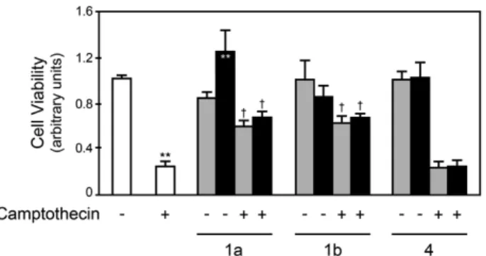
3.4.1 Synthesis of naphtho[2,3-d]isoxazole-4,9-dione-3-carboxylates 1a and 1b, and naphthoquinone 4 ... 47 3.4.2 1a and 1b, but not 4, act as potent inhibitors of cell death ... 47 3.4.3 1a and 1b reduce camptothecin-induced caspase activation in primary rat hepatocytes ... 51 3.4.4 1a and 1b prevent camptothecin-induced cytochrome c release in
primary rat hepatocytes ... 52 3.4.5 1a and 1b modulate Bcl-XL expression in primary rat hepatocytes .. 54
4. Distinct regulatory functions of calpain 1 and 2 during neural stem cell self-renewal and differentiation ... 59
4.1 Abstract ... 61 4.2 Introduction ... 62 4.3 Methods ... 63 4.3.1 Ethics ... 63 4.3.2 Cell culture and treatments ... 64 4.3.3 siRNA and plasmid transfections ... 65 4.3.4 Flow cytometry analysis ... 66 4.3.5 Immunoblotting ... 67 4.3.6 Immunocytochemistry ... 67 4.3.7 Densitometry and statistical analysis ... 68 4.4 Results ... 68 4.4.1 Calpain inhibition decreases proliferation of neural stem cells ... 68 4.4.2 Calpain inhibition increases differentiation of neural stem cells ... 71 4.4.3 Calpain inhibition throughout neural differentiation increases
neurogenesis ... 73 4.4.4 Calpain 1 and 2 are differentially expressed throughout neural
differentiation and do not correlate with cell death ... 76 4.4.5 Calpain 1 represses neural differentiation, while calpain 2 increases
glial differentiation ... 78 4.5 Discussion ... 78 5. Synthetic Condensed 1,4-naphthoquinone derivative shifts neural stem cell
differentiation by regulating redox state ... 85 5.1 Abstract ... 87 5.2 Introduction ... 88 5.3 Materials and methods ... 89 5.3.1 Ethics ... 89
5.3.2 Chemical synthesis of naphtho[2,3-d]isoxazole-4,
9-dione-3-carboxylate 1a ... 90 5.3.3 Cell culture ... 90 5.3.4 Cell treatments ... 90 5.3.5 Cell flow cytometry analysis ... 91 5.3.6 Immunoblotting ... 92 5.3.7 Immunocytochemistry ... 92 5.3.8 Densitometry and statistical analysis ... 93 5.4 Results ... 93 5.4.1 Naphtho[2,3-d]isoxazole-4,9-dione-3-carboxylate 1a induces a shift
from neuronal to glial differentiation of neural stem cells ... 93 5.4.2 The 1a-directed neural stem cell fate switch is independent of
cysteine protease inhibition ... 94 5.4.3 1a increases intracellular levels of reactive oxygen species and
antioxidant response proteins ... 97 5.4.4 Reactive oxygen species mediate the 1a-directed shift in the
differentiation potential of neural stem cells ... 100 5.5 Discussion ... 102 6. Concluding Remarks ... 109 References ... 117
Figures
Figure 1.1. Intrinsic and extrinsic pathways of apoptosis ... 4 Figure 1.2. Calpain signaling pathways in survival and apoptosis ... 12 Figure 1.3. Different ROS levels elicit variable cellular outcomes ... 14 Figure 1.4. Quinones form covalent bonds with biological molecules and generate ROS ... 18 Figure 2.1. Developmental stages and corresponding in vitro NSC populations ... 20 Figure 2.2. Comparison of neurosphere and monolayer culture systems ... 24 Figure 2.3. Redox status influences NSC proliferation and differentiation ... 34 Figure 3.1. Chemical formula of naphtho[2,3-d]isoxazole-4,9-dione-3-carboxylates 1a and 1b, and naphthoquinone 4 ... 43 Figure 3.2. 1a and 1b, but not 4, modulate camptothecin-induced decrease in cell viability in primary rat hepatocytes ... 48 Figure 3.3. 1a and 1b reduce camptothecin-induced nuclear fragmentation in primary rat hepatocytes ... 49 Figure 3.4. 1a and 1b reduce TGF-β1- and rotenone-induced nuclear fragmentation, in different cell types ... 50 Figure 3.5. 1a and 1b reduce camptothecin-induced caspase-3 activation in primary rat hepatocytes ... 51 Figure 3.6. 1a and 1b reduce camptothecin-induced caspase-8 and -9 activities in primary rat hepatocytes ... 53 Figure 3.7. 1a and 1b modulate camptothecin-induced cytochrome c release in primary rat hepatocytes ... 53 Figure 3.8. 1a and 1b modulate Bcl-XL expression in primary rat hepatocytes ... 54
Figure 4.1. Calpeptin treatment decreases cell density and proliferation ... 69 Figure 4.2. Calpeptin treatment modulates cell cycle proteins ... 70 Figure 4.3. Calpeptin treatment decreases Nestin- and increases β-III Tubulin-positive cells ... 72 Figure 4.4. Inhibition of calcium flux decreases Nestin- and increases β-III
Figure 4.5. Neuronal and astrocytic phenotypes after induction of neural differentiation ... 74 Figure 4.6. Calpastatin overexpression increases neuronal but not glial differentiation
... 75 Figure 4.7. Calpain expression levels are altered throughout differentiation of NSC . 77 Figure 4.8. Calpain 1 and 2 silencing regulates the expression of neural differentiation markers ... 79 Figure 5.1. 1a decreases neuronal and increases glial differentiation of mouse NSC . 95 Figure 5.2. 1a-directed NSC fate switch is independent of caspase and calpain inhibition ... 96 Figure 5.3. 1a increases intracellular levels of ROS during NSC differentiation ... 98 Figure 5.4. Antioxidant response pathways are activated by 1a treatment during NSC differentiation ... 99 Figure 5.5. Antioxidants revert Sirt1 and Nrf2 activation in response to 1a treatment ... 101 Figure 5.6. 1a shifts NSC differentiation to glia through ROS production ... 103 Figure 6.1. Graphical abstract ... 115
General Introduction
1. Apoptosis
The term apoptosis was first used by Kerr et al. in 1972 to describe an active, programmed mechanism of “controlled cell deletion” (Kerr et al., 1972). The word derives from the Greek, and stands for the “falling off” of petals from flowers or leaves from trees, an eloquent metaphor of the physiological and homeostatic nature of apoptosis. Indeed, apoptosis can be seen as the complementary process to cell division in living organisms. It balances cell numbers, eliminating unnecessary cells during embryonic development, as part of a turnover process in proliferating tissues, and in pathological situations, preventing malformations and cancer when activated in aberrant cells. Given its importance, deregulation of apoptosis can have dramatic consequences (Ellis et al., 1991). Examples in opposite sides of the spectrum include cellular degeneration, with excessive activation of cell death pathways, and uncontrolled proliferation of cancer cells, where cell death is under-activated (Lowe and Lin, 2000; Vila and Przedborski, 2003). Apoptosis was initially characterized and distinguished from other forms of cell death by a number of morphological features, including rounding up and shrinkage of the cell, loss of contact to neighboring cells and to the extracellular matrix, plasma membrane blebbing, condensation of chromatin, fragmentation of the nucleus and, finally, disintegration into apoptotic bodies, membrane-bound vesicles containing the entire contents of the cell, including intact organelles (Hacker, 2000). Although these features are still broadly used in the scientific community to access and classify apoptotic cell death, there have been a number of efforts towards switching from a morphological to a molecular definition of cell death modes (Kroemer et al., 2005; Galluzzi et al., 2009; Kroemer et al., 2009; Galluzzi et al., 2012b).
1.1. Signaling pathways
Although apoptosis and programmed cell death are still often used as synonyms, the latest revision from the Nomenclature Committee on Cell Death (NCCD) distinguishes 13 distinct regulated cell death modes, including extrinsic apoptosis by death receptors, extrinsic apoptosis by dependence receptors, caspase-dependent intrinsic apoptosis and caspase-incaspase-dependent intrinsic apoptosis (Galluzzi et al., 2012b).
1.1.1. Extrinsic apoptosis
Extrinsic apoptosis is mediated by extracellular ligands that are sensed by specific transmembrane receptors responsible for propagating the signal intracellularly (Fig. 1.1A). These include death receptors, which transduce death signals upon ligand binding, and dependence receptors, which induce apoptosis in the absence of the cognate ligand and otherwise support cell survival (Mehlen and Bredesen, 2011). Extrinsic apoptosis always requires the activation of caspases, cysteine proteases responsible for the initiation and propagation of the death signal (Galluzzi et al., 2012b).
Figure 1.1. Intrinsic and extrinsic pathways of apoptosis. (A) The extrinsic pathway can
be activated upon FAS ligand (FASL) binding to FAS, which leads to the recruitment of FAS-associated protein with a death domain (FADD), cellular inhibitor of apoptosis proteins (cIAPs), c-FLIPs and pro-caspase-8 (or -10). These form the death-inducing signaling complex (DISC), where caspase-8 (and -10) is activated. Caspase-8 can directly cleave and activate caspase-3 (in type I cells) or activate the intrinsic pathway by cleaving the BH3-only protein BID (in type II cells), leading to mitochondrial outer membrane permeabilization (MOMP). Alternatively, the extrinsic pathway may be activated via dependence receptors, which induce apoptosis in the absence of their ligand (NETRIN-1). The deleted in colorectal carcinoma (DCC) mediates the assembly of a DRAL- and TUCAN-containing caspase-9 activating platform. UNC5B, in turn, dephosphorylates and activates death-associated protein kinase 1 (DAPK1), which can directly cleave caspases or favor MOMP. (B) The intrinsic
pathway can be activated by several intracellular stress signals, which converge at the mitochondria and lead to MOMP. This leads to the release of several mitochondrial intermembrane space (IMS) proteins into the cytosol, including cytochrome c (CYTC), direct IAP-binding protein with low pI (DIABLO), high temperature requirement protein A2 (HTRA2), apoptosis-inducing factor (AIF) and endonuclease G (ENDOG). In the cytoplasm, CYTC, apoptosis protease-activating factor 1 (APAF1) and dATP form the apoptosome and activate caspase-9, which then activates caspase-3. DIABLO and HTRA2 sequester and/or degrade inhibitor of apoptosis proteins (IAPs), facilitating caspase activation. AIF and ENDOG mediate caspase-independent apoptosis by mediating DNA fragmentation at the nucleus. HTRA2 also contributes to caspase-independent apoptosis by cleaving several cellular substrates, including cytoskeletal proteins. IM, mitochondrial inner membrane; OM, mitochondrial outer membrane; PTPC, permeability transition pore complex; tBID, truncated BID. Adapted from Galluzzi et al. 2012b.
Caspases comprise a large family of proteins that were traditionally divided into two main functional groups, including proapoptotic caspases (caspase2, 3, 6, -7, -8, -9 and caspase-10, which is present in humans but not in mice) and pro-inflammatory caspases (caspase-1; caspase-4 and -5 in humans and their functional orthologs in mice, caspase-11 and -12) (Li and Yuan, 2008; Salvesen and Ashkenazi, 2011). Caspase-14 is involved in keratinocyte differentiation and plays no known role in apoptosis or inflammation (Denecker et al., 2008). This classification, however, is becoming gradually outdated, as additional functions for each caspase are uncovered. Caspases can be additionally classified according to their position in the signaling cascade into initiator (caspase-1, -2, -4, -5, -8, -9, -10, -11 and -12) and effector caspases (caspase-3, -6 and -7), which differ in prodomain length and mode of activation (Li and Yuan, 2008). Initiator caspases contain protein-protein interaction motifs in the prodomain that are important for interaction with upstream adapter molecules and subsequent activation. Effector caspases have short prodomains and rely on truncation for activation. They are usually cleaved by initiator caspases and can then proceed to cleave multiple cellular substrates. Caspases typically recognize and cleave specific motifs in their substrates, including the P1-P1’ peptide bond of a peptide of sequence P4-P3-P2-P1-P1’, where the P1 residue is Asp, the P1’ residue is small and uncharged, and P4-P3-P2 residues are complementary for interactions with the catalytic groove (Pop and Salvesen, 2009). The number of proteins reportedly cleaved by caspases continues to increase, with
almost 1000 human proteins identified, representing nearly 5% of the genome (Crawford and Wells, 2011).
Upon ligand binding, death receptors at the membrane can activate caspases within seconds, killing the cell in the following hours (reviewed in Ashkenazi and Dixit, 1998; Elmore, 2007). Death receptors belong to the tumor necrosis factor receptor (TNFR) gene superfamily, sharing cysteine-rich extracellular domains. They contain a cytoplasmic sequence of about 80 aminoacids termed the “death domain” (DD), which is important for oligomerization and interaction with cytoplasmic adapter proteins also containing DDs. Death receptor ligands also share common structures and belong to the TNF gene superfamily. Known ligand/receptor pairs include the Fas ligand and its receptor (FasL/FasR), TNF-α/TNFR1, lymphotoxin-α/TNFR1, Apo3L/DR3, Apo2L/DR4 and Apo2L/DR5. Activation of apoptosis by death receptors can be illustrated with the FasL/FasR model. Binding of FasL (also known as CD95L) to FasR (also known as CD95) leads to receptor oligomerization and binding to the DD-containing adaptor Fas-associated death domain (FADD). Receptor-bound FADD then binds procaspase-8 and c-FLIP through shared death effector domain (DED) regions, forming a functional complex named death inducing signaling complex (DISC) (Lavrik and Krammer, 2012). Procaspase-8 is activated at the DISC into functional caspase-8 that proceeds to cleave and activate caspase-3 and -7. Whereas this process is sufficient for triggering the executioner phase of the apoptotic program in the so-called type I cells (like lymphocytes), type II cells (like hepatocytes and pancreatic β cells) additionally require activation of the mitochondrial pathway of apoptosis. Here, caspase-8 cleaves the pro-apoptotic Bcl-2 family member Bid (Kantari and Walczak, 2011). Truncated Bid (tBid) then translocates to the mitochondria, inducing mitochondrial outer membrane permeabilisation (MOMP), followed by caspase-9 activation, which in turn activates caspase-3 and -7, eliciting cell death. TNF-α binding to TNFR1 followed by induction of apoptosis also depends on the formation of DISC, also known as Complex II, and caspase-8 activation. However, depending on cell type and context, TNRF activation can lead to the recruitment of a different subset of proteins, namely TNFR1-associated death domain protein (TRADD), TNFR-associated factor 2 (TRAF2), inhibitor of apoptosis protein (IAP) 1 (cIAP1), cIAP2 and receptor-interacting protein 1 (RIP1), which form the Complex I and mediate cell survival (Cabal-Hierro and Lazo, 2012). Membrane-bound Complex I triggers several cellular
pathways and leads to the activation of the transcription factors nuclear factor κB (NF-κB) and activator protein 1 (AP-1). NF-κB is a generic name for Rel family dimeric transcription factors that are regulated via shuttling from the cytoplasm to the nucleus in response to cell stimulation (Karin and Lin, 2002). At Complex I, RIP is polyubiquitinated and mediates the activation of inhibitor of NF-κB (IκB) kinase (IKK) (Cabal-Hierro and Lazo, 2012). IκBα, which inhibits NF-κB by retaining it in the cytoplasm, is then phosphorylated by IKK, which promotes its proteasomal degradation. Thus, following TNFR-mediated IKK activation, NF-κB is free to translocate to the nucleus, where it induces the transcription of antiapoptotic and antioxidant genes, such as cIAP1, cIAP2, cFLIP, TRAF1, TRAF2, A20, FBX10, Bcl-XL, A1, MnSOD and ferritin heavy chain (Baldwin, 2012). Interestingly, while
proteolytically inactive caspases promote NF-κB activity, active caspases inhibit it by cleaving and inactivating IKK and by cleaving IκBα, turning it into a stable super-repressor.
Dependence receptors are far less studied but have already been implicated in diverse processes, such as development, oncogenesis, and neurodegeneration (Mehlen and Bredesen, 2011). They are bifunctional, transducing survival signals through signaling pathways, such as the mitogen-activated protein kinase (MAPK) or the phosphoinositide 3-kinase (PI3K) in the presence of the cognate ligands and activating apoptosis or subapoptotic events in their absence. Most dependence receptors interact with caspases and some can assemble a complex that activates caspase-9.
Integrating these complex pathways, the NCCD established that extrinsic apoptosis proceeds through one of these three signaling cascades: (i) death receptor signaling and activation of the caspase-8 or -10/caspase-3 cascade; (ii) death receptor signaling and activation of the caspase-8/tBid/MOMP/caspase-9/caspase-3 pathway; or (iii) ligand deprivation-induced dependence receptor signaling followed by (direct or MOMP-dependent) activation of the caspase-9/caspase-3 cascade (Galluzzi et al., 2012b).
1.1.2. Intrinsic apoptosis
Intrinsic apoptosis, also known as the mitochondrial pathway of apoptosis, is activated by a plethora of intracellular signals that converge at the mitochondria,
where pro- and antiapoptotic factors antagonize each other to decide cell fate. Intrinsic apoptosis is, by definition, mediated by MOMP and is characterized by: (i) generalized and irreversible mitochondrial transmembrane potential dissipation; (ii) release of proteins from the mitochondrial intermembrane space into the cytosol (and their possible relocalization to other subcellular compartments); and (iii) respiratory chain inhibition (Galluzzi et al., 2012b). Unlike in extrinsic apoptosis, caspase activation is not always a strict requirement for intrinsic apoptosis, although caspases accelerate the process and contribute to the typical apoptotic morphology. As caspase activity is required in some, but not all, instances of developmental cell death, the NCCD proposes to differentiate between caspase-dependent and caspase-independent intrinsic apoptosis (Fig. 1.1B) (Galluzzi et al., 2012b).
Mitochondrial outer membrane integrity is controlled by members of the Bcl-2 protein family, which can be either pro- or antiapoptotic and share Bcl-2 homology (BH) regions that mediate protein-protein interactions (Youle and Strasser, 2008). Antiapoptotic members have all four BH regions (BH1, BH2, BH3 and BH4) and contain transmembrane domains that mediate their insertion into the outer membranes of mitochondria and into the endoplasmic reticulum (ER) (Kelly and Strasser, 2011). These include Bcl-2, Bcl-w, Bcl-XL, Mcl-1, and A1. Proapoptotic members, which
may also contain transmembrane domains, include multi-domain proteins with up to four BH domains (Bax, Bak, Bok, Bcl-XS, Bcl-gL and Bfk), and the so-called
BH3-only proteins (Bad, Bik, Bid, Hrk, Bim, Puma, Noxa and Bmf). While a few of the proapoptotic proteins are capable of directly inducing MOMP by homo-oligomerisation into proteolipid pores within the outer mitochondrial membrane (Bax and Bak), most promote apoptosis indirectly by engaging other Bcl-2 family proteins (Youle and Strasser, 2008). There is still much debate around these specific interactions and their relative contributions to MOMP, and several models have been proposed. Recently, a unified model has been described, compatible with and incorporating components from previous models (Llambi et al., 2011). This model defines two modes in which antiapoptotic proteins prevent apoptosis: by sequestering BH3-only direct-activator proteins (MODE 1) or the active effectors Bax and Bak (MODE 2). Alternatively, MOMP can result from the opening of the permeability transition pore (PTP), a multiprotein complex at contact sites of the inner with the outer mitochondrial membrane (Brenner and Grimm, 2006).
As a result of MOMP, several intermembrane space proteins are released from the mitochondria into the cytosol. These include proapoptotic cytochrome c (Cyt c), second mitochondria-derived activator of caspase / direct IAP binding protein with a low pI (Smac/DIABLO) and Omi stress-regulated endoprotease / high temperature requirement protein A2 (Omi/HtrA2), which act through caspase activation; and the caspase independent apoptosis-inducing factor (AIF) and endonuclease G (EndoG) (reviewed in Kroemer et al., 2007). Cyt c, together with dATP and apoptosis protease-activating factor 1 (Apaf-1), participates in the formation of a multiprotein complex named apoptosome. The apoptosome, in turn, is a platform for the proteolytic activation of caspase-9, which can then cleave the effector caspases and drive the cell death process. Another level of regulation is provided by IAP family members, which promote survival signaling pathways and interfere with the activation of caspases (Fulda and Vucic, 2012). Smac/DIABLO and Omi/HtrA2, in turn, can sequester and/or degrade IAPs, thereby facilitating caspase activation. AIF and EndoG do not interact with caspases and instead translocate to the nucleus to promote DNA fragmentation and chromatin condensation. Several other molecules are implicated in the complex network of regulation of apoptosis. We will next discuss the role of calpains, another family of cysteine proteases, in this process.
1.2. Calpains and their involvement in apoptosis
A neutral proteinase corresponding to calpain was first isolated from the soluble fraction of rat brain and described by Guroff in 1964 (Guroff, 1964). Since then, hundreds of calpains and related molecules were identified in different species, encompassing almost all eukaryotes and also a few prokaryotes. Fifteen human calpain genes have been identified and designated CAPN1, -2-16 (except -4) (Sorimachi et al., 2011). Two genes encode calpain regulatory small subunits, CAPNS1 and -2, while another encodes calpastatin (CAST), an endogenous specific inhibitor of calpain. Calpains are cysteine proteases characterized by their dependence on calcium (Ca2+) and by the presence of a calpain-like protease domain (CysPc) (reviewed in Goll et al., 2003; Ono and Sorimachi, 2012). They represent the oldest branch of the papain superfamily of cysteine proteases and can be classified according to their structure into classical and non-classical, and according to their expression profile into ubiquitous or tissue-specific.
Calpain 1 and calpain 2 are by far the most studied and are often referred to as “ubiquitous,” “conventional,” or “typical” calpains (Goll et al., 2003). They have also been known as µ-calpain and m-calpain for a long time because of their requirement for micromolar or millimolar Ca2+ concentrations for in vitro activation. The functional proteases are heterodimers composed of a large catalytic ~ 80 kDa subunit (CAPN1 or CAPN2) and a common small regulatory subunit (CAPNS1) of about 28 kDa. CAPN1 and CAPN2 are divided into four regions: an N-terminal anchor helix region, the CysPc protease domain, the C2 domain-like domain (C2L), and penta-EF-hand domains (PEF(L)) (Ono and Sorimachi, 2012). CAPNS1 contains an N-terminal Gly-rich (GR) domain and a penta-EF-hand domain (PEF(S)). EF-hand motifs and C2 domains can bind and be regulated by Ca2+ (Yanez et al., 2012).
Ca2+ is a highly versatile intracellular signal that controls and influences a wide range of cellular processes, such as energy metabolism, gene expression, proliferation and cell death (Laude and Simpson, 2009). Calcium signals are regulated in space, time and amplitude, and cytosolic free calcium concentrations are generally kept low (~ 100 nM), contrasting with the 1 mM concentration found in the extracellular fluid. To maintain these low concentrations, cells extrude calcium through pumps and exchangers at the plasma membrane or sequester it into intracellular organelles, the ER being the main internal store. Ca2+ can enter the cytosol in response to many different stimuli through plasma membrane channels, including many types of voltage-operated channels, receptor-operated channels and second-messenger-operated channels. It can also be released from internal stores through Ca2+ and second messenger-controlled channels, including Inositol-1,4,5-trisphophate receptors (IP3Rs) and ryanodine receptors (Berridge et al., 2003). This
allows the generation of controlled calcium signals that can be variable in amplitude, frequency and duration, and restricted to small microdomains or global across the cell (Laude and Simpson, 2009).
As the available intracellular Ca2+ concentrations are much lower that those required for calpain activation in vitro, there must be other mechanisms apart from Ca2+ binding alone that contribute to calpain activation. Indeed, calcium requirements can be reduced by several mechanisms, including interaction with membrane phospholipids and autolysis of the calpain protein (Storr et al., 2011). Phosphorylation of different calpain residues by protein kinase Cι (Xu and Deng, 2006), MAPK (Zadran et al., 2010b) and protein kinase A (Shiraha et al., 2002) has
also been reported, leading either to calpain activation or to a reduction of its activity. In addition, calpain activity is regulated by the specific endogenous inhibitor calpastatin, an intrinsically unstructured protein which is capable of reversibly binding and inhibiting four molecules of Ca2+-bound calpain (Hanna et al., 2008). After activation, calpains cleave proteins at a limited number of sites and produce large, often catalytically active polypeptide fragments, indicating that calpains play regulatory or signaling, rather than digestive, functions in the cell (Goll et al., 2003). Interestingly, unlike caspases, calpain subsite specificity does not seem to depend on specific amino acid sequences, but rather on the conformation of the polypeptide chain.
There are many known calpain functions, including remodeling of cytoskeletal attachments to the plasma membrane, with implications for cell fusion and motility, control of cell cycle progression, proteolytic modification of molecules involved in signal transduction pathways, regulation of gene expression, involvement in long-term potentiation and substrate degradation in apoptotic pathways (Goll et al., 2003). Consequently, deregulation of calpain activity has been implicated in several pathologic conditions, the so-called calpainopathies, including type 2 diabetes, cataracts, Duchenne’s muscular dystrophy, limb-girdle muscular dystrophy type 2A, Parkinson’s disease, Alzheimer’s disease, rheumatoid arthritis, ischemia, stroke and brain trauma, various platelet syndromes, hypertension, liver dysfunction, and some types of cancer (Zatz and Starling, 2005).
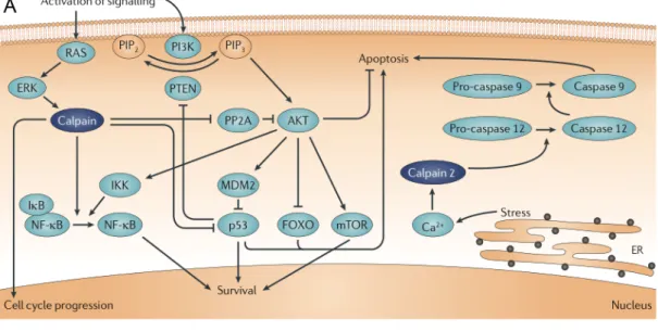
Curiously, depending on cellular context, the nature of apoptotic stimuli and the level of protein expression and localization, calpains can participate in either pro-survival or pro-apoptotic pathways (Fig. 1.2) (Storr et al., 2011). As pro-survivors, calpains have been shown to cleave wild-type p53 (Kubbutat and Vousden, 1997), preventing it from inducing apoptosis; activate NF-кB by cleaving its inhibitor IкBα (Scholzke et al., 2003); interfere with the interaction between protein phosphatase 2A (PP2A) and AKT to prevent forkhead box O (FOXO)-mediated cell death (Bertoli et al., 2009); and cleave caspase-9, rendering it inactive and unable to activate caspase-3 through the apoptosome (Chua et al., 2000). On the other hand, calpains have been shown to promote the activation of p53 after DNA damage, promoting neuronal death (Sedarous et al., 2003). They can additionally activate other substrates involved in apoptosis promotion, including Cdk5, Apaf-1, Jun, c-Jun N-terminal kinase (JNK), Fos and calcineurin inhibitor (cain) (Storr et al., 2011).
Figure 1.2. Calpain signaling pathways in survival and apoptosis. Calpain activity can mediate both pro-survival and apoptotic pathways. (A) In pro-survival signaling, calpains lead to p53 stabilization and nuclear factor-κB (NF-κB) activation through degradation of inhibitors of NF-κB (IκB). They can also modulate forkhead box O (FOXO) transcription factor activity through protein phosphatase 2A (PP2A) and drive cell cycle progression by modulating cell cycle proteins such as cyclin E and p27KIP1. Following endoplasmic
reticulum (ER) stress, calpains can cleave caspase-12, eliciting apoptosis. (B) Calpains can mediate both intrinsic and extrinsic pathways of apoptosis by cleaving several proteins, which can either lead to their activation or inactivation. AIF, apoptosis-inducing factor; Cyto c,
cytochrome c; IKK, IκB kinase; PIP2, phosphatidylinositol-4,5-bisphosphate; PIP3, phosphatidylinositol-3,4,5-trisphosphate. Adapted from Storr et al. 2011.
Interestingly, calpains have been shown to cleave and activate caspases as well. Calpain-induced cleavage of caspase-7 originates two products more reactive than those originated following caspase-3 mediated cleavage (Gafni et al., 2009). ER-localized caspase-12 is also cleaved by calpains, resulting in cell death (Nakagawa and Yuan, 2000). Caspases, on the other hand, can also activate calpains in a positive feedback loop through cleavage of their inhibitor, calpastatin (Wang et al., 1998). Calpains can additionally promote apoptosis through cleavage of Bcl-2 family proteins. Cleavage of the antiapoptotic Bfl-1 and Bcl-XL turns them into proapoptotic
factors (Nakagawa and Yuan, 2000; Valero et al., 2012), while calpain-induced cleavage of Bax turns it into a more potent inducer of apoptosis (Toyota et al., 2003). Interestingly, it has been shown that following DNA damage, poly(ADP-ribose) polymerase 1 (PARP1) activates mitochondrial calpains by inducing alterations in mitochondrial Ca2+ homeostasis (Vosler et al., 2009). Activated calpains can induce caspase-independent apoptosis, as they cleave and release AIF from the mitochondria following MOMP, by decreasing its association with lipids (Polster et al., 2005).
1.3. Reactive oxygen species and their role in apoptosis
Oxygen is paradoxically necessary for aerobic life and toxic to all life forms through the production of reactive oxygen species (ROS) (Mates et al., 2012). Due to high chemical reactivity, ROS can lead to lipid peroxidation, massive protein oxidation and degradation and DNA sequence changes, including mutations, deletions, gene amplification and rearrangements (Mates et al., 2012). ROS may be generated endogenously as products of normal metabolism or following xenobiotic exposure. Intracellular sources include proteins at the plasma membrane, lipid metabolism within peroxisomes, several cytosolic enzymes and, most importantly, mitochondria (Balaban et al., 2005). Indeed, mitochondria generate ~ 90% of cellular ROS as a consequence of oxidative phosphorylation. It is estimated that ~ 0.2% of the oxygen consumed by mitochondria leads to ROS generation, mainly at sites I and III of the respiratory chain (Balaban et al., 2005).
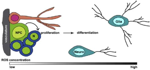
Although ROS have been considered as harmful agents that cause oxidative damage in pathologies and aging, the current notion is that ROS integrate complex signaling pathways and play several important biological roles as second messengers (Murphy et al., 2011; Finkel, 2012). The benefits of antioxidants have become popular knowledge, probably following Harman’s Free Radical Theory of Aging, which proposes that aging results from a decline of cellular functions due to an accumulation of cellular damages caused by metabolism-resultant ROS (Harman, 1956). However, current evidence defies this model, as several longevity-promoting interventions depend on increased levels of ROS, which exert signaling functions to increase stress resistance and longevity, and are impaired by antioxidant supplements (Ristow and Schmeisser, 2011). Taken together, control of redox state appears to be an important requirement for cell homeostasis. In this regard, oxidative stress only occurs when ROS overwhelm the cellular antioxidant defense system (Ray et al., 2012). It has been proposed that ROS-releasing organelles may function as stress sensors, where the intensity and duration of ROS release dictates the cellular outcome in response to the quality and severity of the perceived stress (Finkel, 2012). In this perspective, low intensity ROS release would control metabolic adaptation, as happens under excess nutrient or low oxygen conditions, moderate ROS intensity following danger signals might regulate inflammation, and increased ROS levels would activate cell death inducing pathways, such as apoptotic and autophagic cell death (Fig. 1.3).
Figure 1.3. Different ROS levels elicit variable cellular outcomes. Low levels of
as proliferation and differentiation. Higher levels induce adaptive programs and lead to the upregulation of antioxidant genes. At even higher levels, senescence and apoptosis may be activated. ROS also irreversibly damage cellular components independently of their signaling properties, but only when at high levels. Adapted from Hamanaka and Chandel, 2010.
ROS interact with proteins through oxidation of cysteine (Cys) residues, which contain highly sensitive thiol (−SH) moieties in their side chains (Miki and Funato, 2012). The thiol is oxidized to sulfenyl (−SOH), which in turn can form disulfide bonds with another thiol. Although, these reactions are reversible by various cellular antioxidants, the sulfenyl can be further oxidized into sulfinyl (−SO2H) and
sulfonyl (−SO3H), highly peroxidized moieties that cannot be reduced under normal
intracellular conditions. Cellular antioxidants or free radical scavengers include superoxide dismutase (SOD), catalase, glutathione peroxidase (GPX), thioredoxin (Trx) reductase, nitric oxide synthase (NOS), heme oxygenase-1 (HO1), eosinophil peroxidase (EPO) and metallothionein; and also non-enzymatic antioxidants, such as vitamins C, E and A, β-carotene, polyamines, melatonin, nicotinamide adenine dinucleotide phosphate (NADPH), adenosine, urate, coenzyme Q-10, polyphenols, flavonoids, phytoestrogens, glutathione (GSH), cysteine, homocysteine, taurine, methionine, S-adenosyl-L-methionine, nitroxides and selenium (Mates, 2000).
Several redox systems participate in cell signaling and modulation of cell function. These include the GSH, Trx and pyridine nucleotide redox systems (Circu and Aw, 2010). Pyridine nucleotides are present in the cell as oxidized and reduced nicotinamide adenine dinucleotides in their unphosphorylated (NAD+ and NADH)
and phosphorylated (NADP+ and NADPH) forms (Nakamura et al., 2012). The reduced forms not only serve as electron donors to produce ATP but also regulate the cellular redox status by reducing oxidized GSH or Trx. Among their functions in energy production, regulation of metabolism and maintenance of the cellular redox status, pyridine nucleotides regulate sirtuins. In fact, proteins of the sirtuin family, comprising Sirt1-7 in mammals, use NAD+ as a cofactor and therefore function as sensors of the cellular energy levels and redox status. Sirtuins, which are best known for their roles in metabolism and lifespan/healthspan extension, act in different cellular compartments, from the cytosol, to the nucleus or the mitochondria, where they deacetylate proteins, including histones and transcriptional regulators (Houtkooper et al., 2012). p53 was the first described non-histone target for Sirt1,
which, after DNA damage or oxidative stress, deacetylates and represses p53, preventing it from inducing apoptosis. Sirt3, majorly found at the mitochondria, protects cells from oxidative stress by activating SOD2 and by deacetylating isocitrate dehydrogenase 2 (IDH2) (Someya et al., 2010).
At the transcriptional level, a battery of antioxidant genes is under the regulation of an enhancer named antioxidant responsive element (ARE), allowing a quick cellular response to oxidative conditions. These genes include glutathione S-transferase, NADPH quinone oxidoreductase-1, HO1 and ferritin H (Ray et al., 2012). ARE activation under oxidative stress conditions primarily involves nuclear factor E2-related factor 2 (Nrf2), a member of bZIP transcription factors that is expressed in a variety of tissues (Kang et al., 2005). Under basal conditions, Nrf2 is sequestered in the cytoplasm by its inhibitor, Kelch-like ECH-associated protein-1 (Keap1), which binds actin (Baird and Dinkova-Kostova, 2011). Nrf2 is continuously ubiquitinated and degraded at the proteasome through interaction with the cullin-3 E3-ubiquitin ligase (Cul3) mediated by Keap1. Keap1 contains reactive Cys that function as redox sensors and mediate its dissociation from Nrf2 upon oxidation by ROS. Therefore, in oxidative conditions, Nrf2 is stabilized and translocates to the nucleus, where it dimerizes with members of the small Maf family and binds ARE, activating transcription of cytoprotective genes.
When ROS levels are too elevated for the cell to cope with, apoptosis may be activated. Apoptosis signal-regulated kinase 1 (ASK1) participates in MAPK cascades, activating the JNK and p38 pathways and leading to apoptosis (Ray et al., 2012). Reduced Trx, in turn, binds and inhibits ASK1. However, when Trx is oxidized by ROS, it releases ASK1, allowing its oligomerization, autophosphorylation and subsequent activation. In addition to this mechanism, ROS may also induce sustained activation of these signaling pathways through inhibition of protein tyrosine phosphatases by reversible oxidation of their catalytic-site Cys (Miki and Funato, 2012). For example, following TNF-α stimulation, ROS are actively generated and oxidize MAPK phosphatases, which negatively regulate JNK (Kamata et al., 2005). Activation of Fas also leads to ROS generation, which mediate ubiquitination and proteasomal degradation of the antiapoptotic FLIP, thereby inducing apoptosis (Wang et al., 2008b). ROS have also been shown to directly induce the extrinsic pathway of apoptosis by promoting death receptor clustering and formation of lipid-raft derived signaling platforms (Circu and Aw, 2010).
Apoptosis induction by several agents has been associated with ROS production, loss of mitochondrial membrane polarization and subsequent changes in permeability leading to Cyt c release (Ryter et al., 2007). Indeed, ROS have a well-established role in oxidizing components of the PTP, namely voltage-dependent anion channel (VDAC), adenine nucleotide translocase (ANT) and cyclophilin D, hence impacting mitochondrial anion fluxes (Circu and Aw, 2010). In addition, it has been shown that Cyt c-catalyzed oxidation of cardiolipin, a mitochondria-specific phospholipid, is essential for MOMP and for the subsequent release of pro-apoptotic factors into the cytosol (Kagan et al., 2005). Loss of Cyt c, in turn, leads to a further ROS increase due to disruption of the electron transport chain, thus amplifying the apoptotic cascade (Chen and Lesnefsky, 2006). In addition, it has been suggested that the redox status of Cyt c may influence apoptosis, as only the oxidized form of the protein can induce caspase activation via the apoptosome (Brown and Borutaite, 2008). Another feed-forward loop happens at the level of mitochondrial DNA (mtDNA) stability. Since mtDNA encodes 13 polypeptides of the respiratory chain, mtDNA damage may compromise the integrity of the respiratory chain, leading to further ROS generation and ultimately triggering apoptosis (Circu and Aw, 2010).
Caspases are also prone to oxidation of their catalytic site Cys. Accordingly, oxidation-mediated inactivation of caspase-3, -8 and -9 has already been reported (Borutaite and Brown, 2001; Barbouti et al., 2007). On the other hand, it has been demonstrated that oxidative modifications of caspase-9 by ROS promote intra-mitochondrial autoprocessing of caspase-9 (Katoh et al., 2004) and its interaction with Apaf-1 through disulfide formation, facilitating apoptosome formation and caspase-9 activation (Zuo et al., 2009).
ROS have additionally been shown to induce apoptosis by upregulating proapoptotic and downregulating antiapoptotic Bcl-2 family protein levels, mediating their levels of phosphorylation and ubiquitination (Li et al., 2004). There are many other examples of ROS interplay with apoptotic pathways. It is interesting to note that ROS do not seem to lead to cell demise by modifying and destroying cellular components at random, as initially assumed, but rather through specific targeting and modulation of key components of several regulatory pathways.
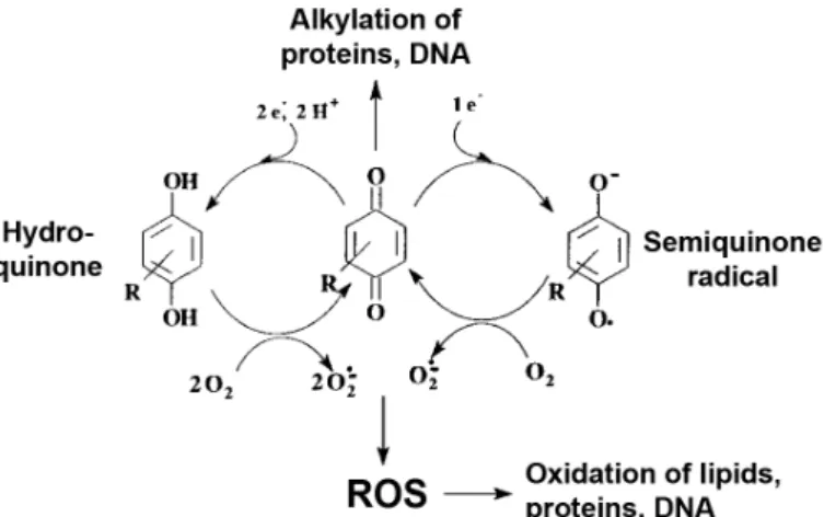
1.4. Modulation of apoptosis by naphthoquinones
Quinones are bioactive compounds that comprise conjugated carbonyl groups in six-membered rings (Kumagai et al., 2012). They are highly reactive molecules and have been assigned inflammatory, anti-inflammatory, and anticancer properties, among others. Quinones interact with biological systems through two reactions: as electron transfer agents, promoting the reduction of oxygen to ROS; and as electrophiles, forming covalent bonds with nucleophilic groups in biological molecules, such as the thiol groups in Cys residues (Fig. 1.4). Naphthoquinones are a subclass of quinones that are widespread in nature, occurring naturally in plants, fungi and bacteria (Medentsev and Akimenko, 1998; Shearer and Newman, 2008; Huang et al., 2010), often used in traditional medicine. They can also be generated in humans through the metabolism of naphthalene (Cho et al., 2006).
Figure 1.4. Quinones form covalent bonds with biological molecules and generate ROS.
Redox cycling and alkylation, the transfer of an alkyl group to another molecule, are two important properties of quinones. They promote the generation of reactive oxygen species (ROS) and the formation of covalent bonds with cellular nucleophiles, such as cysteine residues in proteins. Adapted from Bolton et al. 2010.
Naphthoquinones have been shown to modulate several signaling pathways, eliciting different cellular outcomes. The ability of naphthoquinone derivatives to modulate apoptosis and proliferation has been extensively described, mostly focusing on their anticancer properties (Kumagai et al., 2012). Although most studies only report the general cellular effects of these compounds, such as induction of apoptosis, inhibition of proliferation and protection from toxic agents, some of them provide additional insight into the specific mechanisms through which these outcomes are


![Figure 3.1. Chemical formula of naphtho[2,3-d]isoxazole-4,9-dione-3-carboxylates 1a and 1b, and naphthoquinone 4](https://thumb-eu.123doks.com/thumbv2/123dok_br/15475939.1036027/73.892.297.575.540.775/figure-chemical-formula-naphtho-isoxazole-dione-carboxylates-naphthoquinone.webp)