MicroRNA-221 promotes cell proliferation, migration,
and differentiation by regulation of ZFPM2 in osteoblasts
Xingguo Zheng
1, Jinhua Dai
2, Haijun Zhang
1and Zhibin Ge
1 1Department of Orthopaedics, Ningbo No. 2 Hospital, Ningbo, China 2Department of Clinical Laboratory, Ningbo No. 2 Hospital, Ningbo, ChinaAbstract
Bone fracture is a common medical condition, which may occur due to traumatic injury or disease-related conditions. Evidence suggests that microRNAs (miRNAs) can regulate osteoblast differentiation and function. In this study, we explored the effects and mechanism of miR-221 on the growth and migration of osteoblasts using MC3T3-E1 cells. The expression levels of miR-221 in the different groups were measured by qRT-PCR. Then, miR-221 mimic and inhibitor were transfected into MC3T3-E1 cells, and cell viability and migration were measured using the CCK-8 assay and the Transwell migration assay. Additionally, the expression levels of differentiation-related factors (Runx2andOcn) andZFPM2were measured by qRT-PCR. Western blot was used to measure the expression of cell cycle-related proteins, epithelial-mesenchymal transition (EMT)-related proteins, ZFPM2, and Wnt/Notch, and Smad signaling pathway proteins. miR-221 was significantly up-regulated in the patients with lumbar compression fracture (LCM) and trochanteric fracture (TF). miR-221 promoted ALP, Runx2, and OPN expressions in MC3T3-E1 cells. miR-221 overexpression significantly increased cell proliferation, migration, differentiation, and matrix mineral-ization, whereas suppression of miR-221 reversed these effects. Additionally, the results displayed thatZFPM2was a direct target gene of miR-221, and overexpression of ZFPM2 reversed the promoting effects of miR-221 overexpression on osteo-blasts. Mechanistic study revealed that overexpression of miR-221 inactivated the Wnt/Notch and Smad signaling pathways by regulating ZFPM2 expression. We drew the conclusions that miR-221 overexpression promoted osteoblast proliferation, migration, and differentiation by regulation of ZFPM2 expression and deactivating the Wnt/Notch and Smad signaling pathways. Key words: MicroRNA-221; Bone fracture; Osteoblast differentiation; ZFPM2; Wnt/Notch; Smad
Introduction
Bone fracture is a common and increasing disease, which results from both traumatic injury and disease-related bone fragility (1). In the United States, about six million adults suffer from fractures annually (2). Bone fracture may lead to fever, disability, shock, and treatment is very expensive. Timely and appropriate management of bone fractures can help patients restore original functions. How-ever, some patients still have different degrees of sequelae, such as osteomyelitis, non-union and mal-union, complex regional pain syndrome, and post-traumatic arthritis (3). Bone fracture healing is a physiologically complex process, which involves both biological and mechanical factors (4). Following bone fracture, a series of events occurs, includ-ing cell migration, differentiation, tissue synthesis, and the release of cytokines and growth factors. The recovery process of fracture depends on the activity of osteoblasts (5,6). Osteoblasts are mesenchymal cells, which play a major role in skeletal development and bone formation (7). Osteoblasts are responsible for the synthesis, secretion and mineralization of bone matrix (8). Therefore, it is
necessary to explore the mechanism of osteoblast pro-liferation, migration, and differentiation.
MicroRNAs (miRNAs) are small, non-coding RNA mole-cules, which can mediate the post-transcriptional gene expression (9). A recent study demonstrates that miRNAs are involved in various cellular processes, such as cell proliferation, migration, differentiation, and apoptosis (10). Increasing evidence indicates that miRNAs regulate the differentiation and function of chondrocytes, osteoblasts, and osteoclasts (11). Thesefindings suggest that miRNAs act as key mediators in the processes of bone formation, resorption, remodeling, and repair (12). As Waki et al. (13) reported, several miRNAs, such as miR-140-3p, miR-140-5p, miR-181a-5p, miR-181d-5p, and miR-451a, were signifi -cantly up-regulated in standard healing fractures compared with unhealing fractures. Moreover, more than 15 miRNAs have been reported for bone formation stimulation (14). miR-221 is one of many widely studied miRNAs, which is frequently up-regulated in various cancers (15,16). How-ever, the precise function of miR-221 in bone fracture is
Correspondence: Zhibin Ge:<[email protected]>
still unknown. In this study, we aimed to explore the role of miR-221 in osteoblast proliferation, migration, and dif-ferentiation using MC3T3-E1 cells. This is thefirst study to report the effects of miR-221 on osteoblast growth and differentiation in the bone fracture healing process. This study might provide novel therapeutic strategies for bone fracture.
Material and Methods
Blood sample collection
The blood samples were obtained from three patients with lumbar compression fracture (LCM) and three patients with trochanteric fracture (TF) of Ningbo No.2 Hospital from April 2017 to October 2017. The blood samples were collected at 24 h and 7, 14, and 21 days after surgery or injury and then stored at–80°C until analyzed. The ethical
approval for this study was granted by the Ethics Com-mittee of Ningbo No. 2 Hospital. All participants signed the informed consent.
Cell culture and differentiation induction
The mouse osteoblast-like cells (MC3T3-E1) used in the present study were obtained from American Type Culture Collection (ATCC, USA). The cells were cultured in 100-mm dishes containing a-MEM culture medium with 10% fetal bovine serum (FBS, Gibco, USA), and 1% penicillin and streptomycin under humid environment with 5% CO2at 37°C. The culture medium was changed every three days. For induction of MC3T3-E1 cells differentiation, 1105cells were seeded in a 6-well plate, and incubated in the differentiation medium containing 50mg/mL ascorbic acid and 10 mmol/Lb-glycerophosphate. The control group cells were cultured with conventional medium. The medium was refreshed every three days.
Quantitative real-time polymerase chain reaction (qRT-PCR) analysis
Total RNA was isolated from blood samples using the PAXgene Blood RNA Kit (Qiagen, Germany) and from MC3T3-E1 cells using the common kit of Trizol reagent (Invitrogen, Life Technologies Corporation, USA) accord-ing to manufacturer’s protocols. For examining the expres-sion level of miR-221 in MC3T3-E1 cells, cDNA was synthesized using TaqMan MicroRNA Reverse Transcrip-tion Kit (Invitrogen), and qRT-PCR analysis was carried out using TaqMan Universal Master Mix II (Invitrogen) following the instructions, and U6 (Applied Biosystems, USA) was used to normalized the expression level of miR-221. Data were examined by the 2-DDCtmethod.
miRNAs transfection
The expression plasmids of miR-221 mimic, miR-221 inhibitor, and the corresponding negative controls (NCs) were synthesized by GenePharma Co. (China). Additionally, the overexpression vector of zincfinger protein multitype 2
(ZFPM2) was constructed using the sub-cloning the full-length ZFPM2 coding sequence into pcDNA3.1 plasmid (Sangon Biotech, China). The empty pcDNA3.1 plasmid was used as a negative control. Afterward, MC3T3-E1 cells were transfected with these expression plasmids for 48 h. All cell transfections were detected using Lipofecta-mine 3000 reagent (Invitrogen) according to the manu-facturer’s protocol.
Cell viability assay
Cell viability of MC3T3-E1 cells was detected using the Cell Counting Kit-8 (CCK-8) assay (Beyotime Biotechnol-ogy, China). Briefly, MC3T3-E1 cells were cultured in a 96-well plate, and transfected with expression vectors of miR-221 mimic, miR-221 inhibitor, and pc-ZFPM2. After transfection for 48 h, 10mL CCK-8 was supplemented to each well and the plates were incubated for another 1 h under the routine-culture environment containing 95% air and 5% CO2at 37°C. Finally, a microplate reader (Bio-Rad, USA) was used to measure the absorbance at 450 nm.
Cell migration assay
For the migration assay, the Transwell with a pore size of 8 mM was performed to examine the migration ability of MC3T3-E1 cells. In brief, MC3T3-E1 cells were trans-fected with the above expression vectors. Afterward, these cells were suspended in serum-free medium, and 100-mL cell suspension was added into the upper compartment of a 24-well transwell culture chamber. Meanwhile, 600mL of complete medium was added into the lower compartment. After incubation for 24 h in the conventional culture condi-tions, the Transwell culture chamber was taken out, and washed twice with calcium-free PBS, and cells werefixed with methanol for 30 min. Subsequently, the non-migrated cells were removed carefully using a wet cotton swab from the upper surface of thefilter. The migrated cells on the lower side of thefilter were stained with 1% crystal violet for 20 min, and counted using a microscope (magnification of 400) in a randomfivefields of vision.
Alizarin Red S staining assay
To confirm the important effects of miR-221 on miner-alization of MC3T3-E1 cells, the Alizarin Red S staining assay was performed. Briefly, the cells were washed twice with PBS and fixed with 95% ethanol for 10 min at room temperature. Subsequently, thefixed cells were stained with 1% Alizarin Red S solution (Sigma-Aldrich, USA) for 30 min at 37°C and counted using a light micro-scope (Olympus, Japan). Quantification of Alizarin Red S stain was assessed via extraction with Image J soft-ware (NIH, USA).
Luciferase reporter assay
The 30-untranslated region (30-UTR) of ZFPM2 was
with miR-221 mimic and its corresponding control into cells using Lipofectamine 3000 (Invitrogen). The luciferase assay was confirmed using the dual luciferase reporter assay system (Promega) after transfection for 48 h.
Western blot assay
The proteins from transfected cells used for the western blot assay were isolated using RIPA lysis buffer (Beyotime Biotechnology). The contents of total protein were tested using the BCAt Protein Assay Kit (Pierce, USA) based on the kit instruction. Then, 40mg protein samples were subjected to sodium dodecyl sulfate-polyacrylamide gel electrophoresis (SDS-PAGE) and transferred to polyvinyl-idene difluoride (PVDF) membranes. After blocking with 5% non-fat milk, the membranes were transferred to another container and incubated with the primary antibodies of alkaline phosphatase (ALP, ab83259), Runt-related tran-scription factor 2 (Runx2, ab23981), Osteopontin (OPN, ab8448), E-cadherin (ab40772), N-cadherin (ab18203), Vimentin (ab16700), ZEB1 (ab124512), Snail (ab82846), Osteocalcin (Ocn, ab93876), proliferating cell nuclear anti-gen (PCNA, ab18197), Cyclin A (ab181591), Cyclin E1 (ab71535), cyclin-dependent kinase 2 (CDK2, ab64669), Cyclin D1 (ab134175), CDK4 (ab199728), Wnt3a (ab28472), Wnt5a (ab229200), Notch 1 (ab52627), Notch 2 (ab8926), Notch 3 (ab23426), p-Smad2 (ab53100), Smad2 (ab33875), Smad4 (ab40759), Smad7 (ab216428), and GAPDH (ab181602) at 4°C overnight. After incubation for 2 h at
37°C, the secondary antibody of horseradish peroxidase (HRP)-conjugated goat anti-rabbit IgG (ab205718, 1:2000, Abcam) was added and incubated for 1 h at room tempera-ture. Finally, the signals were captured using ECL reagents (MultiSciences Biotech, China).
Statistical analysis
The results are reported as means±SD. SPSS 19.0
statistical software (SPSS, Inc., USA) was used to analyze the data. One-way analysis of variance (ANOVA) was used to calculate the P values. Po0.05 was considered to be statistically significant.
Results
miR-221 was up-regulated during osteoblast differentiation
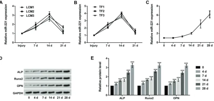
To explore the effect of miR-221 on bone fracture, the blood concentrations of miR-221 in three LCM patients and three TF patients were examined. The results showed that the blood concentrations of miR-221 were obviously increased at 7 and 14 days after surgery, whereas the concentrations of miR-221 was recovered at 21 days after surgery (Figure 1A and B). Additionally, MC3T3-E1 cells were cultured in osteogenic differentiation medium, and the relative expression of miR-221 during the osteoblast differentiation process was detected at different time inter-vals (0, 4, 7, 14, 21, and 28 days) using qRT-PCR.
As shown in Figure 1C, the expression of miR-221 was significantly increased in a time-dependent manner during osteoblastic differentiation. Further, the western blot assay revealed that the protein levels of ALP, Runx2, and OPN were notably up-regulated in a time-dependent manner (Po0.001, Figure 1D and E). These data indicated that up-regulation of miR-221 might have an important role in the process of bone fracture.
Overexpression of miR-221 promoted osteoblast proliferation
Next, MC3T3-E1 cells were transfected with miR-221 mimic, miR-221 inhibitor, and the corresponding controls. After transfection for 48 h, the expression level of miR-221 was measured using qRT-PCR. The results in Figure 2A showed that miR-221 overexpression significantly increased the expression of miR-221 compared with the mimic con-trol group (Po0.01), while miR-221 suppression signifi -cantly decreased the expression of miR-221 compared with the inhibitor control group (Po0.001). We then
measured cell viability using the CCK-8 assay. As shown in Figure 2B, miR-221 overexpression significantly pro-moted cell viability compared with the mimic control group (Po0.05), whereas miR-221 suppression significantly decreased cell viability compared with the inhibitor control group (Po0.05). Western blot assay was performed to further confirm these results by analysis of cell cycle-related proteins (PCNA, Cyclin A, Cyclin E1, CDK2, Cyclin D1, and CDK4). The results showed that miR-221 over-expression increased the over-expression of these proteins, whereas miR-221 inhibition decreased their expression in MC3T3-E1 cells (Po0.01, Figure 2C and D). Taken together, the data indicated that overexpression of miR-221 could promote cell proliferation.
Overexpression of miR-221 promoted osteoblast migration
whereas miR-221 suppression significantly decreased cell migration compared with the inhibitor control group (Po0.05, Figure 3A). Western blot results showed that miR-221 overexpression decreased the expression of E-cadherin (Po0.01), as well as increased the expres-sions of N-cadherin, Vimentin, ZEB1, and Snail (Po0.01). However, miR-221 suppression showed opposite results (Figure 3B and C). These findings indicated that over-expression of miR-221 could promote cell migration and EMT process in osteoblasts.
Overexpression of miR-221 promoted osteoblast differentiation
qRT-PCR results of cell differentiation showed that miR-221 overexpression significantly increased the ex-pression of Runx2 and Ocn on days 7 and 14, whereas miR-221 suppression showed opposite results (Po0.05, Figure 4A and B). Mineralization analysis showed that overexpression of miR-221 significantly increased the mineralized nodule formation compared with the control group (Po0.01, Figure 4C). Furthermore, the results of western blot (Figure 4D-F) revealed that miR-221
overexpression notably increased the protein levels of ALP, Runx2, and Ocn on days 7 and 14, whereas miR-221 suppression showed opposite results except for Ocn expression on day 14, which was unchanged. Above all, the results suggested that overexpression of miR-221 was associated with osteoblast differentiation.
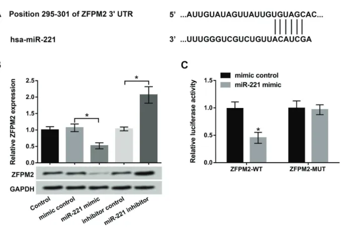
ZFPM2 was a direct target of miR-221
To explore the relationship between miR-221 and ZFPM2, the software programs of TargetScan (www.target scan.org) and microRNA database (www.microrna.org) were used to predict the binding site (Figure 5A). Then, the ex-pression level of ZFPM2 in MC3T3-E1 cells transfected with miR-221 mimic and miR-221 inhibitor was detected by qRT-PCR and western blot. The results showed that the mRNA and protein levels of ZFPM2 were significantly decreased by miR-221 overexpression, as well as promoted by miR-221 suppression (Po0.05, Figure 5B). Meanwhile, dual-luciferase reporter assay results showed that lucifer-ase activity was greatly decrelucifer-ased by co-transfection of miR-221 mimic and ZFPM2-WT (Po0.05). However, co-transfection of miR-221 mimic and ZFPM2-MUT had no
effect on luciferase activity (Figure 5C). In short, the results indicated that ZFPM2 was a direct target gene of miR-221, and miR-221 inhibited ZFPM2 expression in osteoblasts.
Overexpression of miR-221 promoted osteoblast proliferation, migration, and differentiation by targeting ZFPM2
To uncover whether ZFPM2 was involved in the pro-cesses of osteoblast proliferation, migration, and differ-entiation, miR-221 mimic and pc-ZFPM2 were transfected into MC3T3-E1 cells to change miR-221 and ZFPM2 expression. The results in Figure 6A and B showed that miR-221 overexpression significantly increased cell viabil-ity and migration compared with the mimic control group (Po0.05), but overexpression of ZFPM2 reversed these effects by decreasing cell viability and migration (Po0.05). Western blot results revealed that overexpression of miR-221 increased the expression of cell cycle-related proteins (PCNA, Cyclin A, Cyclin E1, CDK2, Cyclin D1, and CDK4), but overexpression of ZFPM2 reversed these effects (Po0.05, Figure 6C and D). Moreover, the expression level of differentiation-related proteins (ALP, Runx2, and Ocn) were increased by miR-221 mimic, but overexpression of ZFPM2 reversed these effects (Po0.05, Figure 6E and F). These findings indicated that overexpression of miR-221 promoted cell viability, migration, and differentiation by regulating the expression of ZFPM2.
Overexpression of miR-221 deactivated Wnt/Notch and Smad signaling pathways
Lastly, we measured the effect of miR-221 and ZFPM2 on the Wnt/Notch and Smad signaling pathways using western blot. Overexpression of miR-221 decreased the protein level of Wnt3a, Wnt5a, Notch 1, Notch 2, Notch 3, p-Smad2, Smad4, and Smad7, but overexpression of ZFPM2 reversed these effects (Po0.05 or Po0.01, Figure 7A-D). These findings indicated that overexpression of miR-221 deactivated Wnt/Notch and Smad signaling pathways by regulation of ZFPM2.
Discussion
Bone fracture is a common medical condition, which is damage in the continuity of the bone, and this disease occurs frequently in children and the elderly (17). The activity of osteoblasts is closely related to the quality of recovery of bone fractures. Osteogenesis is a complex and multistep processes, involving the differentiation of mesenchymal stem cells into osteoblast progenitor cells, preosteoblasts, osteoblasts, and osteocytes, as well as crosstalk between multiple cell types for the formation and remodeling of bone (18). The process is regulated by various signaling networks, such as BMP, Wnt ligands, Notch ligands, transforming growth factor (TGF), tumor necrosis factor, and cytokines. A recent study demonstrated
that miRNAs acted as important regulators of osteogenic signaling pathways (19). In this study, we investigated the effects and mechanism of miR-221 on osteoblasts prolif-eration, migration, and differentiation. The results showed that miR-221 was up-regulated in the patients with LCM and TF, and closely related with the process of osteoblastic differentiation. Moreover, we found that overexpression of miR-221 significantly promoted cell proliferation, migration, differentiation, and matrix mineralization in osteoblasts, and suppression of miR-221 showed contrary results. Further experiments showed that ZFPM2 was a direct target of miR-221, and overexpression of miR-221 pro-moted cell viability, migration, and differentiation by down-regulation of ZFPM2. Finally, the results indicated that miR-221 overexpression blocked Wnt/Notch and Smad7 signaling pathways by regulating ZFPM2 expression.
miR-221 is one of the important miRNAs, which has been widely reported in various cancers, but it has not been fully investigated in osteoblastic differentiation (20). Several other miRNAs have been shown to be up-regulated in osteoblastic differentiation. For example, Chen et al. (21) showed that miR-34a was up-regulated in osteoblastic differentiation of human stromal stem cells. Li et al. (22) demonstrated that miR-216a was remarkably up-regulated
during osteogenic differentiation in human adipose-derived MSCs. Interestingly, miR-31 was reported to be down-regulated during osteoblastic differentiation, however, miR-31 was later identified to be up-regulated during osteoblastic differentiation (23,24). Similar to these studies, we found that miR-221 was up-regulated in LCM and TF patients and also up-regulated during osteoblastic differ-entiation. These data indicated that miR-221 might be involved in the process of osteoblastic differentiation.
Osteogenic differentiation is divided into four stages: cellular commitment, proliferation, matrix maturation, and mineralization. Our study demonstrated that overexpres-sion of miR-221 promoted osteoblast proliferation. In line with thisfinding, Xu et al. (25) showed that transfection of MC3T3-E1 osteoblasts with miR-365 ameliorated dexa-methasone-induced inhibition of cell viability. Cyclins and CDKs are known to be regulators of cell cycle. Anin vitro study has shown that Cyclin E, Cyclin B, Cyclin A, and CDK inhibitors regulate osteoblastic differentiation (26). Our study also found that overexpression of miR-221 increased the expression of cell cycle-related proteins (PCNA, Cyclin A, Cyclin E1, CDK2, Cyclin D1, and CDK4). Thus, these findings indicated that overexpression of miR-221 promoted cell proliferation in osteoblasts.
EMT is a biological process, which is characterized by a transition from epithelial cells to interstitial phenotypes by specific procedures. Mounting evidence has indicated that EMT is involved in the formation of many tissues and organs during development (27,28). Moreover, several signaling pathways, such as TGF-b, Wnt, and Notch, have been reported to induce the EMT process. These signal-ing pathways can activate transcription factors, includsignal-ing Snail, Slug, and ZEB family, which suppress the expression of E-cadherin, resulting in cell invasion and migration (29).
Osteoblast migration improves the repair of bone fracture and growth of bone tissue (30). Our study found that over-expression of miR-221 promoted osteoblast migration by decreasing the expression of E-cadherin and increasing the expression of N-cadherin, Vimentin, ZEB1, and Snail. Understanding the regulatory mechanism of osteo-blast differentiation is very important to develop strategies for treating bone disorders, including bone fracture. Runx2, Osterix, andb-catenin are the vital transcription factors for osteoblast differentiation (6). Runx2 is a main transcription
factor required for the differentiation of osteoblasts from mesenchymal precursors and subsequent bone matrix mineralization (31). Moreover, recent research has proven that Runx2 can directly stimulate the osteoblast marker gene expression, such as Ocn (32). Ocn is a late bone marker, which appears in osteogenic differentiation and mineralization (33). Several studies have shown the involve-ment of miRNAs in osteoblast differentiation. For example, miR-26a and miR-125b are shown to inhibit osteoblast differentiation, whereas miR-33-5p and miR-194 are reported to promote osteoblast differentiation (34,35). An interest-ing study from Zhang et al. (36) reported that miR-221 could inhibit osteogenic differentiation by targeting Runx2 in C2C12 cells. Similarly, Yeh et al. (37) found that miR-221 attenuated the osteogenic differentiation in human annu-lus fibrosus cells. However, the opposite results in the present study revealed that miR-221 promoted osteoblast differentiation by increasing the expression of ALP, Runx2, and Ocn in MC3T3-E1 cells. The different results might be related to the different cell lines used. Further studies are still needed to confirm the hypothesis.
ZFPM2 is a zincfinger protein encoded by the ZFPM2 gene, which is an important regulator of hematopoiesis and cardiogenesis in mammals (38). A recent study revealed that miR-429 could induce MC3T3-E1 osteoblastic cells differentiation by regulation of ZFPM2 expression (39). However, whether miR-221 affects cell proliferation, migra-tion, and differentiation through regulating ZFPM2 expres-sion in MC3T3-E1 cells is still unclear. In our study, we found that ZFPM2 was a direct target of miR-221. More-over, miR-221 decreased the expression of ZFPM2 in osteoblasts. Further experiments revealed that over-expression of ZFPM2 reversed the promoting effects of miR-221 on MC3T3-E1 cells proliferation, migration, and differentiation, indicating that the effects of miR-221 on osteoblastic cells are mediated via regulating ZFPM2.
It has been reported that Wnt, Notch, and Smad sig-naling pathways play important roles in osteoblast differ-entiation (40). Therefore, we explored the effect of miR-221 and ZFPM2 on Wnt/Notch and Smad signaling pathway proteins (Wnt3a, Wnt5a, Notch 1 to 3, Smad2, Smad4, and Smad7), and found that overexpression of miR-221
decreased the protein levels of these proteins, but ZFPM2 overexpression reversed these effects, indicating that miR-221 blocked Wnt/Notch and Smad signaling path-ways by regulation of ZFPM2.
In conclusion, these results revealed that miR-221 was up-regulated during osteoblastic differentiation, and over-expression of miR-221 promoted cell viability, migration, and differentiation by regulating ZFPM2 expression and deactivating the Wnt/Notch and Smad signaling pathways.
Our novelfindings indicate a potential role of miR-221 in osteoblast proliferation, migration, and differentiation.
Acknowledgments
This study was funded in full by the Key Laboratory of Tumor Molecular Biology of Ningbo City (2015A22011) and the Key Subjects of Ningbo No.2 Hospital (No. 2016-55).
References
1. Jin H, Wang B, Li J, Xie W, Mao Q, Li S, et al. Anti-DKK1 antibody promotes bone fracture healing through activa-tion of beta-catenin signaling.Bone2015; 71: 63–75, doi: 10.1016/j.bone.2014.07.039.
2. Kanis JA, Oden A, McCloskey EV, Johansson H, Wahl DACooper C. A systematic review of hip fracture incidence and probability of fracture worldwide.Osteoporos Int2012; 23: 2239–2256, doi: 10.1007/s00198-012-1964-3.
3. Zura R, Xiong Z, Einhorn T, Watson JT, Ostrum RF, Prayson MJ, et al. Epidemiology of fracture nonunion in 18 human bones. JAMA Surg 2016; 151: e162775, doi: 10.1001/ jamasurg.2016.2775.
4. Ghiasi MS, Chen J, Vaziri A, Rodriguez EK, Nazarian A. Bone fracture healing in mechanobiological modeling: A review of principles and methods.Bone Rep2017; 6: 87–100, doi: 10.1016/j.bonr.2017.03.002.
5. Histing T, Stenger D, Kuntz S, Scheuer C, Tami A, Garcia P. et al. Increased osteoblast and osteoclast activity in female senescence-accelerated, osteoporotic SAMP6 mice during fracture healing. J Surg Res 2012; 175: 271–277, doi: 10.1016/j.jss.2011.03.052.
6. Komori T. Regulation of osteoblast differentiation by tran-scription factors.J Cell Biochem2006; 99: 1233–1239, doi: 10.1002/jcb.20958.
7. Guntur AR, Rosen CJ. New insights into osteoblasts and their role in bone formation: the central role of PI3Kinase. J Endocrinol2011; 211: 123–130, doi: 10.1530/JOE-11-0175. 8. Karsenty G, Kronenberg HM, Settembre C. Genetic control of bone formation.Annu Rev Cell Dev Biol2009; 25: 629– 648, doi: 10.1146/annurev.cellbio.042308.113308.
9. Piccoli MT, Gupta SK, Thum T. Noncoding RNAs as regula-tors of cardiomyocyte proliferation and death. J Mol Cell Cardiol2015; 89: 59–67, doi: 10.1016/j.yjmcc.2015.02.002. 10. Bartel DP. MicroRNAs: genomics, biogenesis, mechanism, and function.Cell2004; 116: 281–297, doi: 10.1016/S0092-8674(04)00045-5.
11. Dong S, Yang B, Guo H, Kang F. MicroRNAs regulate osteo-genesis and chondroosteo-genesis.Biochem Biophys Res Com-mun2012; 418: 587–591, doi: 10.1016/j.bbrc.2012.01.075. 12. Suttamanatwong S. MicroRNAs in bone development and their diagnostic and therapeutic potentials in osteoporosis. Connect Tissue Res 2017; 58: 90–102, doi: 10.3109/ 03008207.2016.1139580.
13. Waki T, Lee SY, Niikura T, Iwakura T, Dogaki Y, Okumachi E, et al. Profiling microRNA expression during fracture healing. BMC Musculoskelet Disord 2016; 17: 83, doi: 10.1186/ s12891-016-0931-0.
14. Nakasa T, Yoshizuka M, Andry Usman M, Elbadry Mahmoud E, Ochi M. MicroRNAs and Bone Regeneration. Curr Genomics 2015; 16: 441–452, doi: 10.2174/138920291 6666150817213630.
15. Yang F, Wang W, Zhou C, Xi W, Yuan L, Chen X, et al. MiR-221/222 promote human glioma cell invasion and angiogen-esis by targeting TIMP2.Tumour Biol2015; 36: 3763–3773, doi: 10.1007/s13277-014-3017-3.
16. Xu Q, Li P, Chen X, Liang Z, Jiang Z, Nan L, et al. miR-221/ 222 induces pancreatic cancer progression through the regulation of matrix metalloproteinases.Oncotarget2015; 6: 14153–14164, doi: 10.18632/oncotarget.3686.
17. Petrescu PH, Izvernariu DA, Iancu C, Dinu GO, Berceanu-Vaduva MM, Crisan D, et al. Pathological fracture of the femur in a patient with Paget’s disease of bone: a case report.Rom J Morphol Embryol2016; 57: 595–600. 18. Lian JB, Stein GS, van Wijnen AJ, Stein JL, Hassan MQ,
Gaur T, et al. MicroRNA control of bone formation and homeostasis.Nat Rev Endocrinol 2012; 8: 212–227, doi: 10.1038/nrendo.2011.234.
19. Yuan Z, Li Q, Luo S, Liu Z, Luo D, Zhang B, et al. PPARgamma and Wnt signaling in adipogenic and osteo-genic differentiation of mesenchymal stem cells.Curr Stem Cell Res Ther2016; 11: 216–225, doi: 10.2174/1574888X 10666150519093429.
20. Li T, Li M, Hu S, Cheng X, Gao Y, Jiang S, et al. MiR-221 mediates the epithelial-mesenchymal transition of hepato-cellular carcinoma by targeting AdipoR1.Int J Biol Macromol 2017; 103: 1054–1061, doi: 10.1016/j.ijbiomac.2017.05.108. 21. Chen L, Holmstrom K, Qiu W, Ditzel N, Shi K, Hokland L, et al. MicroRNA-34a inhibits osteoblast differentiation and in vivo bone formation of human stromal stem cells.Stem Cells 2014; 32: 902–912, doi: 10.1002/stem.1615.
22. Li H, Li T, Fan J, Li T, Fan L, Wang S, et al. miR-216a rescues dexamethasone suppression of osteogenesis, pro-motes osteoblast differentiation and enhances bone forma-tion, by regulating c-Cbl-mediated PI3K/AKT pathway.Cell Death Differ2015; 22: 1935–1945, doi: 10.1038/cdd.2015.99. 23. Gao J, Yang T, Han J, Yan K, Qiu X, Zhou Y, et al. MicroRNA expression during osteogenic differentiation of human multi-potent mesenchymal stromal cells from bone marrow.J Cell Biochem2011; 112: 1844–1856, doi: 10.1002/jcb.23106. 24. Baglio SR, Devescovi V, Granchi D, Baldini N. MicroRNA
25. Xu D, Gao Y, Hu N, Wu L, Chen Q. miR-365 ameliorates dexamethasone-induced suppression of osteogenesis in MC3T3-E1 Cells by targeting HDAC4.Int J Mol Sci2017; 18: pii: E977, doi: 10.3390/ijms18050977.
26. Drissi H, Hushka D, Aslam F, Nguyen Q, Buffone E, Koff A, et al. The cell cycle regulator p27kip1 contributes to growth and differentiation of osteoblasts. Cancer Res 1999; 59: 3705–3711.
27. Xu MH, Gao X, Luo D, Zhou XD, Xiong W, Liu GX. EMT and acquisition of stem cell-like properties are involved in spon-taneous formation of tumorigenic hybrids between lung can-cer and bone marrow-derived mesenchymal stem cells.Plos One2014; 9: e87893, doi: 10.1371/journal.pone.0087893. 28. Shimizu M, Kondo S, Urushihara M, Takamatsu M,
Kanemoto K, Nagata M, et al. Role of integrin-linked kinase in epithelial-mesenchymal transition in crescent formation of experimental glomerulonephritis. Nephrol Dial Transplant 2006; 21: 2380–2390, doi: 10.1093/ndt/gfl243.
29. Son H, Moon A. Epithelial-mesenchymal Transition and Cell Invasion.Toxicol Res2010; 26: 245–252, doi: 10.5487/ TR.2010.26.4.245.
30. Riehl BD, Lee JS, Ha L, Kwon IK, Lim JY. Flowtaxis of osteoblast migration under fluid shear and the effect of RhoA kinase silencing. PLoS One 2017; 12: e0171857, doi: 10.1371/journal.pone.0171857.
31. Tamargo J, Caballero R, Delpón E. The renin–angiotensin system and bone.Clin Rev Bone Mineral Metab2015; 13: 125–148, doi: 10.1007/s12018-015-9189-6.
32. Fei L, Wang C, Xue Y, Lin K, Chang J, Sun J. Osteogenic differentiation of osteoblasts induced by calcium silicate and calcium silicate/beta-tricalcium phosphate composite bio-ceramics.J Biomed Mater Res B Appl Biomater2012; 100: 1237–1244, doi: 10.1002/jbm.b.32688.
33. Granéli C, Thorfve A, Ruetschi U, Brisby H, Thomsen P, Lindahl A, et al. Novel markers of osteogenic and adipo-genic differentiation of human bone marrow stromal cells identified using a quantitative proteomics approach. Stem Cell Res2014; 12: 153–165, doi: 10.1016/j.scr.2013.09.009. 34. Mizuno Y, Yagi K, Tokuzawa Y, Kanesaki-Yatsuka Y, Suda T, Katagiri T, et al. miR-125b inhibits osteoblastic differentiation by down-regulation of cell proliferation. Biochem Biophys Res Commun 2008; 368: 267–272, doi: 10.1016/j.bbrc. 2008.01.073.
35. Wang H, Sun Z, Wang Y, Hu Z, Zhou H, Zhang L, et al. miR-33-5p, a novel mechano-sensitive microRNA promotes osteo-blast differentiation by targeting Hmga2.Sci Rep 2016; 6: 23170, doi: 10.1038/srep23170.
36. Zhang Y, Gao Y, Cai L, Li F, Lou Y, Xu N, et al. MicroRNA-221 is involved in the regulation of osteoporosis through regulates RUNX2 protein expression and osteoblast differ-entiation.Am J Transl Res2017; 9: 126–135.
37. Yeh CH, Jin L, Shen F, Balian G, Li XJ. miR-221 attenuates the osteogenic differentiation of human annulusfibrosus cells. Spine J2016; 16: 896–904, doi: 10.1016/j.spinee.2016.03.026. 38. Li TF, Yukata K, Yin G, Sheu T, Maruyama T, Jonason JH, et al. BMP-2 induces ATF4 phosphorylation in chondrocytes through a COX-2/PGE2 dependent signaling pathway. Osteoar-thritis Cartilage2014; 22: 481–489, doi: 10.1016/j.joca.2013. 12.020.