Universidade de Lisboa
Faculdade de Medicina
Ana Rita Cascão Rodrigues
Doutoramento em Ciências Biomédicas
Especialidade em Imunologia
Universidade de Lisboa
Faculdade de Medicina
Ana Rita Cascão Rodrigues
Tese orientada por:
Professor Doutor João Eurico Fonseca
Professor Doutor Luís Ferreira Moita
Doutoramento em Ciências Biomédicas
Especialidade em Imunologia
Todas as afirmações efectuadas no
presente
documento
são
da
exclusiva responsabilidade do seu
autor,
não
cabendo
qualquer
responsabilidade à Faculdade de
Medicina
da
Universidade
de
Lisboa
pelos
conteúdos
nele
A impressão desta dissertação
foi aprovada pela Comissão
Coordenadora
do
Conselho
Científico da Faculdade de
Medicina da Universidade de
Lisboa em reunião de 15 de
Maio de 2012.
Dissertação
apresentada
à
Faculdade
de
Medicina
da
Universidade de Lisboa, para
obtenção do grau de Doutor em
Ciências Biomédicas.
To Science
“The important thing in science is not so much to obtain new facts as to discover new ways of thinking about them.”
Sir William Bragg (1862 - 1942)
XIV
Table of Contents
Index of Figures XVIII
Index of Tables XX
Acknowledgments XXII
List of Abbreviations XXIV
Sumário XXVI Summary XXX Chapter I Introduction -1- 1. Rheumatoid arthritis - 3 - 1.1 Definition - 3 - 1.2 Pathogenesis - 5 - 1.3 Immune cells - 8 - 1.3.1 Macrophages - 8 - 1.3.2 Neutrophils - 8 - 1.3.3 T cells - 9 - 1.3.4 B cells - 10 - 1.4 Cytokines - 10 - 1.4.1 IL-1β - 10 - 1.4.2 TNF - 12 -
XV 1.4.3 IL-6 - 13 - 1.4.4 IL-17 - 14 - 1.5 Treatment options - 14 - 1.5.1 Glucocorticoids - 15 - 1.5.2 Conventional DMARDs - 15 - 1.5.3 Biologic DMARDs - 15 -
2. IL-1β Immunological and inflammatory functions - 18 -
2.1 IL-1 family - 18 - 2.2 IL-1β pathway - 18 - 2.2.1 Priming - 18 - 2.2.2 Processing - 19 - 2.2.3 Secretion - 21 - 2.2.4 Signaling - 21 -
2.3 IL-1β and autoinflammatory diseases - 22 -
2.4 IL-1β and rheumatoid arthritis - 24 -
2.5 Targeting IL-1β in rheumatoid arthritis: the case of anakinra - 27 -
Chapter II Aims -31-
Chapter III Results -35-
Cytokine pattern in very early rheumatoid arthritis favours B cell activation and survival - 39 -
XVI
Identification of a cytokine network sustaining neutrophil and Th17 activation in untreated
early rheumatoid arthritis - 41 -
Caspase-1 is active since the early phase of rheumatoid arthritis - 43 -
Effective treatment of rat adjuvant-induced arthritis by celastrol - 45 -
Gambogic acid is a potent anti-inflammatory and anti-proliferative drug in a rat model of
adjuvant-induced arthritis - 47 -
Chapter IV Discussion -49-
Chapter V Conclusions -67-
XVIII
Index of Figures
Figure 1 – Representative scheme illustrating immune cells and cytokine networks in
rheumatoid joints. - 7 -
Figure 2 – An overview on the multiple roles of IL-1β in rheumatoid arthritis physiopathology. - 12 -
Figure 3 – A simplified scheme showing main pathophysiologic pathways in rheumatoid arthritis and the effects of some currently available biologic agents. - 17 -
Figure 4 – IL-1β processing pathways. - 19 -
Figure 5 – Proposed model for cytokine networks in the very early phase of rheumatoid arthritis. - 54 -
Figure 6 – Proposed mechanism for excessive IL-1β production since early rheumatoid arthritis. - 57 -
Figure 7 – Proposed mechanism for celastrol and gambogic acid anti-inflammatory effects. - 62 -
XX
Index of Tables
Table 1 – The 2010 American College of Rheumatology / European League Against Rheumatism classification criteria for rheumatoid arthritis. - 4 -
Table 2 – The 1987 American College of Rheumatology criteria for the classification of
XXII
Acknowledgments
First and foremost, I would like to acknowledge both my supervisors, Professor Doutor João Eurico Fonseca and Professor Doutor Luís Ferreira Moita, for having accepted me as a PhD student, for all the scientific meetings and discussions, for all the guidance and opportunities. Thank you both for all the support.
Financial support was generously granted by Sociedade Portuguesa de Reumatologia, Pfizer and Fundação para a Ciência e a Tecnologia through the PhD fellowship (SFRH/BD/40513/2007).
My thanks also go to my PhD thesis committee, Professor Doutor Luís Graça, Professor Doutor Carlos São José and Doutora Margarida Carneiro, for all the meetings, discussions and suggestions that improved the work. But most of all, thank you for your unconditional support, motivation and trust.
I would like also to thank all the members from Rheumatology Department, Hospital de Santa Maria, all the medical doctors and nurses, for the effort to collaborate in the development of my work, especially in sample collection and in the organization of clinical data. In particular, a thanking note to Enfª. Lurdes Narciso, Doutora Helena Canhão, Drª. Ana Maria Rodrigues, Dr. Joaquim Polido Pereira, Drª. Elsa Sousa, Drª. Cristina Ponte, Drª. Raquel Marques, Drª. Ana Filipa Mourão (Rheumatology Department, Hospital Egas Moniz) and Dr. Márcio Navalho (Radiology Department, Hospital da Luz) with whom I have worked closer.
I am also very grateful to all the patients and healthy volunteers for their willingness in providing us with samples making this project possible.
I wish to acknowledge Doctor Vineet Gupta from the Department of Medicine, University of Miami for giving us the library of drugs used in this work.
Many thanks to all my colleagues from Instituto de Medicina Molecular that have directly worked with me, especially Rita Moura, Bruno Vidal, Ana Lopes, Helena Raquel, Ana Neves Costa, Ana Luísa Caetano and Saba Abdulghani. This work greatly benefited from the close collaboration and invaluable friendship over the years.
Many thanks also to all my friends for everyday support and friendship.
Finally, I would like to deeply thank my parents and Zé for their unconditional love and guidance in life.
Thank you all for turning this challenging journey into an enjoyable one! Obrigada!
XXIV
List of Abbreviations
ACPA Anti-citrullinated protein antibodies
ACR American College of Rheumatology
AIA Adjuvant-induced arthritis APRIL A proliferation-inducing ligand
ASC Adaptor molecule apoptosis associated speck-like protein containing a caspase recruitment domain
BLyS B-lymphocyte stimulator
CAPS Cryopyrin-associated periodic fever syndromes CARD Caspase recruitment domain
CINCA/NOMID Chronic infantile neurological cutaneous and articular syndrome/neonatal onset multisystem inflammatory disease
DAMP Danger associated molecular pattern DMARD Disease-modifying anti-rheumatic drug
EA Early arthritis
EAE Experimental autoimmune encephalomyelitis ERA Early rheumatoid arthritis
EULAR European League Against Rheumatism
FCAS Familial cold-induced autoinflammatory syndrome
FcγR Fc gamma-receptor
HLA Human leukocyte antigen
IFNγ Interferon gamma
IL Interleukin
IL-1R IL-1 receptor
IL-1Ra IL-1 receptor antagonist IL-1RAcP IL-1 receptor accessory protein IL-1RI IL-1 receptor type I
IL-1RII IL-1 receptor type II
LRR Leucine rich repeat
MAP Mitogen activated protein
MCP-1 Monocyte chemotactic protein-1 MHC Major histocompatibility complex
XXV
MMP Metalloproteinase
MTX Methotrexate
MWS Muckel-Wells syndrome
MyD88 Myeloid differentiation primary response gene 88
NACHT Nucleotide-binding domain or NAIP, CIITA, HET-E and TP1 NF-kB Nuclear factor kappa-light-chain-enhancer of activated B cells
NLR NOD-like receptor
NOD Nucleotide-binding oligomerization domain NSAID Non-steroid anti-inflammatory drug
PAMP Pathogen associated molecular pattern PRR Pattern recognition receptor
PYD Pyrin domain
RANK Receptor activator of nuclear factor
RANKL RANK ligand
RF Rheumatoid factor
RORγ Retinoic acid receptor-related orphan receptor gamma
ROS Reactive oxygen species
VEA Very early arthritis
VEGF Vascular endothelial growth factor VERA Very early rheumatoid arthritis
TCR T cell receptor
TfR1 Transferrin receptor 1
Th T helper
TIR Toll-IL-1 receptor domain
TLR Toll-like receptor
XXVI
Sumário
A artrite reumatóide é uma doença inflamatória crónica imunomediada, com envolvimento predominante da membrana sinovial das pequenas articulações e destruição progressiva de cartilagem e osso, que induz incapacidade funcional e aumento da morbilidade e mortalidade dos doentes. O diagnóstico precoce da artrite reumatóide e a administração imediata de uma terapêutica adequada são cruciais para a prevenção da progressão da doença e da destruição articular, a qual pode ocorrer numa fase muito inicial. A característica mais debilitante da artrite reumatóide é a hiperplasia sinovial. Esta inflamação sinovial é mediada pela interação entre diversas células do sistema imunitário (macrófagos, neutrófilos, células T e B) e complexas redes de citocinas (particularmente interleucina (IL)-1β, fator de necrose tumoral (TNF) e IL-17).
A IL-1β é considerada um dos elementos centrais na patologia da artrite reumatóide, por induzir um aumento na secreção de citocinas e quimiocinas e produção de metaloproteinases (MMPs) bem como expansão e sobrevivência de células T, que por sua vez conduzem a um aumento na produção de autoanticorpos pelas células B, à diferenciação de células T auxiliares (Th)17 e à osteoclastogénese, com consequente erosão óssea. É importante salientar que a produção de IL-1β é fortemente regulada quer a nível da transcrição, quer a nível da pós-tradução, envolvendo o factor nuclear kappa B (NF-kB), o inflamassoma e a caspase-1. É interessante notar que estudos anteriores documentaram que a desregulação destes mecanismos resulta numa resposta inflamatória exacerbada e desnecessária, característica de doenças autoinflamatórias e autoimunes, como é o caso da artrite reumatóide. Estes mecanismos poderão ser particularmente relevantes na fase muito inicial da doença. Contudo, a maioria dos estudos relativos à fisiopatologia da artrite reumatóide têm sido focados na fase estabelecida da doença, sendo o conhecimento acerca das interações celulares e dos mecanismos regulatórios imunológicos que participam na fase inicial da artrite reumatóide ainda muito limitado.
O principal objectivo deste trabalho foi o estudo dos elementos iniciadores do processo inflamatório na artrite reumatóide, incluindo aqueles que estão relacionados com a atividade do inflamassoma. Assim, a primeira tarefa realizada neste estudo foi a análise da libertação de citocinas e quimiocinas na fase muito inicial da artrite reumatóide. Para tal, um grupo de doentes com poliartrite com menos de seis semanas de evolução, não tratada, foi prospectivamente seguido. Estes doentes foram
XXVII
avaliados antes do tratamento, após terapêutica de curta duração com dose baixa de corticosteróides e após metotrexato (MTX). O acompanhamento clínico destes doentes permitiu a identificação de dois subgrupos: um que evoluiu para artrite reumatóide e um outro que evoluiu para outras doenças crónicas inflamatórias articulares ou que incluiu formas autolimitadas de artrite. Adicionalmente, foi analisado um segundo grupo de doentes com artrite reumatóide na fase estabelecida da doença, sob terapêutica com MTX. É interessante notar que, na fase muito inicial da artrite reumatóide, foram observados no soro dos doentes níveis aumentados de citocinas relacionadas com o recrutamento (IL-6), maturação e sobrevivência das células B (a proliferation-inducing ligand (APRIL) e B-lymphocyte stimulator (BLyS)), bem como um perfil de citocinas relacionadas com o recrutamento de neutrófilos (IL-8) e com a polarização (IL-1β e IL-6) e ativação (IL-17A e IL-22) de células Th17, comparativamente com doentes que não evoluem para artrite reumatóide e com doentes com artrite reumatóide estabelecida. Além disso, a análise de amostras de líquido sinovial de doentes tratados com MTX na fase estabelecida da artrite reumatóide revelou também um perfil de citocinas semelhante ao da fase muito inicial desta doença. De salientar que o tratamento com corticosteróides e MTX, embora clinicamente eficaz na redução das manifestações inflamatórias, não pareceu afetar o padrão de citocinas em circulação.
A elevada concentração sérica e sinovial de IL-1β observada desde a fase muito inicial da artrite reumatóide pode ser explicada pelo aumento nos níveis de caspase-1 ativada que foi também observado, quer em doentes com artrite reumatóide com menos de 1 ano de evolução e não tratados, quer nos doentes com artrite reumatóide estabelecida, tratados com MTX. O papel central da IL-1β faria supor que esta citocina seria um alvo ideal na terapêutica da artrite reumatóide. Contudo, os resultados dos ensaios clínicos em doentes com artrite reumatóide estabelecida com um bloqueador do recetor da IL-1β (anakinra) e com um anticorpo monoclonal (canakinumab) não demonstraram o mesmo nível de eficácia dos antagonistas do TNF. Colocámos a hipótese de que a IL-1β desempenhe um papel complementar ao do TNF e mais relevante na fase inicial da doença. Por este motivo, a interferência na fase inicial da artrite com a via reguladora da libertação de IL-1β e de TNF poderia constituir uma opção terapêutica promissora. Para testar esta hipótese, usou-se uma biblioteca de drogas na linha celular macrofágica THP1 no sentido de identificar as drogas que simultaneamente diminuam a produção de ambas as citocinas. As drogas selecionadas foram posteriormente estudadas in vivo num modelo de rato Wistar de artrite induzida por adjuvante para analisar as suas propriedades anti-inflamatórias.
XXVIII
Nestas experiências in vivo foi observado que duas das drogas selecionadas, o celastrol e o ácido gambógico, têm propriedades anti-inflamatórias e anti-proliferativas, tratando eficazmente os ratos artríticos devido ao seu efeito inibitório sobre a IL-1β e o TNF, induzido pela diminuição da ativação quer da caspase-1, quer do NF-kB. Por fim, com o intuito de distinguir entre um papel direto da IL-1β como elemento efetor ou como iniciador do processo inflamatório, pela indução da diferenciação das células Th17, foi também testada in vivo uma droga inibitória da atividade transcricional do retinoic acid receptor-related orphan receptor gamma (RORγ)T (digoxina) no mesmo modelo animal, permitindo a análise do seu efeito anti-inflamatório. Após o tratamento com a digoxina foi observada uma melhoria nos sinais inflamatórios dos ratos artríticos quando a droga foi administrada na fase inicial do desenvolvimento da artrite, tendo-se verificado um efeito menos rápido e menos eficiente na progressão da doença comparativamente com as drogas acima mencionadas. Contrariamente à digoxina, o celastrol e o ácido gambógico são também eficientes quando administrados em fases tardias do desenvolvimento da artrite, sendo este resultado muito importante no contexto da prática clínica. Estes resultados sugerem que a inibição da polarização das células Th17 per se parece ser uma estratégia com uma eficácia limitada, pelo menos no que diz respeito à fase estabelecida da doença.
No seu conjunto, os resultados da presente tese suportam a hipótese de que a IL-1β desempenha um papel mais relevante na fase inicial do que na fase tardia da artrite reumatóide. Estes novos dados têm assim uma implicação clínica importante, ao sugerirem que a introdução precoce de terapêuticas direcionadas às vias reguladoras de IL-1β poderão ser particularmente eficazes na indução da remissão na fase inicial da doença, devendo esta estratégia ser avaliada em doentes com artrite reumatóide.
Palavras-chave: Artrite reumatóide, Inflamassoma, Caspase-1, NF-kB, IL-1β, TNF,
XXX
Summary
Rheumatoid arthritis is a chronic inflammatory and immuno-mediated disease with a significant involvement of the synovial membrane of multiple small joints and progressive destruction of cartilage and bone, which leads to functional impairment and a significant increase of both morbidity and mortality of patients. Early diagnosis and an accurate prompt therapeutic strategy are crucial to prevent rheumatoid arthritis progression and joint destruction that can occur immediately after its onset. The most debilitating characteristic of rheumatoid arthritis is the synovial hyperplasia of joints, which is mediated by the interplay of several immune cells (macrophages, neutrophils, T and B cells) and complex cytokine networks (particularly interleukin (IL)-1β, tumor necrosis factor (TNF) and IL-17).
IL-1β is considered to be a crucial element in rheumatoid arthritis pathology due to its ability to enhance cytokine and chemokine secretion, metalloproteinases (MMPs) production, T cell expansion and survival which further increases antibody production by B cells, T helper (Th)17 cells differentiation, and osteoclastogenesis with consequent bone loss. Importantly, the production of IL-1β is tightly regulated both at the transcriptional and post-translational levels, involving nuclear factor kappa-light-chain-enhancer of activated B cells (NF-kB), inflammasomes and caspase-1. Interestingly, it has been shown that perturbation of these regulatory mechanisms results in an exaggerated inflammatory response that underlies autoinflammatory and autoimmune diseases, such as rheumatoid arthritis. These mechanisms might be particularly relevant in the early phase of the disease. Nevertheless, the majority of the studies concerning rheumatoid arthritis physiopathology have focused on the established phase of this disease, while the knowledge regarding the cellular interactions and the immunologic regulatory cascades involved in its onset are still scarce.
The main goal of this work was to study the early triggers of the inflammatory process in rheumatoid arthritis, including those involved in inflammasome activation. Accordingly, the first task in this study was the analysis of the early cytokine and chemokine environment in the early phase of rheumatoid arthritis. To that end, a cohort of untreated polyarthritis patients with less than six weeks of disease duration was prospectively followed up. These patients were evaluated at baseline (without treatment) and after short-term therapy with low dose of corticosteroids and methotrexate (MTX). The clinical follow up of these patients allowed the identification of two subgroups of patients: one that evolved into rheumatoid arthritis and another which
XXXI
evolved into other chronic inflammatory joint diseases or had self-limited forms of arthritis. Additionally, a second cohort of rheumatoid arthritis patients with established disease and under MTX therapy was also analyzed. Interestingly, we found that in the very early phase of rheumatoid arthritis there are increased levels of circulating cytokines related with the recruitment (IL-6), maturation and survival (a proliferation-inducing ligand (APRIL) and B-lymphocyte stimulator (BLyS)) of B cells, as well as a cytokine profile that supports neutrophil recruitment (IL-8) and Th17 cells polarization (IL-1β and IL-6) and activation (IL-17A and IL-22) when compared with other very early chronic inflammatory joint diseases and with the established phase of the disease. Furthermore, the analysis of synovial fluid samples from established rheumatoid arthritis patients treated with MTX also revealed a pattern of cytokines similar to that of the very early phase of the disease. Of note, treatment with corticosteroids and MTX, although clinically effective in reducing inflammatory manifestations, did not seem to affect the cytokine content in circulation.
The increased concentration of IL-1β observed since the early phase of the disease both in peripheral blood and in the synovial fluid may be explained by the increased levels of active caspase-1 that were also observed both in early untreated rheumatoid arthritis patients with less than 1 year of disease evolution and in established rheumatoid arthritis patients treated with MTX. The crucial role of IL-1β suggests that this cytokine could be a promising target for rheumatoid arthritis treatment. However, clinical trials with IL-1 receptor targeting (anakinra) and with IL-1 monoclonal antibody (canakinumab) have not revealed encouraging results in the established phase of rheumatoid arthritis compared with TNF antagonists. Therefore, we raised the hypothesis that IL-1β plays a complementary role to that of TNF and more relevant in the early stage of the disease rather than in the established phase. Consequently, interference, during the early phase of arthritis, with pathways regulating both IL-1β and TNF could constitute a promising therapeutic option. To test this hypothesis, a drug screen was performed in a THP1 macrophage-like cell line in order to identify those that simultaneously down-regulate the production of both cytokines. The drugs that were selected were further studied in vivo in a wistar rat model of adjuvant-induced arthritis to analyze their potential anti-inflammatory properties. The in vivo experiments revealed that two of the selected drugs, celastrol and gambogic acid, have anti-inflammatory and anti-proliferative properties and are able to effectively treat arthritic rats, by an inhibitory effect of IL-1β and TNF, induced by a down-regulation of caspase-1 and NF-kB activation. Finally, to distinguish between the direct role for IL-1β as an effector or as an upstream initiator, by driving the differentiation of Th17 cells in
XXXII
the inflammatory process, a drug which inhibits retinoic acid receptor-related orphan receptor gamma (RORγ)T transcriptional activity (digoxin) was also tested in vivo in the same animal model in order to observe its anti-inflammatory effects. We found that digoxin was able to ameliorate the inflammatory signs of the arthritic rats only if administered in the early phase of arthritis development, albeit with a slower and less efficient effect on disease progression compared with the drugs above mentioned. In contrast to digoxin, celastrol and gambogic acid are also effective if administered in the later phase of arthritis development, which is crucial for rheumatoid arthritis management. These results suggest that the isolated inhibition of Th17 cells polarization might be a strategy with limited efficacy, at least in the established phase of the disease.
Overall, the results of the present thesis support the hypothesis that IL-1β plays a more relevant role in the early rather than late phase of rheumatoid arthritis. Thus, these original results have important clinical implications, by suggesting that an earlier intervention of therapies targeting IL-1β pathways might be of beneficial clinical use to induce early remission and this strategy should be evaluated in rheumatoid arthritis patients.
Key-words: Rheumatoid arthritis, Inflammasome, Caspase-1, NF-kB, IL-1β, TNF,
Chapter I
Introduction
- 3 -
1. Rheumatoid arthritis
1.1 Definition
Rheumatoid arthritis is a chronic, systemic, autoimmune inflammatory disease that mainly affects the synovium of multiple joints, leading to the progressive destruction of cartilage and bone. This disease is often associated with other co-morbidities, such as cardiovascular diseases, infections and cancer 1, 2. Rheumatoid arthritis causes considerable personal and economic costs as it is associated with serious disability, reduced quality of life, loss of work capacity 3 and diminished life expectancy 4. Epidemiological studies have shown that rheumatoid arthritis affects about 1% of the worldwide population and 75% of those afflicted are women 5, suggesting an influence of hormonal factors in the occurrence of the disease. Moreover, its incidence peaks among those aged between 30 and 50 years old and because it is a chronic condition, its prevalence increases with age.
Importantly, the clinical presentation of rheumatoid arthritis can be elusive and insidious and therefore several months can pass before the clinician makes a definitive diagnosis. The predominant symptoms are symmetrical pain, stiffness and swelling of peripheral joints. The clinical course of the disease is also variable, ranging from mild to severe and highly destructive arthritis. The analysis of the clinical course and of laboratorial and radiological parameters have defined prognostic factors related to progressive joint destruction, such as the presence of rheumatoid factor (RF) and anti-citrullinated protein antibodies (ACPA) as well as the early development of joint erosions. Such prognostic factors can influence treatment decisions, particularly in the early phase of the disease. However, they do not drive treatment decisions by themselves. In fact, tight disease control and treatment driven by a predefined disease activity target are crucial for an effective long-term management of rheumatoid arthritis. A prompt diagnosis and an accurate early therapeutic strategy are essential to prevent rheumatoid arthritis progression and joint erosions, which can occur as early as 4 months after the onset of the disease 6, 7, highlighting the importance of diagnosing rheumatoid arthritis in the earliest possible phase of its course. To contribute to that important goal, the American College of Rheumatology and the European League Against Rheumatism (ACR/EULAR) published the reviewed Rheumatoid Arthritis Classification Criteria in 2010, which refocuses on the number of involved joints, serologic abnormalities, elevated acute-phase proteins and symptoms duration (Table 1) 8.
- 4 -
Table 1 – The 2010 American College of Rheumatology / European League Against Rheumatism classification criteria for rheumatoid arthritis. * The criteria are aimed at classification of newly
presenting patients; ** Differential diagnoses vary among patients with different presentations. If it is unclear about the relevant differential diagnoses to consider, an expert rheumatologist should be consulted; ‡ Although patients with a score of < 6/10 are not classifiable as having rheumatoid arthritis, their status can be reassessed and the criteria might be fulfilled cumulatively over time; § Joint involvement refers to any swollen or tender joint on examination, which may be confirmed by imaging evidence of synovitis; # “Large joints” refers to shoulders, elbows, hips, knees, and ankles; ## “Small joints” refers to the metacarpophalangeal joints, proximal interphalangeal joints, second through fifth metatarsophalangeal joints, thumb interphalangeal joints and wrists; †† Negative refers to international unit (IU) values that are less than or equal to the upper limit of normal (ULN) for the laboratory and assay; low-positive refers to IU values that are higher than the ULN but ≤ 3 times the ULN for the laboratory and assay; high-positive refers to IU values that are > 3 times the ULN for the laboratory and assay. Where rheumatoid factor (RF) information is only available as positive or negative, a positive result should be scored as low-positive for RF. ACPA – anti-citrullinated protein antibody; ‡‡ Normal/abnormal is determined by local laboratory standards. CRP – C-reactive protein, ESR – erythrocyte sedimentation rate; §§ Duration of symptoms refers to patient self-report of the duration of signs or symptoms of synovitis (e.g. pain, swelling, tenderness) of joints that are clinically involved at the time of assessment, regardless of treatment status.
However, since this new revision occurred after the completion of the experimental procedures of this thesis, the criteria used for rheumatoid arthritis classification were the 1987 ACR criteria (Table 2) 9.
- 5 -
Table 2 – The 1987 American College of Rheumatology criteria for the classification of rheumatoid arthritis. A patient is said to have rheumatoid arthritis if he or she meets at least four criteria. Patients with
two clinical diagnoses are not excluded. Designation as classic, definite, or probable rheumatoid arthritis is not to be made.
1.2 Pathogenesis
The etiopathogenesis of rheumatoid arthritis is not yet well understood. The current lack of knowledge is partially due to the fact that rheumatoid arthritis is a complex multifactorial process, which involves an interplay among genotype, environmental triggers and chance. Among the genetic factors, the strongest association with the occurrence of the disease has been established with the major histocompatibility complex (MHC) class II human leukocyte antigen (HLA)-DRB1*0404 and DRB1*0401 alleles 10. Specifically, in the Portuguese population and in some other Mediterranean populations, an association with the DRB1*1001 allele was also found
11. HLA-DRB1 alleles are involved in MHC molecule-based antigen presentation and
are responsible for self-peptide selection and T cell repertoire. As T cell specificity is mediated by an interaction between the T cell receptor (TCR) and the peptide presented by MHC molecules, it has been suggested that rheumatoid arthritis is caused by the presentation of an unidentified arthritogenic antigen 12, either exogenous or endogenous 13. Smoking is the most established environmental risk factor for rheumatoid arthritis 14. Several studies have shown that smoking is a risk factor for the presence of autoantibodies, RFs or ACPAs 15, which are predictor factors for a severe form of rheumatoid arthritis. Interestingly, the observation that autoantibodies may be
- 6 -
present for several years before the clinical onset of rheumatoid arthritis suggests that these risk factors are acting several years before the disease development. Findings from studies of gene-environmental factors interaction showed that smoking increases the risk of rheumatoid arthritis in individuals with susceptible HLA-DRB1 alleles 16. Moreover, smoking and HLA-DRB1 alleles synergistically increase the risk of having ACPA 17. Therefore, the prevailing view is that gene-environment interactions may lead to altered post-translational modifications, such as citrullination, which consequently promote a loss of tolerance to these altered self-proteins. However, there has been no consistent evidence that a single environmental factor could explain per se the onset of this disease.
The most debilitating feature of rheumatoid arthritis is joint destruction, which is derived from an uncontrolled inflammatory process. Inflammation is usually tightly regulated, involving both the mediators which initiate and stop it. In the case of chronic inflammation, such as rheumatoid arthritis, there is an unbalance between the two opposing groups of mediators resulting in the destruction of cartilage and bone.
The articular cartilage is composed of specialized cells, the chondrocytes, embedded in a structured extracellular matrix of proteoglycans and collagen type II. The destruction of cartilage is a consequence of two major events: altered synthesis of matrix components by chondrocytes and increased enzymatic degradation of the matrix, with both mechanisms being influenced by the ongoing inflammatory process. In addition, the formation of a highly invasive synovial tissue, the pannus, leads to further cartilage destruction and bone erosion. The pannus evolves throughout the course of the disease. Initially, it is characterized by the presence of activated mononuclear cells and fibroblasts 18 and increased production of metalloproteinases (MMPs) by synovial-lining cells 19, 20. Subsequently, the cellular pannus can be replaced by fibrous pannus composed of a residual vascularized layer of pannus cells and collagen overlying cartilage 21. Synovial fibroblasts, together with macrophages, also promote bone erosion by the induction of osteoclastogenesis 22. Another characteristic feature of rheumatoid arthritis is the formation of an inflamed synovium, which is characterized by cellular hyperplasia, influx of inflammatory leukocytes, increased angiogenesis and alterations in the expression of adhesion molecules, proteinases, proteinase inhibitors and cytokines. Furthermore, the characteristics of the inflamed synovium also vary with disease progression. During the first weeks of the disease onset, there is a neutrophil influx, tissue oedema and fibrin deposition 23. Next, the synovial lining becomes hyperplastic as it consists of macrophage-like and fibroblast-like synoviocytes, and finally in the sublining layer a pronounced infiltration of
- 7 -
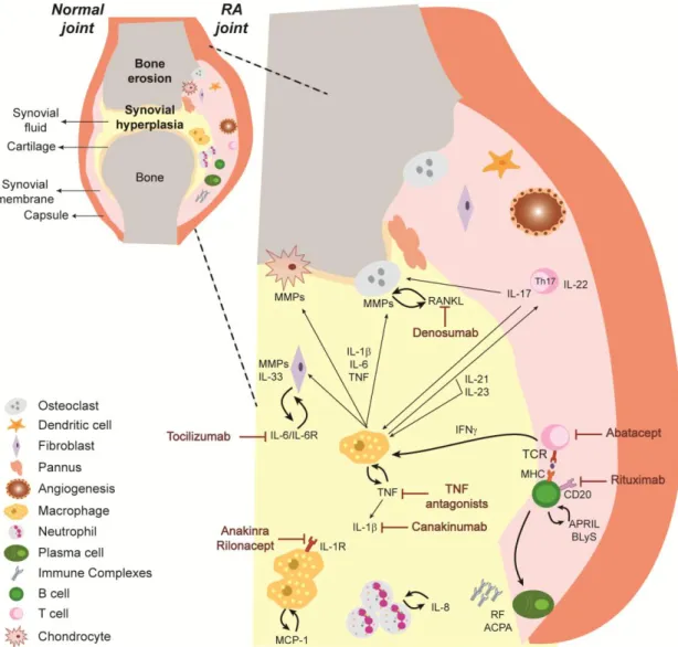
mononuclear cells occurs, namely macrophages, neutrophils, dendritic cells, T cells, B cells and plasma cells. Importantly, synovial-vessel endothelial cells are transformed into high endothelial venules early in the course of the disease, thus promoting the infiltration of inflammatory leukocytes 24 and facilitating the oxygenation of this hypoxic environment 25. Interestingly, several innate immune sensing pathways are found in rheumatoid arthritis joints, namely Toll-like receptors (TLRs), nucleotide-binding oligomerization domain (NOD)-like receptors (NLRs) and molecular components of the inflammasome promoting the detection of tissue damage signals 26, 27. In conclusion, the synovial inflammation characteristic of rheumatoid arthritis is mediated by the interplay of several immune cells and cytokine networks (Figure 1).
Figure 1 – Representative scheme illustrating immune cells and cytokine networks in rheumatoid joints. In rheumatoid arthritis, the inflammatory process leads to a cellular infiltration of the synovial
membrane and synovial fluid, with consequent pannus formation, cartilage and bone destruction. Immune cells are responsible for the secretion of high amounts of several cytokines that amplify inflammation by the recruitment and activation of immune cells. ACPA – Anti-citrullinated protein antibodies, APRIL – A proliferation-inducing ligand, BLyS – B-lymphocyte stimulator, IFNγ – Interferon gamma, IL – Interleukin, IL-6R – IL-6 receptor, MCP-1 – Monocyte chemotactic protein-1, MHC – Major histocompatibility complex, MMP – Metalloproteinase, RANKL – Receptor activator of nuclear factor ligand, RF – Rheumatoid factor, TCR – T cell receptor, TGFβ – Transforming growth factor beta, Th – T helper, TNF – Tumor necrosis factor. (see text for details)
- 8 -
1.3 Immune cells
Rheumatoid arthritis pathophysiology is characterized by the presence of several different interacting immune cells both from the innate immune system, such as macrophages and neutrophils, as well as from the adaptive immune system, namely T and B cells.
1.3.1 Macrophages
Macrophages are pivotal cells in the pathophysiology of rheumatoid arthritis and one of the most abundant cell types in the inflamed synovium. These cells migrate as monocytes from peripheral blood, infiltrate and accumulate in the synovium. Macrophages promote the inflammatory process and joint destruction through the production of several proinflammatory cytokines, such as interleukin (IL)-1β and tumor necrosis factor (TNF), and through osteoclastogenesis 28. Crucial to the mechanism of osteoclastogenesis is the differentiation of monocytes into bone-resorbing osteoclasts, a process mediated by the binding of their receptor activator of nuclear factor (RANK) with its ligand (RANKL) expressed by osteoblasts and some immune cells.
In the synovium, the activation of macrophages can occur by T and B cells, through direct cell-cell contact or indirectly by the production of cytokines 28-30. Moreover, macrophages can also be activated by the immune complexes that deposit in the joints through the low-affinity IgG receptor Fc gamma receptor (FcγR) IIIa 31
. Activation of macrophages is also driven by TLRs and NLRs that recognize a wide range of pathogen associated molecular pattern (PAMP) and danger associated molecular pattern (DAMP) 32.
1.3.2 Neutrophils
Neutrophils are the first to infiltrate the synovium 33. In fact, the synovial tissue of very early rheumatoid arthritis patients can be heavily infiltrated by neutrophils 23. Moreover, circulating neutrophils from these patients have decreased levels of spontaneous apoptosis 34. Synovial neutrophils internalize immune complexes that are present in the joints and become activated. The activated neutrophils release damaging proteases and MMPs, in addition to producing high amounts of reactive oxygen species (ROS), thus contributing to chronic inflammation and host tissue
- 9 -
chemokines, such as IL-1β, IL-8, TNF and B-lymphocyte stimulator (BLyS), which are crucial to amplify inflammation by recruiting and activating more neutrophils and other immune cells such as macrophages, T and B cells 35-37. The delay observed in the apoptosis of neutrophils after rheumatoid arthritis onset can be the result of the action of proinflammatory cytokines, such as TNF, and may create the appropriate conditions for an inflammatory vicious cycle that might contribute to the self-perpetuation of an initial acute arthritis episode in these patients 34.
1.3.3 T cells
The participation of T cells in the etiopathogenesis of rheumatoid arthritis has been supported by several studies 38-43. In fact, the genetic evidence that HLA-DRB1 alleles contribute to rheumatoid arthritis susceptibility suggests a role for T cells in the systemic initiation of the inflammatory process. Moreover, T cells can infiltrate the inflamed synovium and organize themselves into aggregates similar to germinal centre-like structures 38. However, there is still some controversy regarding their precise role. Some studies have shown that T cells do not directly cause synovitis in the joint microenvironment by demonstrating that synovial infiltrating T cells do not show significant proliferation 44, that the phenotype of these cells suggests a recruitment of previously stimulated and mature T cells 45, 46, as opposed to an in situ maturation as previously thought, and also that the synovial cytokine environment has a small contribution of T cell-derived cytokines 47. Therefore, although T cells probably play a part in the systemic initiation of rheumatoid arthritis, their direct role in synovitis and joint destruction is still unclear.
Classically, rheumatoid arthritis has long been classified as a T helper (Th)1 driven disease 41. However, recent data have pointed out that the Th1 contribution was relatively small in the global context of rheumatoid arthritis 48. More recently, this Th1 paradigm was redefined by the increasingly substantiated relevance of the IL-17-secreting Th17 cells subpopulation in the context of this disease. A recent report has demonstrated that self-reactive T cells stimulate antigen-presenting cells to secrete IL-6, which in turn is able to induce the differentiation of naïve self-reactive T cells into Th17 cells 49, via nuclear transcription factor retinoic acid receptor-related orphan receptor gamma (RORγ)T. Other cytokines also participate in the differentiation of Th17 cells such as IL-1β and IL-21. In fact, macrophage-derived and dendritic cell-derived cytokines provide a milieu that supports Th17 differentiation and suppress regulatory T cells polarization, thus shifting T cell homeostasis towards inflammation.
- 10 -
Furthermore, Th17 cells produce IL-17, a cytokine involved in increased production of other cytokines, cartilage-destructive enzymes and also expression of bone destruction-related mediators such as RANKL 50, 51.
1.3.4 B cells
Several reports have also highlighted the participation of B cells in the etiopathogenesis of rheumatoid arthritis 52-57. These cells can contribute to disease onset and perpetuation through their ability to function as antigen-presenting cells to secrete cytokines and to produce autoantibodies, such as RF and ACPA, leading to immune complex formation, which can occur even before the onset of disease symptoms 52. These phenomena might be explained by a deficient removal of autoreactive B cells and an abnormal B cell tolerance checkpoint in the bone marrow that occur in rheumatoid arthritis patients 53. Interestingly, data have shown that peripheral B cells from rheumatoid arthritis patients have altered expression of key molecules, such as high co-stimulatory molecule CD86 and low FcγRIIb (inhibitory receptor for IgG immune complexes required for feedback inhibition), potentially contributing to tolerance breakdown and development of humoral autoimmunity 58.
1.4 Cytokines
Several cytokines have also been implicated in the pathophysiology of rheumatoid arthritis. However, it is still difficult to clearly establish their functional hierarchy and regulatory networks. Specifically, IL-1β, TNF, IL-6 and more recently IL-17 have been identified as key players in established rheumatoid arthritis.
1.4.1 IL-1β
IL-1β is a pleiotropic proinflammatory cytokine produced mainly by macrophages that is crucial for the joint pathology and destruction seen in rheumatoid arthritis 51, 59. Binding of IL-1β to its receptor leads to activation of the mitogen activated protein (MAP) kinase pathway and the nuclear factor kappa-light-chain-enhancer of activated B cells (NF-kB) pathway. Both pathways lead to the downstream secretion of several cytokines, chemokines and MMPs, which are responsible for the inflammatory process and cartilage destruction 59-61. In addition, IL-1β is able to activate several cell types,
- 11 -
such as macrophages, T and B cells, fibroblasts, osteoblasts, endothelial and epithelial cells (Figure 2). IL-1β has been known for a long time to promote T cell responses. For example, IL-1β derived from antigen-presenting cells acts directly on both naïve and memory T cells to enhance their expansion and survival 62. Moreover, increased T cell numbers due to IL-1β also result in an enhanced help for antibody production by B cells
62, thus contributing to rheumatoid arthritis perpetuation. Additionally, recent findings
have also identified a key role for IL-1β in the differentiation of Th17 cells 63, 64, which is
an important T cell subset in rheumatoid arthritis physiopathology. Furthermore, IL-1β together with TNF have been implicated in bone loss by the induction of RANKL expression contributing to osteoclast differentiation via RANK/RANKL interaction 6566. Therefore, IL-1β can be viewed not only as a regulator of innate immune cells but also as a major amplifier of adaptive immune cells.
Importantly, IL-1β is present in high levels in the synovial fluid and tissue of rheumatoid arthritis patients. In fact, some studies suggest that the synovial membrane mRNA levels of IL-1β, TNF and IL-17 are predictors of damage progression 67. In
addition, animal models have demonstrated that targeting of IL-1β and components of its receptor is effective in reducing inflammation, particularly in articular cartilage and bone damage 68, 69. Of note, when transgenic mice expressing human TNF were crossed with IL-1β deficient mice, inflammation still occurred but bone erosions were dramatically reduced 70. Additional studies performed in TNF transgenic mice have also shown that treatment with IL-1 receptor (IL-1R) antagonist fully abolished arthritis even with confirmed high TNF levels, arguing for a possible dominant role for IL-1β 71, 72.
- 12 -
Figure 2 – An overview on the multiple roles of IL-1β in rheumatoid arthritis physiopathology. IL-1β
is a pleiotropic proinflammatory cytokine that modulates the function of several types of cells leading to the secretion of other proinflammatory cytokines and chemokines contributing to cell activation and recruitment to sites of inflammation. Moreover, IL-1β also participates in cartilage and bone destruction promoting the functional impairment characteristic of rheumatoid arthritis patients. IL – Interleukin, MHC – Major histocompatibility complex, MMP – Metalloproteinase, Th – T helper. (see text for details)
1.4.2 TNF
TNF is implicated in the pathogenesis of rheumatoid arthritis, having diverse systemic effects due to its ability to stimulate several cells 51. This proinflammatory cytokine has two receptors; TNF-RI (p55) expressed in most tissues and TNF-RII (p75) expressed in immune cells. Both receptors can act as TNF inhibitors when they are enzymatically cleaved from the cell membrane. TNF is secreted by activated macrophages, neutrophils, T and B cells 73. TNF can contribute to rheumatoid arthritis as a potent inducer of inflammation acting as an autocrine stimulator, as well as a paracrine inducer of other proinflammatory cytokines, such as IL-1β, IL-6 and IL-8 74-76.
- 13 -
facilitating the infiltration of leukocytes into the joints of rheumatoid arthritis patients 77. Furthermore, in vitro studies have already shown that TNF is able to degrade cartilage
78 and bone 79, thus contributing to the debilitating loss of articular joint function.
The serum and synovial fluid concentration of TNF is increased in rheumatoid arthritis patients with active disease 80. It was demonstrated through in vitro studies that cultures of rheumatoid arthritis synovial mononuclear cells are able to spontaneously and chronically produce TNF and other proinflammatory cytokines such as IL-1β, IL-6
and IL-8 81, 82. Transgenic mice expressing a modified human TNFA gene
spontaneously develop peripheral arthritis and show a significant improvement after administration of antibodies specific for human TNF 71. Of note, animal studies have also revealed that blocking TNF is more effective in early stages of arthritis, in contrast to IL-1β, for which the treatment outcome was equally effective in early and established arthritis 69, 83.
1.4.3 IL-6
IL-6 is a multifactorial cytokine that also plays a relevant role in the pathogenesis of rheumatoid arthritis. This cytokine is produced by monocytes, macrophages, T cells and synovial fibroblasts 84. IL-6 possesses diverse biological activities, which include the regulation of immune responses, inflammation, hematopoiesis, hypothalamus-pituitary-adrenal gland axis activity, and the increase of the adrenocorticotropic hormone and cortisol production. The proinflammatory properties of IL-6 comprise the induction of acute-phase proteins in liver cells 85, the increase of neutrophil blood counts 86, the participation in Th17 polarization 49 and plasma B cells differentiation 87,
88
, the increase in the production of chemokines, adhesion molecules 89, and vascular endothelial growth factor (VEGF), which is thought to be the most important angiogenic factor in rheumatoid arthritis 90, as well as the induction of RANKL expression 91 and the production of MMPs and their tissue inhibitors 92-94. These evidences support the concept that IL-6 aggravates the local inflammatory reaction by amplifying inflammatory cell infiltration and by inducing osteoclastogenesis and extracellular matrix turnover.
The serum and synovial fluid concentration of IL-6 is increased in rheumatoid arthritis patients and correlate to disease activity 95, 96. Synovial fibroblastic cells produce augmented levels of IL-6 upon stimulation with inflammatory cytokines, such as IL-1β, TNF and IL-17. Subsequently IL-6 is able by itself to increase the proliferation of these synovial fibroblastic cells 91, 97. The recently shown effective treatment of rheumatoid arthritis patients with the humanized anti-IL-6 receptor antibody
- 14 -
(tocilizumab) 98, 99, which binds to both the soluble and the membrane-bound IL-6 receptors 100, reinforces the important role of IL-6 in the context of this disease. In fact, it has already been observed that tocilizumab treatment not only significantly improves inflammation but also halts radiographic progression 101 in rheumatoid arthritis patients 102.
1.4.4 IL-17
The IL-17 family is composed of 6 members (IL-17A to F) with distinct biological roles. IL-17A (IL-17) is however the most studied one and has long been implicated in several autoimmune diseases. IL-17, produced by Th17 cells, is an important cytokine in the physiopathology of rheumatoid arthritis, mainly due to its participation in joint destruction 103, 104. This cytokine promotes the activation of diverse cells, such as macrophages, synovial fibroblasts, chondrocytes and osteoblasts, and also induces the secretion of proinflammatory cytokines such as IL-1β, TNF, IL-6 and IL-23, which amplify positive feedback loops leading to Th17 cells differentiation 105-107. IL-17 also stimulates the production of chemokines responsible for the recruitment of macrophages, neutrophils, T and B cells to the arthritic joints 108. Additionally, it has been shown that IL-17 can synergize with IL-1β and TNF promoting cartilage and bone damage 109. In fact, this cytokine induces MMPs production by synoviocytes, which are crucial for cartilage destruction 110. Furthermore, in addition to the stimulation of osteoclast-mediated bone resorption, it was recently shown that IL-17 is able to inhibit collagen type I synthesis, further increasing bone destruction 111.
Importantly, IL-17-positive cells were observed in synovial biopsies from rheumatoid arthritis patients 112 and increased levels of IL-17, which correlated with disease activity, were also observed in the serum and synovial fluid samples from these patients 104. Furthermore, in vivo results have also highlighted the relevance of IL-17 in rheumatoid arthritis by showing that the neutralization of IL-17 in mice decreased the severity of antigen-induced and collagen-induced arthritis and that IL-17-deficient mice presented reduced disease severity 113, 114.
1.5 Treatment options
The presentation and the clinical course of rheumatoid arthritis patients as well as the response to the available treatment options are highly variable. Despite the
- 15 -
existence of several treatment options for the management of rheumatoid arthritis, this is still an incurable disease with enormous personal and economic costs. The strategy of disease treatment relies on an early diagnosis and a prompt adequate treatment with the goal of inducing remission in order to preserve joint function and maintain quality of life 115-117. Drug-free remission is at this moment impossible for most patients. Rheumatoid arthritis treatments can be divided into the following categories: glucocorticoids, conventional disease-modifying anti-rheumatic drugs (DMARDs) and biologic DMARDs.
1.5.1 Glucocorticoids
Glucocorticoids (namely prednisone) act rapidly to suppress inflammation and, consequently, pain and swelling 118. Glucocorticoids are useful to control symptoms in the first weeks of disease diagnosis, until slower-action DMARD can start to have an effect. Glucocorticoids can have an effect in limiting joint damage, but long term adverse effects, particularly at higher doses, limits their usefulness 119.
1.5.2 Conventional DMARDs
Conventional DMARDs, which include for instance methotrexate (MTX), hydroxychloroquine, sulfasalazine and leflunomide, have long been used as the main therapeutic option for newly diagnosed rheumatoid arthritis 118, 120. Among the different DMARDs, MTX is the most frequently used and the most likely to induce a long-term response 121-123. DMARDs have a relatively slow onset of action (1-6 months) but they are effective in the long term and are considered to have an acceptable safety 118, 124 either as monotherapy or in combination therapy. However, around 30% of patients are either non-responsive to DMARDs or lose their response secondarily or even develop adverse effects that are incompatible with their use 125, 126 127. Therefore, the recent development of biologic DMARDs is a relevant therapeutic alternative for these DMARD refractory patients.
1.5.3 Biologic DMARDs
Nine different biologic DMARDs are currently approved by the European Medicines Agency and many others are being tested in clinical trials for the treatment of rheumatoid arthritis patients with inadequate response to one or more non-biologic
- 16 -
DMARDs (Figure 3). In rheumatoid arthritis, biologic DMARDs have shown efficacy in improving clinical, functional and radiographic outcomes 117, 128, especially if administrated early in the disease course 116.
The first line of biologic DMARDs is the TNF antagonist class. TNF antagonists approved for the treatment of rheumatoid arthritis include monoclonal antibodies against TNF (infliximab, adalimumab, certolizumab pegol and golimumab) and a soluble TNF receptor (etanercept). All TNF antagonists have similar efficacy and the combination therapy with a TNF antagonist plus MTX has been proven to be superior to the TNF antagonist alone 116, 129, 130. When patients fail to respond to a TNF antagonist they have approximately a 50% chance of responding to a second one, but the probability of efficacy of a third switch is very low 119. There are also approved biologic DMARDs that target specific immune cells such as abatacept (T-cell co-stimulation blocker) and rituximab (anti-CD20 monoclonal antibody for B-cell depletion). Both these drugs, given in combination with MTX, may be useful alternatives for patients who are non-responders to conventional DMARDs (rituximab is not formally approved in that indication) or TNF antagonists 131, 132 and have already shown the ability to inhibit progression of structural damage 133-135. Treatment of rheumatoid arthritis patients with tocilizumab (humanized anti-IL-6 receptor monoclonal antibody) is highly effective 98, 99, with reduced systemic inflammation, synovitis and radiographic progression. This biologic DMARD can be used in the setting of DMARD or TNF antagonist failure 101. There are also biologic agents targeting the IL-1β receptor (anakinra, an IL-1β receptor antagonist), blocking the circulating IL-1β (rilonacept, a fusion protein composed of the IL-1β receptor and IgG) and binding to IL-1β directly (canakinumab, a monoclonal antibody against IL-1β) 136. Specifically, anakinra is
approved for rheumatoid arthritis treatment and it was shown to be effective albeit at a lower efficacy than a TNF antagonist 137, 138. Rilonacept and canakinumab are still undergoing clinical trials but they seem to have also lower efficacy as compared to TNF antagonists 139. Interestingly, denosumab (antibody against RANKL) was also recently shown to significantly reduce structural damage, but had no effect on the clinical signs and symptoms of rheumatoid arthritis 140.
Switching among biologic DMARDs is often considered in patients with inadequate response to initial treatment and the selection of the second biologic DMARD depends in most cases on the reason of the first failure (e.g. a second TNF antagonist is less effective when the first TNF antagonist failed due to lack of efficacy instead of adverse events), but this rationale is somehow empirical and needs further studies 141, 142. Overall, biologic DMARDs have been well tolerated by patients, despite the occurrence
- 17 -
of infections and injection-site reactions, which are among the most common adverse effects. Curiously, these types of problems occur with a similar frequency among the different biologics 143. Moreover, the formation of antibodies to biologic agents is a great concern since it can decrease the efficacy and increase the adverse effects of the treatment. Of note, biologic therapies do not achieve optimal response in all patients and even those who initially respond to treatment may experience diminished efficacy with time. In addition, remission is only possible in 30% of the patients. Therefore, searching for new, more effective and safer treatments for rheumatoid arthritis is still a current medical priority.
Figure 3 – A simplified scheme showing main pathophysiologic pathways in rheumatoid arthritis and the effects of some currently available biologic agents. Treatment options for rheumatoid arthritis
have been developed to stop or at least attenuate disease progression. Recently, several biologic therapeutics, targeting immune cells or cytokines, have been introduced for the treatment of rheumatoid arthritis. ACPA – Anti-citrullinated protein antibodies, APRIL – A proliferation-inducing ligand, BLyS – B-lymphocyte stimulator, IFNγ – Interferon γ, IL – Interleukin, IL-1R – IL-1 receptor, IL-6R – IL-6 receptor, MCP-1 – Monocyte chemotactic protein-1, MHC – Major histocompatibility complex, MMP – Metalloproteinase, RANKL – Receptor activator of nuclear factor ligand, RF – Rheumatoid factor, TCR – T cell receptor, TGFβ – Transforming growth factor β, Th – T helper cell, TNF – Tumor necrosis factor. (see text for details)
- 18 -
2. IL-1β Immunological and inflammatory functions
2.1 IL-1 family
IL-1β is the most well characterized cytokine among the 11 members of the IL-1 family. The IL-1 family also comprises IL-1α, IL-1 receptor antagonist (IL-1Ra), IL-18, IL-33 and IL-1F5 to IL-1F10 144. IL-1β, as discussed above, is a pleiotropic proinflammatory cytokine with immuno- and inflammatory-modulating activities that are crucial for host responses to infection and also to injuries and immunological challenges 145. Several cell types produce this cytokine, although its main source are cells from the innate immune system, such as monocytes and macrophages 145, 146. Under normal conditions IL-1β is expressed at low levels, however, in the presence of an inflammatory stimulus, its secretion increases. The production of IL-1β requires induction in both transcriptional and post-translational steps 145, 146. This dual mechanism responsible for the production of IL-1β is tightly regulated and ensures that its secretion only occurs when it is required, thus preventing an unnecessary exacerbated inflammatory response. The loss of these regulatory steps results in fever, rash and arthritis, characteristic of autoinflammatory syndromes, such as cryopyrinopathies 148. Gout is also associated with altered IL-1β regulation, induced by the stimulus of uric acid crystals 147. Some autoimmune diseases are also likely to be at least partially dependent on IL-1β 149.
2.2 IL-1β pathway
2.2.1 Priming
The production of IL-1β requires two signals, one for IL-1β gene expression and the second for the completion of protein synthesis 150 (Figure 4). IL-1β is expressed as an inactive 31 kDa precursor, designated as pro-IL-1β, in response to PAMPs or cytokines, such as TNF and IL-1β itself, which signal through pattern recognition receptors (PRRs) present in macrophages 151. These signaling pathways lead to the activation of NF-kB, which induces the transcription of IL-1β promoter 152. This
induction of pro-IL-1β gene expression is commonly referred to as the priming step. However, it does not act as a secretion stimulus. Instead, the primed cell must encounter a new PAMP or DAMP to induce the processing and secretion of the active IL-1β.
- 19 -
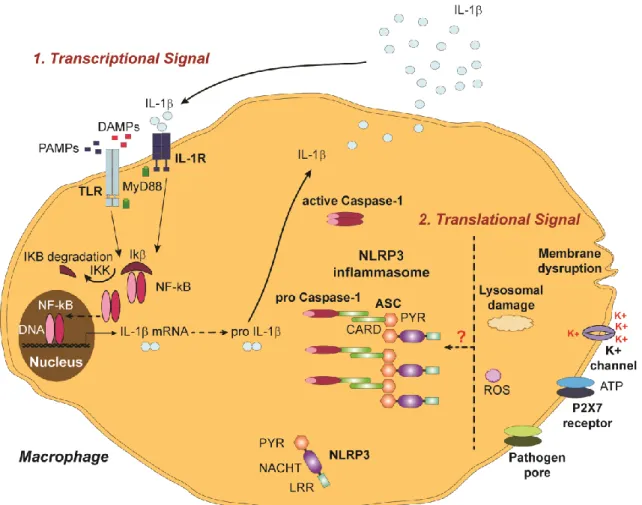
Figure 4 – IL-1β processing pathways. The secretion of mature IL-1β involves a dual mechanism at the
level of IL-1β priming and at the level of its processing/activation, which is tightly regulated in order to prevent uncontrolled and unnecessary exacerbated inflammatory responses. IL-1β priming is mainly regulated by NF-kB and IL-1β processing is strongly regulated by caspase-1 and inflammasome complexes. ASC – Adaptor molecule apoptosis associated speck-like protein containing a caspase recruitment domain, ATP – Adenosine-5'-triphosphate; CARD – Caspase recruitment domain, DAMP – Danger associated molecular pattern, IL – Interleukin, IL-1R – IL-1 receptor, LRR – Leucine reach repeats, MyD88 – Myeloid differentiation primary response gene 88, NACHT – Nucleotide-binding domain or NAIP, CIITA, HET-E and TP1, NF-kB – Nuclear factor kappa-light-chain-enhancer of activated B cells, PAMP – Pathogen associated molecular pattern, PYR – Pyrin domain, TLR – Toll-like receptor, ROS – Reactive oxygen species. (see text for details)
2.2.2 Processing
The 31 kDa pro-IL-1β must be processed to become a 17 kDa active IL-1β that can be secreted by the cell. The pro-IL-1β is cleaved by the intracellular proinflammatory cysteine protease caspase-1 153.
Caspases are also expressed as inactive proenzymes of 30-50 kDa and consist of an N-terminal prodomain followed by a 20 kDa and a 10 kDa conserved domains. The proteolytic activation of caspases includes the cleavage between these two domains and subsequent assembly of two heterodimers each one containing a p20 and p10 subunits and lacking the prodomain 154. This activation of caspase-1 itself occurs via
- 20 -
recruitment to a large multiprotein complex, termed inflammasome, which is present in the cytosol 155.
The inflammasome is a molecular scaffold composed of a cytosolic NLR, adaptor molecules and pro-caspase-1 156. The most well characterized inflammasome is the NLRP3 (also known as cryopyrin or NALP3) inflammasome. The NLRP3 receptor is comprised of a C-terminal PAMP/DAMP sensing leucine rich repeat (LRR), a central nucleotide binding NACHT domain and an N-terminal pyrin domain (PYD) 156. The NACHT (nucleotide-binding domain or NAIP, CIITA, HET-E and TP1) domain is responsible for the oligomerization and, consequently, activation of the inflammasome. The PYD domain of the NLRP3 is responsible for the recruitment of the adaptor molecule, apoptosis associated speck-like protein containing a caspase recruitment domain (ASC) or CARDINAL, by homotypic interactions between their PYD domains. The NLRP3 inflammasome cannot recruit pro-caspase-1 without the adaptor molecule ASC, because it lacks a caspase recruitment domain (CARD). Therefore, pro-caspase-1 is recruited to ASC, by a homotypic interaction of CARD domains, facilitating caspase-1 autocatalytic activation. The activation of the inflammasome itself requires two signals. The first signal is the priming with TLR and NLR ligands or proinflammatory cytokines that enhance NF-kB-driven transcription of inflammasome components 157-159. The NLRP3 is expressed in myeloid cells and is upregulated in response to a diverse panel of agonists. These agonists include exogenous / foreign stimulus, such as asbestos, silica, alum, pathogens and pore-forming toxins, and also endogenous / self-stimulus, such as monosodium urate crystals and calcium pyrophosphate crystals 147, 160-162. The second signal is the activation and assembling of the inflammasome, where ROS 163, 164, K+ efflux 165, pore formation at the cellular membrane and lysosomal disruption 166, 167 seem to play a role. Despite the current limited insight into the precise mechanism of NLRP3 inflammasome activation, it is becoming increasingly evident that one signal alone is insufficient to induce its activation. Also crucial for the regulation of inflammasome activation is its auto-inhibition, which limits the occurrence of an aberrant inflammatory response. To date, some mechanisms have been pointed out as possible pathways to inhibit inflammasome activation, such as NLRs alternative splicing, ASC isoforms 168 and pyrin 169.
Interestingly, there is also evidence that IL-1β activation can be mediated by pathways independent of NLRP3 and caspase-1. For example, it was already demonstrated that neutrophil proteases, such as proteinase 3, are able to cleave pro-IL-1β, in the absence of NLRP3 and caspase-1 activation 170. Data from animal models