Marco André Moras Videira
Dissertation presented to obtain the Ph.D degree in
Biochemistry
Instituto de Tecnologia Química e Biológica António Xavier| Universidade Nova de Lisboa
Supervisor: Dr. Lígia M. Saraiva
Co-supervisor: Dr. Susana A. L. Lobo
Oeiras, May 2018
Staphylococcus aureus: the
paths and crosstalks that
lead to heme
From left to right: Maria Arménia carrondo, Susana Lobo, Lígia Saraiva, Carlos Salgueiro, Marco Videira, Maria Salomé Gomes, Mariana Pinho,
Arsénio Fialho.
14th of May 2018
Third Edition, May 2018
Molecular Mechanisms of Pathogen Resistance Laboratory
Instituto de Tecnologia Química e Biológica
Universidade Nova de Lisboa
Acknowledgments
I would like to express my gratitude to the people that contributed to this work and made this thesis possible:
To my supervisor, Dr. Lígia Saraiva, for inviting me to apply for a PhD student grant and accepting me as member of her group. For her mentoring, constant support, motivation, advices and ideas. For her availability and for pointing the way when it was necessary. Thank you for the suggestions, critics and careful revision of this thesis. I also want to thank you for your friendship and making me a better person. Thank you very much.
To my co-supervisor, Susana Lobo, for her friendship, guidance and helpful brainstorms. For teaching me since the first day I arrived at the lab. And not less important for the many rides home when I was not coming to the lab by car! Thank you.
To my PhD Thesis Committee, Prof. Miguel Teixeira and Dr. Ana Melo, for accepting being part of the committee. For the availability for the meetings and for the discussions. A special thanks to Ana Melo for receiving me in her lab in my degree in biochemistry, showing me how to take my first steps in scientific research and for introducing me to Dr. Lígia Saraiva.
I want to thank Fábio Fernandes and Ana countinho, from Instituto Superior Técnico, for their collaboration in FLIM-FRET and fluorescent anisotropy experiments.
To Martin Warren and Dave Palmer for the collaboration in HPLC-MS experiments, for suggestions and experimental ideas and for the revision of the manuscripts.
I want to thank all the present and former members of our lab, Molecular Mechanisms of Pathogen Resistance, specially Lígia Nobre, Margarida Parente, João Monteiro, Liliana Silva, Sandra Carvalho, Joana Marques, Cátia Família and Patrícia Ferreira for your friendship, helpful discussions, for the laughs and for keeping the good environment in the lab throughout these years.
I also want to thank to ITQB for the facilities and for making fell a home.
To Fundação para a Ciência e Tecnologia (FCT) for the financial support and for awarding me a PhD grant (SFRH/BD/95912/2013).
À minha família pelo carinho e apoio constante ao longo destes anos. Principalmente aos meus pais porque me possibilitaram o meu desenvolvimento científico e pessoal.
Quero agradecer à Camila, minha mulher, pela ajuda preciosa que me deu! Pelo apoio e carinho constante mesmo nos momentos mais difíceis. Pelas discussões e paciência ao longo destes anos. Obrigado por me fazeres feliz!
Thesis Publications
This dissertation is based on the following original publications, listed by chronological order:
Lobo SAL, Scott A, Videira MAM, Winpenny D, Gardner M, Palmer MJ, Schroeder S, Lawrence AD, Parkinson T, Warren MJ, Saraiva LM (2015) Staphylococcus aureus haem biosynthesis: characterisation of the enzymes involved in final steps of the pathway. Mol Microbiol 97(3):472–487.
Videira MAM, Lobo SAL, Silva LSO, Palmer DJ, Warren MJ, Prieto M,
Coutinho A, Sousa FL, Fernandes F, and Saraiva LM (2018) Staphylococcus aureus haem biosynthesis and acquisition pathways are linked through haem monooxygenase IsdG. Submitted
Manuscript in preparation, based on results presented in Chapter 6:
Videira MAM, Família C, Lobo SAL and Saraiva LM (2018) Characterization
of siroheme synthesis in Staphylococcus aureus.
Publications not included in this thesis:
Lobo SAL, Videira, MAM, Pacheco, I, Wass MN., Warren, MJ., Teixeira M, Matias PM & Saraiva, LM. (2017). Desulfovibrio vulgaris CbiKP cobaltochelatase: evolution of a haem binding protein orchestrated by the incorporation of two histidine residues. Environ microbiol, 19 (1), 106-118.
Abstract
S. aureus is one of the leading causes of nosocomial infections and nowadays a major public health concern due to the increase in antibiotic resistant strains. During host invasion, S. aureus is exposed to different oxygen concentrations depending on the site of infection. When oxygen is available, S. aureus uses heme-dependent terminal oxidases for aerobic respiration. However, some locals of infection are oxygen-limited and in this case S. aureus has to use other substrates such as nitrate or nitrite as terminal electron acceptors of an anaerobic respiration chain, in which siroheme-containing enzymes are active. Also during infection, S. aureus uses the heme uptake system to scavenge heme from host hemoglobin and utilize it as iron source. In S. aureus, although the heme uptake system have been intensively studied, the knowledge on the biosynthesis of heme and siroheme remains incomplete. To better understand the metabolism that guarantees S. aureus survival during the infectious processes, this thesis elucidates the way S. aureus synthesizes these two tetrapyrroles. Herein, we Tetrapyrroles are biological molecules widespread through all living organisms and necessary for fundamental cellular processes. Within the wide range of known tetrapyrroles, heme and siroheme are prosthetic groups in proteins responsible for important functions such as gas transport and storage, electron transport, cellular signaling, cellular detoxification as well as for reduction of sulfite and nitrite. Both molecules are synthesized from a last common tetrapyrrole precursor, namely uroporphyrinogen III, forming heme via four enzymatic steps know as the classical heme biosynthesis pathway and generating siroheme via an independent route. In some organisms such as sulfate and nitrate reducing bacteria, heme is alternatively synthesized through the siroheme-dependent pathway.
provide new insights into the heme metabolism of S. aureus through the comprehensive study of three topics: (i) the characterization of the heme biosynthesis pathway of S. aureus and its possible utilization as an antimicrobial target; (ii) clarification on how S. aureus is able to fine-tune the heme that is either synthesized de novo or acquired from the host; and (iii) characterization of the siroheme biosynthesis pathway of S. aureus.
The analysis of S. aureus genome revealed the presence of genes encoding putative enzymes involved in the late steps of heme biosynthesis pathway, namely HemE, HemN, HemY and HemH. The role of these enzymes in the heme biosynthesis pathway of S. aureus was addressed by overexpression, purification and characterization of the enzymes. The results have shown that in S. aureus, the coproporphyrinogen III produced by uroporphyrinogen III decarboxylase HemE is converted to coproporphyrin III by coproporphyrinogen III oxidase HemY, which is then used by coproporphyrin ferrochelatase HemH for iron insertion into the macrocycle, leading to the formation of iron-coproporphyrin III. A S. aureus strain deleted in hemH was constructed and shown to accumulate coproporphyrin III. It was demonstrated that HemY oxidizes coproporphyrinogen III with a specific activity of 24.6 nmol.min-1mg-1 and HemH inserts iron into coproporphyrin III
with an activity of 815 nmol.min-1mg-1. It was shown that the last step of this
pathway is the decarboxylation of coproporphyrin III by the iron-coproporphyrin decarboxylase HemQ. This pathway represents a new transitional route between to previously described pathways, the classical and the siroheme-dependent pathways.
On the search for inhibitors of the pathway, HemY was shown to be impaired by derivatives of diphenyl-ether herbicides making this enzyme a promising candidate for drug discovery, which could be important for tackling the problem of antibiotic resistance.
The second part of the work addressed the link between the systems for heme uptake and heme biosynthesis of S. aureus. Both routes are known
to supply heme to S. aureus, and thus a regulation mechanism was hypothesized to occur to ensure heme homeostasis, so that the toxicity associated with elevated concentration of heme is avoided. To pursue this hypothesis, the late steps of S. aureus heme biosynthesis pathway were reconstituted in an E. coli strain unable to synthesize heme and also devoid of S. aureus heme oxygenases IsdG and IsdI. The results have shown that IsdG, but not IsdI, impairs the growth of the complemented E. coli strain by hampering heme synthesis. IsdG was shown to inhibit the ferrochelatase activity of HemH in a concentration dependent manner. It was shown by fluorescence anisotropy and also FLIM-FRET that IsdG interacts in vitro and in vivo with S. aureus HemH. It was also shown through RT-qPCR and flow cytometry that the cellular content of IsdG is higher in the presence of heme, which suggests that heme increases the content of IsdG promoting interaction with the ferrochelatase enzyme of the heme biosynthesis pathway which results in inhibition of the endogenous produced heme. Furthermore, an in silico analysis have revealed that one third of the genomes encoding heme biosynthesis pathway also contains a gene for an IsdG-type heme oxygenase. This analysis also revealed that IsdG homologues are normally encoded in genomes also containing HemH and/or HemQ. Altogether, this part of the work has demonstrated that bacteria are able to regulate heme homeostasis by linking the heme biosynthesis pathway and the heme uptake system through HemH and IsdG.
Upon infection of the host, S. aureus utilizes the siroheme-containing protein nitrite reductase to reduce nitrite, either using it as part of the respiratory chain to accept electrons when exposed to low oxygen environments or for the detoxification of nitrite produced by neighboring and host enzymes. Analysis of S. aureus genome showed that it contains three genes that may be involved in siroheme synthesis, which two were annotated as cysG in accordance to the siroheme synthase of E. coli. One cysG is located on the nirBD operon and the other cysG is separately localized in a
cluster downstream cysJ. The two gene products were overexpressed, the proteins purified, and in vitro and in vivo assays were performed to analyze their role in S. aureus. Enzymatic assays have shown that the protein encoded by the cysG gene downstream nirBD acts as uroporphyrinogen III methyltransferase (SirA), generating precorrin-2, and the protein encoded by the cysG gene downstream cysJ acts as a precorrin-2 dehydrogenase (SirC), which is able to produce sirohydrochlorin when incubated with precorrin-2 and NAD+. Since one last step was missing and no apparent protein was
present, a new search for a putative sirohydrochlorin ferrochelatase (SirB) was made. Blast analysis revealed the occurrence of a gene annotated as nirR which shares 25% sequence identity and 44% similarity to B. megaterium SirB. Following cloning and expression, the purified protein was shown to act as sirohydrochlorin ferrochelatase. Moreover, functional complementation experiments were performed, where S. aureus SirB in combination with SirA and SirC was able to complement an E. coli strain lacking cysG and thus unable to synthesize siroheme. Therefore, this protein constitutes the last step of the pathway that generates the final product siroheme. Altogether the siroheme pathway active in S. aureus was established for the first time.
To conclude, the results presented in this thesis contribute to a better understanding of the S. aureus physiology and metabolism, by elucidating the way in which the life dependent tetrapyrroles heme and siroheme are formed. Not less important, it was shown how S. aureus maintains the adequate levels of heme avoiding the excess of intracellular free toxic heme that would challenge the pathogen survival.
Resumo
Os Tetrapirróis são moléculas biológicas presentes em todos os organismos vivos e necessários para processos celulares fundamentais. Entre os tetrapirróis mais conhecidos, o hemo e o sirohemo são grupos prostéticos de proteínas com funções importantes, tais como o transporte e armazenamento de gases, o transporte de eletrões, a sinalização celular, a desintoxicação celular e também a redução de sulfito e nitrito. As duas moléculas são sintetizadas a partir de um percursor final comum, o uroporfirinogénio III, onde a formação de hemo ocorre através de quatro reações enzimáticas pela via clássica de biossíntese de hemo e produzindo sirohemo através de uma via independente. Em alguns organismos como bactérias redutoras de sulfato e nitrato, a síntese do hemo ocorre através de uma via dependente de sirohemo.S. aureus é uma das principais causas de infeções nosocomiais e atualmente, é considerado um grave problema de saúde pública devido ao aumento de estirpes resistentes aos antibióticos. Durante a infeção no hospedeiro, S. aureus está exposto a mudanças na concentração de oxigénio nos diferentes locais de infeção. Quando existe oxigénio disponível, S. aureus utiliza oxidases terminais dependentes de hemo para a respiração aeróbia. Contudo, alguns locais de infeção têm concentrações limitadas de oxigénio e neste caso S. aureus utiliza outros substratos, tais como o nitrato ou o nitrito, como aceitadores finais de eletrões numa cadeia respiratória anaeróbia, em que estão ativas enzimas que contêm sirohemo. Também durante a infeção, S. aureus recolhe hemo presente na hemoglobina do hospedeiro, através do sistema de aquisição do hemo, e utiliza-a como fonte de ferro. Embora o sistema de aquisição de hemo tenha sido intensamente estudado em S. aureus, o conhecimento acerca da síntese do hemo e
sirohemo é limitado. Para melhor compreender o metabolismo que permite a sobrevivência de S. aureus durante os processos de infeção, esta tese pretende elucidar as etapas essenciais para a síntese destes dois tetrapirróis em S. aureus. Nesta dissertação, apresentamos novas perspetivas sobre o metabolismo do hemo em S. aureus através do estudo abrangente de três tópicos: (i) a caracterização da via de biossíntese do hemo de S. aureus e a sua possível utilização como alvo antimicrobiano; (ii) a compreensão de como S. aureus equilibra o hemo que é sintetizado de novo ou adquirido através hospedeiro; e (iii) a caracterização da via de biossíntese de siroheme de S. aureus.
Através da análise do genoma de S. aureus verificou-se a presença de genes que codificam enzimas que poderão estar envolvidas nos passos tardios da via de biossíntese do hemo, nomeadamente a HemE, HemN, HemY e HemH. Determinou-se o papel destas enzimas na via de biossíntese do hemo de S. aureus através da sua sobreexpressão, purificação e caracterização. Os resultados mostraram que em S. aureus, o coproporfirinógenio III produzido pela uroporfirinogénio III descarboxilase HemE é convertido em coproporfirina III pela coproporfirinógenio III oxidase HemY, sendo posteriormente utilizado pela HemH para inserção de ferro no macrociclo, levando à formação de ferro-coproporfirina III. Foi construída uma estirpe de S. aureus mutante no gene hemH e mostrou-se que esta acumula coproporfirina III. Foi demonstrado que a HemY oxida o coproporfirinógeno III com uma atividade específica de 24,6 nmol.min-1mg-1
e a HemH insere ferro na coproporfirina III com uma atividade de 815 nmol.min-1mg-1. Mostrou-se também que o último passo é a descarboxilação
de ferro-coproporfirina III pela ferro-coproporfirina descarboxilase HemQ. Esta via representa um novo caminho transicional entre duas vias de biossíntese do hemo descritas anteriormente, a via clássica e a dependente de sirohemo.
Através da pesquisa por inibidores para esta via, demonstrou-se que a HemY é inibida por derivados de herbicidas de difenil-éter que tornam esta enzima um candidato promissor para descoberta de fármacos, o que pode ser importante para enfrentar o problema da resistência a antibióticos.
Na segunda parte do trabalho, estudou-se a associação entre o sistema de aquisição de hemo e a via de biossíntese do hemo em S. aureus. As duas vias são conhecidas por fornecer hemo a S. aureus, e, portanto, colocou-se a hipótese de existir um mecanismo de regulação para garantir a homeostase do hemo, de modo a que a toxicidade associada às elevadas concentrações do hemo seja evitada. Para investigar esta hipótese, os últimos passos da via de biossíntese de hemo em S. aureus foi reconstituída numa estirpe de E. coli incapaz de sintetizar hemo, e em que também estão ausentes as oxigenases de hemo IsdG e IsdI de S. aureus. Os resultados mostraram que a IsdG, mas não a IsdI, prejudica o crescimento da estirpe de E. coli complementada, dificultando a síntese de hemo. Demonstrou-se que a IsdG inibe a atividade de ferroquelatase da HemH estando dependente da sua concentração. Mostrou-se por anisotropia de fluorescência e também por FLIM-FRET que a IsdG interage in vitro e in vivo com a HemH de S. aureus. Também se mostrou através de RT-qPCR e citometria de fluxo que o conteúdo celular de IsdG é maior na presença de hemo, o que sugere que o hemo aumenta o conteúdo de IsdG para promover a interação com a enzima ferroquelatase da via de biossíntese do hemo, resultando na inibição na produção endógena de hemo. Além disso, uma análise in silico revelou que um terço dos genomas que codificam a via de biossíntese do hemo também codificam uma oxigenase de hemo do tipo IsdG. Esta análise também revelou que homólogos da IsdG são normalmente codificados em genomas que também contêm HemH e / ou HemQ. Em conclusão, esta parte do trabalho demonstrou que as bactérias são capazes de regular a homeostase do hemo por ligação da via de
biossíntese do hemo e do sistema de aquisição de hemo através da HemH e da IsdG.
Após a infeção do hospedeiro, S. aureus utiliza a proteína reductase de nitrito, que contêm sirohemo, para reduzir nitrito, utilizando-a como parte da cadeia respiratória para aceitar eletrões quando em ambientes com baixo oxigénio ou para destoxificação de nitrito produzido por enzimas próximas ou do hospedeiro. Através da análise do genoma verificou-se que S. aureus contém três genes que podem estar envolvidos na síntese de sirohemo, sendo que dois destes genes estão anotados como cysG conforme a síntase de sirohemo de E. coli. Um dos genes cysG está localizado no operão nirBD enquanto que o outro gene cysG está localizado separadamente a jusante do gene cysJ. Os produtos dos dois genes foram sobreexpressos, as proteínas foram purificadas e foram efetuados ensaios in vitro e in vivo para analisar o seu papel em S. aureus. Através de ensaios enzimáticos mostrou-se que a proteína codificada pelo gene cysG a jusante do gene nirBD atua como uma uroporfirinogénio III metiltransferase (SirA), ao gerar precorrina-2 e o gene cysG a jusante cysJ atua como uma precorrina-precorrina-2 desidrogenase (SirC), que é capaz de produzir sirohidroclorina quando incubada com precorrina-2 e NAD+. Uma vez que faltava um passo para a produção de
sirohemo e aparentemente nenhuma proteína foi encontrada, efetuou-se uma nova busca por uma proteína ferroquelatase de sirohydroclorina (SirB) hipotética. A análise Blast revelou a presença de um gene anotado como nirR que compartilha 25% de identidade e 44% de similaridade com a SirB de B. megaterium. Após a clonagem e expressão, mostrou-se que a proteína atua como uma ferroquelatase de sirohidroclorina. Além disso, foram feitas experiências de complementação funcional onde se demostrou que a SirB de S. aureus em combinação com a SirA e a SirC é capaz de complementar uma estirpe de E. coli sem cysG e, portanto, incapaz de sintetizar sirohemo. Portanto, esta proteína constituí o último passo da via que sintetiza o produto
final sirohemo. Em suma foi mostrado pela primeira vez uma via de síntese do sirohemo em S. aureus.
Para concluir, os resultados apresentados nesta tese contribuem para uma melhor compreensão da fisiologia e do metabolismo de S. aureus, elucidando o caminho pelos quais os tetrapirrois, hemo e o sirohemo são formados. Não menos importante, mostrou-se como S. aureus mantém níveis de hemo adequados evitando o excesso de hemo intracelular livre que pode comprometer a sobrevivência do patogénico.
Abbreviations
δ-ALA - δ-aminolevulinic acidABM - antibiotic biosynthesis oxygenase Ahb - Alternative heme biosynthesis BHI - Brain Heart Infusion
CA-MRSA - Community-acquired MRSA
ChrA-ChrS - Corynebacterium heme response system CO – Carbon monoxide
Copro III - Coproporphyrin III Copro´gen III - Coprophyrinogen III CPD - Coproporphyrin-dependent CydAB - bd-type menaquinol oxidase CysG - Siroheme synthase
DDSH – Didecarboxysiroheme
DMEM - Dulbecco’s modified Eagle’s medium FAD- flavin adenine dinucleotide
FLIM-FRET - Lifetime imaging based Förster resonance energy transfer FnrL - Fumarate nitrate reductase regulator
Fur - Ferric uptake regulator H2O2- Hydrogen peroxide
HA-MRSA - Hospital-acquired MRSA Hb- Hemoglobin
HMB- Hydroymethylbilane HO- Heme oxygenase
HRMs -Heme regulatory motifs HrrA-HrrS - heme response regulator
HrtAB - Heme-regulated transporter system HssRS -Heme sensor system
Hts- Heme transport system
IC50- half maximal inhibitory concentration Isd - Iron-regulated surface determinant system LB – Luria-Bertani
Lqo - L-lactate dehydrogenase
MpsABC - Membrane potential-generating system MQ - Menaquinone
MQH2 – Menaquinol
Mqo - Malate dehydrogenase
MRSA - Methicillin-resistant Staphylococcus aureus MS – Mass spectrometry
NarGHI - Nitrate reductase
NDH2- Nicotinamide adenine dinucleotide NADH dehydrogenases type 2 NEAT - Near-Iron Transport
NirBD - Nitrite reductase
NreABC - Nitrogen regulation system PBG – Porphobilinogen
Phu - Pseudomonas heme uptake PPD - Protoporphyrin-dependent Proto IX – Protoporphyrin IX
PrrA - photosynthetic response regulator QoxABCD - aa3/ba3-type menaquinol oxidase ROS -Reactive oxygen species
SCV - Small colony variants SAM - S-adenosyl-L-methionine Sdh - Succinate dehydrogenase SHD - Siroheme-dependent
SUMT- Uroporphrinogen III methyltransferase TIM - Triosephosphate Isomerase
TSA - Tryptic soy agar TSB - Tryptic soy broth
Uro´gen III- Uroporphrinogen III
VISA - Vancomycin-intermediate S. aureus VRSA - Vancomcyin-resistant S. aureus
Table of Contents
Acknowledgments ... iii
Thesis Publications ...v
Abstract ... vii
Resumo ... xi
Abbreviations list ... xvii
Table of Contents ... xxi
Introduction
Chapter 1: Staphylococcus aureus
1.1 General features ... 5
1.2 Pathogenesis, clinical relevance and antibiotic resistance ... 6
1.3 S. aureus metabolism ... 9
1.4 References ... 12
Chapter 2: Tetrapyrrole biosynthesis in prokaryotes
2.1 Importance of tetrapyrroles ... 19
2.2 Heme ... 21
2.2.1 Heme biosynthesis ... 24
2.2.2 Regulation of heme biosynthesis ... 30
2.4 References ... 35
Chapter 3: Heme acquisition in prokaryotes
3.1 Heme uptake systems ... 49
3.1.1 Gram-negative heme uptake systems ... 50
3.1.2 Gram-positive heme uptake systems ... 53
3.2 Heme oxygenases ... 56
3.3 References ... 60
Results
Chapter
4:
Staphylococcus
aureus
heme
biosynthesis
characterisation of the enzymes involved in final steps of the
pathways
4.1 Summary ... 73
4.2 Introduction ... 73
4.3 Materials and Methods ... 77
4.4 Results ... 88
4.5 Discussion ... 104
4.6 Acknowledgements ... 108
4.7 References ... 109
4.8 Supplementary data ... 115
Chapter 5: Staphylococcus aureus heme biosynthesis and
acquisition pathways are linked through heme oxygenase IsdG
5.1 Summary ... 127
5.2 Introduction ... 127
5.3 Materials and Methods ... 130
5.4 Results ... 146
5.5 Discussion ... 160
5.6 Acknowledgements ... 165
5.7 References ... 165
5.8 Supplementary data ... 172
Chapter 6: Characterization of siroheme biosynthesis in
Staphylococcus aureus
6.1 Summary ... 179
6.2 Introduction ... 180
6.3 Materials and Methods ... 181
6.4 Results ... 190
6.5 Discussion ... 200
6.6 Acknowledgements ... 201
6.7 References ... 201
Discussion
Chapter 7:
General discussion
7.1 The coproporphyrin-dependent pathway of Staphylococcus
aureus ... 209
7.2 Staphylococcus aureus regulates the heme biosynthesis pathway
by crosstalk with the heme uptake system ... 211
7.3 Staphylococcus aureus synthesizes siroheme ... 214
7.4 Final remarks ... 217
7.5 References ... 222
Chapter 1
Staphylococcus aureus
1.1 General features ... 5
1.2 Pathogenesis, clinical relevance and antibiotic resistance ... 6
1.3 S. aureus metabolism ... 9
1.4 References ... 12
Cha
pter 1
1.1 General Features
Staphylococcus aureus is one of the most important and well-studied species of the genus staphylococci. It was first isolated by Alexander Ogston in 1882 (1) and named Staphylococcus due to its appearance, a bunch of grapes, in Greek. In general, these bacteria grow in pairs, tetrads, short chains or clusters that can be visualized under the microscope (Fig 1.1). Later, in 1884, Rosenbach provided the
first description of the S. aureus species (2, 3).
S. aureus belong to the phylum Firmicutes and are Gram-positive round (cocci) bacteria, measuring around 1 µm diameter. These microorganisms are non-motile, non-spore-forming facultative anaerobes and coagulase and catalase positive (3). The production of coagulase enables S. aureus to clot blood plasma while the expression of catalase confers resistance to H2O2, which is detoxified to H2O and O2. Catalase and
coagulase tests are often used in clinics: catalase test is used to distinguish staphylococci from streptococci and coagulase test is employed to differentiate S. aureus from other coagulase-negative staphylococci (3–5).
S. aureus colonizes asymptomatically the anterior nares of approximately 30% of the human population (6, 7) and the throat (8). Although less frequent, also the skin, the perineum, the intestine, the axillae and vagina are sites of colonization (6, 9, 10). Human transmission of S.
Figure 1.1 Bright field microscope image of S.
aureus occurs by direct contact, contaminated surfaces and through the air (11, 12). Additionally, S. aureus also infects other mammals and birds (12).
1.2 Pathogenesis, clinical relevance and antibiotic resistance
S. aureus is a human commensal bacteria and the pathogenesis of infection is only initiated when the bacteria breaches the skin or tissue via a lesion (13). S. aureus contains several virulence factors that allow the microorganism to adhere and proliferate inside the host. Among the virulence factors identified so far, some examples of proteins required for innate immune evasion are, protein A, responsible for inhibiting phagocytosis through binding to the host IgG, fibronectin-binding proteins Fbp and clumping factor A ClfA, which are proteins involved in platelet activation and aggregation, and are responsible for protecting S. aureus against phagocytosis. Additionally, chemotaxis inhibitory protein of staphylococci, CHIPS, and extracellular adherence protein, Eap, inhibit the recruitment of neutrophils to the infection sites (14, 15, 16).
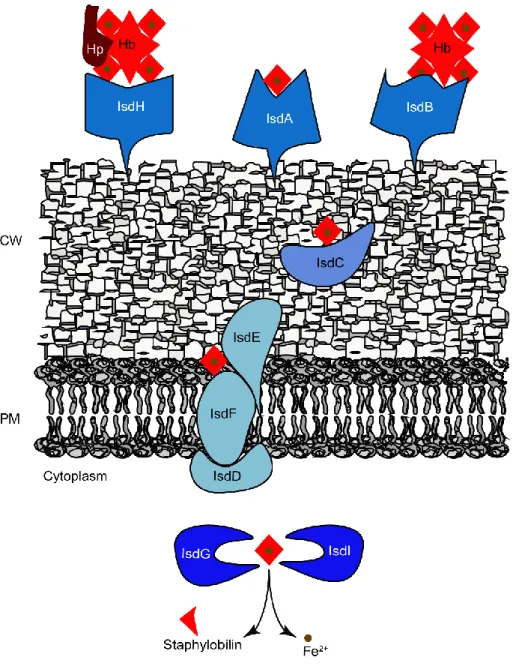
S. aureus also possesses several hemolysins, such as the alpha, beta, and gamma-hemolysins which are able to mediate lysis of red blood and white blood cells (15). Erythrocyte lysis releases hemoglobin that is captured by the iron-regulated surface determinant (Isd) system of S. aureus, removing heme, which is degraded by heme oxygenases to supply nutrient iron, an essential element during the infection process (16, 17). Bacterial heme-uptake systems and the Isd family of S. aureus will be described in detail in Chapter 3 of this dissertation.
S. aureus microorganism is currently a serious human threat and one of the main causes of nosocomial infections (14). Among the diseases caused by S. aureus are skin and soft tissue infections (folliculitis and impetigo), invasive diseases such as osteomyelitis, endocarditis,
Cha
pter 1
bacteremia, pneumonia and toxin mediated diseases such as food poisoning and toxic shock syndrome (6, 10, 18). The occurrence of infections is strengthened by the ability of the bacteria to form biofilms, which allows S. aureus to adhere various surfaces and to resist antimicrobials.
Several S. aureus strains show resistance to antibiotics. The first emergence of drug resistant S. aureus strains was detected only one year after the introduction of penicillin in 1940 (19–21) (Fig 1.2). These strains harbored plasmids encoding penicillinase, a β-lactamase with the ability to destroy the β-lactam ring of penicillin (22). To overcome the penicillin resistance of S. aureus strains, a novel antibiotic, methicillin, was introduced. However, like for penicillin, resistant strains were identified only a few years later (23). The increase of hospital-acquired Methicillin-Resistant Staphylococcus aureus (HA-MRSA) infections led to the introduction of vancomycin. However, the frequent use of this antibiotic caused the appearance of the first vancomycin-intermediate S. aureus (VISA) resistant strains in 1997 (24), followed by the emergence of latter vancomycin-resistant S. aureus (VRSA) strains, that can cope with very high concentrations of vancomycin (25). Yet, vancomycin remains one of the first-line antibiotics to treat severe MRSA infections. More recently, first-linezolid, ceftaroline, and daptomycin have been utilized for the treatment of serious MRSA infections, which are used in combination with vancomycin or other β-lactams (26). Therefore the high increase of antibiotic resistance has been observed in the last years, which makes the pathogen one of major concerns in public health worldwide with a remarkable economic burden (27, 28).
Figure 1.2 Timeline of the emergence of antibiotic resistance in S. aureus. MRSA,
community-associated methicillin-resistant S. aureus; VISA, vancomycin-intermediate S. aureus; VRSA, vancomycin-resistant S. aureus; CA-MRSA community-acquired methicillin-resistant S. aureus. Adapted from (22).
Currently, the strains that exhibit resistance to the whole β-lactam class of antibiotics are generally designated Methicillin-Resistant Staphylococcus aureus (MRSA) strains. The last years have shown a worldwide spread of MRSA present not only in the health care facilities, the so-called HA-MRSA, but also in the community (designated as the community-acquired MRSA (MRSA)) (22). Although in general CA-MRSA strains are more virulent, they show susceptibility to several antibiotics, which suggests that these strains may be descendants strains of the HA-MRSA (22, 29). Nevertheless, an increase of HA-MRSA strains in the community have been reported (30).
In the latest report of the European Antimicrobial Resistance Surveillance Network (EARS-Net 2016), 10 out of 30 countries have MRSA percentages above 25%, while Portugal has approximately 44% (31). Despite the high prevalence of MRSA in Portugal, this percentage has been decreasing in the last years (31).
Cha
pter 1
Hence, it is essential to understand the mechanisms that promote S. aureus survival, to discover novel therapeutic targets.
1.3 S. aureus metabolism
As above mentioned, S. aureus is a facultative anaerobic organism, which means it usually relies on oxygen respiration. However, these microorganisms can use alternative electron acceptors or grow by fermentation (3). Since S. aureus has the ability to invade niches with different oxygen concentrations, it is crucial for this organism to fine-tune the energy metabolism with the O2 availability in these environments.
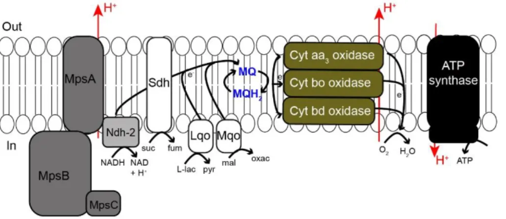
The O2 dependent respiratory chain of S. aureus (Fig 1.3) comprises
two nicotinamide adenine dinucleotide (NADH) dehydrogenases (NDH-2) type 2, (NdhC and NdhF) (32), menaquinones and a branched respiratory system harboring two or three types of oxygen reductases that are heme containing enzymes, namely, a bd-type menaquinol oxidase (CydAB), a heme-copper aa3/ba3-type menaquinol oxidase (QoxABCD), and a bo-type menaquinol oxidase (3). It also contains a F0F1-ATP synthase (AtpBEFHAGDC) but lacks the canonical complex I NADH: quinone oxidoreductase and a Na+-translocating NADH:quinone oxidoreductase (3,
33). Instead, the respiratory chain of S. aureus has a MpsABC (membrane potential-generating system) linked to NDH-2 that is able to generate proton motive force similar to complex I (33). In this chain, electrons are transferred to menaquinones (MQ) generating reduced menaquinone, MQH2, which
transfer the electrons to the terminal oxidases aa3 menaquinol oxidase (composed by cytochromes b-561 and a-602), bo-type menaquinol oxidase (composed by cytochromes b-555) or the cytochrome bd oxidase. Cytochrome b-552 is proposed to be linked to an aa3-type menaquinol oxidase while cytochrome b-557 is suggested to be connected to the bo-type menaquinol oxidase (3, 34, 35). Electrons flow to these cytochromes before
passing to the terminal menaquinol oxidases, where O2 is converted to H2O
(34). Cytochromes aa3 and bo are believed to act as proton pumps (36, 37), while cytochromes aa3 and bd are suggested to support aerobic respiration (34, 38).
Figure 1.3 Scheme of the respiratory chain of S. aureus. L-lactate (Lqo), succinate (Sdh) and
malate (Mqo) dehydrogenases;Mps, membrane potential-generating system; suc, succinate; fum, fumarate; L-lac, L-lactate; pyr, pyruvate; mal, malate; oxac, oxaloacetate, MQ, menaquinone; MQH2, menaquinol; NADH, nicotinamide adenine dinucleotide reduced;NAD+, nicotinamide adenine dinucleotide oxidized; H+, proton; out, outside and in, inside. Adapted from (33).
Due to the high number of heme containing enzymes, mutations in the biosynthesis of heme and menaquinones cofactors leads to impairment of the S. aureus growth, generating the so-called colony variants (SCV) that are persister cells (39). A detail description of the heme cofactors and their crucial role will be presented in Chapter 2.
To grow under anaerobic conditions, S. aureus utilizes nitrate or nitrite as electron terminal acceptors (40, 41). The genome of S. aureus contains a nar operon that encodes a nitrate reductase (NarGHI) that promotes the reduction of nitrate to nitrite using electrons provided by NADH (42) (Fig 1.4). The nitrite produced by NarGHI is converted to ammonia by the dissimilatory nitrite reductase (NirBD), encoded by nirBD, that also uses
Cha
pter 1
NADH as electron donor (Fig 1.4) (3, 42). The nir operon has two additional genes, nasF (also annotated as cysG), proposed to be involved in the synthesis of siroheme and, nirR, that encodes a hypothetical protein with a so far unknown function, but which is speculated to control the nir promoter activity or to be involved in the synthesis of siroheme (3, 42). A detailed description of the synthesis of siroheme will be provided in the next chapter, section 2.4.
Figure 1.4 Genetic map of the genes involved in nitrite and nitrate reduction in S. aureus.
Putative promoters are indicated as angled arrows. Adapted from (42).
S. aureus also contains an oxygen-responsive nitrogen regulation system (NreABC), encoded by the gene cluster nreABC (Fig. 1.4) and positively regulates the expression of the nitrate and nitrite genes in cells exposed to oxygen (42, 43). In cells grown under anaerobic conditions in the presence of nitrate, deletion of this operon resulted in impairment of nitrate and nitrite reduction and consequently in a SCV growth phenotype (42). NreC also controls the expression of the nitrate transporter encoding gene narK (42). Moreover, when growing anaerobically in the presence of nitrate, S. aureus NreC downregulates fermentative genes, such as acetaldehyde dehydrogenase (adhE), L-lactate dehydrogenase (lctE) and alcohol dehydrogenase I (adh1) (42).
The importance of tetrapyrrole molecules, such as heme and siroheme and their associated proteins in the life-time of S. aureus, is patent in the processes mentioned above. Therefore, the de novo synthesis and acquisition pathways that provide these cofactors to S. aureus were studied under the scope of this dissertation and will be highlighted in the next chapters.
1.4 References
1. Ogston A (1882) Micrococcus Poisoning. J Anat Physiol 17(Pt 1):24– 58.
2. Rosenbach FJ (1884) Mikroorganismen bei den Wundinfections-Krankheiten des Menschen. Wiesbaden Germany.
3. Götz F, Bannerman T, Schleifer K (2006) The Genera Staphylococcus and Macrococcus. The Prokayotes, pp 5–75.
4. Ruoff KL (2002) Miscellaneous catalase-negative, Gram-positive cocci: Emerging opportunists. J Clin Microbiol 40(4):1129–1133. 5. Goldstein J, Roberts JW (1982) Microtube coagulase test for detection
of coagulase-positive staphylococci. J Clin Microbiol 15(5):848–851. 6. Wertheim HF, Melles DC, Vos MC, van Leeuwen W, van Belkum A,
Verbrugh H a, Nouwen JL (2005) The role of nasal carriage in Staphylococcus aureus infections. Lancet Infect Dis 5(12):751–762. 7. Kuehnert MJ, Kruszon-Moran D, Hill HA, McQuillan G, McAllister SK,
Fosheim G, McDougal LK, Chaitram J, Jensen B, Fridkin SK, Killgore G, Tenover FC (2006) Prevalence of Staphylococcus aureus nasal colonization in the United States, 2001-2002. J Infect Dis 197(2):172– 179.
8. Nilsson P, Ripa T (2006) Staphylococcus aureus throat colonization is more frequent than colonization in the anterior nares. J Clin Microbiol 44(9):3334–3339.
9. Sollid JUE, Furberg AS, Hanssen AM, Johannessen M (2014) Staphylococcus aureus: Determinants of human carriage. Infect Genet Evol 21:531–541.
10. Mehraj J, Witte W, Akmatov MK, Layer F, Werner G, Krause G (2016) Epidemiology of Staphylococcus aureus Nasal Carriage Patterns in the Community. Curr Top Microbiol Immunol 398:55–87.
Cha
pter 1
Rethinking the Pathogenesis of Community-Associated Methicillin-Resistant Staphylococcus aureus Infection. Clin Infect Dis 46(5):752– 760.
12. Kamholz SL (2016) Human Emerging and Re-emerging Infections. Clin Infect Dis 63(5):713–714.
13. Foster TJ (2005) Immune evasion by staphylococci. Nat Rev Microbiol 3(12):948–58.
14. Liu GY (2009) Molecular Pathogenesis of Staphylococcus aureus Infection. Pediatr Res 65:71–77.
15. DeLeo F, Diep B, Otto M (2009) Host Defense and Pathogenesis in Staphylococcus aureus Infections. Infect Dis Clin North Am 23(1):17– 34.
16. Foster TJ, Geoghegan JA, Ganesh VK, Höök M (2014) Adhesion, invasion and evasion: the many functions of the surface proteins of Staphylococcus aureus. Nat Rev Microbiol 12(1):49–62.
17. Reniere ML, Torres VJ, Skaar EP (2007) Intracellular metalloporphyrin metabolism in Staphylococcus aureus. BioMetals 20(3–4):333–345. 18. Gordon RJ, Lowy FD (2008) Pathogenesis of Methicillin‐Resistant
Staphylococcus aureus Infection. Clin Infect Dis 46(S5):S350–S359. 19. Abraham EP, Chain E (1940) An enzyme from bacteria able to destroy
penicillin. Nature 146(3713):837–837.
20. Kirby WMM (1944) Extraction of a highly potent penicillin inactivator from penicillin resistant staphylococci. Science 99(2579):452–453. 21. Barber M, Rozwadowska-Dowzenko M (1948) Infection by
penicillin-resistant staphylococci. Lancet 252(6530):641–644.
22. Chambers HF, Deleo FR (2009) Waves of resistance: Staphylococcus aureus in the antibiotic era. Nat Rev Microbiol 7(9):629–41.
23. Jevons MP (1961) “Celbenin”- Resistant staphylococci. Br Med J 1:124–125.
Fukuchi Y, Kobayashi I (1997) Dissemination in Japanese hospitals of strains of Staphylococcus aureus heterogeneously resistant to vancomycin. Lancet 350(9092):1670–1673.
25. Weigel LM, Clewell DB, Gill SR, Clark NC, McDougal LK, Flannagan SE, Kolonay JF, Shetty J, Killgore GE, Tenover FC (2003) Genetic analysis of a high-level vancomycin-resistant isolate of Staphylococcus aureus. Science 302(5650):1569–71.
26. Bartash R, Nori P (2017) Beta-lactam combination therapy for the treatment of Staphylococcus aureus and <i>Enterococcus<i> species bacteremia: A summary and appraisal of the evidence. Int J Infect Dis 63:7–12.
27. Stewardson a J, Allignol a, Beyersmann J, Graves N, Schumacher M, Meyer R, Tacconelli E, Angelis G De (2011) The health and economic burden of bloodstream infections caused by antimicrobial-susceptible and non-antimicrobial-susceptiblend 2011 : a multicentre retrospective cohort study. Eurosurveillance 21:1–12.
28. Lee BY, Singh A, David MZ, Bartsch SM, Slayton RB, Huang SS, Zimmer SM, Potter MA, Macal CM, Lauderdale DS, Miller LG, Daum RS (2013) The economic burden of community-associated methicillin-resistant Staphylococcus aureus (CA-MRSA). Clin Microbiol Infect 19(6):528–536.
29. Chambers HF (2001) The changing epidemiology of Staphylococcus aureus? Emerg Infect Dis 7(2):178–182.
30. Grundmann H, Schouls LM, Aanensen DM, Pluister GN, Tami A, Chlebowicz M, Glasner C, Sabat AJ, Weist K, Heuer O, Friedrich AW, on behalf of the ESCMID Study Group C (2014) The dynamic changes of dominant clones of Staphylococcus aureus causing bloodstream infections in the European region: Results of a second structured survey. Eurosurveillance 19(49):1–10.
Cha
pter 1
Antimicrobial resistance surveillance in Europe 2016. Annual Report of the European Antimicrobial Resistance Surveillance Network (EARS-Net). ECDC:1–88.
32. Schurig-Briccio LA, Yano T, Rubin H, Gennis RB (2014) Characterization of the type 2 NADH:menaquinone oxidoreductases from Staphylococcus aureus and the bactericidal action of phenothiazines. Biochim Biophys Acta - Bioenerg 1837(7):954–963. 33. Mayer S, Steffen W, Steuber J, Götz F (2015) The Staphylococcus
aureus nuoL-like protein MpsA contributes to the generation of membrane potential. J Bacteriol 197(5):794–806.
34. Taber HW, Morrison M (1964) Electron transport in staphylococci. Properties of a particle preparation from exponential phase Staphylococcus aureus. Arch Biochem Biophys 105:367–79.
35. Götz F, Mayer S (2013) Both terminal oxidases contribute to fitness and virulence during organ-specific Staphylococcus aureus colonization. MBio 4(6):4–6.
36. Thöny-Meyer L (1997) Biogenesis of respiratory cytochromes in bacteria. Microbiol Mol Biol Rev 61(3):337–76.
37. Tynecka Z, Szcześniak Z, Malm A, Los R (1999) Energy conservation in aerobically grown Staphylococcus aureus. Res Microbiol 150(8):555–566.
38. Hammer ND, Reniere ML, Cassat JE, Zhang Y, Hirsch AO, Hood MI, Skaar EP (2013) Two heme-dependent terminal oxidases power staphylococcus aureus organ-specific colonization of the vertebrate host. MBio 4(4):1–9.
39. Proctor RA, von Eiff C, Kahl BC, Becker K, McNamara P, Herrmann M, Peters G (2006) Small colony variants: a pathogenic form of bacteria that facilitates persistent and recurrent infections. Nat Rev Microbiol 4(4):295–305.
Staphylococcus aureus wild type and mutants. J Bacteriol 123(1):308–316.
41. Somerville GA, Proctor RA (2009) At the crossroads of bacterial metabolism and virulence factor synthesis in staphylococci. Microbiol Mol Biol Rev 73(2):233–248.
42. Schlag S, Fuchs S, Nerz C, Gaupp R, Engelmann S, Liebeke M, Lalk M, Hecker M, Götz F (2008) Characterization of the oxygen-responsive NreABC regulon of Staphylococcus aureus. J Bacteriol 190(23):7847–7858.
43. Kamps A, Achebach S, Fedtke I, Unden G, Götz F (2004) Staphylococcal NreB: An O2-sensing histidine protein kinase with an O2-labile iron-sulphur cluster of the FNR type. Mol Microbiol 52(3):713–723.
Chapter 2
Tetrapyrrole biosynthesis in prokaryotes
2.1 Importance of tetrapyrroles ... 19
2.2 Heme ... 21
2.2.1 Heme biosynthesis ... 24
2.2.2 Regulation of heme biosynthesis ... 30
2.3 Siroheme biosynthesis... 32
2.4 References ... 35
Cha
pter
2
2.1 Importance of tetrapyrroles
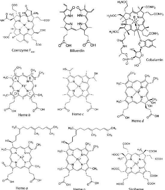
Tetrapyrroles are frequently called the pigments of life and include chlorophylls, coenzyme F430, bilins, cobalamin (vitamin B12), hemes and
siroheme (1) (Fig 2.1).
Tetrapyrroles are molecules involved in many essential biological processes, such as DNA replication, cellular detoxification, metabolism, cell communication, respiration, signal transduction, reduction of nitrite and sulfite, and photosynthesis (2). These compounds derive all from a same universal common tetrapyrrole precursor, namely uroporphyrinogen III (uro´gen III), that is synthesized from δ-aminolaevulinic acid (δ-ALA) (detailed below). The tetrapyrrole structure is formed by four pyrrole aromatic rings often linked by methine groups, in linear (e.g., bilins) or cyclic arrangements (e.g., porphyrins). In the latter, the nitrogen atoms of each pyrrole ring face the center of the macrocycle, which allows the chelation of specific metal ions. Examples of chelated metal ions are: Fe2+ in heme,
siroheme and heme d1; Mg2+ in chlorophylls and bacteriochlorophylls; Ni2+ in coenzyme F430; and Co2+ in cobalamin (1, 3).
Some porphyrins have clinical applications, such as for the treatment of tumors and infections. Porphyrins are light activated at specific wavelengths, transferring electrons to O2 and generating reactive oxygen
species (ROS) which are used to promote cell death (4).
Figure 2.1. Examples of known tetrapyrroles. The rings A, B, C and D and carbons
Cha
pter
2
2.2 Heme
Heme is a hydrophobic molecule and the prosthetic group of numerous proteins involved in crucial life associated processes. One of the best acknowledged function is the storage and transport diatomic gases in hemoglobin and myoglobin (5). Other important roles of heme are for example: i) electron conservation and transfer throughout the respiratory chain in heme containing cytochromes to promote the synthesis of cellular energy (6); ii) catalytic purposes namely in detoxification enzymes such as catalases and peroxidases that reduce H2O2 to H2O (7), and in cytochrome
P450, that catalyzes the oxidation of organic substrates (8); iii) gas sensors, as observed for the CO-sensing CooA transcription factor (9), and as demonstrated for DosS and DosT sensory kinases of Mycobacterium tuberculosis (10); iv) regulation of transcription, translation, protein translocation and protein assembly through the interactions with Heme Regulatory Motifs (HRMs), which are short motifs constituted of cysteine-proline sequences (11).
The iron coordinated to the porphyrin moiety allows heme to function as an electron donor or acceptor due to the change between Fe2+ and Fe3+
redox states. Iron is coordinated by the nitrogen atoms of each pyrrole ring and can be further coordinated by up to two axial ligands to yield a five or six-coordinated heme, respectively (1). The axial ligands are usually histidine, cysteine or methionine from the polypeptide chain. Five-coordinated hemes are generally present in proteins with catalytic, gas sensor and transporter functions as it allows the coordination of a substrate molecule, while six-coordinated hemes are usually present in electron transfer proteins (1, 12). Several types of heme are found in organisms (Hemes a, b, c, d and o) (Fig 2.1), being the more common one heme b, which is also the precursor of all other types of hemes. Heme b is characterized by the presence of vinyl groups attached to rings A and B,
propionic acid groups in rings C and D, and four methyl groups distributed one per ring. Heme a and o are very similar between them, as heme o is an intermediary compound of the heme a synthesis from heme b (1). In heme a and heme o, the vinyl group of ring A is substituted by an isoprenoid side chain; however, in position 8 heme a contains a formyl group, while a methyl group is present in heme o (Fig 2.1). In heme c, the vinyl groups of rings A and B are substituted by thioether bridges that covalently bound to the apoprotein through cysteines (Fig 2.1). The more significant modifications are present in heme d, where two additional hydroxyl groups are observed in ring C (Fig 2.1), and the hydroxyl and propionate side groups in carbon 6 can form a lactone (1, 13). These types of hemes are usually present in bacterial respiratory proteins (1). Other less common forms of heme are heme l, heme m and heme s. Heme l is found in mammalian lactoperoxidases, heme m in mammalian neutrophil enzyme myeloperoxidase while heme s is present in marine worm hemoglobins (1). Although heme has a high biological importance, the intracellular heme content has to be tightly regulated as high levels of free heme are highly reactive and toxic to the cells, for instance causing the formation of ROS, which damage DNA, lipids and proteins (14). Due to its hydrophobic properties, heme has the ability to intercalate into lipid membranes promoting the oxidation of membrane components that leads to cell lysis and death (5, 15). Additionally, heme mediates degradation of proteins to small peptide fragments and is also a strong pro-inflammatory agent (5, 15).
Heme toxicity is well studied in mammalian cells and is estimated that the physiological concentration of free heme ranges from 0.03 to 1 µM (16) and concentrations above 10 µM (16), produce ROS. In mammals the possible causes for accumulation of intracellular free heme are, the increase of heme synthesis, the accumulation of extracellular heme, deficient heme oxygenase activity, impaired heme incorporation in apo-proteins and enhanced hemeprotein degradation (5). Mammals avoid heme toxicity by
Cha
pter
2
degradation of the free heme via heme oxygenase or by means of other proteins such as reduced glutathione and NADPH–cytochrome P-450 reductase (15). Additionally, heme content is controlled by heme binding proteins such as hemopexin, albumin, haptoglobin, and heme-binding protein (HBP23), all of which form non-toxic heme complexes (15). In mammals, other mechanisms for regulation of intracellular heme content consists in the regulation of heme synthesis and in extracellular heme trafficking (5).
In bacteria, the effects of heme toxicity are generally more pronounced in Gram-positive than in Gram-negative bacteria (17, 18). The growth of S. aureus is slightly inhibited at concentrations above 5 µM of hemin (19). Staphylococcus epidermidis seems to tolerate higher concentrations of hemin as concentrations above 10 µM are necessary for reduced growth (20). Pseudomonas aeruginosa and E. coli can cope with 150 µM of hemin without growth impairment (17). Nevertheless, the number of studies on mechanisms of heme toxicity to bacteria are limited. In S. aureus and E. coli, heme was shown to cause DNA damage (21). Also for S. aureus, hemin was reported to induce ion fluxes in a light-independent mode, hence causing oxidative damage (22). Additionally, free iron released during heme degradation promotes bacterial cell damage through the production of hydroxyl free radicals (Fenton chemistry) (18). It was recently demonstrated for S. aureus that, when heme is in excess, heme molecules accumulate in bacterial membranes, that by interaction with oxygen and membrane menaquinones generate superoxide radicals (23).
Bacteria have also evolved mechanisms to prevent heme toxicity that comprise heme efflux pumps, heme sequestration proteins and heme degrading proteins. Numerous Gram-positive bacteria encode a heme-regulated transporter system (HrtAB), an efflux pump that was shown in S. aureus to act in combination with the heme sensor system HssRS. High levels of heme are recognized by HssRS that activates the HrtAB heme
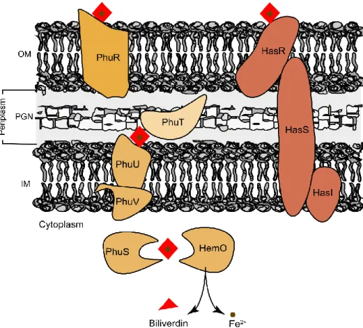
export activity (19, 24). Orthologues of this efflux system were found in Gram-positive bacteria such as Bacillus anthracis, Lactococcus lactis, Corynebacterium diphtheriae and Streptococcus agalactiae (25–28). Alternatively to the heme efflux systems mentioned above, Gram-negative bacteria rely on heme sequestration proteins to face heme toxicity. Examples of these proteins are Yersinia enterocolitica HemS (29), Pseudomonas aeruginosa PhuS (30), Escherichia coli O157:H7 ChuS (31), Yersenia pestis HmuS (32) and Shigella dysenteriae ShuS (33). Interestingly, some of these proteins are also considered part of the heme uptake systems operating in Gram-negatives.
2.2.1 Heme biosynthesis
In bacteria, heme is either synthesized endogenously or captured from the environment by heme uptake systems. Almost every living organism is capable of synthetizing heme; however, bacteria such as those of the genera Dehalococcoides and Thermotoga are not able to produce heme. Furthermore, Enterococcus faecalis, lactic acid bacteria, Bartonella hensaela, Haemophilus influenzaea and some Streptococcus species although possessing hemeproteins apparently do not contain heme biosynthesis related genes (14, 34).
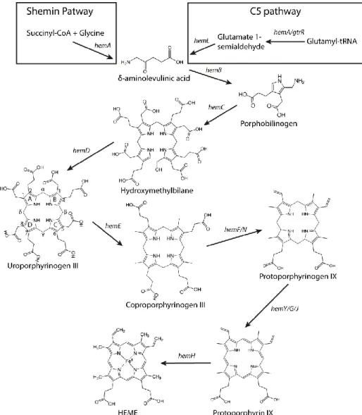
For organisms that produce endogenously heme and/or other tetrapyrroles, its synthesis starts with the formation of the universal tetrapyrrole precursor δ-aminolevulinic acid (δ-ALA) (Fig 2.2) (1, 3, 34–36). So far, two distinct pathways are known for the synthesis of δ-ALA: i) the Shemin or C4 pathway, where succinyl-coenzyme A and glycine are condensed to δ-ALA with the action of Ala synthase encoded by hemA (3, 34, 37). This route is present in metazoans, fungi and alphaproteobacteria; ii) the C5-pathway, that although discovered latter it is likely to be the more ancient route, and which uses charged glutamyl-tRNA for the production of
Cha
pter
2
δ-ALA in a two-step reaction (3, 34, 37). Glutamyl-tRNA is converted into glutamate-1-semialdehyde by glutamyl-tRNA reductase (HemA or GtrR) which is then converted into δ-ALA by glutamate-1-semialdehyde-2,1-aminomutase encoded by the hemL gene (3, 34, 37). The C5-pathway occurs in most bacteria, plants and archaea. The only organisms known to have the two pathways for the synthesis of δ-ALA are Euglena gracilis and Chromobacterium violaceum (34).
Figure 2.2 Scheme of the steps of the biosynthesis of heme b via the classical pathway, also
known as the protoporphyrin-dependent pathway. The rings A, B, C and D and carbons are indicated in uro´gen III according to Fisher nomenclature. Adapted from (3).
Following the generation of δ-ALA, the porphobilinogen synthase enzyme (HemB) promotes the condensation of two molecules of δ-ALA to produce porphobilinogen (PBG) (3, 34).
The next enzymatic step involves the deamination and assembly of four porphobilinogen molecules to yield hydroxymethylbilane, which is performed by hydroxymethylbilane (HMB) synthase (HemC). This enzyme deaminates PBG and promotes the assembly of the four deaminated porphobilinogen molecules into a linear complex (HMB) (3, 34). HMB spontaneously cyclizes to generate uroporphyrinogen I, a non-physiological compound (37). Therefore, HMB is converted into uro´gen III by cyclization and inversion of the acetate and propionate groups of the D pyrrole ring in the presence of uroporphyrinogen III synthase (HemD) (Fig 2.2) (3, 34).
As mentioned in section 2.1, uro´gen III is the last common precursor of all known tetrapyrroles and until this step, all enzymatic reactions from δ-ALA to uro´gen III are highly conserved in organisms that synthesize these pigments. In the classical pathway, following the generation of uro´gen III four extra enzymatic reactions are necessary to catalyze its conversion to the final product heme b (Fig 2.2).
For many years, the classical pathway was considered as conserved in all heme producing organisms. However, a second type of route, named alternative heme biosynthetic pathway was revealed and broke that dogma (38). This new route proceeds via the methylation of uro´gen III through the generation of siroheme. The pathway has been proposed to precede the classical pathway since it utilizes intermediates, such as siroheme, that are more ancient than heme, and because the reactions take place under anaerobic conditions (34). The alternative heme biosynthetic pathway is widespread in archaea, denitrifying and sulphate-reducing bacteria (34, 38).
Cha
pter
2
More recently a third route for the synthesis of heme has been reported to be present mostly in Firmicutes and Actinobacteria (39). This pathway, termed transitional pathway, branches from uro´gen III and contains coproporphyrin III as a new intermediate (39). This novel heme biosynthetic pathway was shown in this thesis to be active in S. aureus as described in chapter 4 of the results section.
Very recently, in 2017 (34), these pathways were renamed as protoporphyrin-dependent (PPD), siroheme-dependent (SHD) pathway and coproporphyrin-dependent (CPD) pathway, respectively, as well as the enzymes involved. However, in this dissertation the enzyme names will be referred according to the classical nomenclature since this still remains the one that is most used.
Protoporphyrin-dependent (PPD) pathway
The synthesis of heme b through the PPD route (previously known as classical heme pathway (Fig 2.2) converts uro´gen III to coprophyrinogen III (copro´gen III) by the decarboxylation of the acetic acid side chains of each pyrrole ring, generating methyl groups, in a reaction that is catalyzed by uro´gen III decarboxylase enzyme (HemE) (3, 34). Bacterial HemE enzymes are homodimers with subunits of 40 kDa and with no apparent associated cofactor (34).
In the next step, the propionate groups of rings A and B of copro´gen III are oxidatively decarboxylated to vinyl groups, yielding protoporphyrinogen IX (proto´gen IX) in a step catalyzed by coproporphyrinogen III decarboxylase. Depending on the oxygen dependence, two family of enzymes catalyze the reaction: the oxygen-dependent HemF (copro´gen III decarboxylase) and the oxygen-inoxygen-dependent HemN (copro´gen III dehydrogenase). HemF is typically a homodimer that requires O2 as a terminal electron acceptor, with the concomitant formation
of CO2 and H2O2 (40, 41). HemN is a monomeric protein which harbors a
[4Fe-4S] cluster and a S-adenosyl-L-methionine (SAM) molecule (42). In the penultimate step of the pathway, proto´gen IX is oxidized to protoporphyrin IX in a reaction that involves six electrons. As in the previous step, the reaction may be performed by an dependent or an oxygen-independent enzyme. The oxygen-dependent proto´gen IX oxidase, HemY, is a homodimer protein containing flavin adenine dinucleotide (FAD) that, like HemF, utilizes O2 as terminal electron acceptor. The oxidation of proto´gen
IX by HemY uses FAD and involves three O2 molecules generating three
H2O2 molecules (34). The oxygen-independent HemG was first reported in
E. coli and is a 21 kDa protein (35). Contrarily to HemY, HemG feeds electrons to the respiratory chain, anticipating the use of several electron acceptors (43). HemG is mostly present in γ-proteobacteria while HemY occurs in eukaryotes and prokaryotes (34, 44). In 2010, a new proto´gen oxidase was identified in α-proteobacteria and δ-proteobacteria and named HemJ, but its full characterization remains to be done (44, 45).
The final step of heme biosynthesis consists in the insertion of iron into protoporphyrin IX to form protoheme, which is commonly designated as heme b (Fig 2.2). This reaction is catalyzed by protoporphyrin ferrochelatase (HemH), a well characterized enzyme that was reported to be a membrane-bound protein in eukaryotes and several prokaryotes, but that is a soluble monomer in Gram-positive bacteria, such as B. subtilis (46–48). Furthermore, protoporphyrin ferrochelatases may contain an iron-sulfur cluster bound to C-terminal region of the protein, which occurs mainly in eukaryotes, but has also been reported for some bacteria, such as, Mycobacterium tuberculosis, Bdellovibrio bacteriovorus, Caulobacter crescentus and Myxococcus xanthus (47, 49). Apart from protoporphyrin, ferrochelatases may use a wide range of substrates such as, mesoporphyrin, deuteroporphyrin and protoporphyrin, and promote the insertion of other cations like Co2+, Zn2+ and Mn2+ (34, 35, 46). Interestingly, in murine and
Cha
pter
2
yeast ferrochelatases, the chelation of metal ions other than iron such as Cu2+, Zn2+, Co2+ and Ni2+ promotes substrate inhibition (50).
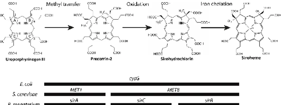
Siroheme-dependent (SHD) pathway
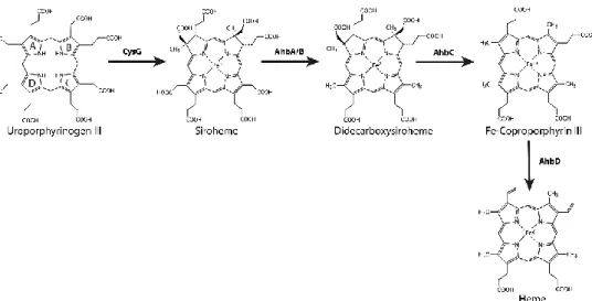
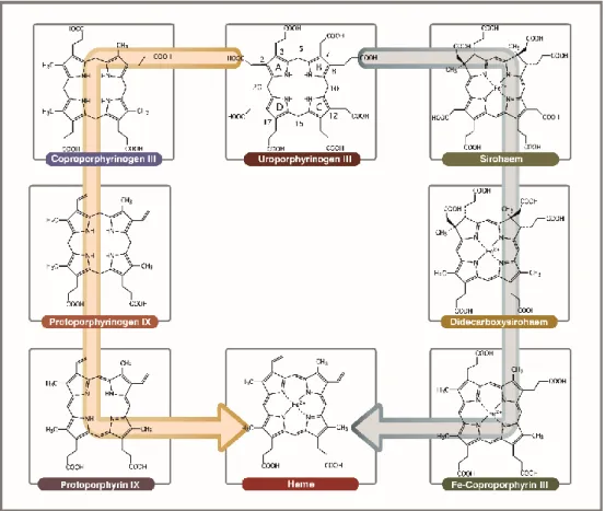
For the formation of heme through the SHD pathway (previously known as alternative heme biosynthetic pathway), uro´gen III is first converted to siroheme via precorrin-2 and sirohydrochlorin in three consecutive reactions involving the siroheme synthase CysG (Figure 2.3) (1, 51). Siroheme is then converted to 12,18-didecarboxysiroheme by the decarboxylation of the acetic acid side chains in rings C and D to methyl groups, in a step involving the alternative heme biosynthesis proteins AhbA and AhbB (38, 52, 53) (Fig 2.3). Both proteins form a heterodimeric complex with a mass of 40 kDa named siroheme decarboxylase complex (52). The next step involves the elimination of the acetic acid residues of rings A and B, generating Fe-coproporphyrin III, also known as coproheme, which is catalyzed by AhbC in a SAM-dependent reaction (38, 53) (Fig 2.3). In the last step, the two propionate side chains of rings A and B of Fe-coproporphyrin III are oxidative decarboxylated into vinyl groups forming the final product, heme b. This reaction is promoted by AhbD (38, 54) (Fig 2.3) and is also SAM-dependent. In D. vulgaris, AhbD was shown to be a monomeric cytoplasmic protein that harbors two iron-sulfur clusters (54).
2.2.2 Regulation of heme biosynthesis
In several biosynthetic pathways, substrates and products may have an active role in the regulatory process. In heme biosynthesis the best known regulatory process involves the control of the first step by its final end product, heme. In Salmonella species and E. coli, heme binds to HemA promoting its degradation by proteases, which impairs the heme biosynthesis (55) and excess of cellular heme levels is avoided. In Corynebacterium diphtheriae, high concentrations of heme leads to repression of the hemA promoter by the Corynebacterium heme response system (ChrA-ChrS) and the heme response regulator (HrrA-HrrS). These two-component regulatory systems also activate the transcription of the hmuO gene, which encodes a heme oxygenase enzyme (56, 57). Furthermore, when heme levels are elevated, HrrA represses the transcription of hemE and hemH (57).
In Acidithiobacillus ferrooxidans, addition of δ-ALA substrate to cells increases heme levels, decreasing the Glu-tRNA synthetase activity and the levels of Glu-tRNA reductase (58).
Figure 2.3 Steps required for the synthesis of heme b via the siroheme-dependent pathway. Adapted