Chitosan-based nanomedicine for rosmarinic acid ocular delivery
Thesis presented to obtain the PhD degree in Pharmaceutical Sciences, Pharmaceutical Technology Specialty,
Faculty of Pharmacy of University of Porto
by
Sara Isabel Macedo Baptista da Silva
Under supervision of Prof. Dr. Bruno Sarmento and co-supervision of Prof. Dr. Domingos Ferreira and Prof. Dr. Manuela Pintado
iv Declaration
The partial reproduction of this thesis is authorized only for research purposes by written declaration of the person concerned.
(Sara Baptista da Silva)
v Inspiration
“Success is a journey, not a destination!”
vi Dedication
“There are only two ways to live life. One is as though nothing is a miracle. The other is as though everything is a miracle.”
Albert Einstein
I dedicate this thesis to the most important persons in my life, my family, Oscar and our daughter, Catarina Isabel. .
vii Acknowledgements
“Lord I can’t say it in words…can you please just listen through my heart”
Unknown
I would like to formally express my deep gratitude to the following people and institutions – who (and which) have meant a lot to me during my PhD program, and made it possible: Fundação para a Ciência e a Tecnologia, for financial support via a PhD fellowship (ref.: SFRH/BD/61423/2009), under the supervision of Professor Bruno Sarmento; said grant permitted timely development of my research program, as well as participation in several international scientific meetings to complement my training and sharing my results.
Laboratory of Pharmaceutical Technology, Faculty of Pharmacy, University of Porto for accepting me as a PhD student, for the hospitality and work conditions available, during my doctoral program.
My most sincerely acknowledges to Escola Superior de Biotecnologia of Universidade Católica Portuguesa (ESB-UCP), for the crucial collaboration in my PhD course, the indubitable hospitality and for providing facilities and logistical to best support my studies. INEB – Instituto de Engenharia Biomédica, ISCS–N – Instituto Superior de Ciências da Saúde – Norte and IBILI-Institute for Biomedical Imaging and Life Sciences – University of Coimbra, for the acceptance, kindness and constant availability cooperation during this project.
Professor Bruno Sarmento my supervisor, who I am sincerely grateful for having accepted me as a PhD student, for the wide support and comprehensive scientific guidance he continuously gave me. His consistent and integrated contribution for my growth, both as a person and as a researcher, has been by all means outstanding. I would like, in particular, to thank him for every effort made in guarantee the best research conditions, going wherever necessary to find the most appropriate support; and for his everlasting encouragement, patience and motivation, as well as his availability to discuss specific and general topics of my dissertation. I am indeed deeply grateful, for having always believed in my abilities, for all the concerns, problems, opportunities and achievements shared along this journey, for the friendship and affection, my heartfelt thanks.
viii
Professor Manuela Pintado my co-supervisor, I would like to thank for always accepting me as a student, having accompanied during these 12 years of academic training, for always believing in my abilities, skills and competences, for her unconditional understanding, patience, friendship and complicity. For her believed and dedication to my work, for all the conversations established and knowledge shared throughout these years and during this doctoral course, as well as for her encouragement, affection and help; and also for providing a healthy, happy and extremely professional working environment, my deepest thanks.
Professor Domingos Ferreira my co-supervisor, I would like to thank for his sympathy, kindness, constant availability and willingness to help during my doctoral program. For every effort to ensure the best institutional reception conditions and every logistical that could ensure the success of my work, my sincere grateful.
Professor Francisco Ambrósio, my deepest gratitude for agreeing to collaborate in this PhD project. For all the help, cooperation, understanding, by all the efforts made the investment of time and resources in better monitoring and performance of this work. For having given me the opportunity to work with his research team and by the unmatched amiability.
My most sincere thanks to Professor Horacina Cavalcante and to the entire Stamford family: Professor Newton, Professor Tania, Thayza and Thathiana, for have accompanied me during my doctoral plan in every possible way, for all the help, dedication, and enormous friendship.
My colegues and friends within the different research groups – José das Neves, Fernanda Andrade, Filipa Antunes, Pedro Fonte, Francisca Araújo, Rute Nunes, Carla Pereira, Manuela Amorim, Ana Oliveira, Raquel Madureira, Débora Campos, Raquel Boia, Filipe Elvas, Tiago Martins, Pedro Tralhão, Maria Madeira, Joana Martins, and so many others. By somehow helping me in developing my studies and shared by so many difficulties and achievements, which certainly has helped me grow as a person and as a professional. My closest friends which are the family that I chose for me every day:
Helena Monteiro, thank you for your friendship which already makes “silver wedding”, for our perpetual oath on time, for always find yourself without seeking, for even far in distance seems like I have been with you yesterday, and always.
ix
Sandra Borges, words will be for sure fall short in expressing my eternal friendship. Thank you for bringing a rain-bow to my life, for the craziness shared. For every concern, worry, and happiness moments joint, for listening, advising, for the constant patience and complicity; For being always there.
Manuela Amorim, I will not be able to thank you everything that you art for me and everything what you already has done for me during this project, much less in a nutshell. Above all thank you for the unconditional friendship, complicity, and confidence, for every laugh and tear shared, for being my true friend.
Inês Cravo Roxo, Joana Barbosa, Franklin Costa (and the little Matilde), Ricardo Freixo, Luciana Silva and Renato Resende thank you for always accompanied my life at its best and worst for every moment spent, I appreciate and reciprocate with eternal friendship. I also have to leave my deepest gratitude to my sweet Francisca Maria, who accompanied my life forever, and thank her for the immense dedication to my family, for their undying affection and unconditional support in every moment of our life, from best to worst; My most sincere and profound thanks.
I would like to thank to my brother Jorge Filipe and my sister Sofia Manuel, being my best friends, for existing and making my life so full field, so complete; For being with me in every dream, fight, step and for never live me alone. I would also have to thank to my nephews (Carlos Eduardo, Filipa Alexandra and Barbara Sofia), for being the best continuity of my brothers, for their love and for being always with me in a health madness. My Grandmother, Isabel Maria, will always be my sunshine, my companion of all hours, the example of strength and light, and her way of being and living will always inspire my life.
Words will be reductive and insufficient to thank my parents for the unconditional love, for making me what I am today, for everything they have given me throughout my life, by making the development of my academic training possible and for always believed in me. Thank you for the unconditional love, strength, support and courage injected in me, today and every day. Thank you for everything, now and forever.
And last, but far from least – Oscar, is my Alter Ego, my companion of dreams and struggles. Oscar is the better half of me, the more aware, responsible and realistic. I grew up with him personal and intellectually. He has accompanied every moment of my life,
x
from the best to the worst. He has attended my academic formation closely and helped in everything he can so that I can move forward successfully. I would like to thank him for the unreserved love, for the amazing person he is, for all time he invested on me, and for the support, encouragement, patience and motivation that he constantly showed. For being my true love, and for have given me the best of my life, our daughter Catarina Isabel. Catarina born during my PhD workplan, she is the miracle of my life, my endless love, my heart, the reason why I want to get over myself and be better every day, to be her best reference.
xi Abstract
Chitosan based nanoparticles prepared by ionic gelation with sodium tripolyphosphate (TPP) were developed and optimized. Chitosan and TPP were tested in different concentrations for the study of the chitosan:TPP best mass ratio until the final proportion of (7:1). The pH of nanoparticles formation medium was adjusted to 5.8, which was the pH value that best favors the interaction of rosmarinic acid and chitosan, thus reaching a maximum leading of rosmarinic acid entrapment. The encapsulation of rosmarinic acid,
Salvia officinalis (sage) and Satureja montana (savory) was also tested in different
theoretical loadings fairly to the initial concentration of chitosan. Nanoparticles were prepared and characterized freshly and lyophilized, and the theoretical loading was further fixed in 40% for rosmarinic acid and 50% for sage and savory nanoparticles. The particle size was observed to be dependent on both chitosan and TPP concentrations and minimum sizes were obtained for the lowest chitosan and TPP concentrations. The mean particle size was 244.0 ± 18.0, 278.4 ± 42.2 and 229.3 ± 29.6 for rosmarinic acid, sage and savory nanoparticles, respectively. No significant differences (P > 0.05) were observed between the different nanoformulations, which was expectable considering that the nanoparticulate system was optimized to encapsulate rosmarinic acid and since it is the major component of natural extracts. The obtained nanoparticles showed values of low dispersity index (< 0.25), corresponding to a narrow particle size distribution such as: 0.193 ± 0.062, 0.268 ± 0.092 and 0.149 ± 0.121 for rosmarinic acid, sage and savory nanoparticles, respectively. Nanoparticles surface charge ranged from 29.5 ± 1.6, 24.9 ± 0.1 and 26.5 ± 3.5 mV for rosmarinic acid, sage and savory nanoparticles, respectively. This indicates that the colloidal systems were stable in storage time. Scanning electron microscopy (SEM) and transmission electron microscopy (TEM) confirmed the particle size distribution, and individual, smooth, spherical and small sizing nanoparticles (ranged from 200-300 nm) containing the rosmarinic acid and extracts were observed. The loading capacity was 5.3 ± 0.4, 8.1 ± 0.6, and 7.8 ± 0.2% for rosmarinic acid, sage and savory nanoparticles, respectively. The higher values for extract nanoparticles were intimate correlated to the lower initial amount of rosmarinic acid in the nanocomplexes. In a similar way, the association efficiencies were higher for extracts than rosmarinic acid, with values of 51.2 ± 3.0, 96.1 ± 0.2 and 98.2 ± 0.1% for rosmarinic acid, sage and savory nanoparticles respectively, quantified by a previously validated high-performance liquid chromatography (HPLC) method. The higher extracts values may be also associated to
xii
lower amount of rosmarinic acid in chitosan nanoparticles, since impair of association efficiency and the initial concentration is documented as a reverse. The rosmarinic acid in
vitro release profile was evaluated in phosphate buffer saline (PBS), pH 7.4, for the
different nanoparticles and a fast release was observed in all the formulations, reaching almost 100% up to 60 min, with no significant differences (P > 0.05). Therefore these nanoparticulate systems may provide a rational strategy for the development of ocular rapid release systems for rosmarinic acid delivery. Differential scanning calorimetry (DSC) and Fourier-transform infrared (FTIR) analysis set that no chemical interactions were found between antioxidants and chitosan, after encapsulation. Antioxidant activity was evaluated by 2,2-azinobis-(3-ethylbenzothiazoline-6-sulphonic acid) (ABTS) and oxygen radical absorbance capacity (ORAC) methods before and after lyophilization to assure that antioxidant activity was not compromised after the dried process. The best antioxidant activity results were obtained by ORAC method and after nanoparticles lyophilized, the values for rosmarinic acid, sage and savory nanoparticles were 3.6520 ± 0.1770, 0.4251 ± 0.0069 and 0.4526 ± 0.0087 µmol/eq Trolox, respectively. Nonetheless, lower antioxidant activity was observed in the nanoparticles comparing to the free compounds due to the partial entrapment effect of the compounds. Considering the mucin particle method, nanoparticles indicate mucoadhesive proprieties, by the increase on nanosize and the consequent surface charge decrease, after mucin incubation, suggesting an increase retention time over the ocular mucosa after instillation. All nanoparticles demonstrate to be safe without relevant cytotoxicity (below 10%, for all the formulations and concentrations), by methylthiazolyldiphenyl-tetrazolium bromide conversion (MTT) and lactate dehydrogenase enzyme release (LDH) assays against retina pigment epithelium (ARPE-19) and human cornea (HCE-T) cell lines. Nonetheless, the results in both cell lines were concentration dependent. Chorioallantoic membrane test (HET-CAM) also confirmed the absence of nanoparticles irritancy in the eye. The permeability study in HCE monolayer cell line showed apparent permeability coefficient (Papp) of 3.41 ± 0.99 x 10⁻⁵ and 3.24 ±
0.79 x 10⁻⁵ cm/s for rosmarinic acid loaded chitosan nanoparticles and free in solution, respectively. In ARPE-19 monolayer cell line the Papp was 3.39 ± 0.18 x 10⁻⁵ and 3.60 ±
0.05 x 10⁻⁵ cm/s for rosmarinic acid loaded chitosan nanoparticles and free in solution, respectively. No significant differences (P > 0.05) were achieved neither between both cell lines nor between loaded and unloaded chitosan nanoparticles, probably due to the rapid release profile of nanoparticles described above. Preliminary ocular in vivo studies were implemented to evaluate the antioxidant effect of rosmarinic acid through intravitreally injection in Wistar rats using an Ischemia-reperfusion (I-R) model. Electroretinograms
xiii
(ERG) and immunohistochemistry showed that at 50 µM rosmarinic acid did not present a significant retina protective effect, suggesting acute pro-inflammatory damages in I-R model, difficult to revert with a single injection. Nonetheless and considering the good results achieved during this project, the rosmarinic acid nanoparticles were safe, mucoadhesive, with high ocular permeability, with good antioxidant activity performance and its therapeutical potential may be important to ocular diseases prophylaxis. It can be also concluded that these natural nanocarriers were promising ocular drug delivery systems and the study highlights the need to explore new drug delivery systems to be applied on ocular surface in order to overlap the limitations on topical drug effectiveness. Keywords: Chitosan, rosmarinic acid, extracts, ocular diseases
xv Resumo
As nanopartículas de quitosano foram desenvolvidas e otimizadas por gelificação iónica com tripolifosfato de sódio (TPP). O quitosano e TPP foram testados em diferentes concentrações para o estudo da melhor relação quitosano:TPP até à proporção final de (7:1). O pH da solução das nanopartículas foi ajustado para 5,8, para favorecer a interação máxima entre o quitosano e o ácido rosmarínico. A encapsulação de ácido rosmarínico, e dos extractos naturais de Salvia officinalis (salva) e Satureja montana (segurelha) também foram testados, considerando diferentes cargas teóricas relativamente à concentração inicial de quitosano. As nanopartículas foram preparadas e caracterizadas diretamente após a sua formulação e após liofilização, e foram posteriormente fixados os valores de carga teórica de 40% para o ácido rosmarínico e 50% para a salva e segurelha. O tamanho das partículas demonstrou ser dependente de ambas as concentrações de quitosano e TPP e os tamanhos mínimos foram obtidos para a menor concentração de quitosano e TPP. O tamanho médio das partículas foi 244,0 ± 18,0, 278,4 ± 42,2 e 229,3 ± 29,6 nm para as nanoparticulas de ácido rosmarínico, salva e segurelha, respetivamente. Não houve diferenças significativas (P > 0,05) observadas entre as diferentes nanoformulações o que seria expectável, considerando que o sistema de nanopartículas foi otimizado para encapsular o ácido rosmarínico, e este é o principal componente dos extratos naturais. As nanopartículas obtidas apresentaram valores de baixo índice de polidispersão (< 0,25), o que corresponde a uma reduzida distribuição do tamanho das partículas, sendo os valores: 0,193 ± 0,062, 0,268 ± 0,092 and 0,149 ± 0,121 para as nanoparticulas de ácido rosmarínico, salva e de segurelha, respetivamente. A carga da superfície das nanopartículas foi: 29,5 ± 1,6, 24,9 ± 0,1 and 26,5 ± 3,5 mV para as nanopartículas de ácido rosmarínico, salva e de segurelha, respetivamente. Isto sugere que os sistemas coloidais são estáveis ao longo do tempo. A microscopia eletrónica de varrimento (SEM) e a microscopia eletrónica de transmissão (TEM) confirmaram os resultados anteriores e demonstraram nanoparticulas individualizadas, lisas, esféricas e pequenas (variando entre 200-300 nm). A capacidade de carga das partículas foi de 5,3 ± 0,4, 8,1 ± 0,6 e 7,8 ± 0,2% para as nanoparticulas de ácido rosmarínico, salva e de segurelha, respetivamente. Os valores mais elevados para as nanopartículas com extrato foram intimamente correlacionados com a menor quantidade inicial de ácido rosmarínico nos nanosistemas. De um modo semelhante, as percentagens de eficiência de associação dos extratos nas nanopartículas foram mais elevadas, em comparação com as do ácido rosmarínico. Os valores foram: 51,2 ± 3,0,
xvi
96,1 ± 0,2 e 98,2 ± 0,1%para as nanopartículas de ácido rosmarínico, salva e segurelha, respetivamente. Estes valores mais elevados associados à nanoencapsulação dos extratos também podem ser associados à menor quantidade de ácido rosmarínico nas nanopartículas de quitosano, uma vez que a concentração inicial é documentada como inversamente proporcional à própria eficiência de associação. O perfil de liberação in vitro do ácido rosmarínico foi avaliado em tampão fosfato (PBS), a pH 7,4 nas diferentes formulações, por um período de 60 min, e não se observaram diferenças significativas (P > 0,05). A rápida libertação do ácido rosmarínico dá indicações que estes sistemas de nanopartículas podem fornecer uma estratégia racional para o desenvolvimento de formulações de libertação imediata para administração ocular do ácido rosmarínico. A eficiência de associação e de liberação in vitro foram realizadas utilizando um método de cromatografia líquida de alta eficiência (HPLC), especialmente desenvolvido e otimizado para garantir a obtenção de resultados precisos e exatos. As análises de calorimetria diferencial de varrimento (DSC) e a espetrofotometria de infravermelho por transformada de Fourier (FTIR) permitiram concluir que não foram encontradas interações químicas entre os antioxidantes e o quitosano, depois do processo de encapsulação. A atividade antioxidante dos nanosistemas foi avaliada pelos métodos de 2,2-azinobis-(3-etil-benzotiazolin-6-ácido sulfónico) (ABTS) e de capacidade de absorção radical (ORAC), antes e depois do processo de liofilização, para garantir que a atividade antioxidante não é comprometida durante o processo de secagem das partículas. Os melhores resultados de atividade antioxidante foram obtidos pelo método de ORAC após liofilização das partículas, os resultados para as nanoparticulas de ácido rosmarinico, salva e segurelha foram: 3,6520 ± 0,1770, 0,4251 ± 0,0069 e 0,4526 ± 0,0087 µmol/eq Trolox, respetivamente. Todavia foi observada uma atividade antioxidante mais baixa nas nanopartículas do que nos compostos livres, devido ao efeito da nanoencapsulação. As partículas demonstraram propriedades mucoadesivas após incubação com mucina, pelo aumento em tamanho e consequente diminuição da carga de superfície. Os resultados indicam que pode ser expectável um aumento do tempo de retenção sobre a mucosa ocular após a instilação. Todas as formulações demonstraram ser seguras para o teste de citotoxicidade 3-(4,5-dimetiltiazol-2yl)-2,5-difenil brometo de tetrazolina (MTT) e para o teste da libertação da enzima lactato desidrogenase (LDH), sem citotoxicidade relevante (abaixo de 10%, para todas as formulações e concentrações), em linhas oculares da retina (epitélio pigmentar da retina - ARPE-19) e da córnea (linha de células da córnea humana - HCE-T). O teste da membrana corioalantóide (HET-CAM Teste) foi utilizado como alternativa aos testes biológicos em coelhos (teste de Draize) e também sugere a
xvii
ausência de irritação das partículas no olho. Os estudos de permeabilidade em monocamada de células da córnea (HCE) revelou um coeficiente de permeabilidade aparente (Papp) de 3,41 ± 0,99 x 10⁻⁵ e 3,24 ± 0,79 x 10⁻⁵ cm / s para as nanopartículas de
ácido rosmarínico e para o ácido rosmarínico livre, respectivamente. O estudo de permeabilidade em monocamada de células da retina (ARPE-19) revelou valores de Papp
de 3,39 ± 0,18 x 10⁻⁵ e 3,60 ± 0,05 x 10⁻⁵ cm/s para as nanopartículas de ácido rosmarínico e para o ácido rosmarínico livre, respectivamente. Não houve diferença significativas (P > 0,05) entre os valores de permeabilidade das nanoparticulas, composto livre e entre ambas as linhas celulares, provavelmente devido ao perfil de liberação rápido das nanopartículas acima descrito. Foram feitos testes preliminares in vivo, em que o ácido rosmarínico foi injetado na cavidade intravítrea de ratos Wistar, num modelo animal isquemia-reperfusão (I-R). Eletrorretinogramas (ERG) e ensaios imuno-histoquímicos revelaram que o ácido rosmarínico (a uma concentração de: 50 µM), por injeção intravítrea, não teve um efeito protector na retina. O que poderá ser devido danos pró-inflamatórias severos no modelo I-R, difíceis de reverter com o estudo de uma única injeção. No entanto, e considerando os bons resultados obtidos neste trabalho, as partículas de quitosano contendo ácido rosmarínico demonstraram ser seguras, mucoadesivas, com elevado potencial de permeabilidade ocular e com um bom perfil de atividade antioxidante, o que permite concluir que estes nanosistemas podem ser importantes para a prevenção de doenças degenerativas oculares. Os resultados desta tese, permitem também concluir que estes nanosistemas naturais são promissores na administração tópica de antioxidantes no olho e ressalta a necessidade de se explorar novos sistemas para ultrapassar as limitações na eficiência da administração tópica de fármacos no olho.
xviii
This work was submitted as a PhD Thesis in partial fulfilment of the requirements for Philosophiæ Doctor (PhD) degree in Pharmaceutical Sciences at the Faculty of Pharmacy, University of Porto.
It was conducted under the guidance of Prof. Dr. Bruno Filipe Carmelino Cardoso Sarmento, PhD, Affiliated Researcher at INEB - Instituto de Engenharia Biomédica and Assistant Professor at Instituto Superior de Ciências da Saúde-Norte (ISCS-N), and under the co-supervision of Prof. Dr. Maria Manuela Estevez Pintado, Assistant Professor at Biotechnology School of Portuguese Catholic University and Prof. Dr. Domingos Carvalho Ferreira, Full Professor of Faculty of Pharmacy, University of Porto.
The research experimental work was conducted at the Laboratory of Pharmaceutical Technology, Faculty of Pharmacy, University of Porto, in collaboration with CBQF - Biotechnology School of Portuguese Catholic University, INEB - Instituto de Engenharia Biomédica, ISCS–N - Instituto Superior de Ciências da Saúde – Norte and IBILI - Institute for Biomedical Imaging and Life Sciences – University of Coimbra.
xix Scope and outline
This thesis was organized in 9 chapters, thus closely reflecting the development of my research work. All chapters were related to each other and the aims and methodology chosen in each chapter were indeed dependent on the conclusions brought about in previous one(s).
Overall, the work described in this thesis encompasses development and characterization of chitosan nanoparticles for the rosmarinic acid, sage and savory encapsulation - to prevent and control degenerative eye diseases.
Part I include Chapter 1, and entail a bibliographic review regarding chitosan biological proprieties, biomedical potential as well as chitosan-based delivery systems. A particular emphasis was put on the key factor of antioxidants in the degenerative eye diseases prophylaxis, as well as in the nanocarriers as a way to improve antioxidant activity performace and efficacy. In Part II - Chapter 2, the project aims and goals were detailed to be a guideline of the work major core. In Part III - Chapter 3 a high-performance liquid chromatography (HPLC) method was developed and optimized to be used throughout the experimental work of this thesis and to allow the best precise quantification of antioxidant content in the natural extracts, the nanoparticles association efficiency and either release and permeability profiles, developed in the following chapters. In Part IV - Chapter 4 a comprehensive development, optimization and physical-chemical characterization of antioxidant-chitosan nanoparticles was presented. The effect of rosmarinic acid content, mass correlation and pH of nanoparticle preparation were evaluated for the ionic gelation optimization process. Complementary methodologies were employed to provide a more rational understanding of the interactions between components and the success of the encapsulation, such as the particle size and zeta potential. In Chapter 5 the nanocarriers were evaluated and characterized regarding the in vitro antioxidant activity potential. In Part V - Chapter 6 the nanocarriers were then tested to guarantee their safety performance, mucoadhesion proprieties and in vitro ocular cell permeability. In Chapter 7, it was performed the first attempting efforts to prove rosmarinic acid therapeutical potential in an ischemia-reperfusion (I-R) animal model. Finally, in Part VI, the overall conclusions were presented in Chapter 8 - and future prospects, based on critical questions arising from this dissertation, were put forward in Chapter 9.
xx
Most information presented in the 9 chapters that constitute this dissertation has been already submitted to international peer review, via publication in scientific journals – according to the following list:
Part I:
Chapter 1 – State of the art
Baptista da Silva S., Costa J., Pintado M., Ferreira D., Sarmento B. (2010). Antioxidants in the prevention and treatment of diabetic retinopathy – A Review. Jornal of Diabetes and Metabolism 1:111. doi:10.4172/2155-6156.1000111.
Baptista da Silva S., Fernandes J., Tavira F., Pintado M., Sarmento B. (2011). The potential of chitosan in drug delivery systems. In Focus on Chitosan Research, Edited by Arthur N. Ferguson and Amy G. O'Neill, Nova Publishers, ISBN: 978-1-61324-454-8. Tavaria, F., Fernandes, J., Santos-Silva, A., Baptista da Silva, S., Sarmento, B. and Pintado, M. (2011). Biological activities of chitin, chitosan and respective oligomers. In Focus on Chitosan Research, Edited by Arthur N. Ferguson and Amy G. O'Neill, Nova Publishers, ISBN: 978-1-61324-454-8.
Andrade F., Antunes F., Nascimento V., Batista da Silva S., Neves J., Ferreira D., Sarmento B. (2011). Chitosan formulations as carriers for therapeutic proteins. Current Drug Discovery Technologies. 8(3):157-172. doi: 10.2174/157016311796799035.
Sarmento B., Andrade F., Baptista da Silva S., Rodrigues F., Neves J., Ferreira D. (2012). Cell-based in vitro models for predicting drug permeability. Expert Opinion on Drug Metabolism and Toxicology. 8(5):607-621. doi: 10.1517/17425255.2012.673586.
Silva N., Baptista da Silva S., Sarmento B., Pintado M. (2013). Chitosan nanoparticles for daptomycin delivery in ocular treatment of bacterial endophthalmitis. Drug Delivery, doi: 10.3109/10717544.2013.858195.
Baptista da Silva S., Borges S., Ramos O., Pintado M., Ferreira D., Sarmento B. (2014). Treating retinopathies: Nanotechnology as a tool in protecting antioxidants agents in
xxi
Systems Biology of Free Radicals and Antioxidants. Springer-Verlag (Germany), ISBN: 978-3-642-30017-2.
Baptista da Silva S., Borges S., Pintado M., Sarmento B. (2014). Formulation of essential oils in pharmaceutical dosage forms - biopharmaceutics and therapeutic potentials, Pharmaceutical Biology. (Accepted for publication).
Vasconcelos T., Baptista da Silva S., Ferreira D., Pintado M., Marques S. (2015). Cell-based in vitro models for ocular permeability studies. In Concepts and Models for Drug Permeability Studies: Cell and Tissue-based in vitro Culture Models. Edited by Bruno Sarmento, Elsevier. (Accepted for publication).
Part III:
Chapter 3 - High-performance liquid chromatographic method validation;
Baptista da Silva S., Oliveira A., Ferreira D., Sarmento B., Pintado M. (2013). Development and validation method for simultaneous quantification of phenolic compounds in natural extracts and nanosystems. Phytochemical Analysis. 24(6): 638-644. doi: 10.1002/pca.2446.
Part IV:
Chapter 4 - Development, optimization and physical-chemical characterization of chitosan-based nanoparticles;
Chapter 5 - In vitro assessment of antioxidant activity of chitosan-based nanoparticles;
Baptista da Silva S., Amorim M., Fonte P., Madureira R., Ferreira D., Pintado M., Sarmento B. (2015). Natural extracts into chitosan nanocarriers for rosmarinic acid drug delivery. Pharmaceutical Biology. doi:10.3109/13880209.2014.935949.
xxii Part V:
Chapter 6 - In vitro evaluation of cytotoxicity, mucoadhesion and ocular permeability of rosmarinic acid into chitosan-based nanoparticles;
Chapter 7 - Therapeutical potential evaluation in ischemia-reperfusion animal model of chitosan based-nanoparticles;
Baptista da Silva S., Ferreira D., Pintado M., Sarmento B. Evaluation of chitosan-based nanoparticles for ocular delivery of rosmarinic acid through in vitro mucoadhesion and permeability studies, submitted for publication.
xxiii List of contents DECLARATION ... IV INSPIRATION ... V DEDICATION ... VI ACKNOWLEDGEMENTS ... VII ABSTRACT ... XI RESUMO ... XV SCOPE AND OUTLINE ... XIX LIST OF FIGURES ... XXVIII LIST OF TABLES ... XXXI LIST OF ABBREVIATIONS ... XXXII PART I - INTRODUCTION ... 1 CHAPTER1-STATE OF ART ... 3 1. INTRODUCTION ... 5 2. CHITOSAN PROPRIETIES AND BIOMEDICAL APPLICATION ... 8 2.1. CHITOSAN-BASED DRUG DELIVERY SYSTEMS ... 10 2.1.1. CHITOSAN SOLUTIONS ... 11 2.1.2. FILMS ... 12 2.1.3. TABLETS ... 14 2.1.4. HYDROGELS... 16 2.1.5. MICROPARTICLES ... 19 2.1.6. NANOPARTICLES ... 21 2.2. CLINICAL TRIAL - SAFETY AND TOLERABILITY OF CHITOSA-N-ACETYLCYSTEINE EYE DROPS IN HEALTHY YOUNG VOLUNTEERS ... 23 3. OXIDATIVE PRODUCTS AND THE CLINICAL IMPORTANCE OF ANTIOXIDANTS
24
3.1.1. TYPES OF ANTIOXIDANTS AGENTS ... 25 3.1.2. PHYSIOLOGY AND PATHOBIOLOGY OF REACTIVE OXYGEN SPECIES IN RETINOPATHIES ... 27
xxiv
3.1.3. OXIDATIVE STRESS IMBALANCE AND RETINAL AFFECTED DISEASES ... 29 4. NANOTECHNOLOGY APPLIED TO ANTIOXIDANTS PROTECTION ... 30 4.1. NANOANTIOXIDANTS PHARMACOTHERAPY ... 35 4.2. SAFETY ISSUES OF ANTIOXIDANT NANOPARTICLES ... 36 5. SUMMARY ... 38 PART II - AIMS AND GOALS ... 39 CHAPTER2-AIMS AND ORGANIZATION OF THE THESIS ... 41 PART III ... 45 ABSTRACT ... 47 CHAPTER3-HIGH-PERFORMANCE LIQUID CHROMATOGRAPHY METHOD VALIDATION ... 49 3. INTRODUCTION ... 51 3.1. EXPERIMENTAL... 52 3.1.1. MATERIALS ... 52 3.1.2. EQUIPMENT AND CHROMATOGRAPHIC CONDITIONS ... 52 3.1.3. PREPARATION OF STANDARD AND SAMPLE SOLUTIONS ... 53 3.1.4. METHOD VALIDATION ... 53 3.1.5. METHOD APPLICABILITY ... 54 3.2. RESULTS AND DISCUSSION ... 54 3.2.1. APPLICATION OF THE CHROMATOGRAPHIC METHOD ... 54 3.2.2. LINEARITY ... 55 3.2.3. PRECISION ... 56 3.2.4. ACCURACY ... 58 3.2.5. SPECIFICITY ... 58 3.2.6. RANGE ... 59 3.2.7. ROBUSTNESS ... 59 3.2.8. DETECTION LIMIT AND QUANTITATION LIMIT ... 59 3.2.9. METHOD APPLICABILITY ... 60 3.3. CONCLUSION ... 61 PART IV ... 63 ABSTRACT ... 65
xxv
CHAPTER 4-DEVELOPMENT, OPTIMIZATION AND PHYSICAL-CHEMICAL CHARACTERIZATION OF CHITOSAN-BASED NANOPARTICLES ... 67 4. INTRODUCTION ... 69 4.1. EXPERIMENTAL... 70 4.1.1. MATERIALS ... 70 4.1.2. PREPARATION OF CHITOSAN-BASED NANOPARTICLES ... 70 4.1.3. ENCAPSULATION OF SAGE, SAVORY AND ROSMARINIC ACID INTO CHITOSAN-BASED NANOPARTICLES ... 71 4.1.4. SIZE AND SURFACE CHARGE ... 71 4.1.5. MORPHOLOGY ... 72 4.1.6. ASSOCIATION EFFICIENCY ... 72 4.1.7. IN VITRO RELEASE OF ROSMARINIC ACID FROM CHITOSAN NANOPARTICLES ... 73 4.1.8. HIGH PERFORMANCE LIQUID CHROMATOGRAPHY ANALYSIS AND ROSMARINIC ACID QUANTIFICATION ... 73 4.1.9. DIFFERENTIAL SCANNING CALORIMETRY ANALYSIS ... 74 4.1.10. FOURIER-TRANSFORM INFRARED ANALYSIS ... 74 4.1.11. STATISTICAL ANALYSIS ... 75 4.2. RESULTS AND DISCUSSION ... 75 4.2.1. PARTICLE SIZE, POLYDISPERSITY AND ZETA POTENTIAL ... 75 4.2.2. MORPHOLOGY ... 77 4.2.3. ASSOCIATION EFFICIENCY AND DRUG LOADING ... 79 4.2.4. IN VITRO ROSMARINIC ACID RELEASE FROM CHITOSAN NANOPARTICLES ... 80 4.2.5. THERMAL BEHAVIOR BY DIFFERENTIAL SCANNING CALORIMETRY ANALYSIS ... 82 4.2.6. SPECTROSCOPY BY FOURIER-TRANSFORM INFRARED ANALYSIS ... 85 4.3. CONCLUSION ... 88 CHAPTER 5 – IN VITRO ASSESSMENT OF ANTIOXIDANT ACTIVITY OF CHITOSAN-BASED NANOPARTICLES ... 89 5. INTRODUCTION ... 91 5.1. EXPERIMENTAL... 91 5.1.1. MATERIALS ... 91 5.1.2. SAMPLE PREPARATION ... 92 5.1.3. CHITOSAN NANOPARTICLES DEVELOPMENT AND OPTIMIZATION ... 92
xxvi
5.1.4. ENCAPSULATION OF SAGE, SAVORY AND ROSMARINIC ACID INTO CHITOSAN NANOPARTICLES ... 93 5.1.5. ANTIOXIDANT CAPACITY ASSESSMENT... 93
5.1.5.1. 2,2-azinobis (3-ethylbenzothiazoline-6-sulphonic acid) method ... 93 5.1.5.2. Oxygen radical absorbance capacity ... 94
5.1.6. STATISTICAL ANALYSIS ... 94 5.2. RESULTS AND DISCUSSION ... 94 5.2.1. ANTIOXIDANT ACTIVITY MEASUREMENT ... 94 5.3. CONCLUSION ... 97 PART V ... 99 ABSTRACT ... 101 CHAPTER 6 - IN VITRO EVALUATION OF CYTOTOXICITY, MUCOADHESION AND OCULAR PERMEABILITY OF ROSMARINIC ACID INTO CHITOSAN-BASED NANOPARTICLES ... 103 6. INTRODUCTION ... 105 6.1. EXPERIMENTAL... 106 6.1.1. MATERIALS AND CELLS ... 106 6.1.2. PREPARATION AND CHARACTERIZATION OF ANTIOXIDANT-CONTAINING CHITOSAN NANOPARTICLES ... 107 6.1.3. SIZE AND SURFACE CHARGE ... 107 6.1.4. ASSOCIATION EFFICIENCY ... 107 6.1.5. MUCOADHESION PROPRIETIES EVALUATION BY MUCIN INTERACTION METHOD .... 108 6.1.6. CELL VIABILITY AND CYTOTOXICITY OF NANOPARTICLES ... 109
6.1.6.1. Cytotoxicity test using chorioallantoic membrane ... 110
6.1.7. PERMEABILITY STUDIES ... 111
6.1.7.1. Cell monolayers culture ... 111 6.1.7.2. Transepithelial electrical resistance ... 111 6.1.7.3. Permeation studies in cell monolayers ... 111 6.1.7.4. High performance liquid chromatography analysis ... 112
6.1.8. STATISTICAL ANALYSIS ... 112 6.2. RESULTS AND DISCUSSION ... 113 6.2.1. PARTICLE SIZE, POLYDISPERSITY AND ZETA POTENTIAL ... 113 6.2.2. ASSOCIATION EFFICIENCY AND LOADING CAPACITY ... 114
xxvii
6.2.3. MUCOADHESION PROPRIETIES EVALUATION BY MUCIN INTERACTION METHOD .... 115 6.2.4. CELL VIABILITY STUDIES ... 116 6.2.5. PERMEABILITY ASSAYS ... 121 6.3. CONCLUSION ... 125 CHAPTER 7-THERAPEUTICAL POTENTIAL EVALUATION IN ISCHEMIA-REPERFUSION ANIMAL MODEL OF CHITOSAN BASED-NANOPARTICLES ... 127 7. INTRODUCTION ... 129 7.1. EXPERIMENTAL... 130 7.1.1. ANIMALS AND DRUG ADMINISTRATION ... 130 7.1.2. INTRAVITREAL ADMINISTRATION OF ROSMARINIC ACID ... 130 7.1.3. ELECTRORETINOGRAM RECORDINGS ... 130 7.1.4. RETINAL ISCHEMIA-REPERFUSION INJURY ... 131 7.1.5. PREPARATION OF FROZEN RETINAL SECTIONS ... 131 7.1.6. TERMINAL DEOXYNUCLEOTIDYL TRANSFERASE-MEDIATED DUTP NICK END LABELING (TUNEL) ASSAY ... 132 7.1.7. IMMUNOHISTOCHEMISTRY ... 132 7.1.8. IMAGE ANALYSIS ... 133 7.2. RESULTS AND DISCUSSION ... 133 7.2.1. DIFFERENTIAL EFFECTS OF ROSMARINIC ACID ON ELECTRORETINOGRAMS ... 133 7.2.2. EFFECT OF ROSMARINIC ACID ON RETINAL ISCHEMIA-REPERFUSION INJURY RAT MODEL -MICROGLIAL REACTIVITY ... 135 7.2.3. EFFECT OF ROSMARINIC ACID ON CELL DEATH AND RETINAL GANGLION CELL LOSS
137
7.3. CONCLUSION ... 138 PART VI ... 141 CHAPTER8-GENERAL CONCLUSIONS ... 143
8. Conclusions ... 145 9. Future prospects ... 151
xxviii List of figures
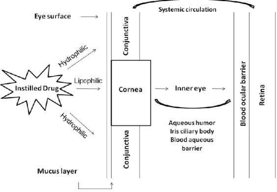
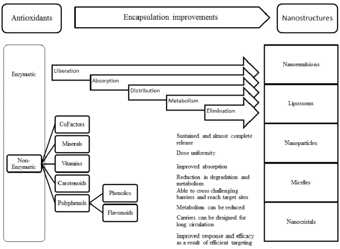
Figure 1. 1. Structure and molecular formula of chitosan. ... 9 Figure 1. 2. Schematic illustration of eye structure and subsequent critical biological barriers that drugs need to overcome after topical administration onto the eye surface. . 28 Figure 1. 3. Schematic view of the ocular permeability, physiological barriers, and transport pathways of a drug applied topically onto the eye. ... 29 Figure 1. 4. Classification of antioxidants and characterization of some nanostructures. Highlights to real benefits of antioxidants encapsulation considering its liberation, absorption, distribution, metabolism, elimination and response. ... 31 Figure 3. 1. Representative chromatogram of: (i). rosmarinic acid (retention time: 48.9 ± 0.1 min) and (ii). quercetin (retention time: 57.9 ± 0.6 min); (a). in standard solutions; (b). in sage extracts; (c). and savory extracts. ... 60 Figure 4. 1. Schematic illustration of ionic gelation process of antioxidant-chitosan based nanoparticles. ... 71 Figure 4. 2. TEM micrographs of fresh chitosan-based nanoparticles loaded: (a). commercial rosmarinic acid; (b). sage; and (c). savory. ... 77 Figure 4. 3. SEM micrographs of lyophilized chitosan-based nanoparticles loaded: (a). commercial rosmarinic acid; (b). sage; and (c). savory. ... 78 Figure 4. 4. Rosmarinic acid in vitro release from rosmarinic acid, sage and savory chitosan-based nanoparticles. ... 81 Figure 4. 5. Thermogram of: I.(a). chitosan empty nanoparticles; (b). free rosmarinic acid; (c). rosmarinic acid and chitosan physical mixture (mixing ratio 1:1); (d). rosmarinic acid encapsulated in chitosan nanoparticles (at a theoretical 40% loading) (d). II.(a). chitosan empty nanoparticles; (b). free sage; (c). sage and chitosan physical mixture (mixing ratio 1:1); (d). sage encapsulated in chitosan nanoparticles (at a theoretical 50% loading). III.(a). chitosan empty nanoparticles; (b). free savory; (c). savory and chitosan physical mixture (mixing ratio 1:1); (d). savory encapsulated in chitosan nanoparticles (at a theoretical 50% loading). ... 84
xxix
Figure 4. 6. Spectrum of: I. (a). chitosan empty nanoparticles; (b). rosmarinic acid in a free form; (c). physical mixture between chitosan unloaded nanoparticles and rosmarinic acid (mixing ratio 1:1); (d). rosmarinic acid encapsulation into chitosan nanoparticles. II.(a). chitosan empty nanoparticles; (b). sage in a free form; (c). physical mixture between chitosan unloaded nanoparticles and sage (mixing ratio 1:1); (d). sage encapsulation into chitosan nanoparticles. III.(a). chitosan empty nanoparticles; (b). savory in a free form; (c). physical mixture between chitosan unloaded nanoparticles and savory (mixing ratio 1:1); (d). savory encapsulation into chitosan nanoparticles. ... 87 Figure 6. 1. Effect of rosmarinic acid, sage and savory-loaded chitosan nanoparticles on cell cytotoxicity of ARPE (A, C and E) and HCE (B, D and F) cell lines after 4 h (black bar) and 24 h (white bar) of incubation. DMEM+cells and DMSO were used as controls. The formulation concentration used was displayed in the tables, relatively to rosmarinic acid (A, B), sage (C, D) and savory (E, F) (results were the mean of 6 replicates, bars represent standard deviation). ... 117 Figure 6. 2. Effect of rosmarinic acid, sage and savory-loaded chitosan nanoparticles on viability of ARPE (A, C and E) and HCE (B, D and F) cell lines after 4 h (black bar) and 24 h (white bar) of incubation. DMEM+cells and DMSO were used as controls. The formulation concentration used was displayed in the tables, relatively to rosmarinic acid (A, B), sage (C, D) and savory (E, F) (results were the mean of 6 replicates, bars represent standard deviation). ... 119 Figure 6. 3. Cumulative transport and TEER cell monolayer measurements of rosmarinic acid loaded chitosan-based nanoparticles and free in solution across HCE-T model cells (Values were means of 6 replicates, bars represent standard deviation). ... 124 Figure 6. 4. Cumulative transport and TEER cell monolayer measurements of rosmarinic acid loaded chitosan-based nanoparticles and free in solution across ARPE-19 model cells (Values were means of 6 replicates, bars represent standard deviation). ... 125 Figure 7. 1. Electroretinogram of mixture response of both rods and cones cell populations either in photopic or scotopic conditions (α and β – waves) recordings of rosmarinic acid administration effect. Left eye: representative traces of individual ERG recorded 24 h before treatment injection (Baseline ERG – blue line) and 24 h after intravitreal injection in I-R model (red line); Right eye: representative traces of individual ERG recorded 24 h
xxx
before treatment injection (Baseline ERG – blue line) and 24 h after intravitreal injection (red line). ... 134 Figure 7. 2. Rosmarinic acid effect on activated microglia induced by I-R injury at 24 h of reperfusion retinal. Retinal sections were stained with antibodies against Iba1 (green) and OX-6 (red). Nuclei were stained with DAPI (blue). Representative images were depicted in: Activated microglia/macrophages (Iba 1 - OX-6 immunoreactive cells), comparing with saline ischemial. ... 136 Figure 7. 3. Rosmarinic acid administration did not altered the cell death induced by I-R injury at 24 h of reperfusion. Cell death was assayed with TUNEL assay. Nuclei were stained with DAPI (blue). Representative images were depicted in: TUNEL+ cells (green) comparing with saline intravitreal and ischemial. ... 138
xxxi List of tables
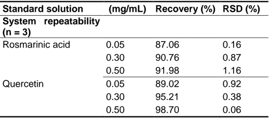
Table 1. 1. Principal properties of chitosan in relation to its use in biomedical applications. ... 10 Table 1. 2. Recent published data of antioxidants nanoencapsulation for several applications. ... 33 Table 3. 1. Results of precision tests for the determination of rosmarinic acid and quercetin in standard solutions. ... 56 Table 3. 2. Accuracy results for different levels of rosmarinic acid and quercetin in standard solutions. ... 58 Table 4. 1. Average hydrodynamic diameter (Z), polydispersity index (PdI), zeta potential and the association efficiency considering the different loading (%) of rosmarinic acid, sage and savory nanoparticles (n = 3). ... 76 Table 4. 2. Association efficiency, theoretical loading, and final rosmarinic acid content in chitosan nanoparticles. ... 80 Table 4. 3. Peak temperatures in the DSC thermograms collected from chitosan, rosmarinic acid, sage and savory, physical mixtures, and nanoparticles. ... 82 Table 5. 1. Antioxidant activity measurements by ABTS and ORAC, considering the 50% loading (m/m) sage and savory nanoparticles; and 40% loading (m/m) of and rosmarinic acid nanoparticles for (n = 3). ... 96 Table 6. 1. Average hydrodynamic diameter (Z), polydispersity index (PdI) and zeta potential of chitosan nanoparticles loaded rosmarinic acid, sage and savory. ... 114 Table 6. 2. Average hydrodynamic diameter (Z), polydispersity index (PdI) and zeta potential of chitosan nanoparticles loaded rosmarinic acid before and after mucin interaction (n = 3). ... 116 Table 6. 3. Cytotoxicity of rosmarinic acid, sage and savory-loaded chitosan nanoparticles for development of irritation symptoms such as vasoconstriction, hemorrhage and coagulation. ... 121
xxxii List of abbreviations
ABTS - 2,2-Azinobis (3-Ethylbenzothiazoline-6-Sulphonic) Acid AE - Association Efficiency
AGE - Advanced Glycation End Products AMD - Age Macular Degeneration ANOVA - One-Way Analysis of Variance
ARVO - Association for Research in Vision and Ophthalmology BAB - Blood-Aqueous Barrier
BRB - Blood-Retinal Barrier CAT - Catalase
CBQF - Centro de Biotecnologia e Química Fina Cupper - Cu
Da - Dalton
DAPI - 4',6-Diamidino-2-Phenylindole DD - Deacetylation Degree
DL - Detection Limit
DMEM - Dulbecco’s Modified Eagle’s Medium DMSO - Dimethyl Sulfoxide
DNA - Desoxyribonucleic Acid DPPH - Diphenyl-1-Picrylhydrazyl DSC - Differential Scanning Calorimetry EMA - European Medicines Agency ERG - Electroretinograms
FDA - Food and Drug Administration
FFUP - Faculdade de Farmácia da Universidade do Porto FTIR - Fourier Transform Infrared
GCL - Ganglion Cell Layer GNP - Gold Nanoparticles GPx - Glutathione peroxidase GSH - Glutathione
HBA - p – Hydroxybenzyl Alcohol HBSS - Hanks’ Balanced Salt Solution HCE-T - Human Cornea Cell Line
xxxiii HPLC - High Performance Liquid Chromatography
HPOX - Hydroxybenzyl Alcohol Incorporated Copolyoxalate IBILI - Institute for Biomedical Imaging and Life Sciences INEB - Instituto de Engenharia Biomédica
INL - Inner Nuclear Layer
IOBA – NHC - Immortalized Epithelial Cell Line from Human Conjunctiva IOP - Intraocular Pressure
IPL - Inner Plexiform Layer I-R - Ischemia-Reperfusion
ISCS-N - Instituto Superior de Ciências da Saúde – Norte LCPUFA - Long-Chain Polyunsaturated Fatty Acid
LDH - Lactate Dehydrogenase
MNP - Magnetically Responsive Nanoparticles MnSOD - Superoxide Dismutase
MSc - Master of Science
MTT - Thiazolyl Blue Tetrazolium Bromide MW - Molecular Weight
NR - Rosmarinic Acid Nanoparticles NSG - Satureja montana Nanoparticles NSV - Salvia officinalis Nanoparticles OCT - Optimal Cutting Temperature
ORAC - Oxygen Radical Absorbance Capacity OS - Oxidative Stress
Papp - Apparent Permeability Coefficient PBS - Phosphate Buffer Saline
PdI - Polydispersity Index PhD - Philosophiæ Doctor PLA - Polylactic Acid PP - Polypropylene
PUFA - Polyunsaturated Fatty Acid PVA - Poly (Vinyl Alcohol)
QL - Quantification Limit
QUEN - Quercetin Nanoparticles RGC - Retinal Ganglion Cells ROS - Reactive Oxygen Species
xxxiv RPE - Retina Pigment Epithelium
rpm - Rotations per Minute
RSD - Relative Standard Deviation SD - Standard Deviation
SEM - Scanning Electron Microscopy SiNPs - Silicate Nanoparticles
SOD - Superoxide dismutase
TEM - Transmission Electron Microscopy TPP - Tripolyphosphate
TUNNEL - Terminal Deoxynucleotidyl Transferase (TdT)-Mediated dUTP Nick End Labeling
UV - Ultra-Violet v - Volume
VEGF - Vascular Endothelial Growth Factor w - Weight
1
PART I - Introduction
“Somewhere, something incredible is waiting to be known”
3
CHAPTER 1 - State of art
5 1. Introduction
In pharmaceutical science there is a continuous blockbuster drug development, and nowadays biomolecules as active agents, are widely explored to develop new therapeutics. Nevertheless, most of these new active compounds are unstable and must be protected from degradation in the physiological environment, due to the poor absorption that constrains the transport across biological barriers. Thus, the efficacy of most drugs clearly depends on the design of appropriate carriers for their physical protection, delivery and controlled release (1). Among the different approaches explored so far, colloidal carriers are particularly interesting, especially those made of mucoadhesive polymers to assure their epithelium permanence (2, 3). For this application, chitosan has had quite impact in the association and delivery of labile macromolecular compounds (4). Chitosan carriers have an exceptional potential for drug delivery, especially for mucosal, since these systems are stable in contact with physiological fluids and barriers. They are also able to control drug release and protect against adverse conditions like mucosal enzymes and biological protective fluids. Due to its mucoadhesion, particle size, particle surface chemistries, charge and the unique absorption enhancing properties, the chitosan potential in the medical field is widely promising. Different formulations such as films, tablets, hydrogels, micro and nanosystems are expected to optimize, characterize and select the drug performance, improved properties and increase stability for great specific applications. Pharmacokinetics and toxicological relevance of chitosan systems are guaranteed by in
vitro model systems in molecular, subcellular and cellular levels, as well as their
therapeutic efficacy and safety performance should also be proven in vivo. One category of compounds in which these chitosan carriers may be a key for success, are the antioxidants. Considering the biology definition, antioxidants are chemical compounds or a substance that inhibits oxidation, counteracting the damage of free radicals effects in a living organism, and for this reason are reactive species (5). Antioxidants are widespread virtually in plant foods, often at high levels, and include phenols, phenolic acids and flavonoids (6). Rosmarinic acid (a-O-caffeoyl-3,4-dihydroxyphenillactic acid) (7) is a phenolic compound, which can provide protection against cancer (7) and have other multitude biological activities, namely adstringent, anti-inflammatory, anti-mutagen, antibacterial and antiviral (7, 8). The latter activity has been tested in the therapy of Herpes simplex infections with rosmarinic acid-containing extracts of Melissa officinalis (7). It is also one of the efficient natural antioxidants (9) since rosmarinic acid displays a
6
huge potential radical scavenging activity, higher than trolox (a derivative of a-tocopherol) (10-12). Rosmarinic acid has also an anti-angiogenic activity to retinal neovascularization in a mouse model of retinopathy (13). Significantly inhibited the proliferation of retinal endothelial cells in a dose-dependent manner, and inhibited in vitro angiogenesis of tube formation. Moreover, rosmarinic acid showed no retinal toxicity. These data suggest rosmarinic acid could be a potent inhibitor of retinal neovascularization and may be applied in the treatment of vasoproliferative retinopathies (13). It is the major component of Salvia officinalis (sage) and Satureja montana (savory) natural extracts. These are plants often used in traditional medicine, and which grow in the poor soils of the Mediterranean basin (14). Besides application as condiment, sage and savory have been used as an anti-diarrhea vector, digestion adjuvant, contribute to heal wounds, play an anti-inflammatory role, disinfectant, fight insomnia and decrease blood pressure. Some of these biological activities have been associated with its high contents of rosmarinic acid and the presence of other relevant phenolic compounds such as quercetin and rutin (9, 14). Beyond the biological huge benefits, antioxidants are extremely sensitive to light, oxygen, are highly reactive with other compounds, in some cases possess poor solubility, inefficient permeability, and are extremely unstable (15-17). For all these reasons their delivery using the conventional dosage forms is a challenge (18). In this context, alternative carriers are being considered, regarding the optimization of pharmacokinetics and pharmacodynamics of antioxidant molecules. Chitosan nanoparticles, due to their proper properties, are on the raw. For this concern the nanotechnology is expected to increase the ability, to retain the antioxidant activity during the preparation process, to optimize the release of the compound from the carrier system, and to ensure a good control of their physical-chemical properties increasing their stability. If these nanocarriers may improve the efficacy performance of the antioxidants, several diseases like cancer, diabetes, hypertension, arterio-sclerosis, cardiovascular disease or ocular anomalous conditions that have a clinical impairment with oxidation processes, may be prevented or better controlled (19). Considering the eye disorders, there are many types of retinopathies conditions that may have an oxidative etiology (20, 21), like retinitis pigmentosa, glaucoma, macular degeneration, retinoblastoma and diabetic retinopathy (19, 21). Multiple factors have been also proposed to explain retinopathies, including genetic disorders, infections by microbial agents, sorbitol pathway hyperactivity, accumulation of advanced glycation end products (AGEs) (22) and protein kinase C activation (23). Nevertheless the precise pathological mechanism remains to be elucidated. Which is clear is the collateral damage of these disorders, that may result on
7
reflectivity changes, bifurcations, tortuosity neovascularization as well as other patterns of blood vessels and even blindness (19, 21). In the case of ocular pathologies the oxidative stress (OS) clinical impairment has a significant impact, since the ocular globe is the organ most affected by OS. Its constantly expose to light and oxygen and its high polyunsaturated fatty acid (PUFA) content that is prone to lipid peroxidation, may be some prominent reasons (24). OS is also associated with increased vascular permeability, disruption of blood-retinal barrier, apoptotic loss of retinal capillary cells, microvascular abnormalities and neovascularization (25). High levels of OS are also usually associated with increased levels of oxidative modified desoxyribonucleic acid (DNA) and nitrosylated proteins, and antioxidant defense enzymes impair (26). When such damages are presented, without effective medical treatment, cells and tissues of the retina become malnourished and progressively degenerate, which leads to damage in cells responsible for vision, leading to its inevitable loss (21). Due to this intimate relationship between OS and the pathogenesis of retinopathies, the use of appropriate antioxidants may have potential on the metabolic and functional abnormalities in retinopathies (27). Nevertheless, for antioxidants assure these conditions they need the nanocarrier support to take them to the right place without losing their functional activity.
8
2. Chitosan proprieties and biomedical application
Chitosan is an alkaline-deacetylated chitin biodegradable polysaccharide commonly derived from the exoskeletons shells of crustaceans and insects (28). Its extraction is a cost-effective way to valorise seafood wastes and it is produced with different deacetylation degrees (DD) (29, 30), and molecular weights (MW) (31). Chitin is synthesized by an enormous number of living organisms and considering the amount of chitin produced annually in the world, it is the most abundant polymer after cellulose (29). Chitin is obtained on an industrial scale from shrimp and crustaceans in general. The pupae of silkworms are also an alternative source of chitin and, consequently, of chitosan (32). Another sources include the production of chitosan from fungi by fermentation (33), from chrysalides (a by-product from the silk industry) (32), or from sources of chitin (e.g. β-chitin) obtained from squid pens (4). However, the most commonly obtained form of chitosan is the α-chitosan from crustacean, which represents approximately 70% of the organic compounds in such shells (4).
For the chitin isolation from the raw materials and subsequent chitosan production, the original sources in solid form are washed with water, desiccated at room temperature and cut into small pieces. Demineralization is carried out at room temperature using hydrochloric acid and the de-proteinization should also be performed (32).
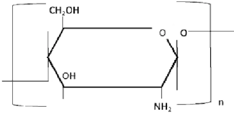
There are small difference in the chemical structure of chitin and chitosan, but these differences are extremely important when drug delivery is thought (34). Considering the similar structures of chitin and chitosan with cellulose, both are made by linear β-(1-4)-linked monosaccharides. However, the functional groups connected to the second carbon in the repeating units differ from one to another (35). Chitin is a linear homopolymer composed of β-(1,4)-linked N-acetyl-glucosamine units (34), while chitosan is a linear co-polymer polysaccharide consisting of β-(1–4)-linked 2-amino-2-deoxy-D-glucose (D-glucosamine) and 2-acetamido-2-deoxy-D-glucose (N-acetyl-D-(D-glucosamine) units, as depicted in Figure 1.1 (4). Chitin is insoluble in water and most common organic solvents used in pharmaceutical technology and consequently not useful in the development of drug delivery systems (36). In contrast, chitosan is a rather insoluble in water and organic solvents, but soluble in dilute aqueous acidic solution (pH < 6.5), which can convert the glucosamine units into a soluble protonated amine form (34). It is readily soluble in dilute organic acids such as acetic, citric and malic acid as well as hydrochloric acid (37). This positive charge of chitosan is very useful since enables negative interaction with polyanions (36). Chitosan is usually characterize in terms of MW, which commonly ranges
9
from ca. 10 to 1000 kDa, and DD, in the range of 50 to 95% (38). The biodegradability and biological properties of chitosan are frequently dependent on the relative proportions of N-acetyl-D-glucosamine and D-glucosamine residues (4) as well as on the MW.
Figure 1. 1. Structure and molecular formula of chitosan. .
Chitosan has biological properties (28, 39), unique and exceptional, that promote its use as a drug carrier (1, 3, 40), particularly to enhance transiently the permeability of mucosal barriers (36), increasing the effect on cell permeability (41), biocompatibility and biodegradability (42), being simultaneously non-toxic, non-antigenic (43) and mucoadhesive (2, 42). Previous reports also indicated that chitosan possess various biological activities, such as antitumor effects (44), anti-hypercholesterolemia (45) and antimicrobial activity against several pathogen and spoilage bacteria that has already been widely demonstrated (46). Currently, chitosan is widely used as a supporting material for tissue engineering applications, cell culture and nerve regeneration (47). Table 1.1, summarizes the main potential biomedical properties of chitosan and associated characteristics. The better understanding of this unique cationic polymer invites new and several applications.
10
Table 1. 1. Principal properties of chitosan in relation to its use in biomedical applications. Potential biomedical applications Principal characteristics
Surgical sutures Biocompatible
Dental implants Biodegradable
Artificial skin Nontoxic, biological tolerance
Bone rebuilding Non-antigenic
Corneal contact lenses Mucoadhesiveness
Time-control release drugs for animals and
humans Renewable
Encapsulating material Film forming
Tissue engineering Hydrating agent
Cell culture Hydrolyzed by lysozyme
Nerve regeneration Efficient against bacteria, viruses, fungi
Anti-tumor
Anti-hypercholesterolemia
2.1. Chitosan-based drug delivery systems
Controlled release technology emerged during the 1980s with a remarkable and increasing importance. This pharmaceutical technology that allows the predictable and reproducible release of a drug into a specific environment over an extended period of time, creating an optimal response, with minimum side-effects and prolonged efficacy, is a borderline of science (48).
The cationic nature of chitosan has been conveniently exploited for the development of new drug delivery systems and actually, a variety of chitosan-based delivery carriers have been described for pharmaceutical field as well as for other medical applications (1). In addition, the further chemical modification of chitosan is a powerful tool to control the interaction of the polymer with drugs, to enhance the load capability and to tailor the release profile of the particles (1). Hence, chemically modified chitosan improves its bulk properties for the preparation of sustained drug release systems (1), which enhance its versatility in the biomedical and biotechnological fields (49).
Chitosan can be employed to formulate a variety of pharmaceutical dosage forms, namely solutions, hydrogels, films, tablets, micro and nanoparticles. It is also a protagonist in
11
other advanced fields, like non-viral vector for DNA and gene delivery (49, 50). Drug delivery applications include oral, nasal, parenteral and transdermal administration, implants and gene delivery. The transmucosal administration of drugs is being largely exploited by chitosan-based systems (30).
2.1.1. Chitosan solutions
The study of chitosan solutions and related properties has much merit considering the multi-potential of the polymer and its many applications in the biomedical field. The preparation of the aqueous solution is also a mandatory step before obtaining any type of secondary materials such as films, gels, sponges, fibers, or particles. Despite this, most of the properties such as flocculation, adsorption and biological activities are accomplished in aqueous solution, the interactions between molecules of chitosan and metal ions, proteins, cells and bacteria have been widely studied in solutions (51). The gross conformation of chitosan in solution may be spherical shape, random coil and rod shape, which is manipulated by two sets of parameters: structure parameters such as MW and DD, and solution parameters such as ionic strength, solvent, temperature and pH. Molecular weight can induce a conformation transitions — the conformations of small MW chitosans are stiffer and more extended than those of higher MW. In general, an increase of ionic strength makes the molecule contract (51). These parameters are obtained from the plots of log intrinsic viscosity, sedimentation coefficient, diffusion coefficient, radius of gyration and log MW of the polymer (51). The most widely used methods to characterize the conformation transition are capillary viscosity, analytical ultracentrifugation, static and dynamic light scattering (51). Commonly, chitosan solutions are easily prepared dissolving chitosan in organic acid like acetic or lactic acid.
Chitosan solutions have demonstrated most of their applications in the mucosal absorption, mainly due to the transiently ability of chitosan molecules to open tight junction of epithelial cells. Globally, it can be concluded that chitosan is able to enhance the paracellular route of absorption by tight junction disruption, inducing the translocation of tight junction proteins from the membrane to the cytoskeleton (52). Regarding the effect of chitosan salts on drug intestinal permeability in vitro, there is general agreement that these polymers are potent absorption enhancers for poorly absorbed drugs such as atenolol and peptides (52). Studies with buserelin, 9-deglycinamide, 8-arginine
12
vasopressin and insulin have evidenced a strong increase in the transport of these drugs in the presence of chitosan glutamate and chitosan hydrochloride (acidic environment) (52). Studies using chitosan glutamate and western blotting of Caco-2 cells fractions, observed translocation of tight junction proteins (ZO-1 protein) from the membrane to the cytoskeleton in response to treatment with chitosan (53).
Yu and his collaborators (54) developed a chitosan solution for nasal insulin delivery. They reported that chitosan concentration, osmolarity, medium and absorption enhancing ability of chitosan solution have significant effect on nasal insulin absorption. This may represent a new administration of insulin, safer, convenient for diabetic patients and with good applicability.
Recent research has also exploited the ability of chitosan solutions to control the spread of cytokine and improve the immunoadjuvant properties in vaccine applications (55). Sustained, local delivery of immunomodulatory cytokines in chitosan solution is under investigation for its ability to enhance vaccine and anti-tumor responses, both pre-clinically and clinically. In short, the study concluded that the chitosan solution maintained a deposit of bioactive antigen and cytokines, induced a cell expansion of the lymph nodes, including an increase in dendritic cells and antigen presenting cells. The resulting increase in the capacity of antigen presentation was exploited to improve both the humoral and cellular responses to the vaccine. The ability of chitosan to form deposits of antigens and cytokines, in addition to its safety profile and inherent versatility makes it a promising platform for the delivery of vaccines and cytokines (55).
2.1.2. Films
Use of porous biomaterials in the film development attracts scientists in particular concerning the biomedicine applications (47). In the field of tissue engineering, porous polymer scaffolds can provide a framework for the seeded cells until they are well organized into a functioning tissue, especially in bone regenerative therapy (47). In the field of surgical, the porous biomaterial is usually used as wound dressing to absorb wound fluid and promote healing (47). On the other side, films, erodible and non-erodible inserts, rods and shields are the most logical delivery systems aimed at remaining for a long resident time in contact with physiologic surfaces like skin and the mucosal (56). Films by themselves or acting as drug carriers have been particularly considered worldwide (31). Porous membrane materials based on chitosan, add advantages of