Daniela Maria Martins Mendes
Clinical and molecular effects of
Hyperbaric Oxygen Therapy in diabetic
foot ulcers healing
Dissertação de candidatura ao grau de Doutor em Medicina, apresentada à
Faculdade de Medicina da Universidade do Porto
III
Artigo 48º, parágrafo 3 – A Faculdade não responde pelas doutrinas expendidas na
dissertação (Regulamento da Faculdade de Medicina do Porto – Decreto 19 337, de 29 de
Janeiro de 1931).
Esta investigação foi realizada na Consulta Multidisciplinar de Pé Diabético dos Serviços de
Endocrinologia, Diabetes e Metabolismo e de Medicina Interna do Centro Hospitalar de Vila
Nova de Gaia/Espinho, EPE; na Unidade de Medicina Hiperbárica do Hospital Pedro Hispano;
no Centro de Medicina Subaquática e Hiperbárica do antigo Hospital da Marinha; no Cancer
Biology Group do Instituto de Patologia e Imunologia Molecular da Universidade do Porto e
V
Orientadora: Professora Doutora Raquel Ângela Silva Soares Lino (Professora Catedrática da
Faculdade de Medicina de Universidade do Porto)
Co-orientador: Professor Doutor Jorge Filipe de Almeida Vieira Lima (Investigador do Instituto
de Patologia e Imunologia Molecular da Universidade do Porto e Professor Afiliado da
Faculdade de Medicina de Universidade do Porto)
A candidata declara que teve uma contribuição determinante na realização do trabalho,
interpretação dos resultados e discussão dos mesmos. Além disso, contribuiu activamente
para a redacção dos trabalhos apresentados.
VII
Corpo Catedrático da Faculdade de Medicina da Universidade do Porto
Professores Efectivos
Doutor Manuel Alberto Coimbra Sobrinho Simões
Doutora Maria Amélia Duarte Ferreira
Doutor José Agostinho Marques Lopes
Doutor Patrício Manuel Vieira Araújo Soares Silva
Doutor Daniel Filipe Lima Moura
Doutor Alberto Manuel Barros da Silva
Doutor José Manuel Lopes Teixeira Amarante
Doutor José Henrique Dias Pinto de Barros
Doutora Maria Fátima Machado Henriques Carneiro
Doutora Isabel Maria Amorim Pereira Ramos
Doutora Deolinda Maria Valente Alves Lima Teixeira
Doutora Maria Dulce Cordeiro Madeira
Doutor Altamiro Manuel Rodrigues Costa Pereira
Doutor Rui Manuel Almeida Mota Cardoso
Doutor António Carlos Freitas Ribeiro Saraiva
Doutor José Carlos Neves da Cunha Areias
Doutor Manuel Jesus Falcão Pestana Vasconcelos
Doutor João Francisco Montenegro Andrade Lima Bernardes
Doutora Maria Leonor Martins Soares David
Doutor Rui Manuel Lopes Nunes
Doutor José Eduardo Torres Eckenroth Guimarães
Doutor Francisco Fernando Rocha Gonçalves
Doutor José Manuel Pereira Dias de Castro Lopes
Doutor António Albino Coelho Marques Abrantes Teixeira
Doutor Joaquim Adelino Correia Ferreira Leite Moreira
Doutora Raquel Ângela Silva Soares Lino
VIII
Professores Jubilados ou Aposentados
Doutor Abel José Sampaio da Costa Tavares
Doutor Abel Vitorino Trigo Cabral
Doutor Alexandre Alberto Guerra Sousa Pinto
Doutor Álvaro Jerónimo Leal Machado de Aguiar
Doutor Amândio Gomes Sampaio Tavares
Doutor António Augusto Lopes Vaz
Doutor António Carvalho Almeida Coimbra
Doutor António Fernandes da Fonseca
Doutor António Fernandes Oliveira Barbosa Ribeiro Braga
Doutor António Germano Pina Silva Leal
Doutor António José Pacheco Palha
Doutor António Manuel Sampaio de Araújo Teixeira
Doutor Belmiro dos Santos Patrício
Doutor Cândido Alves Hipólito Reis
Doutor Carlos Rodrigues Magalhães Ramalhão
Doutor Cassiano Pena de Abreu e Lima
Doutor Daniel Santos Pinto Serrão
Doutor Eduardo Jorge Cunha Rodrigues Pereira
Doutor Fernando de Carvalho Cerqueira Magro Ferreira
Doutor Fernando Tavarela Veloso
Doutor Francisco de Sousa Lé
Doutor Henrique José Ferreira Gonçalves Lecour de Menezes
Doutor Jorge Manuel Mergulhão Castro Tavares
Doutor José Carvalho de Oliveira
Doutor José Fernando Barros Castro Correia
Doutor José Luís Medina Vieira
Doutor José Manuel Costa Mesquita Guimarães
Doutor Levi Eugénio Ribeiro Guerra
Doutor Luís Alberto Martins Gomes de Almeida
Doutor Manuel António Caldeira Pais Clemente
IX
Doutor Manuel Augusto Cardoso de Oliveira
Doutor Manuel Machado Rodrigues Gomes
Doutor Manuel Maria Paula Barbosa
Doutora Maria da Conceição Fernandes Marques Magalhães
Doutora Maria Isabel Amorim de Azevedo
Doutor Mário José Cerqueira Gomes Braga
Doutor Serafim Correia Pinto Guimarães
Doutor Valdemar Miguel Botelho dos Santos Cardoso
Doutor Walter Friedrich Alfred Osswald
XIII
Acknowledgements
Em primeiro lugar, gostaria de agradecer ao Professor Doutor Sobrinho Simões a oportunidade de integrar este Programa Doutoral, o apoio que me prestou ao longo de todo o percurso e a paciência para ouvir/ler alguns desabafos.
Ao Professor Doutor Henrique de Almeida pela ajuda à realização do projecto.
À minha Orientadora, Professora Doutor Raquel Soares, por ter aceitado a orientação deste projecto, nunca ter duvidado da sua conclusão, todo o auxílio prestado e prontidão nos esclarecimentos e proveitosas conversas de que surgiram ideias para o futuro.
Ao meu co-orientador, Professor Doutor Jorge Lima, toda a colaboração e profícuas ideias de projectos conjuntos de interajuda.
A todo o pessoal do Departamento de Bioquímica envolvido neste trabalho, nomeadamente ao Professor Doutor João Moura, Dra. Ilda Rodrigues, Dra. Alice Cortez, Dra. Raquel Costa, Dra. Ângela Castela, o meu muito obrigada.
I would like to thank the valuable help of Professor Edward Boyko and Professor Michael Bennett in reviewing and editing the articles.
Ao Professor Doutor Pedro Barata que, ao tomar conhecimento deste projecto, se entusiasmou e procurou prestar toda a colaboração possível.
Quero agradecer a todo o pessoal do Centro Hospitalar de Vila Nova de Gaia/Espinho, EPE:
- Aos Directores da Unidade de Gestão Integrada de Medicina Dr. Alcino Branco e Dr. Pedro Teixeira - Aos Directores do Serviço de Medicina Interna Dr. João Valente e Dr. Vítor Paixão Dias
- À minha Orientadora de Formação Dra. Olga Gonçalves;
- A todos os elementos da Consulta Multidisciplinar de Pé Diabético: Dra. Manuela Ribeiro, Dra. Merlinde Madureira, Dr. Campos Lemos, Dr. Daniel Brandão; Enf. Aldora Távora, Enf. Ema Lemos, Enf. Isaura Duarte, Enf. JB Sobral; Auxiliares “Paulinha” e “Maria Manuel” e Administrativa Isabel Cordeiro; por todo o apoio que prestaram e paciência que demonstraram para a realização deste trabalho, sem eles não teria sido possível;
- Ao Serviço de Anatomia Patológica por todo o apoio prestado; - À Dra. Margarida Mota, por ser uma das minhas mentoras e amigas;
- E a todos os colegas e pessoas que, de uma forma ou outra, contribuíram para a realização deste trabalho e/ou que sempre acreditaram que ele seria possível.
XIV
Não posso deixar de agradecer ao pessoal da Unidade de Medicina Hiperbárica do Hospital Pedro Hispano: Dr. Óscar Camacho, Dr. Tiago Fernandes, Dra. Elisabete Marques; Enf. Manuel Lopes, Enf. Filipe Alvarilhão, Enf. Edgar Cachada, Enf. Filipe Serrano; Operadores de câmara Alcino Pires, Miguel Monteiro e Manuel Preto; e às administrativas Manuela Malheiro e Ivone Gama; o entusiasmo e a disponibilidade demonstrados e ajuda indispensável à execução deste projecto. Um “profundo” agradecimento adiantado pela prometida prenda de conclusão de Doutoramento.
Um agradecimento ao Centro de Medicina Subaquática e Hiperbárica do antigo Hospital da Marinha, em especial ao Dr. Francisco Guerreiro e ao Dr. António Oliveira-Anão, pela receptividade à colaboração neste estudo, curiosidade e estímulo ao seu desenvolvimento e conclusão; e a todo o pessoal que tão bem me acolheu e boa experiência me proporcionou, nomeadamente restante quadro médico, enfermeiros, mergulhadores e pessoal administrativo, e pelo suporte teórico que me facilitaram. Foi uma honra poder estar presente na despedida à “antiga casa”; desejos de boa sorte e muitas felicidades na “nova casa”!
Ao Dr. Carlos Barosa, por me transmitir o seu amor pelo mar e incansável disponibilidade.
Um agradecimento especial ao Dr. Albuquerque e Sousa que, há vários anos, me levantou a cortina do mundo da Medicina Hiperbárica, pela sua simpatia, interesse incansável, por nunca ter duvidado que este trabalho “chegaria a bom porto” e sempre o ter apoiado, mesmo quando era uma ideia embrionária.
A todos os professores e mentores que tive, nomeadamente Professor Doutor Nuno Grande e Professor Doutor Oliveira Torres.
Um agradecimento especial ao saudoso Dr. Rui Seca, que me incutiu o “bichinho” da Medicina Interna. To all authors that kindly helped me by sending their articles and clarifying my doubts: Caroline Fife, Daniel Mathieu, Ernst Chantelau, Ezio Faglia, Gary Mawyer, Gary Smerdon, Michael Strauss, Miodrag Zivkovic, Paul Cianci and Tovy Malkin
Aos meus Amigos que acreditaram que este Doutoramento seria possível e compreenderam e toleraram as minhas ausências, em especial ao “Alguém” que partiu durante a fase final de escrita, obrigada pelos bons momentos, pelo sentido de humor tão especial e inconfundíveis gargalhadas. À Teresa Vaio pela sua Amizade e por nunca duvidar.
Um obrigado especial a 2 pessoas sempre disponíveis, que me apoiaram inteiramente, sobretudo nas alturas mais difíceis em que algumas coisas pareciam impossíveis.
Não posso deixar de fazer um agradecimento especial a todos os que me guiaram e orientaram ao longo de todo este percurso.
À Zezinha, ao Gustavo e aos outros meninos e meninas que me rodeiam.
XV
Abstract
THE INDEPENDENT CONTRIBUTION OF DIABETIC FOOT ULCER ON LOWER EXTREMITY AMPUTATION AND MORTALITY RISK
Aims: To estimate 3-year risk for diabetic foot ulcer (DFU), lower extremity amputation (LEA) and death; determine predictive variables and assess derived models accuracy.
Material and Methods: Retrospective cohort study including all subjects with diabetes enrolled in our diabetic foot outpatient clinic from beginning 2002 until middle 2010. Data was collected from clinical records.
Results: 644 subjects with mean age of 65.1 (±11.2) and diabetes duration of 16.1 (±10.8) years. Cumulative incidence was 26.6% for DFU, 5.8% for LEA and 14.0% for death. In multivariate analysis, physical impairment, peripheral arterial disease complication history, complication count and previous DFU were associated with DFU; complication count, foot pulses and previous DFU with LEA and age, complication count and previous DFU with death. Predictive models’ areas under the ROC curves ranged from 0.80 to 0.83. A simplified model including previous DFU and complication count presented high accuracy. Previous DFU was associated with all outcomes, even when adjusted for complication count, in addition to more complex models.
Conclusions: DFU seems more than a marker of complication status, having independent impact on LEA and mortality risk. Proposed models may be applicable in healthcare settings to identify patients at higher risk of DFU, LEA and death.
Keywords: Diabetes ∙ Diabetic Foot ∙ Foot ulcer
EFFECT OF HYPERBARIC OXYGEN THERAPY IN DIABETIC FOOT ULCER HEALING: A SYSTEMATIC REVIEW AND META-ANALYSIS
Aims: Conduct a systematic review, with meta-analysis (MA), considering hyperbaric oxygen therapy (HBOT) efficacy for diabetic foot ulcer (DFU) treatment; identify variables affecting outcome; grade overall methodological quality.
Material and Methods: We searched the National Library of Medicine, Rubicon Foundation Research Repository and hand-searched relevant scientific meeting abstract books. Two investigators independently extracted data concerning study characteristics and clinical outcomes, one assessed methodological quality.
XVI
Results: Forty studies were included: 11 randomized controlled trials (RCT), 8 non-randomized comparative trials (NRT) and 22 observational studies. The mean proportion of items satisfied, using CONSORT and STROBE, when pertinent, methodological quality reporting checklists, was 47% and 29% respectively. Wound area reduction of 92-100% in the HBOT group at 6 months was reported, but MA was not possible. Pooled difference in time to healing was 0.8 months less in HBOT group (not statistically significant). More ulcers healed with HBOT than without and the chance of healing a DFU was significantly greater with HBOT. There were also significantly fewer major amputations in those undergoing HBOT. Pooled estimates did not achieve statistical significance when RCTs alone were included in analysis. Ten studies assessed independent variables’ impact on healing; only age and TcPO2 were evaluated in 2 or more studies. Two models proposed to identify subjects that would benefit most of HBOT were never externally validated.
Conclusions: Although the overall methodological quality of these studies is poor, the existing evidence does suggest that HBOT is effective in DFU healing and decreasing the rate of major amputations.
Keywords: Diabetes ∙ Diabetic Foot ∙ Foot ulcer ∙ Hyperbaric Oxygen Therapy ∙ Systematic review ∙
Therapy
MOLECULAR ENVIRONMENT CHARACTERIZATION, HYPERBARIC OXYGEN THERAPY MODULATOR EFFECT AND CLINICAL IMPACT ON DIABETIC FOOT ULCERS’ HEALING
Aims: To compare, in subjects with diabetes mellitus (DM), molecular serum environment (angiogenic, vasculogenic and inflammatory markers) between those with and without active diabetic foot ulcer (DFU); assess hyperbaric oxygen therapy (HBOT) efficacy in DFU reduction and closure, serum markers modulation and microvasculature improvement.
Material and Methods: We conducted a non-randomized trial enrolling a group of patients with DM without active DFU (n=5) and one with active DFU with no significant wound evolution after 8 weeks of standard therapy. We compared those treated with HBOT (n=14) with untreated ones (n=6), due to treatment refusal or contra-indication. Endpoints (determined at 3, 6 and 12 months) included: laboratory markers, clinical outcome, DFU size, percentage of epithelialization.
Results: The sample mean age was 62 and DM duration 18 years. The majority were men, with type 2 DM, insulin-treated, with several complications and acceptable glycaemic and lipid control. After 3 months of HBOT, patients had a significant reduction in leukocyte and C-reactive protein serum levels, and of all DFU measurements. At every time points, the HBOT group achieved better outcomes (less amputation and death) and DFU reduction, with consequent increasing epithelialization percentage.
XVII
Comparison was not possible with the non-HBOT group as only one patient remained alive with active DFU and without major amputation at 3 months. Data points that after 1 month of HBOT, vessel number in the DFU tends to increase.
Conclusions: Our data reinforces the potential molecular and clinical efficacy and benefit of HBOT when added to current standard treatment of DFU.
Keywords: Diabetes ∙ Diabetic Foot ∙ Foot ulcer ∙ Hyperbaric Oxygen Therapy ∙ Therapy
IMPROVING HYPERBARIC OXYGEN THERAPY REFERRAL FOR DIABETIC FOOT ULCER TREATMENT: A NATIONWIDE MODELS’ VALIDATION AND REFINEMENT STUDY
Aims: This study characterizes the population with diabetic foot ulcer (DFU) undergoing hyperbaric oxygen therapy (HBOT) in Portugal over the last 5 years.
Material and Methods: Validation and optimization of the existing models for the prediction of healing of HBOT treated DFU; through a multicentre retrospective cohort study of all patients undergoing such therapy due to DFU from 2008 to mid-2013 in Portuguese continental hyperbaric medicine centres (n=2).
Results: We included 128 individuals. 66.4% underwent HBOT in Lisbon’s former Navy’s Hospital. Subjects from this hospital presented less frequently retinopathy (19.6% versus 74.5%, p<0.001) and previous DFU (29.2% versus 53.5%, p=0.02) and had ulcers of shorter duration (median 1.8 versus 4.0 months, p<0.001) than Pedro Hispano’s Hospital. Overall, 53.1% healed, which was more likely in non-smoking females without arterial disease, previous DFU or history of lower extremity amputation. Both completion of the planned series of treatments and increasing number of treatments had a positive impact on outcome. Available models had low predictive accuracy. We propose an optimized version of the Hawkins’ model that has higher accuracy.
Conclusions: There were some differences in the patients referred to each of these facilities, but healing rates were similar. Further studies are still needed to improve referral criteria for HBOT.
XIX
Sumário
ÚLCERA DE PÉ DIABÉTICO EM INDIVÍDUOS COM DIABETES COMO FACTOR DE RISCO INDEPENDENTE DE AMPUTAÇÃO E MORTALIDADE
Objectivos: Estimar o risco de úlcera de pé diabético (UP), amputação do membro inferior (AMI) e morte a 3 anos; determinar as variáveis preditivas e avaliar a validade do modelos derivados.
Material e Métodos: Estudo de coorte retrospectivo incluindo todos os sujeitos com diabetes seguidos na nossa consulta de Pé Diabético entre o início de 2002 e meados de 2010. Os dados foram colhidos dos registos clínicos.
Resultados: Incluímos 644 sujeitos com idade média de 65.1 (±11.2) e duração de diabetes de 16.1 (±10.8) anos. A incidência cumulativa foi de 26.6% para UP, 5.8% para AMI e 14.0% para mortalidade. Na análise multivariada, limitação física, história de complicação de doença arterial periférica, número de complicações e UP prévia demonstraram associação com UP; número de complicações, pulsos podológicos e UP prévia com AMI e idade, número de complicações e UP prévia com morte. As áreas sob a curva ROC dos modelos derivados variaram entre 0.80 e 0.83. Um modelo simplificado incluindo apenas UP prévia e número de complicações apresentou validade elevada. UP prévia associou-se com todos os outcomes, mesmo quando ajustada para número de complicações ou para modelos mais complexos.
Conclusão: UP parece ser mais do que um marcador de complicações instaladas, tendo um impacto independente no risco de AMI e mortalidade. Os modelos propostos podem ser utilizados em diversos contextos clínicos para identificar os sujeitos em maior risco de UP, AMI e morte.
Palavras-Chave: Diabetes ∙ Pé Diabético ∙ Úlcera
EFEITO DA OXIGENOTERAPIA HIPERBÁRICA NO TRATAMENTO DE ÚLCERAS DE PÉ DIABÉTICO: UMA REVISÃO SISTEMÁTICA E META-ANÁLISE
Objectivos: Realizar uma revisão sistemática e meta-análise (MA), avaliando a eficácia da oxigenoterapia hiperbárica (OHB) no tratamento de úlceras de pé diabético (UP) em indivíduos com Diabetes Mellitus; identificar as variáveis preditivas; avaliar a qualidade metodológica da evidência disponível.
Material e Métodos: Foi realizada uma pesquisa electrónica nos National Library of Medicine e Rubicon Foundation Research Repository e manual dos resumos de reuniões científicas relevantes. Dois
XX
investigadores seleccionaram os estudos e extraíram os dados e desfechos clínicos de forma independente, um avaliou a qualidade metodológica.
Resultados: Foram incluídos 40 estudos: 11 ensaios clínicos randomizados (ECR), 8 não randomizados (ECNR) e 22 estudos observacionais. A média de proporção de items reportados foi 47% e 29% para a CONSORT e a STROBE, respectivamente. A redução da área da ferida foi 92-100% no grupo OHB aos 6 meses (não foi possível MA). O grupo OHB demorou menos 0.8 meses até cicatrização (sem atingir significância estatística). As feridas com OHB cicatrizaram mais frequentemente. Ocorreram significativamente menos amputações major nos indivíduos sob OHB. As estimativas agregadas, incluindo somente os ECNR, não atingiram significância estatística. Apenas 10 estudos analisaram a associação de variáveis independentes com a cicatrização; só a idade e TcPO2 foram avaliadas por 2 ou mais estudos. Os 2 modelos existentes para identificar os sujeitos que mais beneficiarão da OHB nunca foram validados externamente.
Conclusões: Embora a qualidade metodológica dos estudos seja baixa, a evidência existente sugere que a OHB é eficaz na cicatrização das UP em indivíduos com diabetes, diminuindo o risco de amputações major.
Palavras-chave: Diabetes ∙ Pé Diabético ∙ Úlcera ∙ Oxigenoterapia Hiperbárica ∙ Revisão Sistemática ∙
Terapêutica
A OXIGENOTERAPIA HIPERBÁRICA NA EVOLUÇÃO CLÍNICA DE PATIENTES COM ÚLCERA DE PÉ DIABÉTICO ACTIVA: MELHORIA LOCAL E SISTÉMICA
Objectivos: Comparar, em indivíduos com diabetes mellitus (DM), com e sem úlcera de pé diabético activa (UP), o ambiente molecular sérico (marcadores angiogénicos, vasculogénicos, inflamatórios); avaliar a eficácia da oxigenoterapia hiperbárica (OHB) na redução e cicatrização de UP, modelação de marcadores séricos e melhoria da microvascularização.
Material e Métodos: Efectuámos um ensaio clínico não randomizado incluindo um grupo de participantes sem (n=5) e outro com UP sem melhoria após 8 semanas de tratamento convencional. Comparámos os sujeitos tratados com OHB (n=14) com os não tratados (n=6), por recusa ou contra-indicação. Os objectivos (aos 3, 6 e 12 meses) foram: marcadores laboratoriais, desfecho clínico, dimensões da UP e percentagem de epitelização.
Resultados: Na amostra, a idade média foi 62 e duração de DM 18 anos. A maioria eram homens, com DM tipo 2, várias complicações e controlo glicémico e lipídico aceitável. Após 3 meses de OHB, ocorreu uma redução significativa do nível sérico de leucócitos e proteína C-reactiva e de todas as dimensões da UP. Em todos os períodos, o grupo OHB apresentou melhores desfechos (menos amputações e
XXI
morte), redução da UP e consequente aumento da percentagem de epitelização. Não foi possível comparar com o grupo não-OHB visto apenas um indivíduo se manter vivo e sem amputação major aos 3 meses. Os resultados indicam que 1 mês de OHB tende a aumentar o número de vasos no leito da UP.
Conclusões: Os resultados reforçam o potencial efeito e benefício da OHB a nível molecular e clínico, quando adicionada ao tratamento convencional da UP.
Keywords: Diabetes ∙ Pé Diabético ∙ Úlcera ∙ Oxigenoterapia Hiperbárica ∙ Terapêutica
OPTIMIZAÇÃO DA REFERENCIAÇÃO DE INDIVÍDUOS COM DIABETES COM ÚLCERAS DE PÉ DIABÉTICO PARA TRATAMENTO COM OXIGENOTERAPIA HIPERBÁRICA: VALIDAÇÃO E REFINAMENTO DE MODELO COM BASE NACIONAL
Objectivos: Este estudo caracteriza a população com diabetes e úlcera de pé diabético (UP) activa que efectuaram Oxigenoterapia Hiperbárica (OHB) em Portugal nos últimos 5 anos.
Material e Métodos: Validação e optimização dos modelos existentes para a predição de cicatrização de UP tratadas com OHB, através de um estudo de coorte retrospectivo multicêntrico de todos os pacientes tratados com OHB, de 2008 a meados de 2013, nos centros de Medicina Hiperbárica em Portugal continental (n=2).
Resultados: Foram incluídos 128 indivíduos; 66.4% realizam OHB no Antigo Hospital da Marinha de Lisboa. Utentes provenientes deste hospital apresentavam menos retinopatia (19.6% versus 74.5%, p<0.001) e história de UP prévia (29.2% versus 53.5%, p=0.02) e as UP tinham menor duração (mediana 1.8 versus 4.0 meses, p<0.001) do que os do Hospital Pedro Hispano. No total, 53.1% das UP cicatrizaram, mais frequentemente em mulheres, não fumadoras, sem doença arterial periférica, história prévia de UP ou amputação. A realização da totalidade do tratamento prescrito e um maior número de sessões influenciaram positivamente o desfecho. Os modelos preditivos disponíveis apresentaram baixa validade, pelo que propusemos uma versão optimizada do modelo de Hawkins com melhores resultados.
Conclusões: Verificámos diferenças nas características dos indivíduos de cada instituição, mas a proporção de cicatrização foi semelhante. São necessários mais estudos para que se possa melhorar os critérios de referenciação para OHB.
XXIII
Table of contents
ACKNOWLEDGEMENTS
XIII
ABSTRACT
XV
SUMÁRIO
XIX
TABLE OF CONTENTS
XXIII
ABBREVIATIONS AND ACRONYMS
XXVII
LIST OF FIGURES
XXXI
LIST OF TABLES
XXXII
I.
LIST OF PUBLICATIONS
1
-II.
AIMS AND OUTLINE
5
-III.
RATIONALE
7
-IV.
BACKGROUND
11
-A. Diabetic foot 11
-1. Healing cascade 11
-2. Epidemiology of Diabetes: national and international 15
-3. The independent contribution of diabetic foot ulcer on lower extremity amputation and mortality
risk 17
-B. Hyperbaric Oxygen therapy 32
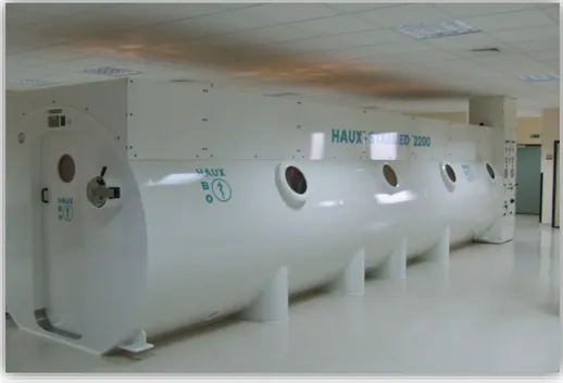
-1. Hyperbaric chamber 32
-2. Effects 35
XXIV
C. Effect of Hyperbaric Oxygen therapy in diabetic foot ulcer healing: a systematic review and
meta-analysis 42
-1. Material and methods 42
-2. Results 44
-3. Discussion 65
-V.
MOLECULAR ENVIRONMENT CHARACTERIZATION, HYPERBARIC OXYGEN
THERAPY MODULATOR EFFECT AND CLINICAL IMPACT ON DIABETIC FOOT ULCERS
HEALING
71
-A. Material and Methods 71
-1. Study design and participants selection 71
-2. Participants and diabetic foot ulcer characteristics 72
-3. Laboratory and molecular analysis 73
-4. Statistical analysis 74
-B. Results 75
-1. Participants characteristics 75
-2. Laboratory markers 76
-3. Clinical outcome 77
-4. Diabetic foot ulcer characterization 79
-5. Percentage of epithelialization 82
-6. Immunohistochemistry 82
-C. Discussion 84
-VI.
IMPROVING HYPERBARIC OXYGEN THERAPY REFERRAL FOR DIABETIC FOOT
ULCER TREATMENT: A NATIONWIDE MODELS’ VALIDATION AND REFINEMENT
STUDY
87
-A. Material and methods 87
-1. Participants selection 87
-2. Data collection 87
-3. Existing models of healing prediction 88
-4. Statistical analysis 89
-B. Results 89
-1. Hyperbaric Oxygen therapy facilities and program characterization 89
-XXV
3. Predictive variables for diabetic foot ulcer healing 92
-4. Predictive model accuracy 93
-C. Discussion 95
-VII.
CONCLUSION
99
-A. Main Conclusions 99 -B. Main Limitations 102 -C. Future research 105-VIII.
REFERENCES
107
-IX.
ANNEX
121
-XXVII
ABBREVIATIONS AND ACRONYMS
ABI Ankle brachial index
ADA American Diabetes Association
AGEs Advanced Glycation End Products
ARR Absolute Risk Reduction
ATA Atmospheres Absolute
AUC Area Under the Operator Receiver Operating Curve
CI Confidence Interval
cm3 Cubic Centimetres
CONSORT Consolidated Standards of Reporting Trials
CRP C-Reactive Protein
DFU Diabetic Foot Ulcer
Dl Decilitre
DM Diabetes Mellitus
DPN Diabetic Peripheral Neuropathy
ECHM European Committee for Hyperbaric Medicine
EPCs Endothelial Progenitor Cells
ESR Erythrocyte Sedimentation Rate
ESRD End Stage Renal Disease
ESWT Extracorporeal Shockwave Therapy
Fi O2 Fraction of Inspired Oxygen
FNH Former Navy’s Hospital
HbA1c Glycated Haemoglobin
HbCO Carboxyhemoglobin
HBI Hallux brachial Index
HBOT Hyperbaric Oxygen Therapy
HIF1 Hypoxia Inducible Factor
Hz Hertz
kPa Kilopascal
ICD-9 The International Classification of Diseases, 9th Revision
LEA Lower Extremity Amputation
LR Likelihood Ratio
XXVIII
MEDLINE National Library of Medicine
Ml Millilitre
mm Hg Millimetres of Mercury
MMPs Metalloproteinases
MOOSE Meta-analysis Of Observational Studies in Epidemiology
NA Not Applicable
NNT Number Needed to Treat
NO Nitric Oxide
NOS Nitric Oxide Synthase
NR Not Reported
NRT Non Randomized Trial
NS No Statistical Significant Association
O2 Oxygen
OR Odds Ratio
PAD Peripheral Arterial Disease
PBS Phosphate Buffered Saline
Pg Pico gram
PHH Pedro Hispano’s Hospital
PlGF Placental Growth Factor
PpO2 Partial Pressure of Oxygen
PPV Positive Predictive Value
PRISMA Preferred Reporting Items for systematic reviews and Meta-Analyses
RAGEs Advanced Glycation End Products Receptors
RCT Randomized Controlled Trial
ROC Receiver operating characteristic
ROS Reactive Oxygen Species
RR Relative Risk
RRR Relative Risk Reduction
SD Standard Deviation
SDF 1-α Stromal Cell Derived Factor 1- Alpha
SIGN Scottish Intercollegiate Guideline Network
STARD Standards for Reporting of Diagnostic Accuracy
STROBE Strengthening of the Reporting of Observational Studies in Epidemiology
XXIX
SWM Semmes-Weinstein Monofilament
TcPO2 Transcutaneous Partial Pressure of Oxygen
TNF-α Tumor Necrosis Factor-alfa
TUC Texas University Classification
UHMS Undersea and Hyperbarical Medical Society
VEGF Vascular Endothelial Growth Factor
VEGFR Vascular Endothelial Growth Factor Receptor
vs Versus
XXXI
List of figures
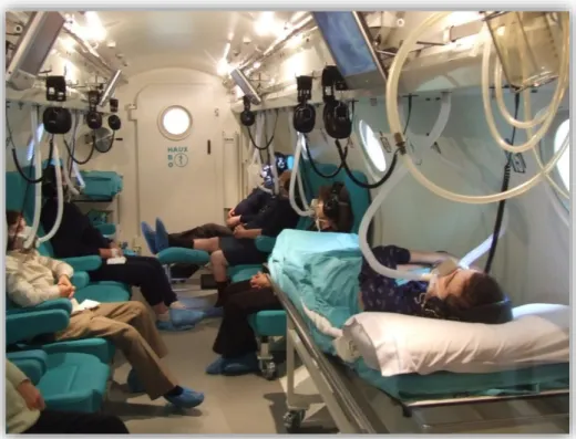
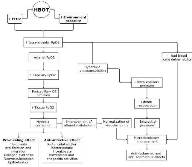
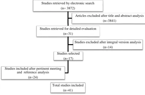
Figure 1. Diabetic foot ulcer pathophysiology ... - 13 - Figure 2. Healing impairment mechanism in the diabetic foot ... - 14 - Figure 3: Receiver operating characteristic curve of predictive models for diabetic foot ulcer (a), lower extremity amputation (b) and death (c) occurrence ... - 24 - Figure 4: Receiver operating characteristic curve of predictive models for diabetic foot ulcer (a), lower extremity amputation (b) and death (c) occurrence using only complication count and previous diabetic foot ulcer ... - 25 - Figure 5: Causes of death ... - 27 - Figure 6: Pedro Hispano’s Hospital multiplace chamber with capacity for 16 seating patients ... - 33 - Figure 7: Former Navy’s Hospital chambers with total capacity of 24 seating patients ... - 33 - Figure 8: Patients undergoing treatment for different pathologies ... - 34 - Figure 9: Monitoring panel ... - 34 - Figure 10. Hyperbaric oxygen therapy mechanisms of action in wound healing ... - 38 - Figure 11: Systematic review flow diagram of article selection process ... - 42 - Figure 12. Boxplot of number of vessels at baseline and 1 month in patients undergoing or not Hyperbaric Oxygen therapy ... - 82 - Figure 13: Predictive models’ receiver operating characteristic curve ... - 94 -
XXXII
List of tables
Table 1: Participants baseline characteristics ... - 20 - Table 2: Variables association with diabetic foot ulcer, lower extremity amputation and death occurrence ... - 22 - Table 3: Characteristics of included studies ordered by study type, quality assessment and sample size ... - 46 - Table 4: Randomized controlled trials methodological quality assessment using the CONSORT checklist ... - 52 - Table 5: Non-randomized trials methodological quality assessment using the STROBE checklist ... - 53 - Table 6: Cohort studies methodological quality assessment using the STROBE checklist ... - 54 - Table 7: Case series methodological quality assessment using the STROBE checklist ... - 55 - Table 8: Diabetic foot ulcer time until complete healing comparison between subjects treated with or without Hyperbaric Oxygen therapy: meta-analysis ... - 56 - Table 9: Diabetic foot ulcer healing rate comparison between subjects treated with or without Hperbaric Oxygen therapy: meta-analysis ... - 57 - Table 10: Lower extremity amputation proportion comparison between subjects treated with or without Hyperbaric Oxygen therapy: meta-analysis ... - 60 - Table 11: Variables association with complete diabetic foot ulcer healing in subjects undergoing Hyperbaric Oxygen therapy ... - 64 - Table 12. Participants baseline characteristics ... - 76 - Table 13. Laboratory markers ... - 78 - Table 14. Clinical outcome ... - 79 - Table 15. Diabetic foot ulcer characteristics at baseline and third month of follow-up ... - 80 - Table 16. Diabetic foot ulcer characteristics at baseline, 6th and 12th month of follow-up ... - 81 - Table 17. Example images of wound bed histology and immunohistochemistry at baseline and month 1 in one patient of each study group with active diabetic foot ulcer ... - 83 - Table 18: Patient characteristics according to Hyperbaric Oxygen therapy unit ... - 91 - Table 19: Patient characteristics by outcome ... - 92 - Table 20: Predictive models area under the receiver operating characteristic curve ... - 94 - Table 21: Optimized models prognostic accuracy measures ... - 95 -
- 1 -
I.
List of publications
Included in the thesis
Section IV, A, 3
Martins-Mendes D, Monteiro-Soares M, Lima J, Soares R.
Diabetic foot patients' 3 and 5 year follow up: ulcer occurrence, amputation and mortality. Rev Clin Esp, 2012, 212: 271-319
Abstract of poster oral presentation in 11th Congress of the European Federation of Internal Medicine, 24-27 October 2012, Madrid, Spain
Martins-Mendes D, Monteiro-Soares M, Boyko EJ, Ribeiro M, Barata P, Lima J, Soares R.
The independent contribution of diabetic foot ulcer on lower extremity amputation and mortality risk. J Diabetes Complications, 2014, [Epub ahead of print]
Impact factor: 2.06
Section V
Mendes D, Fernandes T, Camacho O, Lima J, Soares R.
Clinical and molecular effects of hyperbaric oxygen in diabetic foot ulcers – Preliminary data. Poster presentation in 37th EUBS Annual Scientific Meeting, 24-27 August 2011, Gdansk, Poland
Mendes D, Fernandes T, Camacho O, Lima J, Soares R.
Clinical and molecular effects of hyperbaric oxygen in diabetic foot ulcers – Preliminary data. Poster presentation in 1º Encontro de Doutorandos da FMUP, 14 December 2011, Oporto, Portugal
Mendes D, Monteiro-Soares M, Lemos E, Távora A, Sobral J, Duarte I, Brandão D, Campos-Lemos J, Madureira M, Ribeiro M, Fernandes T, Camacho O, Lima J, Soares R.
- 2 -
Poster presentation in 38th EUBS Annual Scientific Meeting, 10-13 September 2012, Belgrade, Serbia
Mendes D, Monteiro-Soares M, Fernandes T, Camacho O, Lima J, Soares R.
Clinical and microscopic effects of Hyperbaric Oxygen in diabetic foot ulcers’ healing.
Oral presentation in 2º Encontro de Doutorandos da FMUP, 14 December 2012, Oporto, Portugal
Martins-Mendes D, Costa R, Moura J, Rodrigues I, Cortez A, Ribeiro M, Lima J, Soares R.
Hyperbaric oxygen therapy in clinical outcome of patients with diabetic foot ulcers: local and systemic improvement.
Submitted for publication
Section VI
Martins-Mendes D, Monteiro-Soares M, Fernandes T, Camacho O, Ribeiro M, Oliveira MJ, Lima J, Soares R.
Validação de modelo preditivo para optimizar a identificação dos utentes com diabetes e úlcera de pé diabético ativa que beneficiarão de oxigenoterapia hiperbárica.
Revista Portuguesa de Diabetes, 2014, 9 (S1)
Abstract of oral presentation in 11º Congresso Português de Diabetes, 6-9 March 2014, Vilamoura, Portugal
Martins-Mendes D, Monteiro-Soares M, Bennett M, Oliveira-Anão A, Quaresma-Guerreiro F, Fernandes T, Camacho O, Lima J, Soares R.
Improving hyperbaric oxygen therapy referral for diabetic foot ulcer treatment: a nationwide models’ validation and refinement study.
Submitted for publication
In the Diabetic Foot topic but not included in this thesis
Monteiro-Soares M, Mendes D, Guimarães R et al.
Using diabetic foot ulcer development risk stratification systems for the wound healing prediction. 10th Scientific Meeting of the Diabetic Foot Study Group, 28-30 September 2012, Potsdam-Berlin, Germany
- 3 - Monteiro-Soares M, Martins-Mendes D, Guimarães R et al.
Referenciação para uma consulta multidisciplinar de Pé Diabético: análise da qualidade da informação. Revista Portuguesa de Endocrinologia, Diabetes e Metabolismo, 2013, 7(2): 64-104
Abstract of oral presentation in the XIV Congresso Português de Endocrinologia/ 64ª Reunião anual da SPEDM, 24-27 January 2013, Oporto, Portugal
Monteiro-Soares M, Martins-Mendes D, Guimarães R et al.
O impacto do controlo glicémico, avaliado através da Hba1c, na probabilidade de amputação podológico em utentes com diabetes e úlcera activa.
Revista Portuguesa de Diabetes, 2013, 8(S1): 5
Abstract of oral presentation in the Reunião Anual da SPD, 1-2 March 2013, Tomar, Portugal
Monteiro-Soares M, Martins-Mendes D, Dinis-Ribeiro M et al.
The impact of impaired renal function on the prediction of diabetic foot complications: should it be included on foot risk classifications.
Diabetologia, 2013, 56(S1): 1-566
Abstract of poster presentation in the 49th EASD Annual Meeting, 23-27 September 2013, Barcelona, Spain
Monteiro-Soares M, Martins-Mendes D, Vaz-Carneiro A, Sampaio S, Dinis-Ribeiro M.
Classification systems for lower extremity amputation prediction in subjects with active diabetic foot ulcer: a systematic review and meta-analysis.
Diabetes Metab Res Rev, 2014, [Epub ahead of print].
In the Hyberbaric Oxygen Therapy topic but not included in this thesis
Cervaens M, Martins-Mendes D, Camacho O, Barata P.
Oxigenoterapia hiperbárica na recuperação de lesões desportivas. Rev Medicina Desportiva, 2013, 4(5): 20–21
- 5 -
II.
Aims and outline
With this project we aimed to study the effects of hyperbaric oxygen therapy (HBOT) in the treatment of diabetic foot ulcers (DFU), namely the molecular and clinical effects, and consequently to improve the available evidence supporting this therapy.
In order to do so we have developed 4 main studies presented throughout this thesis.
In this Chapter II the structure of the thesis is described, while in Chapter III a short comment concerning the importance of this thesis conduction is performed.
Chapter IV, Section A, diabetic foot epidemiology and healing process impairment are overviewed and the results of a retrospective cohort study, characterizing the population followed in our Outpatient Diabetic Foot Clinic, where the molecular studies were conducted, in terms of demographic and clinical variables; estimating the 3- and 5-year risk of DFU development, lower extremity amputation (LEA) and death; and determining causes and predictive variables for these outcomes occurrence, are presented.
Chapter IV, Section B consists in detailing HBOT mechanisms, physiologic and therapeutic effects, side effects and complications, as well as, its clinical application.
Chapter IV, Section C is composed of a systematic review and meta-analysis (MA) assessing the available evidence concerning systemic HBOT effectiveness for the DFU treatment. With this study, we aimed to a) characterize available evidence assessing HBOT effectiveness for the DFU treatment, b) assess independent variables’ associated with healing, c) calculate pooled measures for minor and major LEA risk and d) identify methodological limitations of the published data.
In Chapter V, HBOT effectiveness in DFU is assessed. A non-randomized human trial is described, assessing molecular environment characterization, along with HBOT modulator effects in order to a)
- 6 -
identify and compare baseline angiogenic, vasculogenic and inflammatory markers in 3 groups of patients: 1) subjects with diabetes and without DFU, 2) with DFU and 3) with DFU treated with HBOT. In the last 2 groups, markers reassessment is performed at 3 months, to detect molecular differences due to HBOT. To understand if those effects persisted long term, in the HBOT group, a re-evaluation was carried out at 6 months. HBOT effectiveness in DFU healing was made by comparing outcome (healing, improvement, LEA and death), DFU dimensions, percentage of wound epithelialization and microvessel density analysis at different time points (3, 6 and 12 months) in the DFU groups.
Chapter VI consists in a nationwide multicentre retrospective cohort study, assessing the variables associated with DFU’s outcome when treated with HBOT, validating and refining a predictive model for DFU outcome in subjects undergoing this therapy, and reporting prognostic accuracy measures.
In Chapter VII, main conclusions and limitations are described in a standardized manner, addressing the studies strengths and limitations, and future research to overcome them is presented.
- 7 -
III. Rationale
Worldwide, diabetes mellitus (DM) is one of the most frequent metabolic disorders [Wild S 2004, IDF 2013] and its global burden is attributed to its several complications, namely DFU with impaired
healing, that frequently requiring LEA [Frykberg R et al, 2006; Margolis D et al, 2008; Sun J et al, 2012].
The occurrence of a DFU bodes poorly for the clinical course of patients with diabetes, with higher rates of re-ulceration, LEA, contralateral LEA and death, compared to persons with diabetes who have not experienced a DFU [Frykberg R et al, 2006].
Given the limited health care resources, it is important to optimize their allocation. To do so, an adequate stratification of subjects with diabetes by their risk of morbidity, namely DFU and LEA, as well as mortality, is crucial. Thus, identification of variables associated with these outcomes is the first step in the pathway for the creation or optimization of preventive/therapeutic programmes.
Even though the cascade of diabetic foot complications-DFU-LEA has been linked to higher mortality risk [Fortington LV, 2013], increasing number of DM complications is also associated with
higher mortality [Brownrigg JR, 2012]. DFU is usually considered a marker of diabetes complication
status, i. e., a marker for neuropathy and associated disease in the foot. Still, some authors hypothesized that DFU occurrence could be, per se, an independent predictive variable of LEA as well as mortality [Boyko EJ et al, 1996].
Nevertheless, adjustment for baseline complications was rarely conducted when assessing the impact of DFU on LEA, and of both on the mortality risk [Boyko EJ et al, 1996]. In addition, simple models
for their prediction (specially using the same core variables) were seldom proposed.
Given the current state of knowledge, we considered essential to estimate the risk for DFU, LEA and death in a cohort of patients with diabetes followed in our Diabetic Foot Outpatient Clinic and determine factors that independently predict LEA and mortality. This was the first step of this thesis, in order to assess whether it was pertinent and valuable to address a treatment modality for DFU in our population.
- 8 -
In our sample of 644 subjects, in a high risk setting, during 2002-2010, the 3-year cumulative incidence for DFU was 26.6%, for minor LEA 2.7% and for major 3.1%, and 14.0% for death. It was possible to derive simple models for prediction of the three outcomes, and we concluded that DFU was independently associated with LEA and death.
Several mechanisms are described in the literature explaining such associations. For example, peripheral arterial disease (PAD) impairs wound healing, due to inadequate circulation, and has been independently associated with both LEA [Adler A et al, 1999] and DFU [Boyko E et al, 1999].
We observed a higher rate of DFU development (>8% annually), but a similar of LEA and death, compared to several studies [Monteiro-Soares M et al, 2011; Schaper N, 2012]. However, measures to improve
these figures are still required, and so methods for DFU adequate treatment and prevention are essential.
This thesis emphasizes DFU therapy, for it is very challenging. Reported life time LEA rates range from 6.4 to 43% [Margolis D et al, 2008; Sun J et al, 2012]. Non-traumatic LEAs are up to 40 times [Fard et al, 2007]
more frequent in subjects with diabetes [Boulton A et al, 2005; Frykberg R et al, 2006]. Despite this great clinical
need, several authors consider the available evidence addressing DFU treatment to be very poor
[Mason et al, 1999].
One of such treatments is HBOT that aims to induce several physiologic and therapeutic effects. Nevertheless, it is an expensive therapy, with limited availability, thus, identification of patients with best possible response is fundamental for proper allocation of limited healthcare resources. Therefore, a systematic review and MA was conducted, evaluating all available evidence assessing the efficacy of systemic HBOT for DFU, identifying the independent predictors of outcome and the methodological limitations in the existing evidence. With such study, we observed that HBOT induces higher chance of DFU healing and lower risk of LEA. However, several limitations in the existing evidence were noticed, namely, small sample sizes, differences in treatment protocols, poor methodological quality and, for all these, a high heterogeneity in the calculated measures. In addition, animal studies point that HBOT, by raising tissue oxygenation in chronically hypoxic tissues, stimulates a number of elements required for wound healing, including angiogenesis, collagen synthesis and reactivation of the oxygen-dependent phagocytic capacity of leukocytes. Combined with the demonstrated ability of even a single HBOT session to mobilise endothelial progenitor cells (EPCs) from the bone marrow in diabetic individuals, all these actions promote active healing in a chronic, non-healing DFU [Gallagher et al, 2006; Thom SR et al 2006; Thom SR et al, 2011; Yang B et al, 2007].
- 9 -
Hence, we considered essential to compare, in subjects with diabetes, the molecular serum environment between those with and without active DFU and assess HBOT modulation and clinical efficacy at 3, 6 and 12 months. Our results showed that HBOT was effective in improving healing. Nevertheless, we were unable to fully understand the molecular mechanisms behind our good clinical results.
With our systematic review, we detected a lack of studies addressing the factors associated with improved healing and only 2 predictive models were retrieved, without ever being externally validated, for the identification of the subjects that would benefit most of HBOT.
In Continental Portugal, there are only two Hyperbaric Medicine Centres treating patients with active DFU, one in Oporto, located in Pedro Hispano’s Hospital (PHH) in Matosinhos (a public civil hospital, referral area from the north to the centre of the country) and one in Lisbon, in the former Navy’s Hospital (FNH) (military hospital, referral area from the centre to the south). Recently, hyperbaric centres in the archipelagos have also started treating DFU patients. In our clinical experience, we have noticed large variations in the clinical criteria when referring patients for HBOT.
Given these disparities, we decided to characterize the Portuguese population undergoing HBOT in the last 5 years, to understand Portugal’s pattern of DFU referral and subsequent outcome, and to propose an optimal simple referral model. Such goal was achieved and, despite several differences were observed between the studied institutions, healing rates were similar.
In sum, with this project we hope to have improved the evidence around DFU treatment with HBOT, in a translational manner. Firstly, assessing potential impact of HBOT in our context by evaluating diabetic foot complications development and death rates. Secondly, evaluating the available evidence about this topic to detect the possible need of further studies. As we concluded that there was a lack of biochemical and clinical studies with adequate methodological report, we performed one addressing molecular markers and clinical evolution and another improving referral protocol.
- 11 -
IV. Background
A. Diabetic foot
1. Healing cascade
Wound healing is a complex biological process involving several coordinated pathways. It is characteristically divided in four different phases: haemostasis, inflammation, proliferation and tissue remodelling [Young A, 2011], that overlap in time.
The process of healing encompasses numerous steps including haemostasis, removal of necrotic material and bacteria, inflammation terminus, extracellular matrix repair, neovascularization, epithelialization and remodeling [Li J et al, 2007; Young A, 2011]. We must emphasize that the majority of these
stages are dependent on oxygen content [Eming SA et al, 2007 A; Sen C, 2008; Tandara A & Mustoe T, 2004; Young A, 2011].
Haemostasis involves arteriolar constriction and posterior dilatation, clot formation through the pathways of the clotting cascade and platelet activation. Platelets also synthesize many factors that regulate various cell types and contribute to the inflammatory phase [Li J et al, 2007; Young A, 2011].
Immediately after a lesion occurs, inflammation begins with different types of leukocytes being attracted to the wound and releasing several mediators, including growth factors, which begin, maintain or finish angiogenic response and also recruiting other cell types. Neutrophils, the first to arrive, ingest and destroy bacteria through phagocytosis, degranulate and release several substances to destroy microorganisms and tissue debris, and produce oxygen reactive species (ROS) that have bactericidal effect [Eming SA et al, 2007 A; Li J et al, 2007; Young A, 2011].
In the proliferative phase, fibroblasts invade the wound bed, proliferate, synthesize extra-cellular matrix components, namely collagen, and later differentiate to a contractile phenotype, the myofibroblast, that connects to surrounding tissues, and is involved in wound contraction [Eming SA et al, 2007 A; Li J et al, 2007; Young A, 2011].
Neovascularization can occur through angiogenesis, by activation and migration of mature resident endothelial cells, and/or vasculogenesis, from undifferentiated angioblasts or EPCs both in
- 12 -
embryogenesis or bone marrow-derived in adults [Stavrou D, 2008; Velasquez OC, 2007]. This is an essential
process for the different phases’ occurrence.
During epithelialization, the epithelial cells migrate from the edges of the wound to cover its surface
[Eming SA et al, 2007 A; Li J et al, 2007; Young A, 2011].
Finally, during tissue remodeling, with synthesis and degradation of several components the scar is altered to achieve a structure similar to unwounded tissue [Eming SA et al, 2007 A; Young A, 2011].
Several biochemical factors are essential for an adequate healing process.
Vascular Endothelial Growth Factor (VEGF) family is composed of 7 elements: VEGF-A to F and Placental Growth Factor (PlGF). VEGF family and their receptors (VEGFR) are mediators of the embryo vascular development and of the adult angiogenesis processes (both physiological and pathological), including wound healing. VEGF-A is the most studied; its transcription is regulated by several factors such as growth factors, pro-inflammatory cytokines, hormones and cellular stress [including hypoxia – namely through action of Hypoxia Inducible Factor 1 (HIF1)] [Eming SA et al, 2007 A; Beldon P 2010].
It was studied, in transgenic mice, the role of PlGF in healing, which promotes faster wound repair, with increase of the granulation tissue vasculatization. Soluble vascular endothelial growth factor receptor-1 (sVEGFR-1), a splicing variant of transmembranar VEGFR-1, is considered a natural inhibitor of VEGF-A. Studies revealed that sVEGFR-1 concentration in chronic wounds fluid is higher than its concentration in wounds that heal [Eming SA et al, 2007 A].
In diabetic patients and animal models, the number and function of circulating EPCs is highly decreased, and these alterations are related to the cardiovascular and healing complications in diabetes [Costa C et al, 2007; Fadini GP et al, 2005; Fadini GP, 2014; Laing T et al, 2007].
Stromal Cell Derived Factor 1- Alpha (SDF1-α) is a cytokine that induces mobilization of bone-marrow derived progenitor cells, mainly through the C-X-C Chemokine receptor type 4 [Velasquez OC, 2007].
Tumor necrosis factor-alfa (TNF-α), a factor with multiple functions, plays a central role in inflammation, therefore, being an intervenient in the inflammatory phase of healing [Schreml S et al, 2010].
DM frequently leads to foot ulcers, through an already exhaustively described pathophysiologic process (Figure 1), and also to a defective wound repair due to alterations in the micro- and macro-vasculature, cellular and molecular environment, as well as to the frequent presence of infection and/or neuropathy [Brem H & Tomic-Canic M, 2007; Costa C et al, 2007; Laing et al, 2007; van Weel et al, 2008].
Diabetic patients and animal models have altered vasculature. In some territories, like retina, angiogenesis is increased, while in others, for instance distal microvasculature of the inferior limbs, it is decreased [Costa C et al, 2007; van Weel et al, 2008]. Studies have shown that diabetic mice, in whom the
healing process is altered, have lower levels of VEGF-A mRNA and of the protein’s intracellular processing during wound repair and decreased angiogenesis [Costa C et al, 2007; Brem H & Tomic-Canic M, 2007].
- 13 -
Additionally, the diabetic environment induces several molecular changes by advanced glycation end products (AGEs) that link to their receptors (RAGEs), including pathways activation, altered dermal fibroblasts and keratinocytes proliferation [Blakytny R & Jude E, 2009; Schreml S et al, 2010]
Figure 1. Diabetic foot ulcer pathophysiology
(Adapted, with permission, from Monteiro-Soares, 2010)
Local hypoxia is common. The optimum oxygen tissue pressure (pO2) is 50-100 mmHg but in many wounds it scarcely reaches 10 to 30 mmHg [Eming SA et al, 2007; van Weel et al, 2008]. On the other hand, one of
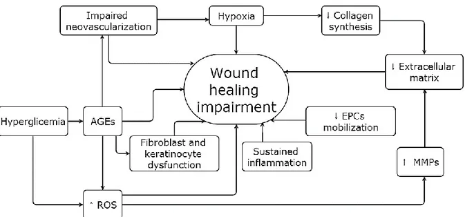
the main reasons for delayed wound healing is a sustained inflammatory reaction [Eming SA et al, 2007 A; Eming SA et al, 2007 B; Schreml S et al, 2010]. For all this (Figure 2), DFUs frequently evolve to chronic wounds,
- 14 -
Figure 2. Healing impairment mechanism in the diabetic foot
: Increase; : Decrease; AGEs: Advanced Glycation End product; EPCs: Endothelial Progenitor Cells; MMPs: Metalloproteinases; ROS: Reactive Oxygen Species
- 15 -
2. Epidemiology of Diabetes: national and international
Approximately 8.3% of the world adult population, corresponding to 382 million people, has DM which turns this condition in one of the most frequent metabolic disorders [IDF 2013].
Incidence and prevalence are continuously rising, consequently carrying high rates of morbid-mortality, with premature deaths, and economic burden [IDF 2013]. In fact, it was reported that 2.5% to
15% of global annual health care budgets are spent on DM, with an annual direct medical cost worldwide of around 241 billion dollars [WHO, 2013; Alavi A et al, 2014].
In Europe, it is calculated that 56 million people have DM, around 28% of deaths in people under 60 years old are due to this disease [IDF 2013]and that the majority of these could be prevented [WHO, 2013; Alavi A et al, 2014].
In Portugal, during 2012, DM caused 4880 potential years of life lost in those less than 70 years and a total of 4867 deaths were attributed to this condition, corresponding to 4.5% of global mortality [OND, 2013].
In Oporto region, DM has a prevalence of 13.9% [OND, 2009], whereas the national one is 12.9% [OND, 2013].
We must highlight that the referral area of the Centro Hospitalar de Vila Nova de Gaia/Espinho, Entidade Pública Empresarial (EPE), Diabetic Foot Outpatient Clinic, where the clinical studies of this thesis were conducted, belongs to this higher prevalence region.
Diabetic foot is one of the major DM complications and causes a considerable costs in health care and patient well-being. It is estimated that around 10 to 25% of subjects with diabetes will develop a DFU
[Frykberg R et al, 2006], during their lifetime, from which 50 to 70% will recur in a 5 year period [Alavi A et al, 2014].
Chronic DFU have a great impact in the subjects’ quality of life. It was stated that such individuals present from 10 to 40% lower quality of life scores when compared to the general population, being comparable to those with chronic lung disease, myocardial infarction, and breast cancer [Armstrong DG et al, 2008].
On the other hand, treating DFU represents substantial costs, with the highest impact being associated with the consequent LEA [Akhtar S et al, 2011] and rehabilitation.
In Portugal alone, 1493 LEA were performed in 2012 due to diabetic foot complications (730 major and 763 minor LEA) [OND, 2013], with major differences between the regions being noticed. In the last
National Observatory report it is stated that fewer major LEA are conducted in the North region, where Oporto is included. Along this thesis some possible explanations will be discussed.
Nevertheless, several studies confirm that treating successfully a DFU is consistently more cost-effective when compared to the expenses of performing a major LEA [Alavi A et al, 2014; Morbach S, 2003].
- 16 -
For all this, the development and/or improvement of therapeutic options for the DFU healing are crucial.
- 17 -
3. The independent contribution of diabetic foot ulcer on lower extremity
amputation and mortality risk
Material and Methods
Subjects
A retrospective cohort study was conducted including all subjects with diabetes followed in Centro Hospitalar de Vila Nova de Gaia/Espinho, Entidade Pública Empresarial (EPE), Diabetic Foot Outpatient Clinic from the 1st of January 2002 until the 31st of May 2010.
Subjects were excluded if they met any of the following criteria: active DFU at the moment of inclusion, inability to ambulate, communication or cognitive impairment (due to aphasia and/or dementia), missing data on any covariate (except for vibration sensation assessed using a tuning fork and HbA1c), follow-up period of less than 3 years, or living outside our referral area.
The Diabetic Foot Clinic is a tertiary care unit, with a multidisciplinary team and specialized diabetic foot care, treating patients from primary care institutions (usually with high risk feet and/or unavailable appropriate care in their residence area) or from other departments and hospitals. The study was approved by the Ethics Committee of our institution and no adverse event occurred in any subject due to participation in this research.
Data collection
Clinical records were reviewed and data collected from 1st until the 30th of June 2013.
All variables were collected in the first podiatric appointment in the clinic, through a structured interview and detailed foot exam, apart from HbA1c, by one of the two department podiatrists who were experienced in the care of diabetic foot complications.
Demographic characteristics (age at the time of inclusion, gender, education level), DM type (classified according to the World Health Organization definition [WHO, 2006]), duration and treatment (diet only,
oral medication or insulin), metabolic control [through glycated haemoglobin (HbA1c)], physical (inability to reach his/hers own feet [Monteiro-Soares M et al, 2012]) and/or visual impairment and smoking
habits (absent, current, former) were recorded.
DM complications [retinopathy, nephropathy, neuropathy, cerebrovascular, cardiovascular, PAD and metabolic (ketoacidosis, hyperosmolar coma or other coma)] were classified in accordance to the Diabetes Complications Severity Index created by Young et al [Young BA et al, 2008], according to their
protocol, using the International classification of diseases, 9th revision (ICD-9) Codes, through clinical record review.
- 18 -
As Young et al concluded that the accuracy of the number of complications was similar to the Complication Severity Index and as the number of complications is easier to calculate we opted to use it. Nephropathy was also staged by the American Diabetes Association (ADA) classification [ADA, 2013],
using the serum creatinine value closest to the date of the first appointment.
Participants’ feet were characterized using the variables more frequently described in DFU development risk stratification systems [5] and with proved association with its occurrence [Monteiro-Soares M et al, 2012], namely, the presence of deformities, onychomycosis, diabetic peripheral neuropathy
(DPN) [using the Texas Verbal Questionnaire [Armstrong DG et al, 1998 A], Semmes-Weinstein monofilament
(SWM) insensitivity and tuning fork vibration sensation], PAD (characterized by total foot pulses and intermittent claudication), oedema and history of previous DFU or LEA. There is a lack of studies assessing the reliability of these measurements [Monteiro-Soares M et al, 2012]. Previous DFU was collected
through foot assessment, patient self-report and all were additionally confirmed by medical record review. All the above described variables in addition to visual and/or physical impairment, presence of onychomycosis and DFU occurrence were collected and defined according to the protocol previously described by Monteiro-Soares et al [Monteiro-Soares et al, 2010 B].
Vibration sensation test (VST) was assessed with a 128 Hz tuning fork applied, perpendicularly with a constant pressure, on a bony part on the dorsal side of the distal phalanx of the first toe.
This procedure was repeated twice and two incorrect answers were classified as altered sensation
[Bakker K et al, 2012]. This procedure was instituted in 2008 and therefore patients that entered into the
study prior to this time do not have this assessment.
Subjects were followed from the time of inclusion to death or completion of the 3-year follow-up. Minor LEA was defined as the surgical removal of toe(s), ray(s) or forefoot. Major LEA was considered amputation of the entire foot by any level of the leg (including the ankle).
HbA1c value was not always available (n= 164) as several patients were followed for their metabolic control mainly by primary care physicians.
DFU and/or LEA occurrence and death dates were registered. Subjects were advised to contact the clinic if any lesion developed and during appointments they were asked if any DFU occurred. Furthermore, complete medical records from the hospital as well as primary care institutions were reviewed in order to detect missed events. LEA and death (date and cause) are automatically registered in the individuals’ computerized clinical file. Death causes were collected using the ICD-9 codes.
- 19 - Statistical analysis
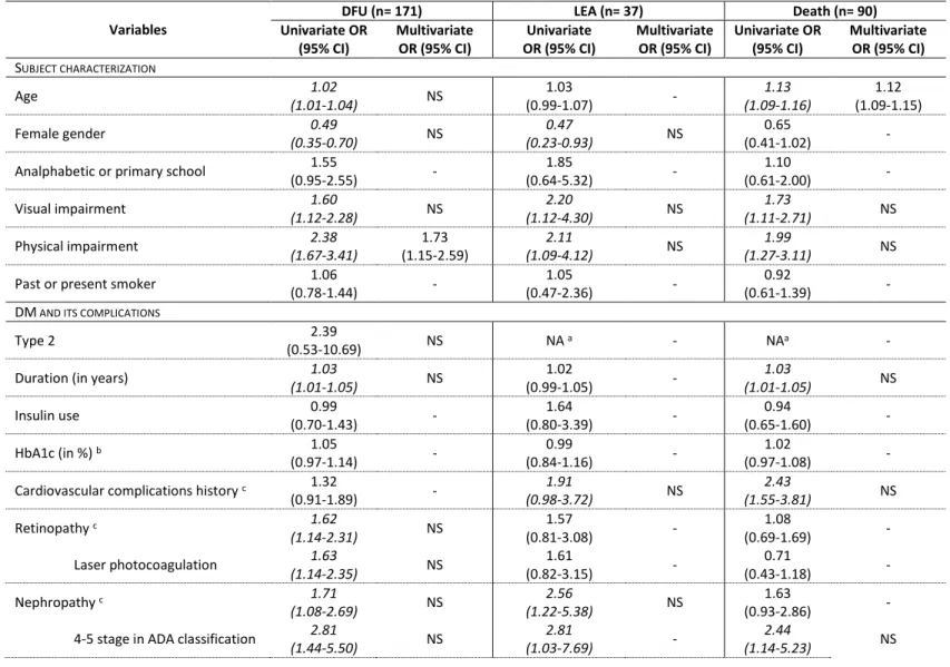
Association between variables and outcomes (DFU, LEA or death) was conducted using univariate logistic regression. Values of p ≤ 0.05 were considered as statistically significant and ≤ 0.1 as pertinent for initial inclusion into the predictive models. Multivariate analysis to estimate odds ratios (OR) for amputation and mortality in relation to DFU adjusted for covariates was performed using logistic regression analysis employing a backwards stepwise algorithm approach. In addition, all multivariable models included age, gender and diabetes duration.
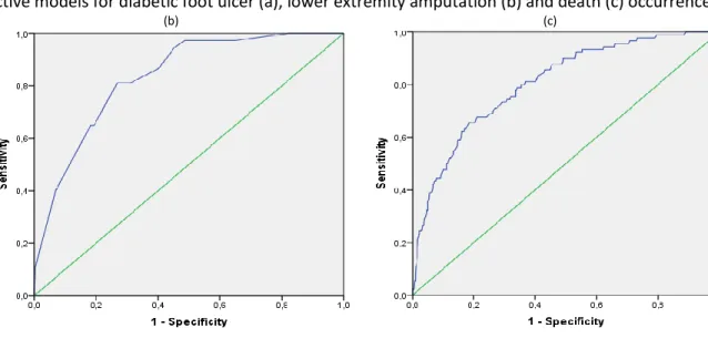
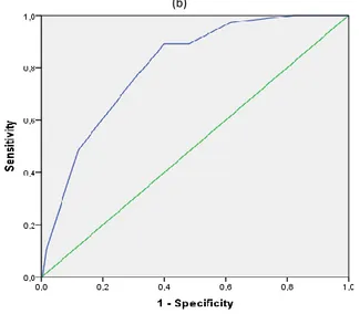
After the model creation for each outcome, a multivariable score was computed for each subject using the β coefficient values and the actual values for the covariates for those subjects. The ability of the score to discriminate between patients who did and did not develop the outcomes of interest was assessed using the area under the receiver operating characteristic curve (AUC) with the 95% confidence interval (CI).
All statistical analyses were conducted using the programme IBM SPSS, version 20.0 (Chicago, IL, USA). Missing and indeterminate results were excluded from analysis.
Results
Participant characteristics
In this study, 644 subjects were included and followed for a median of 36 months (range 1-36). At baseline, patients had a mean age of 65.1 (±11.2) years; mean diabetes duration of 16.1 (±10.8) years; and mean HbA1c of 7.8% (±3.7). The majority had type 2 DM and less than half were on insulin. More than half were female; over 80% were undereducated (primary school level or less) and over a quarter had some form of impairment (visual and/or physical). The most frequent complications were PAD related (63.0%) and the least frequent was metabolic complication history (3.6%). Forty-one percent of our population had a history of previous DFU (Table 1).
Cumulative incidence at 3 years for DFU and the outcomes of interest was as follows: DFU 26.6% (95% CI 23.2-30.0), recurrent DFU 34.5% (95% CI 27.4-48.4), minor LEA 2.7% (95% CI 1.4-4.0), major LEA 3.1% (95% CI 1.8-4.4), total LEA 5.8% (95% CI 3.9-7.5) and death 14.0% (95% CI 11.3-16.7).