U
NIVERSIDADE DE
L
ISBOA
Faculdade de Ciências
Departamento de Biologia Vegetal
Testing short LNA-modified oligonucleotides for Duchenne
Muscular Dystrophy gene therapy
MESTRADO EM BIOLOGIA MOLECULAR E GENÉTICA
Vanessa Borges Pires
Dissertação orientada pela Prof. Doutora Célia da Conceição Vicente Carvalho e coorientada pela Prof. Doutora Ana Rita Barreiro Alves de Matos
Agradecimentos
Primeiro gostaria de agradecer à Professora Maria Carmo-Fonseca por me ter acolhido no seu laboratório e pelo incentivo dado para que apresentássemos os nossos trabalhos à comunidade científica e pelo feedback em relação às nossas prestações em apresentações e no trabalho. Gostaria de agradecer a todos os envolvidos neste projeto, especialmente à minha orientadora, a Doutora Célia Carvalho, por me ter dado a oportunidade de desenvolver este trabalho no Instituto de Medicina Molecular de Lisboa e pela partilha de valiosos conhe-cimentos, pelas críticas construtivas, pela paciência, pelo apoio e incentivo ao longo do ano que me fez crescer como pessoa e investigadora. Gostaria de agradecer à Professora Doutora Ana Rita Matos pela disponibilidade, apoio, motivação e por ser o meu elo de ligação com a Faculdade de Ciências da Universidade de Lisboa nos aspetos mais burocráticos.
Gostaria de agradecer muitíssimo a todos os colegas do Instituto de Medicina Molecular que me apoiaram no decorrer deste ano. Ao pessoal do Biotério do IMM, gostaria de agradecer a disponibilidade que apresentaram para me ajudar nos momentos de dúvidas e o conheci-mento que me concederam, em especial à Dolores Bonaparte pelo ensinaconheci-mento cuidado na manipulação dos animais. Gostaria de agradecer à Joana Coelho, pela disponibilidade, pelos ensinamentos no que diz respeito a testes funcionais em animais e pela muita paciência que teve comigo e com os ratinhos mdx52. Gostaria de agradecer ao Ângelo Chora pela disponi-bilidade e auxílio prestado na realização das injeções intravenosas nas caudas dos ratinhos mdx52. Ao grupo que me acolheu e deu apoio no ano que decorreu, o grupo da Professora Doutora Maria Carmo-Fonseca, um grande obrigado por me receberem, me apoiarem sempre que eu tinha alguma dúvida e me ajudarem de todas as maneiras possíveis. Gostaria ainda de agradecer à Ana Jesus, à Sílvia, à Dinora, ao Sérgio, à Sandra, à Geni e à Noélia pelas muitas discussões e dúvidas que me esclareceram, e à Soraia, Sara, Tomás, Joana Tavares, Catarina, Rita Drago e Rita Mendes de Almeida pelas muitas brincadeiras, pelo apoio e cari-nho constante e pela motivação que me deram ao longo deste ano. Gostaria de agradecer ao António e Ana Margarida, da Bioimagem, pelos conhecimentos e apoio prestado.
Gostaria de agradecer a todos os meus amigos pelo apoio e pensamento positivo, em particular, à Ana Patrícia, à Joana Oliveira e à Ema pela força e motivação e por acreditarem em mim.
Por último, mas porque os últimos são os primeiros, gostaria de agradecer aos meus pais pelo apoio incansável e constante com que sempre me presentearam e por tornarem possível esta etapa na minha vida.
Resumo
A base hereditária de cada organismo vivo é o genoma, uma longa sequência de ácido desoxirribonucleico (DNA), que contém a informação genética organizada em genes. Em eu-cariotas, os genes são compostos por exões, interrompidos por intrões. Os exões contêm a informação genética que é traduzida em proteína. Uma vez que a informação genética se encontra no núcleo da célula, e a maquinaria de tradução está localizada no citoplasma, é necessária a formação de um ácido ribonucleico (RNA) mensageiro temporário (pré-mRNA). Este pré-mRNA é processado no núcleo, por uma série de passos que incluem o capping da extremidade 5’, a remoção de intrões e junção de exões, o splicing e a clivagem e poliadenila-ção da extremidade 3’. O RNA mensageiro resultante (mRNA) é exportado para o citoplasma, tornando-se disponível para os ribossomas, onde a tradução em proteína decorre.
O splicing é um processo altamente complexo e regulado no qual estão envolvidas cente-nas de proteícente-nas. O splicing alternativo, que ocorre quando um único gene origina mais do que uma sequência de mRNA, é um mecanismo importante para a regulação da expressão génica. Uma vez que o splicing e o splicing alternativo são mecanismos extremamente importantes e conservados na Natureza, a sua disrupção pode levar a doenças. Apesar de a disrupção des-tes mecanismos estar subjacente a muitas doenças hereditárias e adquiridas, a modulação do splicing através da utilização de oligonucleotídeos antisense pode ter aplicações terapêuti-cas. A modulação de splicing pode ser conseguida in vitro com o uso de compostos químicos ou oligonucleotídeos antisense (AONs) que se podem ligar a uma determinada sequência do pré-mRNA e regular o splicing do gene, quer para restaurar a função do gene ou para inibir a expressão génica. A modulação do splicing oferece esperança para o combate de muitas doenças genéticas, que são atualmente incuráveis. O exemplo mais notável desta aplicação terapêutica é na Distrofia Muscular de Duchenne (DMD), causada principalmente por muta-ções nonsense e de frame-shift no gene DMD, localizado no cromossoma X, que codifica para a proteína distrofina. As distrofias musculares constituem um grupo heterogéneo de doenças genéticas musculares caracterizadas por um progressivo enfraquecimento, atrofia e degeneração muscular. As distrofias musculares associadas a deficiências no gene da pro-teína distrofina, podem apresentar vários fenótipos, desde o mais grave e mais comum – a Distrofia Muscular de Duchenne (DMD), até ao mais ligeiro – a Distrofia Muscular de Becker (BMD). A DMD mais severa, apresenta uma incidência de 1 em cada 3500 recém-nascidos do sexo masculino, apresentando a BMD, a mais ligeira, uma incidência de 1 em cada 18 500. A distrofina é a proteína responsável pela manutenção da estabilidade da membrana das fibras musculares. Mutações neste gene levam à perda de função da proteína, sendo esta patologia
caracterizada pela ausência de distrofina funcional no músculo cardíaco, esquelético e liso. Apesar dos esforços, nenhuma terapia eficaz e clinicamente aplicável foi ainda desenvol-vida, sendo no entanto possível atrasar o onset da doença recorrendo a terapias com gluco-corticoides. Têm sido investigadas terapias genéticas de correção da reading frame, sendo as abordagens mais promissoras as que visam o skipping de exões com mutações frame-disrupting do pré-mRNA, que apesar de introduzirem deleções, conseguem gerar transcritos in-frame permitindo a síntese de uma proteína, que mesmo sendo mais pequena, consegue ser funcional. Na terapia desta patologia, o skipping de um exão mediado por oligonucle-otídeos antisense (AONs) pode restaurar a open reading frame (ORF) e permitir a síntese de uma proteína parcialmente funcional. O skipping de um exão pode ser induzido pela li-gação de oligonucleotídeos antisense, direcionados para um ou ambos os locais de splice ou sequências internas do exão. Teoricamente, a correção da reading frame pode ser conseguida com o skipping de um ou mais exões que flanqueiem a deleção, ou com o skipping de exões in-frame que contenham mutações nonsense, ou com o skipping de exões duplicados. Os oli-gonucleotídeos antisense Drisapersen (com a estrutura química 2’-O-metil) e Eteplirsen (com a estrutura química morfolino) estão atualmente a ser testados em ensaios clínicos de Fase III como terapia para a Distrofia Muscular de Duchenne, e a aguardar aprovação da Agência Europeia de Medicamentos (EMA) e da Federal Drug Administration (FDA). Estes oligonucle-otídeos antisense conseguem restaurar a reading frame da distrofina promovendo o skipping do exão 51 do gene da distrofina, sendo esta abordagem aplicável a aproximadamente 13% dos pacientes que sofrem desta patologia.
Uma vez que os oligonucleotídeos antisense, que têm sido estudados e que avançaram para ensaios clínicos, têm mostrado um sucesso limitado no que diz respeito a eficácia clí-nica, neste trabalho procuramos testar a utilização de um diferente tipo de oligonucleotídeos: oligonucleotídeos modificados com Locked Nucleic Acid (LNA) – LNA-AONs, como nova estra-tégia para alcançar a correção do gene mutado, resgatando a expressão da proteína distrofina, induzindo o skipping direcionado do exão 51. Procurámos descobrir se estes LNA-AONs po-dem ser utilizados para terapias de modulação de splicing com aumento da eficiência. Os nucleótidos LNA possuem uma unidade de açúcar alterada que forma uma ponte metileno. Esta modificação permite propriedades melhoradas em termos de aumento de estabilidade de hibridação com a sequência alvo, de alta sensibilidade, boa descriminação de incompati-bilidades (mismatches), baixa toxicidade e estabilidade metabólica aumentada, ajustando às propriedades necessárias para terapia humana.
Neste trabalho, linhas celulares derivadas de mioblastos de pacientes com DMD e indiví-duos normais (Mamchaoui, K. et al, 2011) foram utilizados para indução in vitro de skipping do exão 51 do pré-mRNA da distrofina e o modelo animal mdx52 (Aoki, Y. et al, 2012), ratinhos que apresentam uma deleção do exão 52 no gene DMD, foi utilizado para indução in vivo de skipping do exão 51 do pre-mRNA da distrofina nos tecidos musculares. Pesquisámos skipping do exão 51 ao nível dos transcritos, através da utilização de técnicas de RT-PCR e pesquisá-mos a restauração da produção da proteína, através da utilização de técnicas de Western Blot e Imunofluorescência. Os nossos resultados mostraram skipping eficaz do exão 51 e
ração da produção da proteína distrofina em mioblastos distróficos de pacientes, transfectados com oligonucleotídeos modificados com LNA utilizando concentrações tão baixas como 5nM. No modelo animal, o ratinho mdx52, após cinco injeções intravenosas na cauda, a cada duas semanas, com 1mg/kg de LNA-AON, duas semanas após a última injeção, não foi detetado skipping do exão 51 por RT-PCR, nem proteína via Western Blot. No entanto, aglomerados de fibras positivas para a distrofina foram detetadas por imunohistoquímica em ratinhos mdx52 tratados,e não em ratinhos injectados como controlo, levando a crer que são fibras em que a produção de distrofina foi induzida terapeuticamente devido ao LNA-AON usado. Ocasional-mente, observamos a presença de fibras positivas isoladas para a distrofina em ratinhos não tratados, no entanto nestes casos estamos perante fibras revertentes, ou seja, fibras isoladas ocasionais que ocorrem naturalmente e parecem expressar distrofina corretamente localizada. Estas fibras revertentes poderão ser exemplos onde, por acaso, mutações secundárias adici-onais ou eventos intrínsecos aberrantes de splicing proporcionam o skipping de um ou mais exões adicionais de forma a restaurar a reading frame correta original, permitindo a produção de uma proteína funcional. Para que ocorra uma reversão do fenótipo da DMD para um fenó-tipo menos severo, como o da BMD, ou para evitar distrofias musculares em humano e ratinho, é necessária a expressão de níveis de 20 a 30% da distrofina normal no tecido muscular.
O objetivo deste projeto foi testar se LNA-AONs poderiam ser utilizados para terapias de modulação splicing com maior eficiência, em relação aos AONs já estudados. Nós demons-tramos que o resgate de expressão de distrofina realizando o skipping do exão 51 é viável em linhas celulares derivadas de mioblastos in vitro, e em ratinhos mdx52 distróficos in vivo. Os resultados apresentados, obtidos com o modelo celular, parecem muito promissores, a fim de alcançar uma boa recuperação da proteína distrofina em mioblastos de doentes DMD bem como os alcançados em em ratinhos mdx52. Seguidamente será necessária otimização do sistema de entrega dos oligonucleotídeos para o tecido muscular de forma generalizada, uma vez que o tratamento de todo o organismo é um desafio e que os tecidos envolvidos são pós-mitóticos, sendo aproximadamente 30 a 40% do corpo humano constituído por músculo. A melhoria da eficiência de modulação de splicing e da biodistribuição de oligonucleotídeos anti-sense (AONs) pode reduzir a dose terapêutica e intervalo das administrações, minimizando a potencial toxicidade, efeitos off-target, e custos. Com este trabalho mostrámos a aplicabilidade de pequenos oligonuleótidos LNA modificados para terapia genética da Distrofia Muscular de Duchenne, abrindo caminho para uma pesquisa de métodos mais eficientes para terapia gené-tica por modulação de splicing em tratamento sistémico de uma doença genégené-tica hereditária.
Palavras-chave: Distrofia Muscular de Duchenne, Terapias de Modulação de Splicing,
Oligonucleótidos modificados com LNAs, Skipping de exões, Splicing
Abstract
Genetic disorders are caused by abnormalities in an individual’s DNA. Novel therapeutic strategies for this types of diseases have been emerging, especially on therapies that involve the modulation of disease-related gene expression. Modulation of splicing using antisense oligonucleotides (AONs) is feasible in vitro and can have possible therapeutic applications.
The most notable example is in Duchenne Muscular Dystrophy (DMD), a genetic hereditary disease caused mainly by frame-shifting or nonsense mutations in the DMD gene in chromo-some X, which encodes for the dystrophin protein, essential for membrane stability of muscle fibers. In DMD cells, antisense-mediated exon skipping can restore the open reading frame and allow synthesis of partly functional dystrophin proteins instead of non-functional proteins, trans-forming DMD in the milder Becker Muscular Dystrophy (BMD). The antisense oligonucleotides Drisapersen (2’-O-methyl chemistry) and Eteplirsen (morpholino chemistry) are currently being tested in clinical trials as a therapy for DMD. These aim to restore the dystrophin reading frame by promoting skipping of exon 51, an approach that is applicable to approximately 13% of DMD patients. Locked Nucleic Acid (LNA) modified oligonucleotides carry an altered sugar moiety that forms a methylene bridge allowing improved properties in terms of increased duplex stabil-ity, high sensitivstabil-ity, good mismatch discrimination, low toxicity and increased metabolic stabilstabil-ity, fitting the properties needed for human therapy. An oligonucleotide wuth this chemistry is cur-rently awaiting for clinical application as HCV infection therapy after good results in clinical trials. We aim to test if LNA oligonucleotides (LNA-AON) can be used for splicing modulation therapies, with increased efficiency.
In this work, myoblast derived cell lines from patients and mdx52 mice were used for in vitro and in vivo induction of skipping of DMD-exon 51, respectively. We looked for skipping of exon 51 at the transcript level (RT-PCR) and for restoration of protein production (Western Blot and Immunofluorescence). Our results show effective skipping of exon 51 and restora-tion of dystrophin protein producrestora-tion in dystrophic myoblasts transfected with the LNA-AON at concentrations as low as 5nM. In the animal model, the mdx52 mouse, after five biweekly tail intravenous injections of 1mg/kg with the LNA-AON, we were able to visualize clusters of dystrophin positive fibers therapeutically induced by the LNA-AON on cryosections of selected muscles.
With this work we showed the applicability of short LNA-modified oligonucleotides for Duchenne Muscular Dystrophy gene therapy, paving the way for a search of more efficient methods for gene therapy splicing modulation in systemic treatment of an inherited genetic disease.
Keywords: Duchenne Muscular Dystrophy, Splice Modulation Therapy, LNA Oligonucleotides,
Exon Skipping, Splicing
Contents
List of Figures xi
List of Tables xi
Introduction 1
Splicing Mechanics . . . 1
Duchenne Muscular Dystrophy . . . 3
Antisense Oligonucleotides (AONs) and Splice-Switching Oligonucleotides (SSOs . . 5
Antisense-mediated Exon Skipping: Applicability in DMD . . . 7
Clinical Trials: Antisense Oligonucleotides (AONs) and DMD . . . 7
A new approach: LNA-modified oligonucleotides (LNA-AON) . . . 8
Materials and Methods 9 Myoblast Derived Cell Lines Culture and Differentiation . . . 9
LNA-AON Structure and Transfections . . . 9
Animals . . . 10
LNA Treatment of mdx52 Mice . . . 10
Blood Analysis and Muscle Functional Testing . . . 10
RNA Isolation from Cell Extracts . . . 11
RNA Isolation from Muscle Samples . . . 11
RT-PCR Protocol . . . 12
Protein Extraction from Cell Extracts . . . 13
Protein Extraction from Muscle Samples . . . 13
Western Blot Protocol . . . 13
Immunocytochemistry . . . 14
Immunohistochemistry . . . 14
Microscope Image Acquisition and Analysis . . . 14
Statistical Analysis . . . 14
Results and Discussion 15 In vitro Evaluation of the Splicing Modulation . . . 15
Exon 51 Skipping at the Transcript Level . . . 15
Restoration of Protein Production . . . 17
In vivo Evaluation of the Splicing Modulation in mdx52 mouse model . . . 21
Blood Analysis and Muscle Functional Testing . . . 21
Exon 51 Skipping at the Transcript Level . . . 22
Restoration of Protein Production . . . 22
Final Remarks . . . 26
Bibliography 29
Supplemental Information 35
List of Figures
1 Splicing Process . . . 2 2 Differences between a normal, Duchenne patients and Becker dystrophin protein 4 3 The dystrophin-associated protein complex . . . 5 4 Chemical structure of biological and synthetic oligonucleotides . . . 6 5 Efficacy of exon 51 skipping in a patient myoblast derived cell line (DM8036
patient cell line) . . . 16 6 Exon 51 skipping is not detected after two weeks differentiation in patient
my-oblast derived cell lines (DM8036 patient cell line) . . . 17 7 Restoration of protein production after exon 51 skipping in a myoblast derived
cell line (DM8036 patient cell line) . . . 18 8 Restoration of protein production after exon 51 skipping in myoblast derived cell
lines (DM8036 patient cell line) . . . 20 9 No amelioration of the skeletal muscle function was observable in the mdx52 mice 22 10 Efficacy of exon 51 skipping in treated mdx52 mice . . . 23 11 Restoration of protein production after systemic injections in mdx52 mice . . . . 24 12 Restoration of protein production after systemic injections in mdx52 mice . . . . 25 13 Efficacy of exon 51 skipping in treated mdx52 mice injected once with eye
intra-venous injection of 10mg/kg . . . 35 14 Exon 51 skipped dystrophin did not ameliorate skeletal muscle function in mdx52
mice injected once with eye intravenous injection of 10mg/kg . . . 36 15 Schematic representations of the LNA-AON target site and the restoration of
protein production in the cell and animal model . . . 36
List of Tables
1 Exon Skipping Applicability in DMD Patients . . . 7 2 Primer sequences used in this study. . . 12
Introduction
Genetic disorders are caused by abnormalities in an individual’s DNA. Novel genetic therapeu-tic strategies for this types of diseases are emerging. We will focus especially on therapies that involve the modulation of disease-related gene expression, since pre-mRNA splicing is an essential step for eukaryotic gene expression1. The modulation of splicing can be achieved in vitro with the usage of chemical compounds or antisense oligonucleotides (AONs) that can bind to a target site in pre-mRNA and interfere with RNA splicing, either to restore gene function or to inhibit gene expression1. Splicing modulation offers hope for combatting many genetic dis-eases that are currently untreatable. Different approaches have been developed with the aim of treating some genetic disorders like ataxia-telangiectasia, methylmalonic academia, dystrophia myotonica1. The disease in which research on a splicing modulation therapy is more advanced is Duchenne Muscular Dystrophy (DMD).
Splicing Mechanics
The hereditary basis of every living organism is the genome, a long sequence of deoxyribonu-cleic acid (DNA), that contains the genetic information organized in genes2. In eukaryotes, the
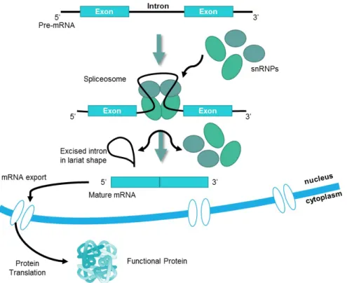
genes are composed of exons and are interrupted by introns2. The exons contain the genetic information that is translated by the cell into protein, while the introns do not contain protein encoding information2,3,4. Since the genetic information is enclosed in the cell nucleus, and the translating machinery is located in the cytoplasm, a temporary messenger ribonucleic acid (RNA) molecule (mRNA) needs to be generated4. The pre-mRNA is processed in the nucleus, by a series of steps that include 5’ end capping, the removal of introns and joining of exons, splicing and 3’ end cleavage and polyadenylation2,4,5,6, resulting in a mRNA that is exported from the nucleus to the cytoplasm, becoming available to the ribosomes where translation into protein ensues4,6(Figure 1).
Splicing is a highly complex, tightly regulated, process in which hundreds of proteins and RNAs are involved3,4,5. Splicing requires four loosely defined sequence elements in the
pre-mRNA, which are the 5’ splice site (consensus in mammals: AG/GURAGU), the branch point (YNYURAC), a variable stretch of pyrimidines termed polypyrimidine tract and the 3’ splice site (YAG/N; where "/" denotes the exon-intron boundary)2,7. The splice sites are sequences imme-diately surrounding the exon-intron boundaries that include the sites of breakage and reunion of exons2. The initial process of splice site recognition, by sequence motifs in introns and exons, commits the pre-mRNA substrate to the splicing pathway and are important for proper process-ing of pre-mRNA into mRNA2. The 5’ and 3’ splice sites and the branch point sequences are
2
Figure 1: Schematic representation of the splicing process.
recognized by components of the splicing apparatus that assemble to form a large complex – the spliceosome2. The splicing reaction proceeds via two sequential transesterification reac-tions: a lariat is formed when the intron is cleaved at the 5’ splice site and the 5’ end is joined to a 2’ position at the adenosine (A) at the branch site in the intron, the intron is released as a lariat when it is cleaved at the 3’ splice site, and the left and right exons are then ligated together2. Since introns can be very large in vertebrates, recognition of the splice site involves additional interactions across the exon between the 3’ splice site and the downstream 5’ splice site, this process is known as exon definition2.
When a gene gives rise to a single type of spliced mRNA, there is no ambiguity in as-signment of exons and introns, however, the vast majority of mammalian genes are spliced and follow patterns of alternative splicing4,2,5,8. Alternative splicing occurs when a single gene gives rise to more than one mRNA sequence, and explains how a huge proteome (>100,000 proteins) can arise from a relatively small number of genes (25,000 on the human genome), being also an important mechanism for regulation of gene expression4,2,5,8. There are vari-ous modes of alternative splicing, including intron retention, alternative 5’ splice site selection, alternative 3’ splice site selection, cassette exon inclusion or skipping and mutually exclusive selections of the alternative exons2,5,8.
Alternative splicing is often associated with weak splice sites, meaning that the splicing signals located at both ends of introns diverge from the consensus splicing signals2. The
se-quences surrounding alternative exons are often more evolutionary conserved than sese-quences flanking constitutive exons2. The regulation of alternative splicing is a complex process that
3
may recognize RNA elements in specific exonic and intronic sequences, near the splice site, and enhance or suppress splice site selection2. The exonic alternative splicing sequences that enhance splice site selection are referred to as exonic splicing enhancers (ESEs)2. When the exonic sequences promote suppression of splice site selection, these are called exonic splicing silencers (ESSs)2. Similarly, many elements can affect splice site selection through intronic se-quences, being referred to as intronic splicing enhancers (ISEs) and intronic splicing silencers (ISSs)2. The effect of splicing enhancers and silencers is mediated by sequence-specific RNA binding proteins, many of which may be developmentally regulated and/or expressed in a tissue-specific manner4,2,5,8. Since splicing and alternative splicing are such important and well conserved mechanisms, in their disruption underlie many inherited and acquired genetic diseases4,5,8,9.
Modulation of splicing can be achieved in vitro by oligonucleotides containing sequences complementary (antisense) to unique sequences within the pre-mRNA, which can bring about the exclusion or inclusion of an exon, modifying splicing and, thus, gene expression10,5,6,11. This has potential therapeutic applications and antisense-mediated splicing modulation to block aberrant splice sites, to correct disrupted alternative splicing levels, to include aberrantly skipped exons and to induce exon skipping to knockdown protein levels are currently targets of intensive research4,11,10,12. The most notable example of this approach is in Duchenne Muscular Dys-trophy (DMD), where an antisense-mediated exon skipping therapy is currently finishing phase III trials and awaiting FDA and EMA approval and viewed as the most promising approach to allow treatment of this incurable disease in the near future10,4.
Duchenne Muscular Dystrophy (DMD)
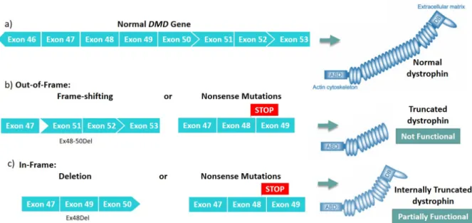
Duchenne Muscular Dystrophy (DMD) is an inherited X-linked recessive allelic disorder13. DMD, the most common form of muscular dystrophy, with an incidence of 1 in 3500 new-born males, is characterized by progressive deterioration of muscle function, with most patients not living beyond age of 30 due to cardiac and respiratory complications14,13. This disease occurs mainly due to frame-shifting deletion or nonsense mutations in the DMD gene that en-code for the dystrophin protein, and comprises 79 exons that produce the longest primary transcript15,13. Another DMD gene disorder is Becker Muscular Dystrophy (BMD), with an in-cidence of 1 in 18500 newborn males, presents a large spectrum of severities, from borderline DMD to almost asymptomatic cases13,16. In DMD, the disruption of the reading frame leads to the absence of functional dystrophin protein15, while BMD typically results from shortened but in-frame transcripts of the DMD gene that allow expression of an internally truncated but partially functional protein13,16,17 (Figure 2). Chamberlain determined how much dystrophin was needed to prevent dystrophic pathology in transgenic mice (20-30%) and explored the per-centage of muscle fibers that would need to be converted to a dystrophin positive phenotype to achieve a substantial correction of the pathology18,19,20. Their results indicated that a ma-jority of fibers must accumulate approximately 20% of wt levels of dystrophin for a significant correction of the muscle pathology18.
4
Figure 2: Differences between a normal dystrophin protein (a) and the dystrophin produced in Duchenne patients (b) or Becker patients (c).
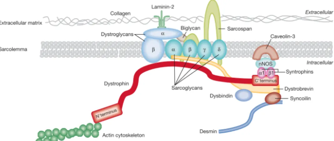
In muscle cells, the dystrophin protein associates with numerous proteins (dystroglycans, sarcoglycans, sarcospan, α-dystrobrevins, syntrophins, syncoilin and others) to form the dys-trophin associated protein complex (DAPC)21. The DAPC plays a structural role in maintain-ing muscle integrity, stabilizmaintain-ing the sarcolemma durmaintain-ing repeated cycles of contraction and re-laxation, and transmitting force generated in the muscle sarcomeres to the extracellular ma-trix21,13. The dystrophin, via the transmembrane dystroglycan protein and its associated pro-tein complex, including the sarcoglycans, is able to link the actin cytoskeleton to the extracel-lular matrix13 (Figure 3). The muscle isoform of dystrophin is a 427kDa protein that binds to cytoskeletal actin via its N-terminal actin-binding domain 1 (ABD1) and to β-dystroglycan via its C-terminal domain, with the central rod domain, consisting of 24 spectrin-like repeats, in between21,13. In order to be functional the dystrophin protein requires its N- and C-terminal domain and much of the rod domain appears to be partially dispensable (at least 8 integral repeats are needed for functionality), dystrophin deficiency leads to the loss of the associated protein complex21. In the absence of dystrophin, the muscle membrane becomes susceptible to damage and muscle fiber deterioration occurs, resulting in cycles of regeneration that lead to replacement of muscle fibers by fibrotic or adipose tissues, with the subsequent loss of muscle fibers and muscle function13,4.
In DMD, the mainstays of therapy are glucocorticoid corticosteroids (prednisone and de-flazacort) and palliative care (respiratory support and management of cardiac complications), which slow down disease progression, but do not prevent the progressive loss of muscle fibers and muscle function with increasing disability13,19,10.
To date there is no cure for this disease, however novel therapies for DMD have already started to arise towards a variety of gene corrective, gene replacement and surrogate gene ap-proaches13. Since this disease results from mutations in a single gene, gene therapy to replace the defective gene is a very attractive approach10. However, a major challenge for therapy of
5
Figure 3: The dystrophin-associated protein complex in muscle linking the internal cytoskeleton to the extracellular matrix22.
DMD is the need to devise a treatment strategy that targets whole-body musculature, includ-ing limb muscles, respiratory muscles (intercostal and diaphragm muscles), cardiac muscle and smooth muscle of the gastroesophageal tract, being this approach generally referred to as systemic therapy19,10. Whole-body treatment is particularly challenging in DMD because the
tissues involved are post-mitotic and approximately 30 to 40% of the human body consists of muscle10. Although adeno-associated virus (AAV) is one of the few viral vector systems that
efficiently infects muscle, it has a small cloning capacity that is easily exceeded, since the DMD gene presents 79 exons that correlate with a 13,993bp transcript10,13. Thus, it is not
surpris-ing that research also focuses on ways to restore gene expression at the mRNA level, more specifically by modulating its final presentation10.
Antisense Oligonucleotides (AONs) and Splice-Switching
Oligonu-cleotides (SSOs)
Targeting splicing by antisense oligonucleotides (AONs), small synthetic RNAs, DNAs or analogs, which hybridize specifically to their target sequences, allows RNA modifications that are not possible with RNA interference or other antisense techniques that destine the RNA for destruc-tion10,23,24,25. The appeal of targeting RNA is easy to appreciate: the nucleotide sequence pro-vides an opportunity to design sequence-specific and therefore gene-specific drugs24. AONs can cause inhibition or redirection of splicing and inhibition of protein synthesis through var-ious mechanisms, including disruption of the cell’s splicing machinery, interference with the ribosomal complex, and/or by activation of RNase H1-mediated degradation of the oligo-RNA heteroduplex26,27,25. Splicing modulation is accomplished by the use of AONs, termed splice-switching oligonucleotides (SSOs), which aim to modify the splicing pattern of a pre-mRNA24,25. These SSOs target nuclear pre-mRNA molecules to change exon splicing and generate an al-ternative protein isoform28. SSOs were first described for correction of aberrant splicing in human β-globin pre-mRNAs29, but have progressed furthest in the research for a treatment of DMD28.
6
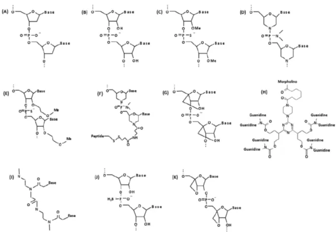
Figure 4: Chemical structure of biological and synthetic oligonucleotides. (A) DNA;
(B) RNA; (C) 2’O-methylphosphorothioate (2’O-MePS); (D) Morpholino (PMO); (E) 2’-methoxyethoxy (2’-MOE); (F) PMO with peptide conjugate (PPMO); (G) Locked Nucleic Acid (LNA); (H) Vivo-morpholino (vPMO); (I) Peptide nucleic acid (PNA); (J) Boranophosphate-oligodeoxy-nucleoside (BH3-ODN); (K) Oxetane-modified AONs26.
The first hurdle that first generation AONs had to overcome was regarding drug delivery26. Since these AONs do not easily cross the lipid bilayer of the cell membrane, they cannot read-ily penetrate to their intracellular targets at significant concentrations to be effective26. Another problem associated with first generation AONs is off-target toxic effects, because DNA and RNA can be immunostimulatory, binding and activating receptors involved in innate immunity in a sequence- and chemistry-dependent manner26. Furthermore, to achieve biochemical efficacy, a large proportion of RNA targets must be hybridized and silenced26. In order to overcome these challenges, new AONs have been conceived such that the ribose backbone (normally present in RNA and DNA) is replaced with other chemistries26. A variety of AON chemistries have been developed (Figure 4), which can be so distinct from classical nucleic acid structures that are not anymore targeted by nucleases or DNA/RNA-binding proteins, hence avoiding nu-clease degradation, facilitating stronger base-pairing with target mRNA sequences, increasing stability, helping to prevent most off-target toxic effects, and enabling easier entrance into the cell26.
7
Antisense-mediated Exon Skipping: Applicability in DMD
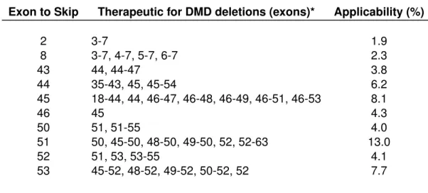
Antisense-mediated exon skipping aiming for reading frame restoration for DMD is a mutation specific approach and so a personalized therapy. However the majority of DMD patients have deletions that cluster in hotspot regions, being the skipping of a small number of exons applica-ble to a relatively large number of patients30. In theory, single and double exon skipping would be applicable to 79% of deletions, 91% of small mutations, and 73% of duplications, amounting to 83% of all DMD mutations30. Exon 51 skipping, which is currently being tested in clinical trials, would be applicable to the largest group (13%) of all DMD patients30 (Table 1). However, 17% of DMD patients, which carry larger deletions (>36 exons) or deletions in the actin-binding N-terminus or the C-terminus of dystrophin are not eligeble to be treated by a exon skipping approach30,31.
Table 1: Summary of the Exon-Skipping Applicability in DMD Patients31,30.
DMD deletions were reported in the Leiden DMD mutation database (www.dmd.nl/).
Exon to Skip Therapeutic for DMD deletions (exons)* Applicability (%)
2 3-7 1.9 8 3-7, 4-7, 5-7, 6-7 2.3 43 44, 44-47 3.8 44 35-43, 45, 45-54 6.2 45 18-44, 44, 46-47, 46-48, 46-49, 46-51, 46-53 8.1 46 45 4.3 50 51, 51-55 4.0 51 50, 45-50, 48-50, 49-50, 52, 52-63 13.0 52 51, 53, 53-55 4.1 53 45-52, 48-52, 49-52, 50-52, 52 7.7
Clinical Trials: Antisense Oligonucleotides (AONs) and DMD
In 2013, clinical trials with two competitive SSO drugs were underway to treat DMD, the two separate SSO compounds, Eteplirsen (AVI-4658, initiated by AVI Biopharma, now Sarepta Therapeutics) and Drisapersen (PRO051, initiated by Prosensa/GlaxoSmithKlein), that intend to cause skipping of dystrophin exon 5128,32,33. These SSOs cover the same target sequence, differing in size (eteplirsen containing 10 more nucleotides) and in their chemical compo-sition, as eteplirsen is based on phosphoramidate morpholino chemistry (PMO), and dris-apersen is based on phosphorothioated 2’-O-methyl RNA chemistry (2OMe-PS)28. 2OMe-PS oligonucleotides comprise chemically synthesized, negatively charged, single stranded RNA molecules with a phosphorothioate (PS) backbone that hybridizes to the target exon, being this PS moiety very stable and resistant to intracellular endonucleases and exonucleases, and in
8
order to further increase the resistance to nuclease activity, in C2’ the hydrogen is replaced by a methyl group31. Conversely PMO oligonucleotides are developed with replacements of the deoxyribose moiety with a morpholine ring and of the phosphodiester link by an uncharged phosphorodiamidate link31. Both drugs triggered exon 51 skipping and production of some dystrophin protein following intramuscular injections33. Despite its success in this first clinical trial, a phase III clinical trial with Drisapersen failed to meet the primary endpoint of a sta-tistically significant improvement in the 6 Minute Walking Distance Test (6MWT) compared to placebo33. It was later identified a confounding variable present in the study - the age of the patients, patients with ages inferior to 7 years old performed better than the older patients (per-sonal communication). The AONs in clinical trial are finishing phase III and currently awaiting approval from the Federal Drug Administration (FDA) and European Medicines Agency (EMA). A number of other SSOs (Prosensa, 2013) targeting different exons within the dystrophin gene are also in early clinical and preclinical development for skipping of exons 44, 45, 52, 53 and 5528.
A new approach: LNA-modified oligonucleotides (LNA-AON)
Since the AONs already in clinical trials have shown limited success regarding clinical efficacy, we aimed to test if Locked Nucleic Acid (LNA)-modified oligonucleotides (LNA-AON) could be used for splicing modulation therapies with increased efficiency. LNAs are a general and versatile tool for specific high-affinity recognition of stranded DNA (ssDNA) and single-stranded RNA (ssRNA)34. In LNA-AONs, a varying number of natural nucleotides are replaced
with nucleotide analogs carrying an altered sugar moiety, in which the ribose 2’-O- and 4’-C-atoms are connected via a methylene bridge35. LNA belongs to the so-called “third generation”
of modified nucleotides, with improved properties in terms of increased duplex stability, high sensitivity, good mismatch discrimination, low toxicity and increased metabolic stability35.
Sev-eral studies reported that no adverse effects of LNA-AONs on cell vitality have been observed at their biologically effective concentration or dosages35. Miravirsen is the first LNA-AON de-veloped as a therapy. It antagonizes miR-122 in patients chronically infected with the hepatitis C virus (HCV), and was developed after observations that this virus could only replicate in the presence of miRNA-122, a liver specific microRNA that plays a pivotal role in regulating hep-atic functions such as lipid metabolism and stress response. Current clinical trials suggest that this may be an effective and safe strategy for this patients36. Numerous studies demonstrate the enormous potential of LNA-AONs in basic and applied research, as well as in molecular medicine and therapeutics35.
With this work we expect to show the applicability of short LNA-modified oligonucleotides for Duchenne Muscular Dystrophy gene therapy, paving the way for a search of more efficient methods for gene therapy splicing modulation in systemic treatment of an inherited genetic disease.
Materials and Methods
Myoblast Derived Cell Lines Culture and Differentiation
Myoblast derived cell lines from a patient (DM8036 cell line with a deletion of exons 48-50 in the DMD gene) and a control individual (KM155 cell line) were kindly provided by Vincent Mouly from the Institut de Myologie UPMC Université Paris 6, France (Mamchaoui et al. 2011)37. The myoblast derived cell lines were maintained with Skeletal Muscle Cell Growth Medium (PromoCell, Cat. No. C-23060), a medium optimized for expansion of human skeletal muscle cells that contains low-serum (5% v/v), at 37oC with 5% carbon dioxide.
To induce differentiation, i.e. the fusion of myoblasts to myotubes with typical multinucleated syncythia, we monitored the cell density by microscopy and induced differentiation when cell confluence was approximately 80%, nearly 24h after seeding the cells at high density replacing the medium. Differentiation medium was DMEM (Gibco, Cat. No. 41966-029) containing ITS (Sigma, Cat. No. I3146-5ML), a general cell supplement, containing a mixture of recombinant human insulin, human transferrin, and sodium selenite. Cells were monitored by microscopy and after two days of differentiation if the presence of syncythia was observable, the cells were collected for RNA purification or protein extraction.
LNA-AON Structure and Transfections
From our group previous (unpublished) experiments, that compared different sequences and lengths of LNA-AONs, this LNA-AON was selected because it presented the greatest capability of inducing skipping of exon 51 in myoblast derived cell lines. It has the sequence 5’- AGGAA-GATGGCATTTC -3’ (DNA LNA-AON) and contains a fully phosphorothioate modified backbone, presenting 60% LNA, with two LNA-modified nucleotides at the 3’-end and one LNA-modified nucleotide at the 5’-end. The 16mer LNA-AON purchased from Exiqon.
For experiments, DM8036 cells were seeded in a P24wells plate (TPP, Cat. No. TPP92024; RNA purification and immunocytochemistry) or in a P12wells plate (Corning, Cat. No. CORN3513; protein extraction) after adding the transfection reagent (Lipofectamine RNAimax, Invitrogen, Cat. No. 13778-075) and the LNA-AON, in the intended concentration, in each well. For a growth area of 1.9cm2 (P24wells plate), 1µL of Lipofectamine RNAimax was used in 15µL of Optimem (Gibco, Cat. No. 31985-047). Different final concentrations of LNA-AONs were tested ranging from 5 to 500nM. DM8036 cells mock transfected with optimem were used as a negative control, and KM155 cells (dystrophin expressing control individual cell line) mock transfected with optimem were used as a positive control.
10
Animals
Animal procedures were performed in accordance with the guidelines of the European Commu-nity guidelines (Directive 2010/63/EU), Portuguese law on animal care (1005/92), and approved by the Instituto de Medicina Molecular Internal Committee and the Portuguese Animal Ethics Committee (Direcção Geral de Veterinária). Exon 52-deficient X chromosome-linked muscular dystrophy mouse model (mdx52 mice) was kindly provided by Shin’ichi Takeda from the Na-tional Center of Neurology and Psychiatry, in Japan (Aoki et al. 2012)38 and C57BL/6J mouse model, purchased from Charles River, were used in this study.
LNA Treatment of mdx52 Mice
Mdx52 mice (n= 5/6 per group) were treated with five tail intravenous injections of 1mg/kg LNA-AON or saline solution (controls), in an approximate volume of 100µL each injection, at biweekly intervals (every 2 weeks). Treatment started when animals were 5-week old. One week after the last injection, functional and behavioral testing of the animals was performed using Grip Strength, Wire Hang, OpenField and RotaRod tests to assess the motor deficient phenotype in mice. The animals were examined 2 weeks after the final injection and euthanized via eutasil (CEVA) and cervical dislocation. Cardiac puncture was performed for terminal blood collection for analysis of specific biomarkers (creatine phosphokinase (CPK), blood urea nitrogen (BUN), creatinine, aspartate transaminase (AST) and alanine transaminase (ALT)). Muscles (Gastroc-nemius - GC, Tibialis Anterior - TA, Heart - H and Diaphragm - D) were isolated immediately, snap frozen in liquid nitrogen and stored at -80oC for immunohistochemistry, Western Blotting and reverse transcription PCR (RT-PCR).
Blood Analysis and Muscle Functional Testing
Blood, left at room temperature (RT) for 1h was centrifuged at 13500 rpm for 10min (Eppendorf) and the plasma was collected and sent to analysis of biochemical markers: creatine phosphok-inase (CPK), blood urea nitrogen (BUN), creatinine, aspartate transamphosphok-inase (AST) and alanine transaminase (ALT), by VetinLab (Veterinary Clinical Analysis, Lisbon).
The functional and behavioral testing of the animals was performed using different Grip Strength, Wire Hang, OpenField and RotaRod tests to assess motor deficient phenotype in mice39,40, during the light period of the cycle, in a silent room, under dim light.
a. Grip Strength (SOP: DMD_M.2.2.001, SMA_M.2.1.002) – widely-used non-invasive method
designed to evaluate mouse limb strength. We performed 3 assays per trial and 3 trials in total of the tests:
• Wire Hang Test – the animals are placed in a wire grid, that is inverted, the duration of the test is 1min and the latency to fall is registed;
• Automatic Grip Strength – the mouse grasps a horizontal metal bar or grid while being pulled by the tail. The bar or grid is attached to a force transducer (PCE
11
instruments, Cat.No. PCE-FM50) that provides the peak pull-force achieved.
b. OpenField – The simplest test of locomotor activity that involves observing and recording
an animal’s movements around an open-field arena. The OpenField protocol used was adapted from Coelho et al 201440. The animals were placed in a designated corner of a square apparatus, surrounded by vertical walls (66cm x 66cm x 66cm) – open-field arena. They freely explored the maze for 5 min. Their movements were recorded and analyzed using the video-tracking software – SMART . The reference point used by theR
software to determine the position of the animal was the center of the mouse’s dorsum. Measurements of locomotor activity: the total distance traveled and the average speed were determined. At the end of the 5 min test, the animal was removed from the open-field arena and placed into its home cage.
c. RotaRod - used to assess sensorimotor coordination and motor learning in rodent
mod-els. The subjects are placed in a rotating rod and the latency to fall is recorded, an habituation period is needed.
Habituation Period (2-3 days)
Animals are placed on the rod at 8-12 rpm fixed rotation until they are able to stand unaided on the rod (3 trials per day separated by 30min each).
Test day Fixed Rotation Protocol
The animals are placed on a rod which accelerates to and then constantly rotates at the required velocity (12rpm).
Accelerating Protocol
The animals are placed in a rod that accelerates quickly from 0-4 rpm and then gradually from 4-40 rpm during a period of 5min. Attention: If animals fall before 7 rpm is reached they are placed back on the rod and it does not count as a fall.
3 trials are averaged to give the latency to fall of each animal.
A trial is complete when the animal falls or the time period ends. All the information regard-ing functional and behavioral testregard-ing of the mice was obtained from the websites http://www.treat-nmd.eu/research/preclinical/dmd-sops/ and http://sbfnl.stanford.edu/cs/bm/sm/.
RNA Isolation from Cell Extracts
Total RNA was extracted using PureZol (BioRad, Cat. No. 732-6890), according to the manu-facturer’s instructions. DNAse I treatment (Roche, Cat. No. 4716728001) and acidic phenol ex-traction with UltraPureTM Phenol:Cloroform:Isoamyl Alcohol (25:24:1, v/v; Invitrogen, Cat. No. 15593-031) were performed to additionally purify the RNA. Quantification and purity evaluation by A260/280 ratios were assessed using the spectrophotometer Nanodrop2000 (ThermoSci-entific)
RNA Isolation from Muscle Samples
Total RNA was obtained from 30mg fragments of frozen muscle. The Minibeadbeater (BioSpec Products) was used to disrupt and homogenize the tissue, using 1.0mm diameter zirconia-silica
12
beads (BioSpec Products), 3min. Total RNA was purified with RNeasy Fibrous Tissue MiniR
Kit (Qiagen, Cat. No. 50974704), according to the manufacturer’s instructions.
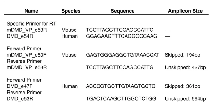
RT-PCR Protocol
The primer sequences for the PCR were designed using Primer3 (http://bioinfo.ut.ee/primer3-0.4.0/), BLAST was performed in Ensembl (http://www.ensembl.org/index.html) and to check for self- and hetero-dimers OligoAnalizer 3.1 (http://eu.idtdna.com/calc/analyzer) was used. Primers are listed in Table 2.
Six hundred nanograms of RNA template was used for a 20µL retrotranscription reaction using Transcriptor High Fidelity cDNA Sinthesis Kit (Roche, Cat. No. 5081963001), according to the manufacturer’s instructions at 55oC for 90min, followed by 5min at 85oC for degradation of
the reverse transcriptase (BioRad MyCyclerTM Thermal Cycler). For a 20µL retrotranscription reaction, 1.2nmol of a specific primer (listed in Table 2) was used. The cDNA product was diluted 1/7.5 and 4µL were then used as the template for PCR in a 10µL reaction volume with KAPA2GTMFast Ready Mix (KapaBiosystems, Cat. No. KK5609). The cycling conditions were
95oC for 5min, then 45 cycles: 95oC for 15sec, 58oC for 30sec, 72oC for 15sec.
Table 2: Primer sequences used in this study.
Name Species Sequence Amplicon Size
Specific Primer for RT
mDMD_VP_e53R Mouse TCCTTAGCTTCCAGCCATTG — DMD_e54R Human GGAGAAGTTTCAGGGCCAAG — Forward Primer
mDMD_VP_e50F Mouse GAGTGGGAGGCTGTAAACCAT Skipped: 194bp Reverse Primer
mDMD_VP_e53R TCCTTAGCTTCCAGCCATTG Unskipped: 427bp
Forward Primer
DMD_e47F Human ACCCGTGCTTGTAAGTGCTC Skipped: 361bp Reverse Primer
DMD_e53R TGACTCAAGCTTGGCTCTGG Unskipped: 594bp
PCR products were separated on a 2% (wt/wt) agarose gel. The molecular weight marker 1Kb Plus DNA ladder (Invitrogen, Cat. No. 10787-018) was used. Digital images were obtained using the Chemidoc XRS+ system (BioRad) and analyzed using the Image Lab 5.2 software (BioRad).
13
Protein Extraction from Cell Extracts
The protein extraction protocol was adapted from Winter et al. 201241. Cultured cells on P12wells plates were washed briefly with phosphate-buffered saline (PBS) at RT, and lysed and homogenized in 40µL treatment buffer (100mM Tris-HCl pH 6.8, 20% sodium dodecyl sul-fatel (SDS)), in a surface area of 3.8cm2(P12wells plate) for 5min. Protein concentration was determined using the PierceTMBCA Protein Assay kit (ThermoScientific, Cat. No. 1513-7485), according to the manufacturer’s instructions. This kit is a detergent-compatible formulation based on bicinchoninic acid (BCA) for the colorimetric detection and quantification of total pro-tein. Bovine Serum Albumin (BSA) is used as a protein standard for the determination of the protein concentration and the absorbance of the protein extracts is measured at 562nm. After protein quantification 1/100µL Benzonase (Sigma, Cat. No. E1014-25KU) and 0.5M MgCl2
(final concentration of 14mM) was added, this endonuclease degrades all forms of DNA and RNA (single stranded, double stranded, linear and circular), even in the presence of SDS. After incubation for 15min at room temperature (RT), the homogenate was completed with 13.3µL of a 0.04% bromophenol blue, 20% dithiothreitol (DTT) and 80% glicerol solution to contain 75mM Tris-HCl pH 6.8, 15% sodium dodecyl sulfate, 5% dithiothreitol, 20% glycerol and 0.01% bromophenol blue and boiled at 98oC for 5 min.
Protein Extraction from Muscle Samples
Muscles were homogenized using the treatment buffer previously described (100mM Tris-HCl pH 6.8, 20% sodium dodecyl sulfatel) and 1.0mm diameter zirconia-silica beads (BioSpec Prod-ucts) in the Minibeadbeater (BioSpec ProdProd-ucts), 3min. Protein concentration determination and completion of the protein extracts was performed as above. After 5min at 98oC, the ho-mogenate was then sonicated using an ultrasonic bath at 35kHz (VWR Ultrasonic Cleaner), and centrifuged (Eppendorf) at 20 000g, 4oC for 30 minutes.
Western Blot Protocol
1-5µg of protein was loaded on a 7% polyacrylamide gel and run for 90min at RT: 10min at 60V + 80min at 100V (BioRad). Precision Plus ProteinTM Standard (BioRad, Cat. No. 161-0373) was used. Gels were blotted to nitrocellulose membranes (Whatman Protran BA 85 Nitrocel-lulose 0.45um 200x200mm, Cat. No. 10401191), in a Tank Transfer System (BioRad Mini Trans-Blot R Cell) with 300mA for 90min at RT (BioRad Power Pac BasicTM). The membranes
were stained with Ponceau S (Sigma, Cat. No. P3504-10G) to confirm the efficiency of the transference of the protein to the membrane. The membranes were marked and cutted to in-cubate separately the primary antibody from the loading control. Blots were blocked with 5% non-fat dried milk in Tris-buffered saline (TBS) plus 0.05% Tween-20 (TBST) followed by an overnight incubation at 4oC with a rabbit polyclonal anti-dystrophin antibody (dilution 1/1000; Abcam, Cat. No. ab85302) in 5% non-fat dried milk in TBST. Goat Anti-Rabbit IgG (H+L)-HRP (dilution 1:3000; 60min at RT; BioRad, Cat. No. 1706515) was used as a secondary antibody.
14
The polyclonal antibody rabbit anti-Lamin A/C (dilution 1:10000; overnight at 4oC; H-110, Santa
Cruz, Cat. No. sc-20681) was used as a loading control. Digital images were captured using the Chemidoc XRS+ system (BioRad) and analysed using the Image J software. To estimate the molecular weight of the truncated dystrophin protein the websites web.expasy.org/translate and www.bioinformatics.org/sms2/protein-mw.html were used.
Immunocytochemistry
For microscopy analysis, cells were transfected and seeded onto 0.1% gelatin coated glass cov-erslips (10x10mm2) on P24wells plates. After 7 days differentiation, the cells were fixed with 3.7% paraformaldehyde (PFA) in phosphate-buffered saline (PBS), 10min at RT, and permeabi-lized with 0.5%Triton x100 in PBS, 10min at RT. The cells were incubated for 30min at RT in a blocking solution containing 1%BSA and 0.05%Tween20 in PBS, all antibodies were diluted in this blocking solution. The cells were stained with a polyclonal rabbit anti-dystrophin antibody (dilution 1/100, 60min incubation at RT; Abcam, Cat. No. ab85302). Rhodamine (TRITC)-conjugated affinipure donkey anti-rabbit was used as a secondary antibody (dilution 1/200, 60min incubation at RT, Tetramethyll Rhodamine Isothiocyanate (TRITC); Jackson ImmunoRe-search Laboratories Inc., Cat. No. 711-025-152). 0.1µg/mL 4’,6-diamidino-2-phenylindole (DAPI) was used to stain the nuclei, 10min incubation at RT; Sigma, Cat. No. D9542-5MG). Vectashield Mount Medium (Vectorlabs, Cat. No. H-100) was used as a mounting medium, the borders of the coverslips were sealed with nail polish.
Immunohistochemistry
At least five 10µm cryosections were cut from the muscles of interest (tibialis anterior, gastroc-nemius, diaphragm and heart) and fixed in ice cold acetone, 10min and air dried. Incubation with blocking solution, staining with antibodies anti-dystrophin and TRITC, and DAPI staining were performed as above.
Microscope Image Acquisition and Analysis
Digital images were captured using a Zeiss LSM 710 Confocal Point-Scanning Microscope, with 20x (immunohistochemistry) and 40x (immunocytochemistry) objectives using the lasers Diode 405-30 (405nm) and DPSS 561-10 (561nm). Digital images of maximum intensity projections, of 3 stacks spaced by 1µm, were analyzed with the software Image J and LSM 5 Image Browser (Zeiss).
Statistical Analysis
Statistical differences were assessed with a Mann-Whitney-Wilcoxon Test, using custom R (ver-sion 3.2.1) scripts. All data are reported as mean values ±standard deviation (SD). The level of significance was set at P < 0.05. The software Graphpad Prism 6 was used for dose-response curves presentation and EC50 determination.
Results and Discussion
In vitro Evaluation of the Splicing Modulation
A myoblast derived cell line from a patient (DM8036 cell line)37, that does not produce the dystrophin protein due to a deletion of exons 48-50 in the DMD gene, was transfected with a LNA-AON targeting exon 51, for in vitro induction of skipping of DMD-exon 51 and consequent restoration of protein production. A myoblast derived cell line from normal a individual (KM155 cell line)37 was mock transfected and used as a positive control. Skipping of exon 51 allows for restoration of the reading frame of the dystrophin mRNA, correcting the frameshift and subsequently leading to the production of a truncated but partially functional dystrophin protein. From previous (unpublished) experiments, that compared different sequences and lengths of LNA-AONs, was selected a LNA-AON for this work because it presented the greatest capability of inducing skipping of exon 51 in myoblast derived cell lines. Different concentrations of the LNA-AON were tested, ranging from 5-500nM. In order to examine the exon 51 skipping at the transcript level and explore restoration of protein production, RT-PCR techniques and Western Blot and Immunocytochemistry techniques were employed.
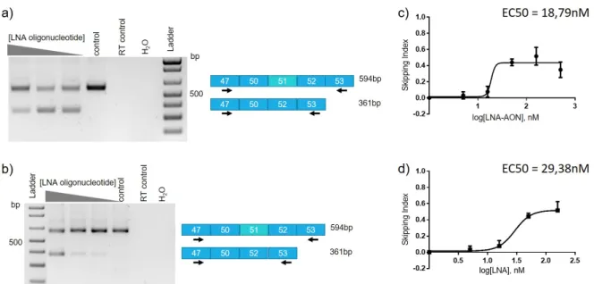
Exon 51 Skipping at the Transcript Level
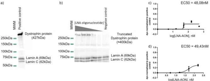
Myoblast derived cells (DM8036 patient cell line) were transfected with different concentrations of LNA-AON. Differentiation was induced 24h after transfection and extraction and purification of total RNA was performed after 2 days differentiation. Before harvesting the cells, differ-entiation was monitored by phase-contrast microscopy where multinucleated myotubes were observable. Retrotranscription was performed with a specific primer for DMD-exon 54 and the PCR protocol used was optimized do detect skipping of exon 51 in total mRNA, with only one round of amplification. In all the literature reviwed, amplification was done with nested PCR42,43. In our hands nested PCR also worked, but is not usefull for a semi-quantitative ap-proach. Actualy Spitali et al 201044 compared different techniques for quantification of exon skipping levels in AON-treated mdx mouse muscle, and demonstrated that with a two-round amplification PCR (nested PCR), the skipping levels were generally overestimated. With our protocol, no skipped isoform bands were observed in the negative control, which consisted of the same cells mock transfected. The results obtained, with the RT-PCR, show effective skip-ping of exon 51 at the transcript level (Figure 5a and b) in dystrophic myoblasts transfected with the LNA-AON at concentrations as low as 5nM.
We also tryed the approach of qRT-PCR to quantify the skipping of exon 51 in total mRNA,
16
Figure 5: Efficacy of exon 51 skipping in a myoblast derived cell line (DM8036 patient cell line). a) RT-PCR results after transfection of the LNA-AON with different concentrations
(500, 158, 50nM and a negative control mock transfcted) and two days differentiation. b) RT-PCR results after transfection of the LNA-AON with different concentrations (50, 15.8, 5nM and a negative control mock transfected) and two days differentiation. c) and d) Dose-response curves obtained considering the skipping index and the LNA-AON concentration transfected. In d) the values for the 500nM were excluded from the calculations.
however construction of exon junction primers that could efficiently discriminate between the exon junctions of exons 47-51 (unskipped transcript) versus exons 47-52 (skipped transcript) proved difficult: we could obtain an amplification band with primers targeting the skipped iso-form even in the negative control. Since the dystrophin mRNA is a low abundance transcript45, detections of small variations of alternative spliced transcripts could be difficult at the RNA level if primers and the RT-PCR strategy are not optimized.
From the results obtained, a dose-response curve was originated using the software Graph-pad Prism 6. The calculations were made considering the Skipping Index and the concentra-tion of the LNA-AON (Figure 5c and d). The Skipping index was calculated accordingly with the equation: SkippingIndex = exon51exclusion/(exon51exclusion + exon51inclusion). To calculate the
Skipping Index, the relative quantification of the intensityof the skipped and unskipped bands in the agarose gel was assessed using the software ImageLab 5.2. From the dose-response curve obtained (Figure 5c), the calculation of the half maximal effective concentration (EC50)
was performed, this value refers to the concentration of a drug which induces a response halfway between the baseline and maximum, i.e. 50% of the maximum effect is observed. The EC50 value obtained was of 18,79nM. But since we can observe that the 500nM
concentra-tion presents a lower efficiency than the 158nM concentraconcentra-tion, not presenting the maximum effect observed and that the program was not taking into account the decrease at the highest concentration tested, we excluded the 500nM concentration values and calculated a new dose-response curve (Figure 5d). From this new curve, we obtained a EC50 value of 29,38nM, which
17
might be a certain toxicity of this 500nM concentration for cell viability i.e. the preferential death of the cells with more uptake of the LNA-AON.
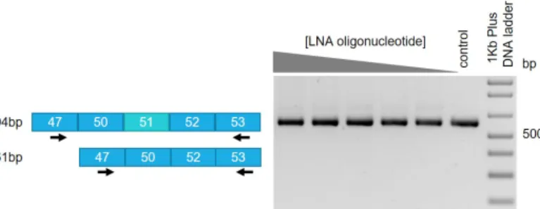
In order to examine if the effect of the LNA-AON is long lasting, the myoblast derived cells were transfected once with the different concentrations of the LNA-AON and were incubated in differentiation medium for two weeks. After two weeks differentiation, total RNA analysis with the optimized RT-PCR protocol, showed no detection of skipping of exon 51 (Figure 6). This experiment suggests that repeated administration of the treatment is necessary for a continued effect since this therapy is at the RNA level.
Figure 6: Exon 51 skipping is not detected after two weeks differentiation in myoblast derived cell lines (DM8036 patient cell line). The concentrations of transfected LNA-AON ranged from 5-500nM (500, 158, 50, 15.8, 5 and a negative control mock transfected).
From studies performed by Tennyson, we know that the human dystrophin gene with its 79 exons spanning over 2300kb requires approximatly 16h to be transcribed46, and that the half-life of the dystrophin mRNA is 15.6 ±2.8h in cultured human fetal myotubes45; this estimative
was obtained using actinomycin D which has the ability to inhibit transcription, by binding DNA at the transcription initiation complex and preventing elongation of the RNA molecule by RNA polymerase. There are not considerable experimental evidences of AON turnover, it would be very important to develop this experiments for further improvement of activity and safety of AONs47.
Restoration of Protein Production
Myoblast derived cells (DM8036 cell line) were transfected with different concentrations of LNA-AON. A myoblast derived cell line from normal individuals (KM155 cell line) was mock trans-fected and used as a positive control for dystrophin expression. Differentiation was induced 24h after transfection and for Western Blot, protein extracts were prepared after 2 days differentia-tion. Before harvesting the cells, differentiation was monitored by phase-contrast microscopy where multinucleated myotubes were observable. With the Western Blot protocol used, in the positive control, there was detection of only one band in the membrane, corresponding to the size expected for the control protein (427kDa) (Figure 7a), using 1µg of total protein extract loaded in the polyacrylamide gel. In the negative control, myoblast derived cells from patients (DM8036 cell line) mock transfected, there was no detection of the dystrophin protein. The estimated size for the truncated dystrophin protein is of approximately 400kDa, having in con-sideration the deletion of exons 48-50 and the skipping of exon 51. Restoration of dystrophin
18
protein production in dystrophic myoblasts transfected with LNA-AONs was detected (Figure 7b), with concentrations as low as 15.8nM of transfected LNA-AON.
From the results obtained, calculation of a dose-response curve was originated using the software Graphpad Prism 6. This calculations were made considering the dystrophin protein intensity of bands of the dystrophic myoblasts (DM8036 cell line) normalized to the positive control (KM155 cell line) and the concentration of the LNA-AON (Figure 7c and d). The relative quantification of the intensity of the bands in the membranes was assessed using the software ImageJ, the background was subtracted to the intensity of each band and each dystrophin level was obtained in relative quantification to the loading control (the lamin A band was chosen), for each sample, being afterwards normalized to the dystrophin relative quantification of the positive control. From the dose-response curve (Figure 7c), the EC50 value obtained was of
48,08nM. Since in the Western Blot results, the 500nM concentration presented a lower effi-ciency than the 158nM concentration (as we have seen already in the RT-PCR assay), we ex-cluded the 500nM concentration values from the graphic and calculated a new dose-response curve (Figure 7d). From this new curve, we obtained a EC50 value of 49,43nM, that does not
differ greatly from the previously obtained. It would be important to carry out an Western Blot experiment to see if after two weeks differentiation there is still detection of dystrophin protein restoration in dystrophic myoblasts (DM8036 cell line) transfected with the different concentra-tions of LNA-AON, but that question could be even better addressed by a microscopy approach.
Figure 7: Restoration of protein production after exon 51 skipping in a myoblast de-rived cell line (DM8036 patient cell line). a) Western blotting analysis of the KM155 normal
individual cell line, used as a positive control. b) Western Blotting analysis of the different con-centrations of LNA-AON transfected in the DM8036 patient cell line. c) and d) Dose-response curves obtained considering the intensity of the dystrophin bands relative quantites to the load-ing control, normalized to the positive control and the LNA-AON concentration transfected. In c) the values for the 500nM were excluded from the calculations.
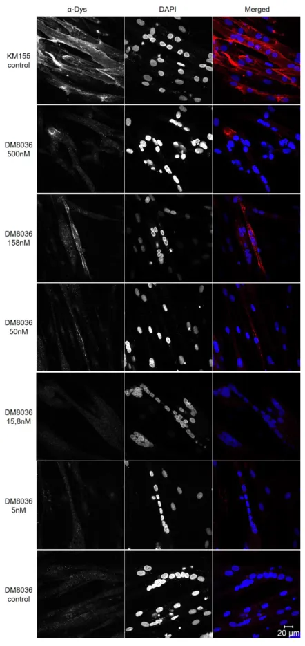
By microscopy of immunolabeled cells we can address, beside the presence of dystrophin protein, its correct localization in the plasma membrane of differentiated cells. In the Immuno-cytochemistry protocol performed, after transfection with the different concentrations of the LNA-AON, induction of differentiation 24h after the transfection, fixation of the cells occurred
19
7 days after differentiation. In order to perceive if the cells were differentiated, i.e. if fusion of myoblasts had originated multinucleated myotubes, DAPI was used to stain the nuclei. Using myoblast derived cells from control individuals mock transfected (KM155 cell line - positive con-trol), we could ascertain if the antibody used was detecting the dystrophin protein and where it was localized. In the positive control, dystrophin positive fibers are observed and the local-ization of the staining is in the periphery of the multinucleated cells. In the negative control, dystrophic myoblasts from patients (DM8036 cell line) mock transfected, there was no detec-tion of dystrophin. Detecdetec-tion of dystrophin positive fibers in dystrophic myoblasts (DM8036 cell line) transfected with LNA-AONs was detected, via Immunocytochemistry (Figure 8), with concentrations as low as 50nM. From all the experiments performed, the concentration that presented a higher efficiency of DMD-exon 51 skipping and dystrophin protein restoration was 158nM LNA-AON.We can see that the presence of dystrophin in treated cells is not as high as in control cells and that it does not occur in all the cells in the coverslip, but the localization in the plasma membrane is as expected, which makes these seem promising results.
20
Figure 8: Restoration of protein production after exon 51 skipping in myoblast derived cell lines (DM8036 patient cell line). Fluorescence immunocytochemistry analysis of the
different concentrations of LNA-AON transfected in the DM8036 patient cell line, KM155 control individual cell line was used as a positive control. Polyclonal antibody ab85302 was used to detect dystrophin, DAPI was used to stain the nuclei.
21
In vivo Evaluation of the Splicing Modulation in mdx52 mouse model
The mdx52 mouse model, that presents a deletion of DMD-exon 5238, was used for in vivo studies of splicing modulation by a LNA-AON targeting skipping of DMD-exon 51, to restore the reading frame of the dystrophin mRNA and subsequently lead to production of a truncated but partially functional dystrophin protein. Mdx52 mice (n= 5-6 per group) were treated with five tail intravenous injections of 1mg/kg LNA-AON or saline solution (control) at biweekly intervals (every 2 weeks). One week after the final injection, functional and behavioral testing of the mice was performed to assess motor phenotype (Grip Strength, Wire Hang and OpenField). The mice were euthanized two weeks after the final injection and terminal blood was collected for analysis of specific biomarkers: creatine phosphokinase (CPK), blood urea nitrogen (BUN), creatinine, aspartate transaminase (AST) and alanine transaminase (ALT). Muscles (Gastroc-nemius - GC, Tibialis Anterior - TA, Heart - H and Diaphragm - D) were isolated, snap frozen and stored for Immunohistochemistry, Western Blotting and RT-PCR analysis.
Blood Analysis and Muscle Functional Testing
In order to examine the functional phenotype, a battery of physiological and blood tests were performed after the five biweekly intravenous injections with the LNA-AON (Figure 9). High levels of serum creatine phosphokinase (CPK), an important enzyme in heart, brain and skele-tal muscle, are present in muscular dystrophies, such as DMD, due to damage of the muscle tissue leading to leakage of CPK into the blood. In normal situations, low levels of CPK are present in the blood (see Supplemental Figure14b). If a reduction of the CPK levels was to be present, this would suggest the protection of muscle fibers against degeneration. To further monitor any potential toxicities in the major organs induced by the treatment with the LNA-AON, we compared a series of standard serum markers as indicators of liver and kidney dysfunction in treated and untreated mdx52 mice (Figure 9a). Creatinine and blood urea nitrogen (BUN) are indicators of renal health, and aspartate transaminase (AST) and alanine transaminase (ALT) are measured clinically as biomarkers of liver health. AST is also used as a biomarker of muscle damage. No significant differences were detected between untreated and treated mdx52 mice groups in the levels of AST, ALT, BUN and creatinine (Figure 9a). These data do not allude towards a toxic effect of the LNA-AON tested in vivo. We also could not find a significant reduction of CPK in treated mice, but there was a problem with getting the results from all the animals in this experiment.
In this study, this reduction was not observed when comparing treated and untreated mdx52 mice (Figure 9a). A significant difference (p = 0.01732, Mann-Whitney-Wilcoxon Test) between the treated and untreated mdx52 mice was observable in the OpenField test, the average velocity of the treated mdx52 mice is greater than the untreated mdx52 mice, but no significant improvement was observed in forelimb grip strength in treated mdx52 mice compared with untreated mdx52 mice (Figure 9b).
22
Figure 9: No amelioration of the skeletal muscle function was observable in the mdx52 mice. a) Measurement of biochemical markers [creatine phosphokinase - CPK (U/L), blood
urea nitrogen - BUN (mg/dL), creatinine (mg/dL), aspartate transaminase - AST (U/L) and ala-nine transaminase - ALT (U/L)] levels. b) Functional and behavioral testing of the mice was performed using Grip Strength (GF and GF/g), Wire Hang (sec) and OpenField (cm/sec and sec), in treated and untreated mdx52 mice. Data (treated n=6, untreated n=5) are presented as mean ±SD.
Exon 51 Skipping at the Transcript Level
Total RNA from muscles was extracted with an RNeasy Fibrous Tissue Mini Kit, from approx-R
imately 30mg of tissue. Retrotranscription was performed with a specific primer for DMD-exon 53 and the PCR protocol used was the optimized protocol described in the section "Evaluation of the Splicing Modulation".
The negative controls (not shown) presented no band. A positive control was not used because we did not have one. As showed in Figure 10a, we did not detect any band corre-sponding to the skipped exon 51 isoform. This result can be interpreted by comparison with the result from Figure 6, because detection of skipping of exon 51 was not observable at the RNA level after two weeks differentiation in myoblast derived cell lines transfected once with the LNA-AON, it is comprehensible that detection of skipping of exon 51 at the RNA level in the mdx52 mice was not observable, since the acquisition of the samples for RT-PCR analysis was made two weeks after the last injection with the LNA-AON.
Restoration of Protein Production
Transverse cryosections of the different muscles collected were performed for immunohisto-chemical staining of dystrophin in treated and untreated mdx52 mice. C57BL/6J mice were used as a positive control. Dystrophin-positive clusters of fibers were detected in tibialis ante-rior and cardiac muscles of treated animals as exemplified in Figures 11 and 12), via immuno-histochemistry. One could think that this dystrophin-positive fibers were likely to be naturally occurring revertant fibers, i.e. occasional isolated fibers that appear to express correctly