Universidade de Lisboa
Faculdade de Medicina de Lisboa
Using Genomics for Drug Discovery in
Medulloblastoma
Cláudia Maria Coelho de Faria
Orientadores
Prof. Doutor James T. Rutka
Prof. Doutor João Lobo Antunes
Doutoramento em Medicina
Especialidade de Neurocirurgia
Todas as afirmações contidas no presente documento são da exclusiva responsabilidade do seu autor, não cabendo qualquer responsabilidade à Faculdade de Medicina de Lisboa pelos conteúdos nele apresentados.
A impressão desta dissertação foi aprovada pelo Conselho
Científico da Faculdade de Medicina da Universidade de Lisboa em
reunião de 22 de Abril de 2014.
To my family for their love and support every day of my life
Preface
I have always wanted to be a neurosurgeon but the decision to pursue a career in science came later in my life. I did my residency in Neurosurgery at Hospital de Santa Maria from 2004 to 2010 and I devoted the last years of training to Pediatric Neurosurgery. My interest in brain tumor research arose from my patients, particularly from those difficult cases where, despite a successful operation, the tumor recurred thus emphasizing the importance of understanding the tumor biology for patient management.
Having this in mind, and with the invaluable support of Professor João Lobo Antunes, I applied to the Programme for Advanced Medical Education in 2009, a PhD program for medical doctors, sponsored by Fundação Calouste Gulbenkian, Fundação Champalimaud, Ministério da Saúde e Fundação para a Ciência e Tecnologia. The initial six months of the program included classes and seminars on various topics, from basic science to translational research and epidemiology. The goal of this educational period was to provide the students with solid scientific basis to develop research projects oriented for specific clinical questions.
When I joined the program I had the naïve assumption that I couldn’t stay away from my natural environment, the operating room without loosing the skills acquired during six years of intense surgical training. The switch from a purely clinical thinking to an integrated clinical and scientific thinking was hard but it became a milestone in my medical career. Having a clear clinical question in mind I have decided to develop my research project in a renowned international brain tumor research centre, The Arthur and Sonia Labatt Brain Tumour Research Centre, at the Hospital for Sick Children in Toronto, Canada.
The choice to move to Toronto was simple. During my residency I had visited for a period the Divison of Neurosurgery at the Hospital for Sick Children in Toronto and I met Dr. James Rutka, pediatric neurosurgeon, senior scientist and Director of The Arthur and Sonia Labatt Brain Tumour Research Centre. Knowing my interest in brain tumor research, he invited me to visit the Centre, showed me the facilities, explained the main research areas and ongoing projects, and introduced me to some of the researchers. When, two years later, I had to choose a research institute to develop my project, I had no doubt that the Labatt Brain Tumour Research Centre was the right place and, therefore, I was extremely grateful for being accepted in Dr. Rutka’s laboratory.
I have decided to focus on medulloblastoma, the most common malignant brain tumor in the pediatric population. I was puzzled with the heterogeneous clinical outcome of patients with medulloblastoma treated with standard protocols and I felt that better therapeutic options were urgently needed. The “boom” of genomic data emerging from the study of large cohorts of medulloblastoma samples had recently started. I was fortunate to work at the Labatt Brain Tumour Research Centre when major discoveries on medulloblastoma took place and also to give my contribution to some of those projects. The results from my own research project opened new avenues for targeted therapies and I hope they can translate into clinical trials for children with medulloblastoma in a near future.
My overall experience in Toronto exceeded my best expectations. During my “research residency”, as I like to refer to my training in the laboratory, I have learned a wide variety of techniques, I became familiar with the latest technologies in the field and, most importantly, I met extraordinary people from around the globe and created a network of collaborations that will certainly be of great value for my future projects. The exceptional environment at the Labatt Brain Tumour Research Centre have been truly inspiring and, now that I am back to my home institution, I hope to pursue a career as a clinician-scientist to better help my patients both in the operating room and in the laboratory.
Acknowledgments
I would like to express my deepest gratitude to Dr. James Rutka, my PhD supervisor, who welcomed me to his laboratory and guided me in my training as a surgeon-scientist. I am forever grateful for the opportunities he gave me throughout my stay in Toronto. It was a privilege to be part of the Rutka lab and a member of the Labatt Brain Tumour Research Centre.
I wish to express my profound gratitude to Professor João Lobo Antunes, the Director of my Department and Co-supervisor of my PhD, for his mentorship and guidance for over 10 years. He has been an example of a dedicated surgeon, a compassionate physician and an excellent teacher. He recognized and stimulated my scientific curiosity long before I had realized its importance in my career. I am thankful for his encouragement and support of my research, and also for his vision for the future.
I am forever thankful to Dr. José Miguéns, my mentor in Pediatric Neurosurgery, for his support of my research and for his generosity taking care of all the pediatric patients in our Department while I was in Toronto. His exceptional dedication to his patients and clinical thoroughness, have been an inspiration and an example to follow.
I am thankful to the Programme for Advanced Medical Education and particularly to Professor Leonor Parreira for her generous support and advice. The outstanding scientific training provided by the Programme was crucial for my research work in Toronto. I wish to acknowledge the sponsors of the Programme, Fundação Calouste Gulbenkian, Fundação Champalimaud, Ministério da Saúde e Fundação para a Ciência e Tecnologia, for their institutional and financial support.
I am grateful to Dr. Michael Taylor for his guidance and mentorship in my research projects. He always gave me insightful comments and suggestions.
I am thankful to Professor José Pimentel, Head of the Laboratory of Neuropathology at Hospital de Santa Maria, for supporting my research and for helping me establishing fruitful national and international collaborations.
I wish to thank the current and past members of the Rutka lab who contributed their time and advice in helping me achieve my research goals including Dr. Christian Smith, Dr. Roberto Diaz, Dr. Sameer Agnihotri, Dr. Arnold Etame, Dr. Adrienne Weeks, Dr. Yuzo Terakawa, Dr. Jong Hee Chang, Mr. Brian Golbourn, Ms. Amanda Luck, Ms. Nesrin Sahba and Mr. Jim Loukides. I wish to express my gratitude in particular to Dr. Christian Smith for his mentorship and support.
I would like to acknowledge my wonderful collaborators from Dr. Michael Taylor’s lab Dr. Adrien Dubuc, Dr. Marc Remke, Dr. Vijay Ramaswamy, Dr. Stephen Mack, Dr. Xiaochong Wu, Dr. Livia Garzia, Dr. Paul Northcott and Mr. Xin Wang, and from the German Cancer Research Centre (DKFZ) in Heidelberg Dr. Stefan Pfister, Dr. Marcel Kool and Dr. Andrey Korshunov, who contributed with many insightful comments and technical expertise in this work.
Table of Contents
Preface... 5 Acknowledgments... 7 Table of Contents ... 9 Abbreviations ... 13 List of Figures ... 19 List of Tables... 21 Abstract ... 23 Resumo... 27 Chapter 1 Introduction ... 311.1 The Genomic Landscape of Medulloblastoma... 31
1.1.1 WNT Medulloblastomas ... 33
1.1.2 SHH Medulloblastomas ... 34
1.1.3 Group 3 Medulloblastomas ... 35
1.1.4 Group 4 Medulloblastomas ... 36
1.2 The Role of HGF/cMET Pathway Signaling in Human Medulloblastoma ... 37
1.2.1 The HGF/cMET Pathway Signaling ... 38
1.2.1.1 Structure of HGF and cMET ... 38
1.2.1.2 cMET Signal Transduction... 39
1.2.1.3 Regulation of cMET Signaling ... 41
1.2.2 HGF, cMET and Cancer... 42
1.2.3 HGF/cMET Signaling in Medulloblastoma ... 45
1.2.3.1 HGF/cMET Pathway in Cerebellar Development ... 45
1.2.3.2 HGF/cMET Pathway in Medulloblastoma Formation... 46
1.2.4 Inhibitors of HGF/cMET Pathway in Cancer Therapy ... 48
1.2.4.1 HGF and cMET Antagonists ... 50
1.2.4.2 Monoclonal Antibodies Directed Against HGF and cMET ... 50
1.2.4.3 Small Molecule Tyrosine Kinase Inhibitors ... 51
1.2.5 Conclusion and Future Directions... 52
1.3 Novel Therapies and Next Generation Clinical Trials in Medulloblastoma... 53
1.3.1 Advances in Medulloblastoma Clinical Trials ... 53
1.3.1.2 High-risk patients... 56
1.3.1.3 Recurrent medulloblastoma ... 58
1.3.2 Emerging Targeted Therapies in Medulloblastoma ... 60
1.3.3 Conclusions and Future Perspectives... 62
1.4 Hypothesis and Aims ... 65
Chapter 2 cMET Inhibition as a Molecular Therapy for Metastatic SHH Medulloblastoma .. 67
2.1 Introduction ... 67
2.2 Methods and Materials ... 69
2.2.1 Tumor Material and Patient Characteristics... 69
2.2.2 Expression Profiling and Molecular Subgrouping ... 69
2.2.3 Analysis of Somatic Copy Number Alterations... 69
2.2.4 Cell Lines and Animal Models... 69
2.2.5 Cell Culture Assays for cMET and PDGFRß Signaling ... 70
2.2.6 Migration and Invasion Assays ... 70
2.2.7 Immunoblotting... 70
2.2.8 Cell Proliferation Assays... 71
2.2.9 Active Caspase Assays... 71
2.2.10 Foretinib Pharmacokinetic Studies... 71
2.2.11 Medulloblastoma Xenografts and Transgenic Mouse Models... 73
2.2.12 Immunohistochemistry... 74
2.2.13 Statistical Analysis ... 74
2.3 Results ... 75
2.3.1 cMET and PDGFRß are Highly Expressed in SHH Medulloblastomas... 75
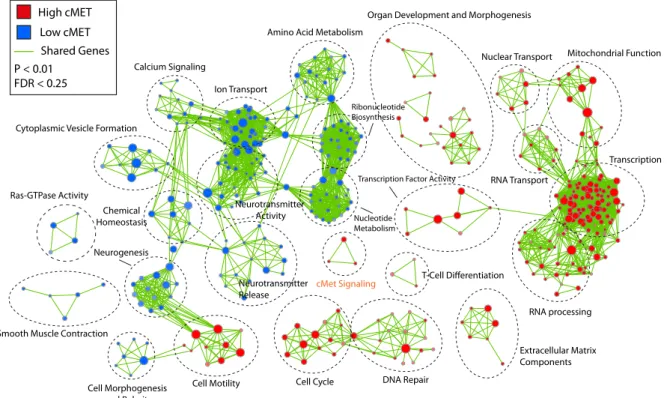
2.3.2 Identification of Biological Pathways and Processes Associated with High cMET and Low cMET SHH Medulloblastomas... 76
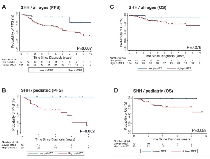
2.3.3 High p-cMET Levels Correlate with Recurrence and Poorer Survival in SHH Medulloblastomas ... 79
2.3.4 Foretinib Inhibits cMET and PDGFRß Pathway Activity ... 84
2.3.5 Foretinib Induces Medulloblastoma Regression In Vivo ... 90
2.3.6 Characterization of Foretinib Pharmacokinetics and Brain Permeability... 93
2.3.7 Foretinib Reduces SHH Medulloblastoma Growth and Dissemination in Mouse Xenografts ... 96
2.3.8 Foretinib is Effective Against the Primary and the Metastatic Compartments in a Transgenic Model of Metastatic SHH Medulloblastoma... 98
2.4 Discussion... 100
Chapter 3 The Connectivity Map Identifies Novel Small Molecules to Target Group 3 Medulloblastoma ... 103
3.1 Introduction ... 103
3.2 Materials and Methods ... 104
3.2.1 Connectivity Map Analysis... 104
3.2.2 Medulloblastoma Cell Lines ... 104
3.2.3 Cell Proliferation Assays... 105
3.2.4 Medulloblastoma Mouse Xenografts ... 105
3.2.5 Immunoblotting... 106
3.2.6 RNA Extraction and Gene Expression Analysis... 106
3.2.7 Statistical Analysis ... 106
3.3 Results ... 107
3.3.1 The C-MAP Identifies Novel Candidate Drugs to Treat Medulloblastoma... 107
3.3.2 C-MAP Candidate Drugs Piperlongumine, Alsterpaullone, Rottlerin and Flunarizine Reduce Proliferation of Group 3 Medulloblastoma Cell Lines .. 110
3.3.3 In Vivo Antitumor Effect of Piperlongumine, Alsterpaullone and Rottlerin in Group 3 Medulloblastomas ... 112
3.3.4 Alsterpaullone Shows In Vitro and In Vivo Specificity for Group 3 Primary Medulloblastoma Stem Cells ... 116
3.3.5 Alsterpaullone Inhibits MYC and other Cell Cycle Related Genes... 119
3.4 Discussion... 124
Chapter 4 Concluding Remarks and Future Directions ... 127
References ... 131
Abbreviations
°C degrees Celsius
µl microliter
µm micrometer
µM micromolar
ADAM a desintegrin and metalloprotease ALK anaplastic lymphoma kinase
ALP alsterpaullone
ANOVA analysis of variance
APC adenomatous polyposis coli BAD BCL-2 antagonist of cell death
BLI bioluminescence imaging
CBL casitas B-lineage lymphoma CBP crebs binding protein
CCND1/2 cyclin dependent kinase 1/2
CCNU lomustine
CDK cyclin-dependent kinase CDK6 cyclin-dependent kinase 6
CDKN2A cyclin-dependent kinase inhibitor 2A gene
CDKN2B cyclin-dependent kinase inhibitor 2B gene CGNP cerebellar granule neuron precursor
C-MAP Connectivity Map
CNA copy-number aberrations
COX-2 cyclooxygenase-2
cm centimeter
cm2 square centimeter
cMET hepatocyte growth factor receptor CNA copy number alterations
CSI craniospinal irradiation
CTNNB1 catenin (cadherin-associated protein) beta 1 DAPI 4',6-diamidino-2-phenylindole
DDX3X DEAD-box RNA helicase
DKK1/2 Dickkopf 1 and 2
DMEM Dulbecco’s modified Eagle’s medium
DMSO dimethyl sulfoxide
DNA deoxyribonucleic acid
DR4/5 death receptors 4 and 5
EDTA ethylenediaminetetraacetic acid
EFS event-free survival
EGFR epidermal growth factor receptor EGL external granule layer
ERBB2 v-erb-b2 avian erythroblastic leukemia viral oncogene homolog 2 ERK1/2 extracellular signal-regulated kinases 1 and 2
FAK focal adhesion kinase
FBS fetal bovine serum
FDA Food and Drug Administration FDR false discovery rate
FFPE formalin-fixed paraffin-embedded tissue Flt-3 FMS-like tyrosine kinase 3
FSTL5 follistatin-related protein 5
FZ flunarizine
GAB 1 GRB2-associated adaptor protein
g gram
G-CSF granulocyte colony stimulating factor GFP green fluorescent protein
GLI 1/2 glioma-associated oncogene homolog 1 and 2
GO gene ontology
GRB2 growth factor receptor-bound protein 2 GSEA gene set enrichment analysis
Gy gray
HART hyperfractionated accelerated radiotherapy HDAC histone deacetylase inhibitors
H&E hematoxylin and eosin
HFRT hyperfractionated radiotherapy HGF hepatocyte growth factor
HOX homeobox protein
hr hours
HZ hertz
IGF1R insulin-like growth factor 1 receptor IGL internal granule layer
IHC immunohistochemistry
IPT immunoglobulin-like also found in plexins and transcriptional factors
ITO indium tin oxide
JNK1/2/3 jun amino-terminal kinases 1, 2 and 3 LEF1 lymphoid enhancer-binding factor 1 KEGG Kyoto encyclopedia of genes and genomes
Kg kilogram
Ki-67 antigen identified by monoclonal antibody Ki-67 Kit stem cell factor receptor
LC/MS/MS liquid chromatography-tandem mass spectrometry LLOQ lower limit of quantitation
MACC1 metastasis-associated in colon cancer-1
MAGIC Medulloblastoma Advanced Genomics International Consortium MALDI matrix-assisted laser desorption/ionization
MALDI-TOF matrix-assisted laser desorption/ionization with time of flight mass spectrometer
MAPK mitogen activated protein kinase
MGMT O6-methylguanine-DNA methyltransferase
MTS (3-(4,5-dimethylthiazol-2-yl)-5-(3-carboxymethoxyphenyl)-2-(4-sulfophenyl)-2H-tetrazolium)
MYC v-myc myelocytomatosis viral oncogene homolog (avian) MYCN v-myc myelocytomatosis viral related oncogene, neuroblastoma
derived (avian)
mM millimolar
mg milligram
MHC major histocompatibility complex
mL milliliter
mm millimeter
mRNA messenger ribonucleic acid mTOR mammalian target of rapamycin NCI National Cancer Institute
NF-κB nuclear factor kappa-light-chain-enhancer of activated B cells
ng nanogram
nm nanometer
ns nanosecond
OCT optimal cutting temperature
OS overall survival
P p-value
PAGE polyacrylamide gel electrophoresis PAI-1 plasminogen activator inhibitor type 1 PARP poly (ADP-ribose) polymerase
PCR polymerase chain reaction
PDGF-BB platelet derived growth factor receptor beta ligand PDGFR platelet derived growth factor receptor
PDGFRß platelet derived growth factor receptor beta
PFAM protein families
PFS progression-free survival PI3K phosphoinositide 3-kinase PKCε protein kinase Cε
PL piperlongumine
PLC phospholipase C
PSI present in the plexins, semaphorins and integrins
p-MET phosphorylated MET
PTCH1 human homolog of Drosophila patched PTEN phosphatase and tensin homolog PVDF plyvinylidene fluoride
PVT1 Pvt1 ongogene (non-protein coding) Ret rearranged during transfection RIPA radioimmunoprecipitation assay
RNA ribonucleic acid
Ron recepteur d’origine nantais
SAM significance analysis of microarrays
SB sleeping beauty
SCR stem cell rescue
SDS sodium dodecyl sulfate SEM standard error of the mean
SFRP1 secreted frizzled-related protein 1
SHH sonic hedgehog
SHP2 SRC homology protein tyrosine phosphatase 3 SMO smoothened, frizzled family receptor
SNCAIP synuclein, alpha interacting protein SNP single nucleotide polymorphism
SOS son of sevenless
SPH serine protease homology
SPINT1/2 serine protease inhibitor Kunitz-type 1 and 2 STAT3 signal transducer and activator of transcription 3 STRT standard fractionated radiotherapy
SUFU suppressor of fused
TF tissue factor
TGF-β transforming growth factor beta Tie-2 angiopoietin receptor 2
TMA tissue microarray
TNF tumor necrosis factor
TP53 tumour suppressor protein p53
TRAIL tumor necrosis factor-related apoptosis–inducing ligand
TSP-1 thrombospondin 1
VCP vincristine, CCNU and prednisone VEGF vascular endothelial growth factor WHO World Health Organization
WIF WNT inhibitory factor 1
List of Figures
Chapter 1Figure 1.1: Features of the four medulloblastoma subgroups, including molecular genetics and
clinical outcome. ... 33
Figure 1.2: HGF and cMET structures... 39
Figure 1.3: The HGF/cMET signaling pathway... 40
Figure 1.4: Mechanisms of cMET signaling regulation... 42
Chapter 2 Figure 2. 1: cMET and PDGFRß expression accross medulloblastoma subgroups... 75
Figure 2.2: cMET as a signature gene in SHH medulloblastoma... 77
Figure 2.3: Subgroup-specific copy number aberrations in primary medulloblastomas. ... 78
Figure 2.4: Biological pathways and processes associated with high cMET and low cMET SHH medulloblastomas... 79
Figure 2.5: cMET pathway activation as a hallmark of SHH medulloblastomas. ... 80
Figure 2.6: Clinicopathological correlations of SHH medulloblastomas according to p-cMET status... 81
Figure 2.7: cMET pathway activation identifies subgroups of SHH medulloblastoma with distinct clinical outcomes. ... 82
Figure 2.8: Prognostic impact of leptomeningeal dissemination according to p-cMET status in SHH medulloblastomas... 83
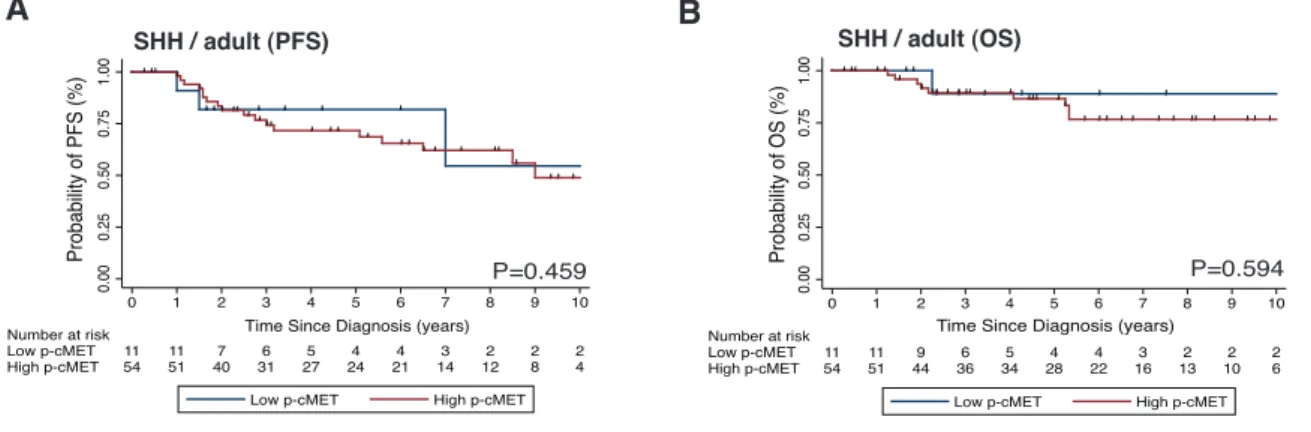
Figure 2.9: Prognostic impact of p-cMET status in adult SHH medulloblastomas. ... 84
Figure 2.10: cMET pathway targeting by foretinib in human medulloblastoma cell lines... 85
Figure 2.11: PDGFRß pathway inhibition by foretinib in human medulloblastoma cell lines. ... 86
Figure 2.12: Anti-proliferative and pro-apoptotic effects of foretinib in human medulloblastoma cells. ... 87
Figure 2.13: Effect of cMET and PDGFRß inhibition in medulloblastoma migration... 88
Figure 2.14: Effect of cMET and PDGFRß inhibition in medulloblastoma invasion... 89
Figure 2.15: Foretinib induces regression of Daoy medulloblastoma flank xenografts. ... 91
Figure 2.16: Foretinib reduces tumor growth in ONS76 medulloblastoma flank xenografts.. 92
Figure 2.18: Distribution of foretinib in preclinical mouse models... 95 Figure 2.19: Foretinib decreases tumor growth and metastases in intracranial xenografts models of medulloblastoma... 97 Figure 2.20: Foretinib prevents metastases formation and increases survival in a transgenic mouse model of metastatic SHH medulloblastoma. ... 99
Chapter 3
Figure 3.1: Cytotoxic effect of piperlongumine, alsterpaullone, rottlerin and flunarizine in Group 3 medulloblastoma cell lines... 111 Figure 3.2: Piperlongumine, alsterpaullone, and rottlerin, reduce tumor growth in D458 medulloblastoma xenografts... 113 Figure 3.3: Representative H&E staining of D458 medulloblastomas treated with piperlongumine, alsterpaullone and rottlerin... 114 Figure 3.4: Survival of D458 intracranial xenografts treated with piperlongumine, alsterpaullone, rottlerin and flunarizine... 114 Figure 3.5: Piperlongumine and alsterpaullone increase survival of D425 medulloblastoma xenografts. ... 115 Figure 3.6: Piperlongumine and alsterpaullone induce apoptosis and inhibit AKT pathway activation. ... 116 Figure 3.7: In vitro efficacy of alsterpaullone targeting medulloblastoma stem cells derived from a patient with a Group 3 medulloblastoma... 117 Figure 3.8: In vivo efficacy of alsterpaullone treating mice with primary cerebellar xenografts.
... 118 Figure 3.9: Genomic profiling of Group 3 medulloblastoma cell lines treated with alsterpaullone demonstrates down-regulation of cell cycle related genes, including
MYC... 120 Figure 3.10: Biological pathways and processes up- and down-regulated by alsterpaullone.
... 121 Figure 3.11: Alsterpaullone reverses Group 3 medulloblastoma gene expression signature. 122 Figure 3.12: The gene expression profile of Group 3 medulloblastomas is not affected by piperlongumine... 123
List of Tables
Chapter 1Table 1.1: Summary of the HGF/cMET inhibitors. ... 49 Table 1.2: Clinical trials in medulloblastoma. ... 54
Chapter 2
Table 2.1: Foretinib concentrations in mouse brain, mouse plasma and the brain/plasma ratio after administration of 30, 60 and 100 mg/kg by oral gavage... 94
Chapter 3
Table 3.1: Top 20 drugs with predicted efficacy by the Connectivity Map analysis (P<0.05), for each medulloblastoma subgroup... 108 Table 3.2: Top 15 drugs specific for Group 3 medulloblastomas, as predicted by the Connectivity Map analysis (P<0.05)... 109
Abstract
Medulloblastoma is the most common malignant brain tumor in childhood. The standard of care to treat patients with medulloblastoma includes surgical resection, craniospinal irradiaton (in children older than 3 years of age) and high dose chemotherapy. Overall survival rates for patients submitted to conventional treatment protocols have reached 70-80% but the majority of survivors suffer from severe long-term side effects, including developmental, cognitive, neurological and neuroendocrine deficits.
For many years medulloblastoma was considered a single entity, a small, round blue-cell tumor on histology, originating in the cerebellum. Recently, integrative genomic studies have identified at least four distinct molecular subgroups – WNT, sonic hedgehog (SHH), Group 3 and Group 4 – which display disparate transcriptional profiles, genetic abnormalities, patient demographics and clinical outcome. Patients with WNT medulloblastomas have a very good outcome. Patients with SHH and Group 4 tumors have an intermediate prognosis. Group 3 medulloblastomas are characterized by frequent amplifications or aberrant expression of the oncogene MYC, a high percentage of metastasis at diagnosis and a very poor prognosis despite aggressive therapy. Although disseminated disease at diagnosis is a known factor of poor survival, the mechanisms of dissemination are not fully understood and metastatic lesions are highly genetically divergent from their matched primary tumor. Therefore, we asked whether we could use genomics to discover novel and less toxic small molecule inhibitors to target medulloblastoma in a subgroup-specific manner.
To answer our research question we used two approaches: 1) targeting the hepatocyte growth factor (HGF)/cMET pathway, known to be important in medulloblastoma pathogenesis, with the cMET inhibitor, foretinib and 2) performing a gene expression-based
in silico drug screen to identify small molecule inhibitors with selective antitumor effect in
Group 3 medulloblastomas.
Aberrant signaling through the (HGF)/cMET pathway is involved in tumor progression and metastasis in several human cancers. cMET is a transmembrane receptor which becomes activated through phosphorylation of tyrosine residues upon binding of its ligand HGF. The recruiment of cytoplasmic effector proteins upon cMET activation triggers multiple downstream effector pathways including mitogen activated protein kinase (MAPK) and phosphoinositide 3-kinase (PI3K). In medulloblastoma, cMET activation has been associated with tumor growth and invasion. To determine the subgroup-specific role of cMET
in medulloblastoma, we analyzed the mRNA expression of three large non-overlapping cohorts of patients with primary medulloblastomas (discovery cohort from Boston, n = 199; validation cohort 1, a multicentre cohort obtained from Heidelberg, n = 439; validation cohort 2, a multicentre cohort obtained from the Medulloblastoma Advanced Genomics International Consortium - MAGIC, n = 285) and demonstrated that cMET is a marker of SHH medulloblastoma. Immunohistochemical analysis of activated cMET (phosphorylated cMET) in another independent patient cohort (n = 385) validated these findings and revealed that cMET activation correlates with increased tumor relapse and a poor survival in pediatric patients with SHH medulloblastomas, thus defining a subset of patients that may benefit from cMET targeted therapy.
We selected foretinib, an FDA approved multikinase inhibitor with high affinity to the cMET receptor, to evaluate the effect of cMET targeting in SHH medulloblastoma. Foretinib suppressed cMET activation, decreased proliferation and induced apoptosis, both in medulloblastoma cell lines and in SHH medulloblastoma flank xenografts. Importantly, we characterized the pharmacokinetics of foretinib and demonstrated that it penetrates the blood-brain barrier and is well tolerated through intrathecal administration. Treatment of mouse intracranial xenografts and of an aggressive transgenic mouse model of metastatic SHH medulloblastoma (Patched+/- mouse with the Sleeping Beauty transposon system) with foretinib reduced primary medulloblastoma growth and invasion, decreased the incidence of metastases by 36% and increased survival by 45%.
To investigate potential novel small molecules to target Group 3 medulloblastomas we used a bioinformatic platform called the Connectivity Map (C-MAP). The C-MAP contains a collection of gene expression profiles from several human cell lines treated with a large number of compounds already FDA approved. Comparative analysis between the oncogenic gene signature of interest and the C-MAP database may highlight patterns of gene expression change and identify small molecules with potential predicted activity within a given cancer. We queried the C-MAP using the subgroup-specific gene expression signatures of the four medulloblastoma subgroups. Piperlongumine, a natural product extracted from the fruit of the
Piper longum, was the top candidate drug for non-WNT tumors. We then selected compounds
predicted to have specific antitumor activity for Group 3 medulloblastomas and the cyclin-dependent kinase (CDK) inhibitor alsterpaullone ranked number one. To validate our findings we used two established Group 3 medulloblastoma cell lines (D425 and D458) and a medulloblastoma stem cell line derived from a patient with a Group 3 tumor (M441). The
C-MAP predicted drugs were able to decrease cell proliferation in vitro and to reduce tumor growth and increase survival in Group 3 medulloblastoma mouse xenografts. Alsterpaullone showed the highest efficacy both in vitro and in vivo in the patient derived medulloblastoma stem cell line. Interestingly, the chemical genomic profiling of Group 3 medulloblastoma cells treated with alsterpaullone confirmed inhibition of cell cycle-related genes and showed down-regulation of MYC, the hallmark oncogene of Group 3 tumors.
We used a gene expression-based approach to identify molecular targeted therapy to treat medulloblastoma. Our results demonstrate the preclinical efficacy of two small molecule inhibitors: foretinib in metastatic SHH medulloblastomas and alterpaullone in Group 3 tumors. Given the dismal outcome and lack of options for these patients, the results from our studies provide strong rationale for advancing both compounds as targeted agents into clinical trials for patients with these highly aggressive subgroups of medulloblastoma.
Keywords: medulloblastoma subgroups, genomics, HGF/cMET pathway, foretinib,
Resumo
O meduloblastoma é o tumor cerebral maligno mais comum na população pediátrica. O tratamento convencional dos doentes com meduloblastoma inclui resseção cirúrgica máxima, radioterapia do crânio e do neuroeixo (em crianças com idade superior a 3 anos) e quimioterapia de alta dose. A sobrevida global em doentes submetidos a protocolos de tratamento convencional atinge os 70-80% mas a maioria dos sobreviventes apresenta sequelas graves dos tratamentos incluindo problemas no desenvolvimento, defeitos cognitivos, neurológicos e neuroendócrinos.
Durante muito anos o meduloblastoma foi considerado uma entidade única, um tumor de pequenas células azuis na observação histológica, oriundo do cerebelo. Recentemente, estudos de genómica integrada identificaram pelo menos quatro subgrupos moleculares de meduloblastoma – WNT, sonic hedgehog (SHH), Grupo 3 e Grupo 4 – que apresentam características distintas no que diz respeito ao perfil transcriptómico, anomalias genéticas, dados demográficos e sobrevida. Doentes com meduloblastomas do tipo WNT têm muito bom prognóstico. Doentes com tumores do tipo SHH e Grupo 4 têm um prognóstico intermédio. Os meduloblastomas do Grupo 3 são caracterizados por amplificações frequentes ou expressão aberrante do oncogene MYC, uma elevada incidência de metástases na altura do diagnóstico e um mau prognóstico apesar de terapias agressivas. Embora a presença de doença disseminada na altura do diagnóstico seja um factor conhecido de baixa sobrevida, os mecanismos de disseminação são pouco conhecidos e as lesões metastáticas são geneticamente divergentes do seu tumor primário. Assim, questionámos se seria possível usar a genómica na descoberta de pequenas moléculas inibidoras dirigidas aos subgrupos moleculares de meduloblastoma.
Para responder à nossa pergunta utilizámos duas abordagens: 1) uma terapia dirigida à via de sinalização do factor de crescimento dos hepatócitos (HGF)/cMET, conhecida pelo seu importante papel na patogénese do meduloblastoma, usando um inibidor do receptor cMET, foretinib e 2) um rastreio farmacológico in silico com base na expressão genética para identificar pequenas moléculas inibidoras com efeito antitumoral selectivo nos meduloblastomas do Grupo 3.
A sinalização aberrante através da via de transdução de sinal (HGF)/cMET está envolvida na progressão tumoral e metastização de vários cancros humanos. cMET é um receptor transmembranar que é activado através da fosforilação de resíduos de tirosina, após
interacção com o seu ligando HGF. O recrutamento de proteinas efectoras citoplasmáticas após a activação de cMET desencadeia a activação de múltiplas vias de sinalização incluindo MAPK (mitogen activated protein kinase) e PI3K (phosphoinositide 3-kinase). No meduloblastoma a activação de cMET tem sido associada a crescimento e invasão tumoral. Para determinar a importância de cMET nos diferentes subgrupos de meduloblastoma, analizámos a expressão de mRNA em três grupos independentes de doentes com meduloblastomas primários (grupo de descoberta de Boston, n = 199; grupo de validação 1, grupo multicêntrico obtido de Heidelberg, n = 439; grupo de validação 2, grupo multicêntrico obtido do Medulloblastoma Advanced Genomics International Consortium - MAGIC, n = 285) e demonstrámos que cMET é um marcador do meduloblastoma do tipo SHH. A análise imunohistoquímica de cMET activado (ou cMET fosforilado) noutro grupo independente de doentes (n = 385) validou estes resultados e revelou que a activação de cMET se correlaciona com o aumento de recidivas tumorais e com uma baixa sobrevida em doentes pediátricos com meduloblastoma do tipo SHH, definido assim um subgrupo de doentes que pode beneficiar de terapêutica dirigida a cMET.
Seleccionámos foretinib, um inibidor de múltiplas cinases aprovado pela FDA e com elevada afinidade para cMET, para avaliar o efeito da terapia dirigida a cMET no meduloblastoma do tipo SHH. Foretinib bloqueou a activação de cMET, reduziu a proliferação celular e induziu apoptose, tanto em linhas celulares de meduloblastoma como em tumores implantados no flanco de ratinhos. Importa salientar que caracterizámos a farmacocinética do foretinib e demonstrámos que este fármaco penetra a barreira hemato-encefálica e é bem tolerado quando administrado por via intratecal. O tratamento com foretinib em ratinhos com meduloblastomas implantados no cerebelo e num modelo de ratinho transgénico com uma forma agressiva de meduloblastoma metastizado do tipo SHH (ratinho transgénico Patched+/- com sistema de transposão do tipo Sleeping Beauty) reduziu o crescimento e invasão em meduloblastomas primários, diminuiu a incidência de metástases em 36% e aumentou a sobrevida em 45%.
Para investigar potenciais novas pequenas moléculas dirigidas aos meduloblastomas do Grupo 3, usámos uma plataforma bioinformática chamada Connectivity Map (MAP). O C-MAP contém uma colecção de perfis de expressão génica de múltiplas linhas celulares humanas tratadas com um elevado número de compostos aprovados pela FDA. A análise comparativa entre assinaturas genéticas oncogénicas de interesse e a base de dados C-MAP pode detectar padrões de mudança de expressão génica e identificar pequenas moléculas com
potencial actividade num determinado cancro. Assim, questionámos o C-MAP usando as assinaturas genéticas específicas dos quatro subgrupos de meduloblastoma. Piperlongumine, um produto natural extaído do fruto do Piper longum, foi identificado como o melhor candidato para os diferentes subgrupos de meduloblastoma, à excepção do tipo WNT. De seguida, seleccionámos compostos com potencial actividade antitumoral específica para os meduloblastomas do Grupo 3, tendo um inibidor das CDKs (cyclin-dependent kinases), alsperpaullone, sido o melhor classificado. Para validar os nossos resultados usámos duas linhas celulares estabelecidas representativas dos meduloblastomas do Grupo 3 (D425 e D458) e uma linha celular pluripotente obtida de um doente com um tumor do Grupo 3 (M441). Os fármacos predictos pelo C-MAP foram eficazes na diminuição da proliferação celular in vitro bem como na redução do crescimento tumoral e no aumento da sobrevida em ratinhos com meduloblastomas do Grupo 3 implantados no cerebelo. O composto alsterpaullone revelou-se o mais eficaz, quer in vitro quer in vivo, na linha celular pluripotente obtida de um doente. É interessante salientar que a caracterização do perfil químico e genómico de células de meduloblastoma do Grupo 3 tratadas com alsterpaullone confirmou a inibição de genes relacionados com o ciclo celular e a regulação negativa de MYC, o oncogene característico dos meduloblastomas do Grupo 3.
Usámos no nosso estudo uma abordagem com base na expressão genética para identificar terapêuticas dirigidas para o tratamento do meduloblastoma. Os resultados apresentados demonstram a eficácia pré-clínica de duas pequenas moléculas inibidoras: foretinib para o tratamento do meduloblastoma metastático do tipo SHH e alsterpaullone para o tratamento de meduloblastomas do Grupo 3. Tendo em conta o prognóstico sombrio e a falta de opções terapêuticas nestes doentes, os resultados dos nossos estudos constituem forte evidência para que estes compostos sejam incluídos como terapias dirigidas em ensaios clínicos para doentes diagnosticados com estes subgrupos agressivos de meduloblastoma.
Palavras-chave: subgrupos de meduloblastoma, genómica, via de sinalização HGF/cMET,
Chapter 1
Introduction
1.1 The Genomic Landscape of Medulloblastoma
iMedulloblastoma is the most common malignant brain tumor in childhood, comprising 20% of all primary pediatric brain tumors, although it can arise from infancy to adulthood1. The incidence of medulloblastoma in children has been estimated between 5.0 and 6.6 per million per year2,3 and in adults between 0.57 and 0.58 per million per year4. The current treatment protocol for medulloblastoma includes surgical resection, radiation (for children older than 3 years of age) and chemotherapy with 5-year survival rates ranging between 50 and 80%. Among long-term survivors, a major concern is the sequelae induced by treatment including endocrinologic, neurocognitive and behavioral dysfunction5.
Medulloblastoma arises in the cerebellum and originates from primitive pluripotent precursor cells of the ventricular zone and the external granular layer6. Medulloblastoma formation is strongly associated with dysregulation of pathways involved in the normal development of the cerebellum, including the Sonic hedgehog (SHH), Wingless (WNT) and Notch pathways7.
The World Health Organization (WHO) classified medulloblastoma as a grade IV embryonal neoplasm comprising five histological variants: classic, desmoplastic/nodular, anaplastic, large cell and medulloblastoma with extensive nodularity8. Classic medulloblastomas are composed of densely packed, poorly differentiated cells, and areas of
i
Claudia C. Faria, Christian A. Smith and James T. Rutka (2013). New Molecular Targets and Treatments for Pediatric Brain Tumors, Evolution of the Molecular Biology of Brain Tumors and the Therapeutic Implications, Dr. Terry Lichtor (Ed.), ISBN: 978-953-51-0989-1, InTech, DOI: 10.5772/53300.
Claudia C. Faria, Yuzo Terakawa and James T. Rutka (2014). Neurogenetic Basis of Pediatric Neurosurgical Conditions, Principles and Practice of Pediatric Neurosurgery, 3rd ed, Leland Albright, Ian F. Pollack, P. David
Homer-Wright rosettes. The desmoplastic/nodular variant includes pale nodular areas (pale islands) with patterns of neuronal differentiation surrounded by hyperchromatic cells densely packed. Anaplastic medulloblastomas are characterized by large pleomorphic nuclei. Tumor cell “wrapping” and mitotic figures are abundant. Large cell tumors are composed of enlarged round cells with prominent nuclei and pronounced mitotic and apoptotic figures. Tumors with both large cell and anaplastic regions can be encountered. Medulloblastoma with extensive nodularity shows a lobular architecture with long reticulin-free areas between the nodules. Clinical studies suggest a favorable outcome associated with desmoplastic medulloblastoma and a significant worst prognosis with the anaplastic subtype9.
Medulloblastoma has been reported in the setting of several hereditary cancer syndromes including Li-Fraumeni syndrome, Gorlin syndrome, Turcot syndrome and Rubinstein–Taybi syndrome10. The Li-Fraumeni syndrome is associated with germline mutations in the TP53 gene. Among many other cancers, patients with Li-Fraumeni syndrome have an increased risk to develop embryonal tumors (including medulloblastoma) and choroid plexus carcinoma. Gorlin syndrome (nevoid basal cell carcinoma syndrome) patients have germline mutations in the PTCH1 gene, a component of the SHH signaling pathway, and up to a 5% risk of developing medulloblastoma. Germline mutations in the SUFU gene, a downstream mediator of the SHH pathway, have also been shown in patients with medulloblastoma11. Germline mutations of the APC gene, a component of the WNT signaling pathway, are seen in patients with Turcot syndrome who have an increased risk for colorectal carcinoma and brain tumors, predominantly medulloblastoma. Children with Rubinstein-Taybi syndrome have a complex developmental disorder that includes broad thumbs and toes, characteristic facies, severe developmental delay, and a predisposition to malignancy, including medulloblastoma, oligodendroglioma and meningioma. This syndrome is secondary to deletion/mutation of the Crebs binding protein (CBP) gene which functions in three pathways involved in medulloblastoma pathogenesis (SHH, WNT and TP53)12.
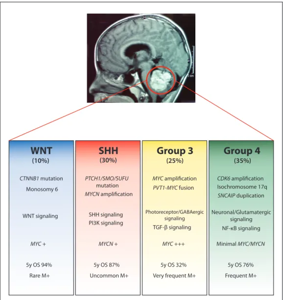
Recent efforts of multiple independent groups have reached to a consensus that medulloblastoma comprises four distinct molecular variants named WNT, SHH, Group 3 and Group 413. The subgroups have different demographics, genetic profiles and prognosis14. This
may explain why patients with the same histological disease have different clinical outcomes and a variable response to current treatments including surgery, whole-brain radiation and intensive chemotherapy. Figure 1.1 summarizes the main features of the four medulloblastoma subgroups.
1.1.1 WNT Medulloblastomas
WNT medulloblastomas are frequent in older children and teenagers and are rarely seen in infants. Patients within this subgroup have usually a very good outcome with survival rates over 90%. However, Remke et al. showed that this is only true for pediatric cases as adult WNT patients exhibit survival rates of approximately 80%15. Histologically, WNT tumors are almost always of the classic variant and they rarely disseminate.
Figure 1.1: Features of the four medulloblastoma subgroups, including molecular genetics
and clinical outcome.
WNT
SHH
Group 3
Group 4
(10%) (30%) (25%) (35%) CTNNB1 mutation Monosomy 6 MYC + 5y OS 94% Rare M+ Uncommon M+ 5y OS 87% 5y OS 32% Very frequent M+ 5y OS 76% Frequent M+ WNT signaling PTCH1/SMO/SUFU mutation MYCN amplification SHH signaling PI3K signaling MYCN + MYC amplification PVT1-MYC fusion MYC +++ TGF-β signaling Photoreceptor/GABAergic signaling CDK6 amplification Isochromosome 17q SNCAIP duplication Neuronal/Glutamatergic signaling NF-κB signaling Minimal MYC/MYCNThis subgroup is enriched in genes of the WNT pathway. Specifically gene expression changes involve a combination of increased expression of inhibitors of WNT signaling (e.g.
WIF1, DKK1, DKK2, AXIN2) and potential promoters of WNT signaling (e.g. WNT16, LEF1)16. CTNNB1 mutation is the most common genetic event in WNT medulloblastomas and promotes the accumulation of β-catenin in the nucleus. Positive nuclear immunostaining for β-catenin is now routinely used as a marker of WNT pathway activation and is associated with better survival rates17. After CTNNB1, the DDX3X (DEAD-box RNA helicase) gene is
the most frequently mutated in medulloblastoma and occurs in 50% of WNT tumors18. WNT medulloblastomas often carry TP53 mutations and they confer a more favourable outcome19. Almost all patients with WNT medulloblastomas have monosomy 6 (deletion of one copy of chromosome 6), although the role of this chromosomal aberration remains undefined20.
Recently, a mouse model of WNT medulloblastoma was generated and the cell of origin for this subgroup was elucidated. Mice with transgenic expression of a CTNNB1 mutation in the context of a TP53 deletion developed classic medulloblastoma, arising from progenitor cells in the lower rhombic lip of the dorsal brainstem21.
The overall favourable outcome of WNT tumors suggests that this subgroup of patients may be considered for the de-escalation of therapy in future clinical trials.
1.1.2 SHH Medulloblastomas
SHH medulloblastomas have an intermediate prognosis with a 5-year overall survival (OS) of approximately 75%. Age distribution in these tumors is bimodal. They are frequently found in infants and adults but are rare in childhood. Prognostic factors such as desmoplasia and metastatic status are age-dependent. Desmoplasia is associated with a poor outcome only in children, while metastasis at presentation constitutes a negative prognostic factor only in adults22. SHH medulloblastomas include tumors of the four main histological variants
(classic, nodular desmoplastic, large-cell anaplastic and medulloblastoma with extensive nodularity), although nodular desmoplastic histology is almost exclusively seen in this subgroup.
SHH-driven medulloblastomas exhibit aberrant expression of SHH pathway genes including PTCH1, SUFU, SMO and GLI2. Germline mutations affecting PTCH1 or SUFU predispose patients with Gorlin syndrome to develop medulloblastoma. Amplifications and somatic copy number aberrations (CNAs) of SHH target genes such as MYCN and GLI2 are also seen in this subgroup. An interesting association of SHH medulloblastoma and
deregulation of the PI3K signaling pathway has been recently reported. The alterations include amplifications of insulin-like growth factor 1 receptor (IGF1R) and focal deletions of
PTEN23. When compared to WNT medulloblastoma genome, the SHH subgroup contains significant more gains and losses of chromosomal regions, including deletion of chromosome 9q and 10q. In an attempt to simplify the molecular subgrouping of medulloblastomas, different laboratories used formalin-fixed paraffin-embedded tissues (FFPE) to test a variety of markers for SHH medulloblastomas including SFRP1, GLI1 and GAB116,24,25.
Of note is the fact that the transcriptomes of pediatric and adult SHH tumors have different expression profiles with increased levels of genes related to extracellular matrix function in the first group and elevated levels of HOX family genes and genes involved in tissue development in the second group22,26. The clinical and molecular distinction of infant and adult SHH medulloblastomas suggests a disparate underlying biology and raise the question of possible different responses to current targeted therapies.
The cells of origin of SHH medulloblastoma are the cerebellar granule neuron precursors (CGNPs) of the external granule cell layer (EGL) of the cerebellum27 and those of the cochlear nuclei of the brainstem28. SHH medulloblastomas can also be initiated from neural stem cells in the subventricular zone29.
Several mouse models of SHH-dependent medulloblastoma are available for basic and preclinical research. The most common initiating events in these models include Ptch1 inactivation and smoothened (Smoa1) activation18.
1.1.3 Group 3 Medulloblastomas
Group 3 medulloblastomas have the worst prognosis of all subgroups. These tumors are restricted to children and infants, frequently have a large-cell anaplastic histology and 40 to 45% are disseminated at the time of diagnosis. One of the features of this subgroup is the amplification of MYC. In a recent study including a cohort of over 1,000 medulloblastoma samples, Northcott et al. identified recurrent fusions involving MYC and PVT1 in about 60% of MYC-amplified tumors23. PVT1 gene is adjacent to MYC on chromosome 8q24.21 and
encodes several microRNAs that seem to promote MYC oncogenic properties30. This study also identified for the first time the TGF-β pathway as a potential target in Group 3 medulloblastoma. The genome of Group 3 tumors is highly unstable often exhibiting gains in chromosomes 1q, 7 and 17q (frequently isochromosome 17q), as well as deletions in chromosomes 10q, 11, 16q and 17p.
Recently, mouse models of MYC-driven Group 3 medulloblastoma, combined with Trp53 inactivation, were published. The cells of origin of these tumors were prominin 1-positive, lineage-negative neural stem cells and the CGNPs of the EGL31,32. Since TP53 mutations are not seen in Group 3 medulloblastomas other candidates to cooperate with MYC in the process of tumorigenesis are being studied.
1.1.4 Group 4 Medulloblastomas
The most common medulloblastoma subgroup is Group 4 and, curiously, it is the least well understood. Group 4 tumors can be found across all age groups although children have an intermediate prognosis while adults may do significantly worse15. The expression of follistatin-related protein 5 (FSTL5) was identified as a marker of high-risk Group 4 patients33. These tumors are usually of the classic variant and show dissemination in one third of the patients. Recurrent amplifications of MYCN and cyclin-dependent kinase 6 (CDK6) are frequent in this subgroup as well as isochromosome 17q (i17q). Recently, a novel and frequent somatic CNA was described in Group 4 medulloblastomas. It is a duplication of
SNCAIP, a gene involved in Parkinson’s disease and located on chromosome 5q23.2.
Duplication of SNCAIP is restricted to Group4α, a subtype of Group 4 medulloblastoma with a mostly balanced genome23. The NF-κB pathway was also identified as a new targetable pathway in this subgroup of tumors. However, there are currently no mouse models of Group 4 tumors and its cell of origin is still unknown.
Despite the potential for prognostic subgrouping based on molecular signatures or immunohistochemistry, clinical application of these findings is in its infancy. No consensus on a prognostic algorithm currently exists, although efforts to merge clinical and molecular features for risk stratification in clinical trials is underway.
1.2 The Role of HGF/cMET Pathway Signaling in Human
Medulloblastoma
iiMedulloblastoma is associated with dysregulation of pathways that normally lead to cerebellum development. Over the past few years, different signaling pathways have been shown to play a critical role in medulloblastoma formation and progression. The hepatocyte growth factor (HGF)/cMET signaling pathway has been implicated in different processes including development and tumorigenesis but only recently has it been demonstrated in medulloblastoma pathogenesis. The receptor tyrosine kinase cMET is normally activated by ligation through its ligand HGF, secreted as a precursor that is proteolytically cleaved in an active form by the serine protease hepatocyte growth factor activator. HGF is a member of the plasminogen-related growth factor family and was originally identified as a growth factor for hepatocytes and as a fibroblast-derived cell motility or scatter factor. The interplay between cMET and its ligand mediate downstream events that, in the central nervous system, play a critical role in cerebellar granule cell precursors proliferation and survival. Dysregulation of this pathway can promote tumorigenesis through cell migration, invasion and metastasis, angiogenesis and prevention from apoptosis. cMET has been found to be overexpressed in a variety of malignancies where its activation can occur by HGF ligation or through ligand independent mechanisms, including mutations and amplifications.
It was shown that medulloblastoma tumor cell lines and surgical tumor samples express HGF and cMET. Furthermore, overexpression of cMET is associated with poor clinical outcome. Treatment of medulloblastoma cell lines with HGF induced tumor cell proliferation, anchorage-independent growth and reduced apoptosis in response to chemotherapy. SPINT2, a tumor supressor gene silenced by promoter methylation in medulloblastoma, was identified by our group as a key regulator of HGF/cMET pathway34. Several therapeutic strategies aiming to target and limit the signaling cascade of HGF/cMET were examined with the cMET inhibitors being the most promising. Targeting the HGF/cMET pathway, alone or in combination with standard therapies is likely to improve present treatments in MET-dependent malignancies such as medulloblastoma.
ii
Claudia Faria, Christian Smith and James Rutka (2011). The Role of HGF/c-Met Pathway Signaling in Human Medulloblastoma, Molecular Targets of CNS Tumors, Dr. Miklos Garami (Ed.), ISBN: 978-953-307-736-9, InTech, DOI: 10.5772/23296.
1.2.1
The HGF/cMET Pathway Signaling
The HGF/cMET pathway has been associated with normal development, organ regeneration and cancer. cMET is a high affinity tyrosine kinase receptor for hepatocyte growth factor (also known as scatter factor, capable of inducing dissociation and motility). cMET is generally expressed in epithelial cells and is activated by HGF produced in surrounding mesenchymal cells or released into the circulation.
During embryogenesis the HGF/cMET signaling is necessary for the development of the placenta, liver, kidney and neuronal tissue but also for the directional migration of skeletal muscle cells35. In adult tissues, this pathway has been implicated in regeneration and wound healing36,37. Therefore, the HGF/cMET axis is a key player in cell proliferation, survival and migration and, when dysregulated, can give origin to a variety of cancers.
1.2.1.1
Structure of HGF and cMET
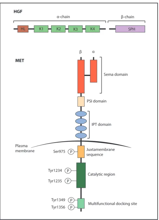
HGF is a multidomain protein similar to plasminogen, a circulating proenzyme that promotes the lysis of fibrin blood clots in its active form as plasmin. HGF is synthesized as a single-chain inactive precursor and it is converted by serine proteases into an active form with two chains (α and β chain) linked by a disulfide bond. HGF consists of six domains: an amino-terminal hairpin loop domain, four kringle domains (K1-K4) and a serine protease homology (SPH) domain which lacks enzymatic activity (Figure 1.2).
cMET, the HGF receptor, is a disulfide-linked heterodimer which results from cleavage of a precursor into an extracellular α chain and a transmembrane β chain. The extracellular region of cMET is composed of three domains: the Sema domain (homologous to the Sema domain of the semaphorins and plexins) that includes the entire α chain and part of the β chain; the PSI domain (also present in the plexins, semaphorins and integrins); and four IPT domains (immunoglobulin-like also found in plexins and transcriptional factors). The intracellular region of cMET consists of three portions: a juxtamembrane sequence that has the role to downregulate kinase activity upon phosphorylation of Ser975; a catalytic region that activates kinase activity following phosphorylation of Tyr1234 and Tyr1235; and a carboxy-terminal multifunctional docking site that contains two docking tyrosines (Tyr1349 and Tyr1356) essential for downstream signaling38.
Figure 1.2: HGF and cMET structures.
1.2.1.2
cMET Signal Transduction
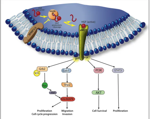
To activate the cMET receptor, the single chain HGF precursor is cleaved into a heterodimeric active form by a protease called HGF activator (HGFA)39. This process is regulated by a protein family of serine protease inhibitors called SPINT1 and SPINT240,41. Inhibiting the activation of HGF by HGFA, SPINT1 and -2 limit signaling through the HGF/cMET pathway.
Following HGF binding, the kinase activity of cMET is switched on. This process starts with receptor dimerization and trans-phosphorylation of two tyrosine residues in the catalytic region (Tyr1234 and Tyr1235) and is followed by phosphorylation of two additional tyrosines in the carboxy-terminal tail (Tyr1349 and Tyr1356). These tyrosines create docking sites for a variety of adaptor proteins and direct kinase substrates including the growth factor receptor-bound protein 2 (GRB2), Grb2-associated adaptor protein (GAB1), son of sevenless (SOS), SRC homology protein tyrosine phosphatase 3 (SHP2), phosphatidylinositol-3-kinase (PI3K) and signal transducer and activator of transcription 3 (STAT3). This leads to the activation of downstream signaling pathways that include the mitogen-activated protein kinase (MAPK), PI3K/AKT and STAT pathways, which mediate cMET-dependent cell proliferation, survival, migration and invasion (Figure 1.3).
The activation of MAPK cascade will sequentially activate different protein kinases whose terminal effectors include extracellular signal-regulated kinases (ERK1 and ERK2), jun amino-terminal kinases (JNK1, JNK2 and JNK3) and p38. These downstream elements will activate cell cycle regulators leading to cell proliferation and will promote alterations in cytoskeletal functions that control cell migration and invasion. PI3K/AKT activation mediates cell survival and resistance to apoptosis through inactivation of the pro-apoptotic protein BCL-2 antagonist of cell death (BAD) and degradation of the pro-apoptotic protein p5342.
Upon activation of STAT3 by the cMET receptor at the plasma membrane, it translocates to the nucleus to operate as a transcription factor regulating the expression of genes implicated in cell proliferation and differentiation43.
Other molecules that interact with the cMET receptor include the epidermal growth factor receptor (EGFR), the α6β4 integrin, the semaphoring receptors of the plexin B family and the variant of the hyaluronan receptor CD44 (that links the extracellular matrix and the intracellular cytoskeleton)44-46. This crosstalk of cMET with different surface proteins
highlights the dynamic environment at the plasma membrane and contributes to cMET associated biological responses47.
1.2.1.3
Regulation of cMET Signaling
It has been shown that the signaling network around the tyrosine kinase receptor cMET is more complex than the known process of recruiting signaling effectors at the plasma membrane and subsequently stimulating intermediates in the cytosol. In fact, this view has been expanded by the finding that cMET signals can also originate from endosomal compartments and by a series of other events.
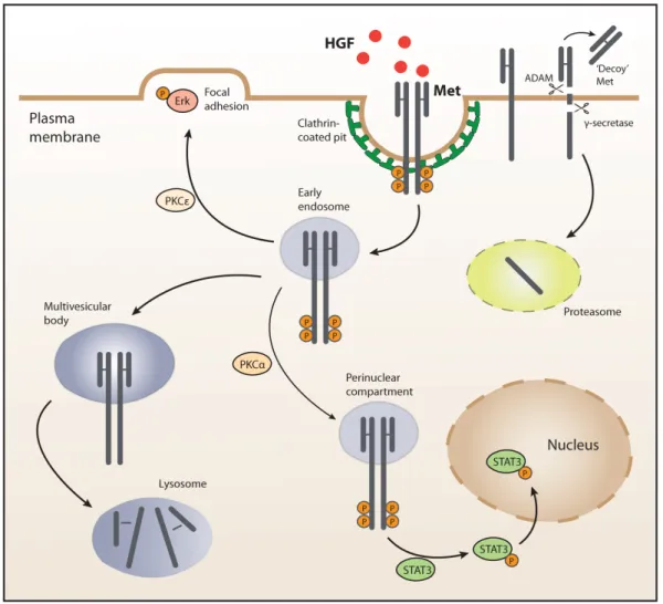
Upon HGF binding, cMET is internalized by clathrin-mediated endocytosis and recruited into peripheral early endosomes. This process is mediated by protein kinase Cε (PKCε) that promotes the transfer of active ERK to focal adhesions and, subsequently, the HGF-induced cell migration. From the peripheral endosomes, cMET travels along the microtubule network to late perinuclear compartments in a process mediated by PKCα. This juxtanuclear accumulation of cMET is a determinant step for activation and nuclear translocation of STAT348,49.
Downregulation of cMET signaling involves trafficking and degradation of ligand-activated receptors in the lysosomes. This process is initiated by the association of cMET with casitas B-lineage lymphoma (CBL) and endocytic adaptors. Following endocytosis, cMET
accumulates in multivesicular bodies that later fuse with lysosomes and lead to protein degradation. cMET can also undergo sequential proteolytic cleavage at two juxtamembrane sites. The first cleavage occurs in the extracellular domain and is mediated by a desintegrin and metalloprotease (ADAM) originating a ‘decoy’ fragment that sequesters the ligand and interferes with the receptor’s activity. The second cleavage is performed in the intracellular domain, by a γ-secretase and yields a fragment that is destroyed in the proteasome50 (Figure
1.4).
Figure 1.4: Mechanisms of cMET signaling regulation.
1.2.2
HGF, cMET and Cancer
The dysregulation of HGF/cMET signaling has emerged as a key player in several human malignancies, particularly in invasion and metastasis. Human cell lines overexpressing either HGF and/or cMET become tumorigenic and metastatic when implanted into nude
mice51. Moreover, transgenic mice expressing the receptor or the ligand develop metastatic tumors52. On the contrary, downregulation of HGF or cMET expression in human tumor xenografts decreases tumor growth53. There are three biological mechanisms underlying the tumorigenicity of cMET: a) the establishment of HGF/cMET autocrine loops; b) the overexpression of HGF or cMET; and c) the presence of activating mutations in the cMET receptor54.
An autocrine mechanism of cMET activation is found in some human tumors. For example, osteosarcomas and rabdomyosarcomas are derived from mesenchymal cells which physiologically produce HGF. Glioblastomas and breast carcinomas are derived from ectodermal tissues that normally express cMET but not HGF. Experimental models of HGF/cMET autocrine loops were also able to generate invasive tumors in vitro and in transgenic mice55
The most frequent mechanism of cMET dysregulation found in human tumors is the overexpression of the receptor or its ligand. A large number of studies showed that HGF and cMET are expressed in a wide variety of human tumors and in their metastasis. These include carcinomas of the breast, colon, lung, ovary, liver, kidney, upper gastrointestinal tract, pancreas and prostate but also sarcomas, haematopoietic malignancies, melanomas and glioblastomas42.
It has also been shown that high expression levels of cMET and its ligand correlate with increased aggressiveness of tumors and patients poor prognosis42. For example, in colorectal cancer patients, cMET is a powerful prognostic factor for early stage invasion and metastasis56. Moreover, in a study including 74 clinical samples of low-grade and high-grade gliomas the authors described a correlation of HGF and cMET expression levels with tumor grade57.
The compelling evidence that links cMET with human cancer relies in the cMET-activating mutations found in hereditary renal papillary carcinoma. These mutations were also found in sporadic tumors such as renal carcinoma, gastric cancer, childhood hepatocellular carcinoma and in head and neck squamous cell carcinomas47,55.
The association between cancer and blood coagulation disorders has been known for many years. In fact, approximately 50% of all patients with malignant tumors and up to 90% of those with metastasis have coagulopathies58. Interestingly, Boccaccio et al. showed in a mouse model that activation of the oncogene cMET induced cancer and a trombohemorrhagic syndrome through transcriptional upregulation of the procoagulation factors plasminogen activator inhibitor type 1 (PAI-1) and cyclooxygenase-2 (COX-2). Upon a first phase
characterized by a hypercoagulation state due to cMET signaling activation, the mice developed a hemorrhagic diathesis due to exhaustion of the hyperactivated hemostatic system59. At the early step of this process, hypoxia induces transcription of cMET that, subsequently, activates the transcription of genes involved in hemostasis, such as PAI-1 and
COX-2. The activation of the coagulation cascade will lead to fibrin deposition around cells
forming an extracellular matrix that will promote angiogenesis and cell migration60.
HGF/cMET signaling has also a role in angiogenesis either by direct influence of cMET activation in vascular endothelial cells, or by regulation of the expression levels of other angiogenic factors in tumor cells. It was previously shown that the HGF/cMET interaction stimulates proliferation and migration of endothelial cells in vitro and induces blood vessel formation in vivo61. Zhang et al. described the “angiogenic switch” in tumor cells upon HGF stimulation by simultaneous upregulation of the proangiogenic vascular endothelial growth factor (VEGF) and downregulation of thrombospondin 1 (TSP-1), an angiogenesis inhibitor62. This process has distinct mediators: while VEGF is modulated by MAPK, PI3K and STAT3, TSP-1 is targeted only by MAPK. An interesting example of this regulation was found in human glioma cells where stimulation with HGF increased the expression levels of VEGF and tumor-associated angiogenesis. The use of HGF and cMET inhibitors in experimental tumor models significantly reduced tumor growth and tumor vessel formation57. Recently, a new key player of the HGF/cMET pathway was described. Metastasis-associated in colon cancer-1 (MACC1) was identified by genome-wide expression analysis in primary and metastatic colon carcinomas. Its expression in human tumor samples was found to be an independent prognostic factor for metastasis formation and metastasis-free survival. The experimental studies showed that MACC1 promotes proliferation, invasion and HGF-induced scattering in vitro and tumor growth and metastasis in xenograft models63. The authors proposed a positive feedback mechanism where MACC1 acts as a transcriptional regulator of the cMET gene. The stimulation of the cMET receptor with HGF causes the translocation of MACC1 from the cytoplasm into the nucleus. There, MACC1 activates the transcription of the cMET gene by binding to its promoter64. The increased amounts of cMET
receptor will be able to bind more HGF molecules thereby enhancing the pathway signaling and promoting cell proliferation, migration and metastasis.