Promotes Angiogenesis via iNOS/Netrin-1/PKC Pathway
Rafeeq P. H. Ahmed, Khawaja Husnain Haider, Jiang Shujia, Muhammad Rizwan Afzal, Muhammad Ashraf*
Department of Pathology and Laboratory Medicine, University of Cincinnati, Cincinnati, Ohio, United States of America
Abstract
Background:We hypothesized that genetic modification of mesenchymal stem cells (MSCs) with Sonic Hedgehog (Shh) transgene, a morphogen during embryonic development and embryonic and adult stem cell growth, improved their survival and angiogenic potential in the ischemic heart via iNOS/netrin/PKC pathway.
Methods/Principal Findings:MSCs from young Fisher-344 rat bone marrow were purified and transfected with pCMV Shh plasmid (ShhMSCs). Immunofluorescence, RT-PCR and Western blotting showed higher expression of Shh inShhMSCs which also led to increased expression of angiogenic and pro-survival growth factors inShhMSCs. Significantly improved migration and tube formation was seen in ShhMSCs as compared to empty vector transfected MSCs (EmpMSCs). Significant upregulation of netrin-1 and iNOS was observed inShhMSCs in PI3K independent but PKC dependent manner. Forin vivo
studies, acute myocardial infarction model was developed in Fisher-344 rats. The animals were grouped to receive 70ml basal DMEM without cells (group-1) or containing 16106 EmpMSCs (group-2) and ShhMSCs (group-3). Group-4 received
recombinant netrin-1 protein injection into the infarcted heart. FISH andsry-quantification revealed improved survival of
ShhMSCs post engraftment. Histological studies combined with fluorescent microspheres showed increased density of
functionally competent blood vessels in group-3 and group-4. Echocardiography showed significantly preserved heart function indices post engraftment withShhMSCs in group-3 animals.
Conclusions/Significance:Reprogramming of stem cells with Shh maximizes their survival and angiogenic potential in the heart via iNOS/netrin-1/PKC signaling.
Citation:Ahmed RPH, Haider KH, Shujia J, Afzal MR, Ashraf M (2010) Sonic Hedgehog Gene Delivery to the Rodent Heart Promotes Angiogenesis via iNOS/Netrin-1/PKC Pathway. PLoS ONE 5(1): e8576. doi:10.1371/journal.pone.0008576
Editor:Annarosa Leri, Harvard Medical School, United States of America
ReceivedSeptember 14, 2009;AcceptedNovember 2, 2009;PublishedJanuary 5, 2010
Copyright:ß2010 Ahmed et al. This is an open-access article distributed under the terms of the Creative Commons Attribution License, which permits
unrestricted use, distribution, and reproduction in any medium, provided the original author and source are credited.
Funding:This work was supported by National Institutes of Health grants#R37-HL074272; HL-080686; HL087246 to M.A. and HL087288; HL089535 to Kh.H.H. The funders had no role in study design, data collection and analysis, decision to publish, or preparation of the manuscript.
Competing Interests:The authors have declared that no competing interests exist. * E-mail: [email protected]
Introduction
Sonic hedgehog (Shh) gene is one of the highly conserved mammalian hedgehog genes and has a wide distribution in a variety of tissues during embryonic development [1]. The post-natal intrinsic activity of Shh remains intact albeit with sub-optimal functioning and is reactivated under tissue ischemia and in various pathologies, including tumors [2,3]. Such post-natal reactivation of the embryonic signaling pathways implying Shh in response to muscle injury in the animal models incurred enhanced angiomyogenic response [4,5]. In one of the experi-mental studies, more than 15-fold increase in Shh mRNA expression was observed in the ischemic myocardium [6]. An outside intervention to overexpress Shh in the heart activated its downstream signaling cascade and strongly induced Patched1 (Ptc1) expression in the cardiomyocytes which indicated an active participation of Shh in the myocardial repair process. Interesting-ly, activation of Shh signaling caused upregulation of pro-angiogenic growth factors including vascular endothelial growth factor (VEGF) and angiopoietin-1 which resulted in an increased angiogenic response and globally improved the heart function.
Bone marrow derived stem cells (BMSCs) which have been shown to improve heart function, attenuate infarct size expansion and contribute to myocardial regeneration both in the experi-mental as well as in clinical settings [7–10]. In addition, BMSCs are excellent carriers of therapeutic genes to the heart [11–13]. In the present study we took advantage of the anti-apoptotic and pro-angiogenic role of Shh signaling and combined Shh transgene delivery to the infarcted heart by transplantation of mesenchymal stem cells (MSCs) which were non-virally transfected to overex-press Shh. The anticipated objective of our multipronged strategy was to achieve intracrine, autocrine, and paracrine effects of Shh protein which was secreted from MSCs overexpressing Shh (ShhMSCs) and regenerated the damaged tissue, induced revascu-larization and concomitantly prevented remodeling of the heart by preserving the existing myocardium.
over expression in MSCs initiated PKC signaling which was characterized by the activation of PKM, a catalytic fragment of PKC. ShhMSCs showed better survival post-engraftment as compared with the empty vector transfected control MSCs (EmpMSCs). Moreover, we report that Shh upregulated angiogenic genes such as netrin-1, iNOS, VEGF and angiopoietins, which play a significant role in Shh induced angiogenesis. Shh-induced upregulation of netrin-1 and iNOS was PKC dependent. We further observed that PKM was upregulated inShhMSCs and was sensitive to cyclopamine and chel pretreatment of the cells.
Materials and Methods
Detailed methods are available in Text S1. Table S1 shows the antibodies used for immunohistology and Western blotting, and Table S2 shows the primers used for RT-PCR. Young female Fischer-344 rats (n = 30) each weighing 180–200 g were used in this study. The present study conformed to the Guide for the Care and Use of Laboratory Animals published by the US National Institutes of Health (NIH Publication No. 85-23, revised 1996) and protocol approved by the Institutional Animal Care and Use Committee, University of Cincinnati. All surgical manipulations were carried out under general anesthesia.
Results
In vitroStudies
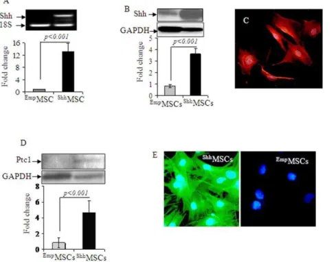
Shh plasmid was successfully constructed using commercially available pCMV Script Vector using mRNA isolated from 14-day rat embryo and the plasmid construct was sequenced for correct insertion of Shh transgene (Figure S1A & Figure S1B). RT-PCR and Western blotting revealed significantly elevated expression of Shh gene (12-fold) and protein (4.5-fold) inShhMSCs as compared
with EmpMSCs (Figure 1A & 1B). Fluorescent immunostaining showed that more than 65% cells stained positive for Shh transgene overexpression (Figure 1C). At 72-h after transfection with Shh, Ptc1 gene expression was 2.7-fold higher inShhMSCs as compared with EmpMSCs. These findings were confirmed by Western blot (Figure 1D) and fluorescent immunostaining (Figure 1E). Flow cytometry showed that Shh overexpression did not alter surface marker expression inShhMSCs (Figure S1C).
Shh Induced Angiogenic Growth Factor Expression in MSCs
In addition to upregulation of Ptc1, overexpression of Shh in MSCs stimulated the expression of secretable angiogenic growth factors including Ang-1 and VEGF. Our custom-made real-time PCR based array of 72 genes revealed multiple angiogenic growth factors showing more than 2-fold increase as compared toEmpMSCs (Table S3). Western blot studies using cell lysate protein samples fromShhMSCs andEmpMSCs confirmed these findings (Figure 2A).
Shh Caused Endothelial Mobilization and Tube Formation
We investigated the ability of human umbilical vein endothelial cells (HUVECs) to migrate toward conditioned medium from Shh
MSCs (ShhCM) using a modified Boyden chamber assay (Figure 2B). Incubation of HUVECs withShhCM for 4-h stimulated increased chemotaxis of HUVECs in a Transwell system as compared withEmpMSCs. In vitro angiogenic response of HUVECs toShhCM was determined by tube formation assay on matrigel which showed that morphological changes were most obvious at 6-h after incubation of the cells with ShhCM as compared with conditioned medium from empty vector transfected MSCs (EmpCM) (Figure 2C). Quantification of the number of branch points per low
Figure 1. In vitro characterization ofShhMSCs.(A) RT-PCR and (B) Western blotting showed significant amplification of Shh transgene and Shh
power microscopic field showed that the number of tubular structures were higher inShhCM as compared to the cells treated with EmpCM (p,0.01), and 10% FBS supplemented DMEM (p,0.001) (Figure 2D). The formation of tubular structures was abolished by prior treatment of the cells with anti-Shh (p,0.01 vs shh
CM without anti Shh antibody) and anti Netrin-1 antibodies (p,0.01 vs shhCM without anti-Shh antibody). Basal DMEM without FBS supplementation failed to induce any morphological changes in terms of tube formation (Figure 2D).
Shh Overexpression and Molecular Signaling in MSCs iNOS gene expression was significantly increased inShhMSCs as compared to EmpMSCs (Figure 3A) which was confirmed by Western blot (Figure 3B). Measurement of NO activity by using a colorimetric NO assay kit showed that Shh overexpression was
associated with a concomitant increase in NO production in Shh
MSCs (Figure 3C). For every 100mg protein, the amount of NO produced in 100-min was 15mmoles inEmpMSCs and was 60mmoles for ShhMSCs. These results showed that iNOS expression in ShhMSCs was biologically active and contributed to the production of NO.
Next we investigated whether PI3-kinase/Akt pathway was involved in Shh-induced effects in MSCs. PI3K gene was successfully knocked down in MSCs by transfection with PI3K siRNA as compared to scrambled siRNA (Sc siRNA) transfected cells which was indicated by abrogation of pAkt expression (Figure S2). During Western blot studies, subsequent Shh transfection of the respective siRNA transfected cells showed that PI3K/Akt abrogation failed to block Shh induced iNOS expression (Figure 3D; lane-3). On the contrary, treatment of MSCs with
Figure 2. Expression of secretable angio-competent growth factors fromShhMSCs.A. Shh overexpression in MSCs induced significant
2.5mM chel or 1mM cyclopamine prior to Shh transfection significantly abolished iNOS expression inShhMSCs (Figure 3D). We also found that Shh overexpression also induced panPKC fragment PKM (45 kd; a proteolytic subunit of PKC) inShhMSCs which was completely abolished by pretreatment of the cells with
2.5mM chel or 1mM cyclopamine (Figure 3E). Prolonged activation of PKC can result in its proteolysis to the constitutively active catalytic fragment protein kinase-M, which would dissociate from the sarcolemma and phosphorylates proteins such as myosin that are inaccessible to membrane-bound protein kinase-C. PKM
Figure 3.ShhMSCs upregulated iNOS and netrin-1 expression.(A) RT-PCR and (B) Western blot showing significantly higher level iNOS gene and protein expression respectively inShhMSCs as compared withEmpMSCs. (C) iNOS activity assay showed increased NO production (in 100-min/ 100mg protein) inShhMSCs as compared withEmpMSCs. This result was in accordance with higher level expression of iNOS inShhMSCs thus indicating its functionally active status. (D) Transfection ofShhMSCs with Sc siRNA and PI3K specific siRNA failed to abrogate iNOS expression. On the other hand, pretreatment ofShhMSCs with 2.5
mM chel or 1mM cyclopamine significantly blocked iNOS expression inShhMSCs. (E) Western blot and densitometry of changes in PKM expression inShhMSCs showed significantly higher level expression of 45 kDa fragment of PKC (PKM) inShhMSCs as compared to EmpMSCs which was not blocked by PI3K specific RNA interference using Sc siRNA as a control. However, PKM fraction was sensitive to 2.5
mM chel or 1mM cyclopamine. (F) Western blot showing significantly higher level protein expression of netrin-1 inShhMSCs (Lanes-2 & 3) as compared with EmpMSCs (Lane-1). Netrin-1 expression was not abrogated by transfection ofShhMSCs with PI3K siRNA or Sc siRNA (Lanes-2 & 3). However, netrin-1 expression inShhMSCs was abrogated by pretreatment ofShhMSCs with 2.5
induces relaxation of smooth muscle fibers contracted at sub-maximal Ca2+
concentrations [14]. However, PI3K specific RNA interference failed to abolish PKM (Figure 3E).
Shh Upregulated Netrin-1 and iNOS Expression
Real-time PCR showed more than 60-fold increase in netrin-1 mRNA levels (Table S3). Western blot showed higher level of netrin-1 in ShhMSCs as compared to EmpMSCs and similar to iNOS expression, pretreatment ofShhMSCs with 2.5mM chel and 1mM cyclopamine significantly abolished netrin-1 expression (Figure 3F). These results indicated that PKC was essential for Shh mediated upregulation of both iNOS and netrin-1.
ShhCM Improved Cell Survival
Using release of LDH as an indicator of cellular injury, LDH release assay showed that treatment of native MSCs (Figure S3) and H9C2 cardiomyocytes (Figure S4) withShhCM was cytopro-tective and prevented cell death under oxidant stress.
In vivoStudies
Myocardial infarction model was developed in female Fisher-344 rats by permanent ligation of the coronary artery. Trans-plantation of the cells was carried out 10 days after the development of myocardial infarction model in order to allow local inflammatory response to subside in the infarcted heart. All animals survived full length of studies and there were no deaths related with cell transplantation. Four animals/group were harvested for molecular studies on day-4 after their respective treatment. Real-time PCR for sry-gene showed that the trans-planted male donor cells survived significantly higher inShhMSCs transplanted animal hearts (group-3) as compared withEmpMSCs transplanted animal hearts (group-2;p,0.01) (Figure 4). No sry -gene signals were observed in basal DMEM injected animal hearts (group-1) which served as a negative control. The presence of surviving male donorShhMSCs were visualized by FISH staining using fluorescently labeled rat y-chromosome specific probe (Figure 4B).
ShhMSCs Attenuated Infarction Size and Enhanced
Angiogenic Potential
Eight weeks after their respective treatment, infarction size was attenuated in group-2 and group-3 as compared with group-1 (n = 4 per group). However, only group-3 showed significant attenuation of infarction size (2363.9%) in comparison with group-1 (45.562.8%;p= 0.01) and group-2 (3664.01%;p,0.05) (Figure 5). The vascular structures in the infarcted myocardium (n = 4 per group) were visualized by fluorescence immunostaining specific for von Willebrand Factor-VIII (vWFactor-VIII) which showed significantly higher blood vessel density (number of capillaries/0.74 mm2) in both infarct and peri-infarct regions in group-3 in comparison with group-1 and group-2 (Figure 6A-B). Capillary density in group-3 was 7467 and 114615 in the infarct and peri-infarct areas respectively as compared to group-2 (6068.8p= 0.2 and 7068.6; p= 0.002) and group-1 (21.561.5; p,0.001 and 50.764.7;p,0.001). Between group-3 and group-2, blood vessel density changed insignificantly in the infarct region (p= 0.2) but the change was significant in the peri-infarct region (p= 0.002). Counter immunostaining with anti-vascular smooth muscle actin for arteriolar density (the number of arterioles/ 0.74 mm2) analysis showed higher blood vessel maturation (staining positively for both vWF-VIII and vascular smooth muscle actin) in group-3 (Figure 6A–B). The percentage of mature blood vessels in infarct and peri-infarct areas was 88.866.1 and
96.560.7 in group-3 as compared to 85.464.6 and 9161 in group-2 and 87.264.4 and 92.661.2 in group-1 (Figure 6C). Although the percentage of the mature blood vessels did not show any significant difference between the three treatment groups, the total number of mature blood vessels was highest in group-3, thus indicating a progressive maturation of the newly formed capillary network in the presence of Shh overexpression. Vascular diameters were also different between the three groups in the infarct (Figure 6D) and peri-infarct (Figure 6E) areas. Setting up the number of pixels as arbitrary units to determine blood vessel diameter, the percentage of blood vessels with,100 pixels was significantly smaller in group-2 in both infarct (18.7%) and infarct (16.1%) areas as compared with infarct (31.9%) and peri-infarct (34.8%) in group-3 (Figure 6D). On the other hand, the percentage of medium sized blood vessels showed insignificant difference in infarct areas of group-2 (43.7%) and group-3 (45.7%) (Figure 6E). In total, (sum of blood vessels in infarct and peri-infarct areas) blood vessels with a diameter ranging from,100 pixels (33%), 100–200 pixels (39%) and.200 pixels (27%) were observed in group-3 as compared with group-2 ,100 (17%),
Figure 4.ShhMSCs survival in the infarcted heart.(A) Real-time
100–200 (43%) and .200 (39%). These results indicated that Shh
MSCs were more efficient in induction of mature and medium sized blood vessels as compared withEmpMSCs. More important-ly, despite extensive neovascularization observed in our experi-ments, we did not witness the formation of hemangioma-like structures subsequent to engraftment of ShhMSCs. Structure elucidation by hematoxylin/eosin staining revealed extensive neovascularization with peculiar vascular lacunae filled with red blood cells both in the infarct and peri-infarct regions (Figure 6F). Similar angiogenic response in the infarcted heart was achieved by injection of recombinant netrin-1. Capillary density at 40x magnification (0.74 mm2) in recombinant netrin-1 treated animal hearts (group-4) was 6065.9 in infarct and 96.6623.3 in peri-infarct areas which was significantly higher as compared with group-1 in the infarct (p = 0.002) and peri-infarct areas (p,0.001) (Figure 7A). Similarly, arteriolar density was also higher in recombinant netrin-1 treated animal hearts (group-4) in the infarct and peri-infarct areas was 37.563.1 and 72.563 in group-4 respectively as compared with group-1 (p,0.05) and group-2 (p,0.05) (Figure 7B).
ShhMSCs Improved Regional Blood Flow in the Infarcted
Heart
To investigate whether vascular density in the infarct and peri-infarct regions were paralleled by increased regional blood flow, myocardial perfusion analysis was performed using fluorescent microspheres (n = 3 per group; Figure 7C). Blood perfusion in the infarcted myocardium was restored after implantation ofShhMSCs (group-3) in comparison with DMEM injected animals (group-1). The average blood flow quantified in the infarcted heart in group-3 was 0.0160.001 ml/mg/min which was significantly improved as compared to group-1 0.0002 ml/mg/min. Although this represented a significant 100-fold increase in the myocardial regional blood flow in comparison with group-1, this increase in group-3 was insignificant as compared with the normal heart (0.0260.001 ml/mg/min).
ShhMSCs Improved Heart Function
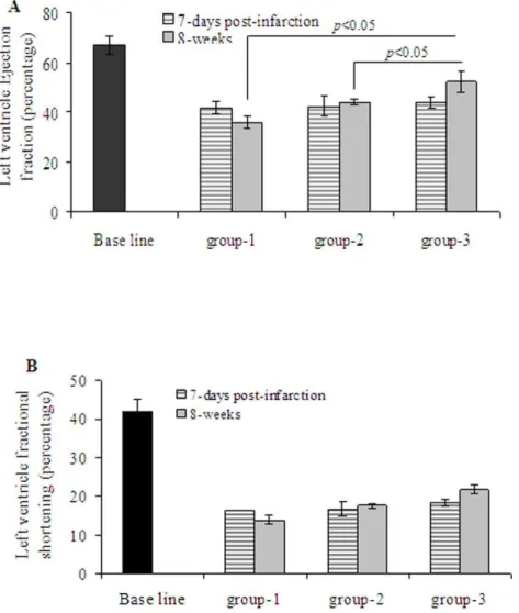
Assessment of LV contractile and remodeling indices was performed on day-7 after myocardial infarction (one day prior to cell transplantation) and 8 weeks after cell transplantation (n = 7 per group). Echocardiography showed deterioration of indices of LV contractile function in all the three groups at 7-days after myocardial infarction without any significant difference (p.0.05 between all the groups) (Figure 8). Thus, at 7-days post-infarction, LV ejection fraction (LVEF) and LV fractional shortening (LVFS) in group-1 were (41.962.55% and 16.361.1), group-2 (42.364.4% and 16.761.8) and group-3 (43.962.2% and 18.360.8) as compared with the baseline (67.363.4 and 4263) respectively. At 8-weeks after their respective treatment, we observed significantly deteriorated LVEF (35.862.3%) and LVFS (13.861.17%) in group-1 (Figure 8A). On the contrary, there was significant attenuation of LVEF and LVFS in group-2 (44.161.2%; 17.660.6%) and group-3 (52.364.4%; 21.861.2%) as compared to the corresponding values of baseline echocardi-ography (67.363.4%; 21.861.2%). As a marker of LV remodel-ing, LV end diastolic diameter (LVEDD) and LV end systolic diameter (LVESD), anterior wall thickness and posterior wall thickness were assessed as shown in Table S4. Echocardiography performed before and after their respective treatment in the experimental and control groups clearly demonstrated that intramyocardial administration ofShhMSCs had favorable impact on preservation of LV function in rats with chronic myocardial infarction.
Discussion
We have shown that in vitro reprogramming of MSCs maximizes their survival and angiogenic potential in the infarcted heart. The salient findings of the study are: 1- Genetic modification of MSCs with Shh transgene results in multifold increased iNOS upregulation and NO production, an effect which was associated with higher level of angiogenic growth factor expression including VEGF, Ang-1 and netrin-1. 2- The induction of iNOS in ShhMSCs occurred in PKC dependent manner. 3-Shh
CM mediated increased angiogenic response was abrogated in the presence of Shh and netrin-1 specific antibodies. 4-Intramyocardial engraftment of ShhMSC induced significant angiogenesis, improved regional blood flow and significantly preserved global heart function. We propose that the increased angiogenesis is due to the production of NO with simultaneous upregulation of multiple angio-competent factors including netrin-1. The results from the present study also indicated that genetically engineeredShhMSCs promoted migration of endothelial progen-itor cells which may be attributed to the dramatic upregulation of MMP-9 (4-fold) inShhMSCs.
Sonic hedgehog (Shh) together with Desert hedgehog (Dhh) and Indian hedgehog (Ihh) are three members of the hedgehog gene family identified in the mammals [15]. Hedgehog signaling involves binding of hedgehog with its ptch-1 receptor which in turn releases its inhibitory effect on Smoothened (smo). Activatedsmothen initiates signaling events which lead to regulation of transcriptional factors belonging to theGlifamily and its relevant downstream genes [16]. Shh is a secretary protein and therefore Shh gene delivery has been assessed in experimental animal models of the infarcted heart [6]. Results from these studies indicate that postnatal reconstitution of Shh signaling resulted in tissue preservation and repair, and that gene therapy with Shh not only prevented fibrosis, it also promoted angiogenesis and regional blood flow. Enhanced angiogenesis may be attributed to the multiple effects of localized Shh transgene expression. Activation of Shh pathway upregulates the expression of
Figure 5. Shh overexpression attenuated infarction size expansion. Masson’s trichome staining of formalin fixed paraffin embedded of the histological sections from (A) group-1 (B) group-2 and (C) group-3 animal hearts was carried out to visualize the area of fibrosis. Infarction size was significantly attenuated in group-3 after ShhMSCs engraftment as compared with groups-1 and 2.
Figure 6.ShhMSCs improved blood vessel density in the infarcted heart.(A–B) Blood vessel density analysis for myocardial angiogenesis at
8-weeks after respective treatment in different groups of animals. The histological sections were immunostained for vWFactor-VIII (red) and smooth muscle actin (green) for visualization of blood vessels. The number of blood vessels per surface area (0.74 mm2) was significantly higher in the infarct and peri-infarct areas in group-3 (p,0.05) as compared with group-1 and group-2. (C) The percentage of mature blood vessels (indicated by double fluorescent immunostaining for vWFactor-VIII and smooth muscle actin) showed no significant difference between the three treatment groups. However, (D–E) showed that average size of blood vessel diameter (based on number of pixels as arbitrary unit) was more uniform in peri-infarct region of group-3. Blood vessels in the infarct and peri-infarct areas with diameter of,100 pixels (33%), 100–200 pixels (39%) and.200 pixels (27%) diameter was observed in group-3 as compared with group-2,100 (17%), 100–200 (43%) and.200 (39%). (F) Photomicrographs of hematoxylin-eosin stained histological sections at 8-weeks after their respective treatment in group-3 and group-2. Red blood cells could be seen in some of the blood vessels as indicated by green arrows showing the functional status of the blood vessels.
multiple angiogenic cytokines, including VEGF, angiopoietins, SDF-1aand IGF-1 and development of capillary network [4,17]. Our results were in harmony with these data and further showed uniquely that Shh gene overexpression up-regulated iNOS, netrin-1 and HGF in addition to the already reported cytokines. HGF is a mitogen of mesenchymal origin, and is also reported to stimulate NO production and endothelial cell motility through upregulation of iNOS [18]. Put together, these molecular changes lead to a significant increase in biologically active NO production in Shh
MSCs.
iNOS is expressed following inflammatory or growth factor stimulation of cells unlike its counterpart eNOS (endothelial nitric oxide synthase) and nNOS (neuronal nitric oxide synthase) which are constitutively expressed. Activation of iNOS can produce copious amounts of NO for longer duration [19]. Generated from the NOS enzyme activity, NO is recognized as an important
regulator of cardiovascular system functionality. Whereas NO inhibits proliferation of smooth muscle cells, gene delivery of iNOS to endothelial cells is protective for the cells via NO release without an influence on their proliferation [20]. Secondly, endothelial cell migration is an essential component of several vascular processes including the maintenance of endothelial integrity and angiogenesis. It is suggested that endothelial cells must exhibit a phenotype of non-directional motility as a prerequisite for responding to stimuli for migration. The same study proposes that NO plays an obligatory role in eliciting this phenotype. More recent studies have shown that NO generated from iNOS activity modulates the expression of matrix metalloproteinase-9 (MMP-9), an enzyme responsible for degradation of extracellular matrix, which significantly influenced cell migration [21]. A few previous studies have reported increased MMP-9 the Shh-expressing cells, which was attenuated by the inhibition of EGF receptor activation or blocking the EGF receptor
Figure 7. Recombinant netrin-1 treatment improved blood vessel density in the infarcted heart.(A) At 8-weeks after recombinant netrin-1 delivery to the heart (group-4), the number of blood vessels per surface area (0.74 mm2) was significantly higher as compared with group-1 in both infarct as well as peri-infarct regions. (B) Double fluorescent immunostaining for vWFactor-VIII (red) and smooth muscle actin (green) showed that like Shh overexpression in the heart, netrin-1 protein delivery resulted in increased arteriolar density (blood vessels double positive for vWFactor-VIII and smooth muscle actin) in group-4. (C) Functional status of blood vessels in the infarcted heart was determined by fluorescent microsphere method for regional blood flow studies assessment. Regional blood flow was significantly improved in group-3 animal hearts as compared with group-1. However, regional blood flow changed insignificantly as compared with that of normal un-infarcted heart.
and ligand interaction [22]. In doing so, Shh has been shown to directly stimulate EGFR signaling. In the present study, prior treatment of cells with cyclopamine and chel abrogated the expression of iNOS and angiogenic growth factors including VEGF, Ang-1 and netrin-1 thus suggesting that these pro-angiogenesis relevant molecular events were PKC dependent.
Netrins are important in axonal guidance, regulation and maintenance of central nervous system and are also involved in the development of mammary gland, lung, pancreas, and blood vessel [23]. Even though there is some controversy, most recent studies suggested that netrin-1 functions as a pro-angiogenic factor. In vivo studies also indicated that netrins promoted neovasculariza-tion and reperfusion in a murine model of peripheral vascular disease and also induced migration, proliferation and tube formation during in vitro studies involving multiple endothelial cell lines [24]. Characterizing a mechanism for netrin-1 induced angiogenesis, a critical role for NO has been elucidated subsequent to feed-forward ERK1/2 and eNOS activation in endothelial cells treated with netrin-1 [25]. Our results showed that netrin-1 expression increased significantly inShhMSCs both at the protein and RNA levels in PKC (PKM) dependent fashion.
Another interesting finding in the present study was the presence of PKM fragment of PKC in ShhMSCs at 72-h after transfection with Shh plasmid. PKM is a constitutively active 40-kDa catalytic fragment of PKC [26]. The regulatory role of PKC in Shh signaling has been reported previously [22]. In a study involving NIH 3T3 cells, PKC-d was integral to hedgehog signaling to promote proliferative activity of the cells [27]. In our present study, treatment of ShhMSCs with both the PKC inhibitor chel and Shh inhibitor cyclopamine abolished PKM fragment which indicated that PKM was downstream of Shh and that PKC was essential for its upregulation. White et al. (2007) while experimenting on intact sea urchin spermatozoa showed PKC, most likely through its cleavage into active catalytic product PKM, was the central signaling mediator associated with maintenance of sperm mobility [26]. PKC inhibitors such as chel and calphostin-C, as well as staurosporine, were found to rapidly arrest the motility of sea urchin spermatozoa freshly released into seawater. At the same time, these inhibitors prevented the motility-associated increase in phosphorylation of several PKC substrates. Pretreatment of ShhMSCs with these inhibitors abrogated PKM with concurrent abrogation of iNOS and netrin-1.
Figure 8.ShhMSCs transplantation preserved global function of the infarcted heart.Echocardiographic assessment of the heart function
Another novel finding of our study supported by molecular and histological data was the improved survival of ShhMSCs post engraftment. In view of the reported data that massive loss of the transplanted cells occurs after transplantation remains a major determinant of the effectiveness of heart cell therapy,ShhMSCs are at an advantage in terms of their survival after engraftment. Histological studies at eight weeks after cell transplantation showed a marked reduction in infarct size and preservation of host myocardium in ShhMSCs transplanted group as compared with Emp
MSC and DMEM groups. The cytoprotective effects were accompanied by a significant increase in capillary density and a higher number of mature blood vessels (smooth muscle actin positive cells) inShhMSCs group. These results are consistent with earlier studies using direct injection of Shh plasmid into the infarcted heart [4,6,24].
In conclusion ShhMSCs showed upregulation of several angiogenic cytokines and signaling molecules including Ang-1, VEGF, IGF, HGF, iNOS and netrin-1. Additionally, ShhMSCs also generated NO at significantly higher levels. These molecular changes were mediated by iNOS/netrin/PKC signaling pathway downstream of Shh gene overexpression which combined with stem cell transplantation could be a promising strategy for the treatment of an infarcted heart.
Supporting Information
Text S1
Found at: doi:10.1371/journal.pone.0008576.s001 (0.06 MB DOC)
Figure S1 Construction of Shh plasmid. (A) Vector Map used in construction of Shh-plasmid. The vector backbone was purchased from commercial source (Stratagene, USA) and Shh mRNA was isolated from 14-day rat embryo, used for cDNA synthesis and cloned into pCMV Script vector. (B) Sequence of pCMV Shh-vector using T7 and T3 primers showing sequence of Shh gene insert. (C) Flow cytometry for surface marker expression showed that overexpression of Shh transgene did not alter the expression of surface markers inShhMSCs (indicated by red line) including CD44, CD59, CD105 and CD106 as compared with the Empty vector transfected MSCs (EMPMSCs; indicated by black line). Found at: doi:10.1371/journal.pone.0008576.s002 (2.61 MB TIF)
Figure S2 Abrogation of PI3K inShhMSCs using PI3K specific siRNA. Western blot showing successful abrogation of PI3K in
Shh
MSCs following transfection with PI3K specific siRNA. Shh
MSCs transfected with scrambled siRNA (Sc siRNA) and Emp
MSCs with siRNA transfection were used as controls. Successful abrogation of PI3K was indicated by loss of Akt phosphorylation.
Found at: doi:10.1371/journal.pone.0008576.s003 (0.85 MB TIF)
Figure S3 Cytoprotective effects of conditioned medium from Shh
MSCs (ShhCM) on native MSCs. LDH release assay showed that ShhCM was significantly more protective for native MSCs against oxidant stress as compared with conditioned medium from Emp
MSCs (EmpCM).
Found at: doi:10.1371/journal.pone.0008576.s004 (1.05 MB TIF)
Figure S4 Cytoprotective effects of conditioned medium from Shh
MSCs (ShhCM) on native H2C9 cardiomyocytes. LDH release assay showed that ShhCM was significantly more protective for H2C9 cardiomyocytes against oxidant stress as compared with conditioned medium fromEmpMSCs (EmpCM).
Found at: doi:10.1371/journal.pone.0008576.s005 (1.17 MB TIF)
Table S1 Primary antibodies used for Western immunoblotting and immunohistochemistry.
Found at: doi:10.1371/journal.pone.0008576.s006 (0.03 MB DOC)
Table S2 Primers used for classic and real-time PCR.
Found at: doi:10.1371/journal.pone.0008576.s007 (0.03 MB DOC)
Table S3 Fold change in different growth factor and cytokine expression inShhMSCs as compared withEmpMSCs.
Found at: doi:10.1371/journal.pone.0008576.s008 (0.03 MB DOC)
Table S4 The heart function indices measured by echocardiog-raphy on (A) day-7 and (B) 8-weeks after cell transplantation. Found at: doi:10.1371/journal.pone.0008576.s009 (0.03 MB DOC)
Author Contributions
Conceived and designed the experiments: RPA KHH MA. Performed the experiments: RPA JS MRA. Analyzed the data: RPA KHH JS MRA MA. Contributed reagents/materials/analysis tools: KHH MA. Wrote the paper: KHH MA. Financial support: KHH, MA.
References
1. Bitgood MJ, McMahon AP (1995) Hedgehog and Bmp genes are coexpressed at many diverse sites of cell-cell interaction in the mouse embryo. Dev Biol 172: 126–38.
2. Pola R, Ling LE, Aprahamian TR, Barban E, Bosch-Marce M (2003) Postnatal recapitulation of embryonic hedgehog pathway in response to skeletal muscle ischemia. Circulation 108: 479–85.
3. Massard C, Deutsch E, Soria JC (2006) Tumour stem cell-targeted treatment: elimination or differentiation. Ann Oncol 17: 1620–4.
4. Pola R, Ling LE, Silver M, Corbley MJ, Kearney M (2001) The morphogen Sonic hedgehog is an indirect angiogenic agent upregulating two families of angiogenic growth factors. Nat Med 7: 706–11.
5. Straface G, Aprahamian T, Flex A, Gaetani E, Biscetti F (2008) Sonic Hedgehog Regulates Angiogenesis and Myogenesis During Post-Natal Skeletal Muscle Regeneration. J Cell Mol Med {Published ahead of print}.
6. Kusano KF, Pola R, Murayama T, Curry C, Kawamoto A (2005) Sonic hedgehog myocardial gene therapy: tissue repair through transient reconstitution of embryonic signaling. Nat Med 11: 1197–204.
7. Kajstura J, Rota M, Whang B, Cascapera S, Hosoda T (2005) Bone marrow cells differentiate in cardiac cell lineages after infarction independently of cell fusion. Circ Res 96: 127–37.
8. Rota M, Kajstura J, Hosoda T, Bearzi C, Vitale S (2007) Bone marrow cells adopt the cardiomyogenic fate in vivo. Proc Natl Acad Sci U S A 104: 17783–8.
9. Assmus B, Honold J, Schachinger V, Britten MB, Fischer-Rasokat U (2006) Transcoronary transplantation of progenitor cells after myocardial infarction. N Engl J Med 355: 1222–32.
10. Stamm C, Westphal B, Kleine HD, Petzsch M, Kittner C (2003) Autologous bone-marrow stem-cell transplantation for myocardial regeneration. Lancet 361: 45–6.
11. Jiang S, Haider H, Idris NM, Salim A, Ashraf M (2006) Supportive interaction between cell survival signaling and angiocompetent factors enhances donor cell survival and promotes angiomyogenesis for cardiac repair. Circ Res 99: 776–84. 12. Haider HK, Jiang S, Idris NM, Ashraf M (2008) IGF-1-Overexpressing Mesenchymal Stem Cells Accelerate Bone Marrow Stem Cell Mobilization via Paracrine Activation of SDF-1{alpha}/CXCR4 Signaling to Promote Myocar-dial Repair. Circ Res 103: 1300–1308.
13. Gnecchi M, He H, Liang OD, Melo LG, Morello F (2005) Paracrine action accounts for marked protection of ischemic heart by Akt-modified mesenchymal stem cells. Nat Med 11: 367–8.
14. Andrea JE, Walsh MP (1992) Protein kinase C of smooth muscle. Hypertension 20: 585–95.
15. Zardoya R, Abouheif E, Meyer A (1996) Evolution and orthology of hedgehog genes. Trends Genet 12: 496–7.
17. Soleti R, Benameur T, Porro C, Panaro MA, Andriantsitohaina R (2009) Microparticles harboring Sonic Hedgehog promote angiogenesis through the up-regulation of adhesion proteins and pro-angiogenic factors. Carcinogenesis 30: 500–508.
18. Purdie KJ, Whitley GS, Johnstone AP, Cartwright JE (2002) Hepatocyte growth factor-induced endothelial cell motility is mediated by the upregulation of inducible nitric oxide synthase expression. Cardiovasc Res 54: 659–68. 19. Alderton WK, Cooper CE, Knowles RG (2001) Nitric oxide synthases:
structure, function and inhibition. Biochem J 357: 593–615.
20. Cooney R, Hynes SO, Duffy AM, Sharif F, O’Brien T (2006) Adenoviral-mediated gene transfer of nitric oxide synthase isoforms and vascular cell proliferation. J Vasc Res 43: 462–72.
21. Sun MH, Han XC, Jia MK, Jiang WD, Wang M (2005) Expressions of inducible nitric oxide synthase and matrix metalloproteinase-9 and their effects on angiogenesis and progression of hepatocellular carcinoma. World J Gastroenterol 11: 5931–7.
22. Heo JS, Lee MY, Han HJ (2007) Sonic hedgehog stimulates mouse embryonic stem cell proliferation by cooperation of Ca2+/protein kinase C and epidermal growth factor receptor as well as Gli1 activation. Stem Cells 25: 3069–80. 23. Barallobre MJ, Pascual M, Del Rio JA, Soriano E (2005) The Netrin family of
guidance factors: emphasis on Netrin-1 signalling. Brain Res Rev 49: 22–47. 24. Wilson BD, Ii M, Park KW, Suli A, Sorensen LK (2006) Netrins promote
developmental and therapeutic angiogenesis. Science 313: 640–4.
25. Park KW, Crouse D, Lee M, Karnik SK, Sorensen LK (2004) The axonal attractant Netrin-1 is an angiogenic factor. Proc Natl Acad Sci U S A 101: 16210–5.
26. White D, de Lamirande E, Gagnon C (2007) Protein kinase C is an important signaling mediator associated with motility of intact sea urchin spermatozoa. J Exp Biol 210: 4053–64.