UNIVERSIDADE DE LISBOA
Faculdade de Medicina Veterinária
SURGICAL MANAGEMENT AND OUTCOME FOLLOWING ADRENALECTOMY: A RETROSPECTIVE CASE STUDY IN 16 DOGS (2008-2018)
MARTA MARIANO DA SILVA
CONSTITUIÇÃO DO JÚRI ORIENTADOR
Doutora Maria Teresa da Costa Mendes Victor Villa de Brito Dr. David Robinson
Doutor Luís Miguel Alves Carreira
Doutora Lisa Alexandra Pereira Mestrinho CO-ORIENTADOR
Doutora Lisa Mestrinho
2018 LISBOA
UNIVERSIDADE DE LISBOA
Faculdade de Medicina Veterinária
SURGICAL MANAGEMENT AND OUTCOME FOLLOWING ADRENALECTOMY: A RETROSPECTIVE CASE STUDY IN 16 DOGS (2008-2018)
DISSERTAÇÃO DE MESTRADO INTEGRADO EM MEDICINA VETERINÁRIA
MARTA MARIANO DA SILVA
CONSTITUIÇÃO DO JÚRI ORIENTADOR
Doutora Maria Teresa da Costa Mendes Victor Villa de Brito Dr. David Robinson
Doutor Luís Miguel Alves Carreira
Doutora Lisa Alexandra Pereira Mestrinho CO-ORIENTADOR
Doutora Lisa Mestrinho
2018 LISBOA
ACKNOWLEDGEMENTS
To start with, I would like to thank Dr. David Robinson, my supervisor and mentor, that along with the other clinical directors of KVG, welcomed me into their practice and helped me grow immensely as a veterinary surgeon trainee. Without him this work would not have been feasible, and I sincerely appreciate his kindness in allowing me to use KVG’s clinical records to produce this work. I am grateful for all his support and mentorship throughout this period and thank him for inspiring me to be an excellent professional like himself.
Also, I would like to thank Dr. Lisa Mestrinho, my co-supervisor and extraordinary Professor, for inspiring in me enthusiasm for surgery and the theme that lead to this dissertation – the first adrenalectomy I have ever watched was performed by her when I was in my 5th year of studies.
Her availability and advice were vital and much appreciated, as well as, her help in the collection of additional cases in order to enrich my study. I also thank to Prof. António Ferreira, clinical director of the HEV, for allowing me to use HEV’s clinical cases, to Prof. Sandra Jesus for her kindness in providing me with CT images to use as illustrations, and to Prof. Telmo Nunes for his insightful advice and sympathy. Additionally, I thank all the veterinarians and colleagues who so nicely helped me gather the data required.
Next, I would like to thank to Mário Almeida for being an extraordinary partner in life, who constantly provided me with much cherished support and motivation throughout this journey, and with whom I am very grateful to share my life with.
Thanks to my dear friends and colleagues from the FMV-UL - José, Catarina, Cláudia, and Mariana - for their treasured companionship throughout these 6 years of both unbelievable ecstatic times and moments of unbearable frustration. Each and every single one of you inspired me and taught me something in different ways, for which I will be always grateful. I cannot conceive the possibility of having lived this experience without you, my dream team, and hope we will continue to accompany each other’s journeys, undoubtedly filled with success. Additionally, a special thank you to my dearest best girlfriends, who have accompanied me for many years now, Maria Ana and Bárbara. Having your friendship as a constant in life is refreshing and feels as a blessing. I also thank to my dearest friends, Luís and Rúben, for being like older brothers and for their insight, adventurous spirit, and unending friendship.
A massive thank you to Mariana and Hugo, for their incredible support and friendship, and for being role models to follow. I am truly grateful for your help and patience to answer my million dilemmas and for being as like my Portuguese family in the UK. I also could not be more thankful to all KVG team for their warm welcome and acknowledgment, and for providing me with several chances to practice in a variety of areas of small animal veterinary medicine.
Thanks to all the Vets, for taking a lot of their valuable time to teach me, answer my questions, and giving me innumerous opportunities to get experienced, by letting me “steal” many of their procedures, namely routine surgeries, and participate by assisting in others. A special thank you to Richard Jones, Mr. J, who in his always uplifting sense of humour, taught me all of his remarkable consulting strategies and made sure I was gathering the broadest experience possible. I feel truly lucky and grateful for your mentorship. To all the nurses, a special thank you for all you taught me and for your constant encouragement and incredible professionalism. Thanks to Louise Jackson, for her availability and kindness, and always making sure I had the due support.
Lastly, and most importantly, I wish to thank my family, of whom I am deeply proud. A special thank you to my mother, for being a role model of strength and independence, and for investing in me, making sure that I always had everything I needed to thrive. To her and to my stepfather, I also thank their valuable support and perceptive advice throughout all my life and this cycle in particular. To my father I thank him for his support and believe in me, and for his effort in investing in my education and future. And to my brother for being one’s true partner in crime and by never letting me feel alone. With you I have learned that with all our differences come different roles to play, none of each with more or less value than the other. Finally, to my dearest grandparents, who despite their humble origins and difficulties, had a vision of empowerment through education, which they successfully passed on to their daughters and grandchildren, undoubtedly at the expense of their outstanding willingness and personal sacrifice. For that and their everlasting unconditional love, I dedicate this dissertation to them. My only wish is to make you proud of me. I hope I will be wordy.
ABSTRACT
SURGICAL MANAGEMENT AND OUTCOME FOLLOWING ADRENALECTOMY: A RETROSPECTIVE CASE STUDY IN 16 DOGS (2008-2018).
Primary neoplasms of the adrenal gland might represent more than 1-2% of all canine tumours and can originate various worrisome clinical presentations; hence why adrenalectomy is generally the treatment of choice. Identification of prognostic factors with occasional uncertainty or contradictions among different authors renders further investigations welcomed. A retrospective study was conducted in 16 dogs undergoing adrenalectomy with the aim to describe the clinical features, surgical management and outcome.
Review of clinical records and interviews with owners and veterinarians involved were performed to register clinical variables, such as, signalment, relevant history, clinical signs, laboratory, imaging and surgical findings, histopathology results, and outcome. The median survival time was calculated through Kaplan-Meier estimate.
Intra- (92%) and postoperative (67%) complications, and perioperative mortality (31%) rates were comparable to recent studies; as was the median survival time (419 days), with 64% of long-term survivors living for more than 1 year, up to 3 years, approximately.
This case series emphasizes that if dogs survive the immediate perioperative period, long-term outcome is generally good with possibility of prolonged survival times, as local or distant tumour recurrence appears to be low. This study also promotes awareness of adrenal
incidentalomas (25%) and emergency clinical presentations (19%). Outcome predictors such
as age of patients with phaeochromocytomas, size of tumour, surgeon’s experience in dealing with caval invasion, presence of metastasis at surgery, acute adrenal haemorrhage, major intraoperative haemorrhage, and postoperative disseminated intravascular coagulopathy must be considered in the approach to these cases.
RESUMO
MANEIO CIRÚRGICO E RESULTADO APÓS ADRENALECTOMIA: UM ESTUDO RETROSPETIVO DE CASOS EM 16 CÃES (2008-2018).
Neoplasias primárias das glândulas adrenais poderão representar mais do que 1-2% de todos os tumores caninos e podem originar vários quadros clínicos preocupantes; e por isso é que a adrenalectomia é geralmente o tratamento de escolha. A identificação de fatores de prognóstico com incerteza ou contradições ocasionais entre diversos autores ditam que investigações adicionais sejam bem-vindas.
Um estudo retrospetivo foi conduzido em 16 cães submetidos a adrenalectomia, para descrever o quadro clínico, maneio e resultado cirúrgico.
Foi feita a revisão de historiais clínicos e entrevistas a donos e veterinários envolvidos de forma a registar variáveis clínicas como identificação do animal, historial relevante, sinais clínicos, achados laboratoriais, imagiológicos e cirúrgicos, resultados de histopatologia, e resultado. A mediana dos tempos de sobrevivência foi calculada através da estimativa de Kaplan-Meier. As taxas de complicações intra- (92%) e pós-cirúrgicas (67%), e de mortalidade (31%) foram comparáveis a estudos recentes; assim como o tempo mediano de sobrevivência (419 dias), com 64% dos sobreviventes a longo prazo a viveram por mais de 1 ano, até 3 anos, aproximadamente.
Esta série de casos enfatiza que se os cães sobreviverem o período peri-cirúrgico imediato, o resultado a longo prazo é geralmente bom com possibilidade de tempos de sobrevivência prolongados, uma vez também que a taxa de recorrência local ou distante aparenta ser baixa. Este estudo promove também a consciencialização de incidentalomas das adrenais (25%) e de quadros clínicos de emergência (19%). Fatores de prognóstico tais como idade dos pacientes com feocromocitomas, tamanho do tumor, experiência do cirurgião em lidar com invasão da veia cava, presença de metástases na altura da cirurgia, hemorragia aguda adrenal, hemorragia intra-cirúrgica de maior importância, e coagulopatia intravascular disseminada pós-cirúrgica, devem ser considerados na abordagem a estes casos.
Palavras-chave – cão, tumor da glândula adrenal, adrenalectomia, incidentaloma, rotura,
TABLE OF CONTENTS
ACKNOWLEDGEMENTS... iii
ABSTRACT ... v
RESUMO... vi
TABLE OF CONTENTS ...vii
LIST OF FIGURES ... ix LIST OF TABLES... ix LIST OF GRAPHICS ... ix LIST OF ABBREVIATIONS ... x TRAINEESHIP REPORT ... 1 I. INTRODUCTION ... 2
1. NEOPLASMS OF THE ADRENAL GLANDS AND DIAGNOSTIC APPROACH ... 3
1.1 Types of neoplasms ... 3
1.1.1 Neoplasms of the adrenal cortex ... 3
1.1.2 Neoplasms of the adrenal medullary secretory cells... 5
1.1.3 Neoplasms of cells of the sympathetic nervous system in the adrenal medulla ... 6
1.1.4 Metastatic neoplasms to the adrenal medulla ... 6
1.1.5 Bilateral tumours, Concurrent PDH (pituitary-dependent hyperadrenocorticism) and Concurrent endocrine neoplasias ... 7
1.2 Signalment ... 9
1.2.1 Adrenocortical tumours ... 9
1.2.2 Phaeochromocytomas ... 10
1.3 History, Clinical signs and Physical examination ... 10
1.3.1 Adrenocortical tumours ... 10
1.3.2 Phaeochromocytomas ... 11
1.4 Diagnostic testing ... 13
1.4.1 Basic bloodwork, Urinalysis and Blood pressure measurement ... 13
1.4.1.1 Adrenocortical tumours ... 13 1.4.1.2 Phaeochromocytomas ... 14 1.4.2 Functional testing ... 14 1.4.2.1 Adrenocortical tumours ... 15 1.4.2.2 Phaeochromocytoma ... 17 1.4.3 Imaging ... 19 1.4.3.1 Radiography... 19 1.4.3.2 Ultrasonography ... 20
1.4.3.3 Computed tomography and Magnetic resonance imaging ... 22
1.4.4 Incidental discovered adrenal mass ... 23
1.4.5 Fine-needle aspiration ... 25
2. TREATMENT OF ADRENAL GLAND NEOPLASMS ... 26
2.1 Conservative management ... 26 2.2.1 Adrenocortical tumours ... 26 2.2.2 Phaeochromocytomas ... 27 2.2 Surgical management ... 28 2.2.1 Indications ... 28 2.2.2 Perioperative management ... 28 2.2.2.1 Adrenocortical tumours ... 29 2.2.2.2 Phaeochromocytomas ... 31 2.2.3 Surgical approaches ... 34
2.2.3.2 Flank/Paracostal/Paralumbar/Retroperitoneal approach ... 34
2.2.3.3 Laparoscopy approach ... 35
2.2.4 Surgical technique ... 36
2.2.4.1 CVC Venotomy and Thrombectomy ... 37
3. OUTCOME FOLLOWING ADRENALECTOMY ... 39
3.1 Complications ... 39
3.2 Mortality ... 40
3.3 Survival ... 41
3.4 Prognosis ... 42
II. OBJECTIVES ... 45
III. MATERIAL AND METHODS ... 46
1. Case selection criteria ... 46
2. Medical records review ... 46
3. Procedures ... 46 3.1 Imaging assessment ... 46 3.2 Perioperative management ... 47 3.3 Surgical technique ... 48 4. Outcome ... 50 5. Statistical analysis ... 50 IV. RESULTS ... 51 1. Signalment ... 51 2. Preoperative evaluation ... 51 2.1 Relevant history ... 51 2.2 Clinical presentation ... 51 2.3 Functional testing ... 52 2.4 Imaging assessment ... 52
3. Surgical procedures and findings ... 53
4. Histopathological evaluation ... 53
5. Outcome ... 54
V. DISCUSSION ... 58
VI. CONCLUSION ... 67
LIST OF FIGURES
Figure 1. Resection of a right-sided adrenal gland tumour invading the CVC ... 38
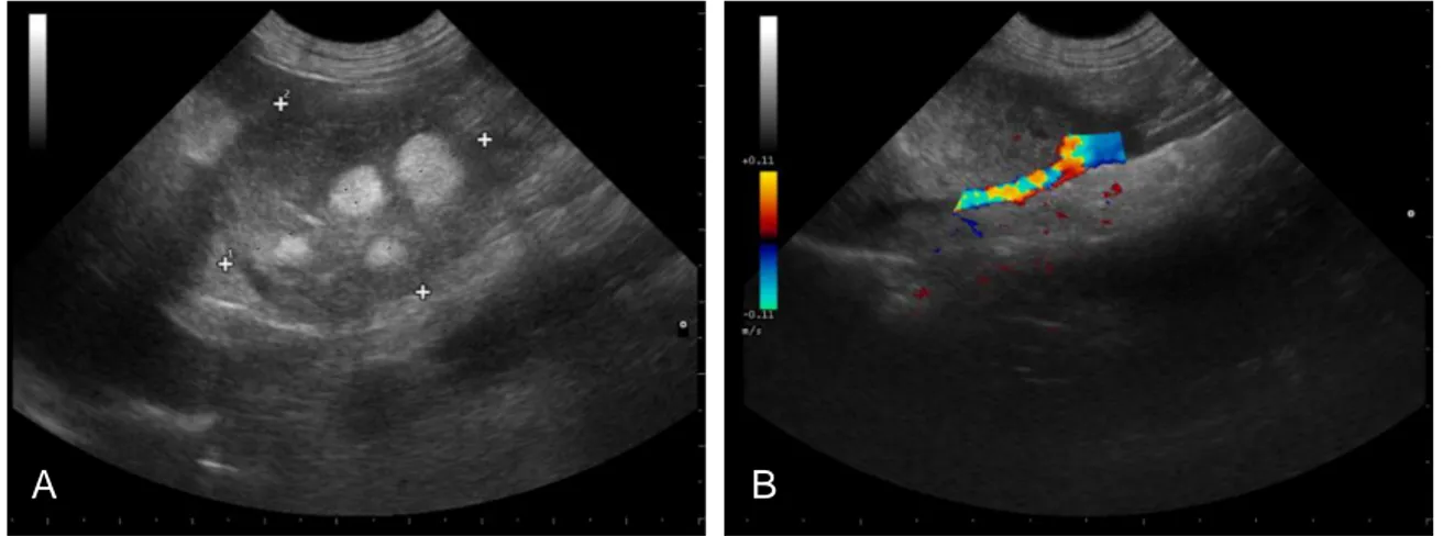
Figure 2. Ultrasonographic pictures demonstrating the measurement of an adrenocortical myelolipoma (A) and verification of vascular invasion using Doppler (B) (Kindly dispensed by Dr. David Robinson, KVG) ... 47
Figure 3. Computed-tomography pictures after administration of intravenous contrast representing a left-sided adrenocortical adenoma with a concurrent caval “true” thrombus (Kindly dispensed by Prof. Dr. Sandra Jesus, HEV) ... 47
Figure 4. Intraoperative view after removal of a “true” CVC thrombus in a patient with a left-sided adrenocortical adenoma (Original photography) ... 49
Figure 5. Intraoperative view of the surgical field after removal of a right-sided adrenal gland phaeochromocytoma (A) and the tumour itself following excision (B) (Original photography) ... 49
LIST OF TABLES
Table 1. Clinical manifestations of HAC and respective frequency at initial presentation ... 11Table 2. Clinical signs in dogs with phaeochromocytomas divided by cause-related categories ... 12
Table 3. Common abnormal laboratory findings associated with HAC ... 13
Table 4. Reported laboratory abnormalities in dogs with phaeochromocytomas ... 14
Table 5. Major differential diagnoses of an incidentally discovered adrenal gland mass ... 24
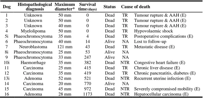
Table 6. Summary data for all 16 dogs undergoing adrenalectomy ... 55
LIST OF GRAPHICS
Graphic 1. Kaplan-Meier life table analysis for overall median survival time of 419 days in 16 dogs undergoing adrenalectomy (Censored cases are represented by vertical bars) ... 56LIST OF ABBREVIATIONS
AAH = Acute adrenal haemorrhage ACTH = Adrenocorticotropic hormone
ACTHST = Adrenocorticotropic hormone stimulation test ACVIM = American College of Veterinary Internal Medicine ADH = Adrenal-dependent hyperadrenocorticism
AT = Adrenocortical tumour BP = Blood pressure
CT = Computed tomography CVC = Caudal venae cava
DIC = disseminated intravascular coagulopathy e.g. = exempli gratia
eACTH = Endogenous adrenocorticotropic hormone
FMV-UL = Faculty of Veterinary Medicine of the University of Lisbon HAC = Canine hyperadrenocorticism
HDDST = High dose dexamethasone suppression test
HEV = Hospital Escolar Veterinário/Veterinary Teaching Hospital of the FMV-UL i.e. = id est
KVG = Kingston Veterinary Group LA = Laparoscopic adrenalectomy
LDDST = Low dose dexamethasone suppression test MEN = Multiple endocrine neoplasia
MRI = Magnetic resonance imaging MST = Median survival time
NAD = Non-adrenal disease
NTR = Non-adrenal tumour-related
PDH = Pituitary-dependent hyperadrenocorticism PT = Portugal
PTE = Pulmonary thromboembolism PU/PD = Polyuria/polydipsia
TR = Adrenal tumour-related
UCCR = Urinary corticoid:creatinine ratio UK = United Kingdom
US = Abdominal ultrasound
TRAINEESHIP REPORT
As part of the Integrated Master’s Degree in Veterinary Medicine from the Faculty of Veterinary Medicine of the University of Lisbon, I fulfilled a 6-month training period between the 4th of September 2017 and the 16th of March 2018, in a total of roughly 1100 hours, at
Kingston Veterinary Group, Hull, United Kingdom. Throughout that period, I assisted and participated in numerous procedures carried out in different areas of small animal veterinary medicine. In the Surgery department, led by Dr. David Robinson, who was also my mentor and supervisor, I cooperated by helping in several orthopaedic procedures (e.g. closing-wedge osteotomy, fabello-tibial suture, tibial tuberosity transposition, fracture repairs, pancarpal arthrodesis, placement of transcondylar screw for correction of incomplete ossification of humeral condyle, stabilization of coxofemoral luxation using the toggle pin method), soft tissue surgeries (e.g., exploratory laparotomies, laparoscopies, cystotomies, splenectomies, gastrotomy, enterotomy, perineal hernia repair, anal sacculectomy, incisional/excisional biopsies, brachycephalic obstruction airway syndrome surgery, patent ductus arteriosus ligation, bilateral thyroidectomy, cholecystectomy, adrenalectomy, grid keratectomy, temporary eyelid tacking) and postoperative care of the patients. Furthermore, I was also given the opportunity to develop my surgical skills by performing several surgical procedures under the supervision of senior surgeons, such as dog, cat and rabbit castrations, dog and cat ovariohysterectomies, umbilical hernia repair, dewclaw removal, aural haematoma repair, superficial nodule/mass excision, mastectomy, pyometra, cystotomy, and dentistry procedures such as teeth extraction, scaling and polishing. The same applies in terms of Anaesthesia, considering I was able to practice procedures such as induction, intubation and general anaesthetic monitoring. In the Internal Medicine department, I worked on my clinical case solving skills and participated in the care and treatment of the inpatients, practicing procedures such as drugs administration, blood sampling, blood typing, collection and transfusion, venous and urinary catheterization, cystocentesis and running several diagnostic tests. In addition, I accompanied clinicians during consultations and was also able to take the lead and develop my consulting skills. Finally, with regards to the Diagnostic Imaging department, I assisted the surgeons during endoscopy (rhinoscopy, bronchoscopy, gastroscopy) and ultrasound, magnetic resonance imaging and computed tomography scans. Moreover, I was able to practice patient positioning for radiographic examination. During and after these procedures, I was taught by senior vets how to interpret the results and to take conclusions towards diagnostic and treatment planning in specific clinical cases.
I. INTRODUCTION
Primary neoplasms of the adrenal gland were reported to account for 1 to 2% of all canine tumours, though nowadays it is arguable that this fraction might have been undervalued (Lunn & Page, 2013; Myers, 1997). In the past years, both medical and veterinarian practitioners have witnessed an escalation in discovery of incidental adrenal lesions (incidentalomas), along with an uncertainty regarding the right manner to address them. The widespread availability of advance diagnostic imaging methods (especially ultrasonography, but also computed tomography and possibly, on a minor role, magnetic resonance imaging) hold the responsibility in this matter (Baum, Boston, & Case, 2016; Cook, Spaulding, & Edwards, 2014; Myers, 1997). The most common types of tumours are, in descending order, cortical adenomas, carcinomas, medullary phaeochromocytomas, and secondary neoplasic lesions (Labelle & De Cock, 2005; Lunn & Page, 2013; Reusch, 2015). Any of these can be associated with a rather vast variety of worrisome clinical scenarios such as endocrine/neuroendocrine derangements (Barthez, Marks, Woo, Feldman, & Matteucci, 1997; Gilson, Withrow, Wheeler, & Twedt, 1994; Reusch & Feldman, 1991; van Sluijs, Sjollema, Voorhout, van den Ingh, & Rijnberk, 1995), local compression or invasion of near-by structures like the ipsilateral kidney and major vessels (e.g., CVC), metastatic disease, and a surgical emergency caused by acute rupture and consequent haemoabdomen or -retroperitoneum (Barrera, Bernard, Ehrhart, Withrow, & Monnet, 2013; Kyles et al., 2003; Lang et al., 2011; Vandenbergh, Voorhout, van Sluijs, Rijnberk, & van den Ingh, 1992; Whittemore, Preston, Kyles, Hardie, & Feldman, 2001).
Generally, adrenalectomy is the treatment of choice, even though it is a technical demanding procedure, associated to considerably high perioperative complications and mortality rates. Yet, more recent studies reported lower mortality, varying from 12 to 26% (Barrera et al., 2013; Kyles et al., 2003; Lang et al., 2011; Massari, Nicoli, Romanelli, Buracco, & Zini, 2011; Schwartz et al., 2008). Furthermore, good long-term outcome with prolonged survival times is possible, provided that dogs survive the perioperative period (Anderson et al., 2001; Barrera et al., 2013; Barthez et al., 1997; Gilson, Withrow, & Orton, 1994; Kyles et al., 2003; Lang et al., 2011; Massari et al., 2011; Schwartz et al., 2008; van Sluijs et al., 1995).
Several authors have appointed a number of variables with or without prognostic significance for dogs with adrenal tumours undergoing adrenalectomy (Anderson et al., 2001; Barrera et al., 2013; Herrera et al., 2008; Kyles et al., 2003; Lang et al., 2011; Massari et al., 2011; Schwartz et al., 2008). Yet, because of the existence of a certain degree of uncertainty or contradictions between studies regarding some of the identified factors, further investigations on the subject are welcomed.
1. NEOPLASMS OF THE ADRENAL GLANDS AND DIAGNOSTIC APPROACH 1.1 Types of neoplasms
1.1.1 Neoplasms of the adrenal cortex
Adrenocortical tumours (ATs), namely, adenomas and carcinomas, are the most frequently seen adrenal gland neoplasms (Barrera et al., 2013; Kyles et al., 2003; Labelle & De Cock, 2005; Lang et al., 2011; Massari et al., 2011; Oblak, Bacon, & Covey, 2016; Schwartz et al., 2008). Adenomas are classically single, well-demarcated nodules, partially or completely encapsulated by fibrous connective tissue and a compressed border of cortical parenchyma. While small adenomas tend to maintain a yellow colour similar to the normal cortex, larger ones vary from yellow to red, deform the gland’s external surface, and may extend into the medulla. Though similar, discrete adenomas differ from nodular hyperplasia predominantly due to the latter inexistence of encapsulation signs along with the typical presence of multiple nodules on both adrenal glands (Rosol & Gröne, 2016). Histologically, adenoma tumour cells are well-differentiated with resemblances of normal zonae fasciculata or reticularis cells. Organised in broad trabeculae or nests divided by small vascular spaces, the cells present abundant lightly eosinophilic cytoplasm, frequently vacuolated and filled with several lipid droplets. Foci of mineralisation, haematopoiesis, and adipose tissue accumulations can be demonstrated, as well as, fibrin thrombi allocated in large dilated blood-filled sinusoids (telangiectasis) (Labelle et al., 2004; Rosol & Gröne, 2016).
Cortical carcinomas arise less commonly in comparison to adenomas (Labelle & De Cock, 2005; Rosol & Gröne, 2016). Carcinomas tend to be larger and are more likely to occur bilaterally. They are constituted by variegated, yellow to red, friable, tissue that incorporates the disturbed adrenal. This malignant AT is commonly locally fixed due to extensive invasion of surrounding structures, namely, the CVC and aorta, resulting in potential large tumour thrombi (Rosol & Gröne, 2016). Histologically, the normal architecture of the gland is often completely destructed by the malignant neoplasm. Tumoural cells are highly pleomorphic – not only typically large and polyhedral with prominent nuclei and densely eosinophilic or vacuolated cytoplasm, but also smaller cells may be present –, form clusters, and are subdivided by fibrovascular stroma. Areas of haemorrhage may be demonstrated as result of rupture of thin-walled sinusoids. The growth pattern varies between tumours and within the same one. Moreover, this feature may be used to distinguish between carcinomas in regard to their type of invasion. Specifically, the existence of a trabecular growth pattern in a minimum of 1/3 of the tumour was significantly more frequent in carcinomas with metastases compared with carcinomas with vascular invasion alone (Labelle et al., 2004). In their study, Labelle et al. (2004) also found that ~50% (14/26) of dogs with carcinomas had metastases, mainly spreading
to the liver and lungs, but also, kidneys, ovary and mesenteric lymph nodes. Other reported locations include thyroid vessels, serosal surfaces of liver and pylorus, and small intestines (Anderson et al., 2001; Barrera et al., 2013). Furthermore, Labelle et al. (2004) noted that metastases may be seen with or without vascular invasion, and vice versa. Vascular invasion, specifically caval thrombi, may be formed via direct penetrating entry into the CVC lumen through the adrenal capsule and vascular wall; or via the common trunk (former phrenicoabdominal vein) creating a stalked thrombus (Kyles et al., 2003). In addition to local and distant spread, the clinical presentation may still be complicated by compression of other organs by a large tumour (i.e., mass-effect), or by an haemoabdomen due to non-traumatic rupture of the neoplasm (Rosol & Gröne, 2016; Whittemore et al., 2001).
Whereas differentiation between the adenomas and carcinomas might be challenging, combination of immunohistochemical assessment of the Ki-67 proliferation index with an established group of histologic criteria may simplify the distinction. Features significantly associated with carcinomas included having > 2 cm in diameter, peripheral fibrosis, capsular invasion, trabecular growth pattern, haemorrhage, necrosis, and significantly higher Ki-67 proliferation index; as opposed to extramedullary haematopoiesis, fibrin thrombi, cytoplasmic vacuolation, which were features significantly associated to adenomas (Labelle et al., 2004). Occasionally, cortical adenomas and carcinomas are endocrinologically active and produce excessive quantities of hormones (glucocorticoids, mineralocorticoids, sex-hormones, steroid precursors) in autonomously and randomly manner. Cortisol-producing tumours are by far the most common in dogs and represent approximately 15% of canine hyperadrenocorticism (Cushing’s syndrome). As a consequence of the negative feedback inhibition of pituitary ACTH (adrenocorticotropic hormone) secretion exerted by the hypercortisolaemia (or other intermediates), the contralateral cortex atrophies leaving only the adrenal capsule and the zona glomerulosa. In the remaining zonae (fasciculata and reticularis) only a few secretory cells may be identified. The same atrophy may be demonstrated in the remnants of compressed cortex delimitating producing adenomas (Behrend, 2015; Reusch, 2015; Rosol & Gröne, 2016). In contrast, aldosterone (Johnson et al., 2006; Rijnberk et al., 2001) and sex-hormone (e.g., 17-hydroxyprogesterone, progesterone, estradiol, androstenedione) (Benitah, Feldman, Kass, & Nelson, 2005; Norman, Thompson, & Mooney, 1999; Ristic, Ramsey, Heath, Evans, & Herrtage, 2002; Syme et al., 2001) secreting-lesions are found very rarely in dogs (Behrend, 2015; Reusch, 2015). Likewise, production of other intermediates (e.g., deoxycorticosterone and corticosterone) is uncommonly reported, as well as, tumours that release multiple types of hormones and with different functions: solely deoxycorticosterone (Gójska-Zygner, Lechowski, & Zygner, 2012; Reine, Hohenhaus, Peterson, & Patnaik, 1999);
deoxycorticosterone and cortisol (Davies et al., 2008); corticosterone and aldosterone (Behrend et al., 2005; Frankot, Behrend, Sebestyen, & Powers, 2012); corticosterone, aldosterone, and cortisol (Machida et al., 2008).
Lastly, although infrequently seen, myelolipomas are another type of benign adrenocortical neoplasms, endocrinologically inactive, composed of nodular collections of well-differentiated fat and hematopoietic tissue cells with foci of bone formation. Their origin is unclear, however, it is theorised that they result from metaplastic transformation of cortical cells (Rosol & Gröne, 2016; Tursi, Iussich, Prunotto, & Buracco, 2005).
1.1.2 Neoplasms of the adrenal medullary secretory cells
After adrenocortical adenomas and carcinomas, phaeochromocytomas are the most common canine adrenal tumours, and the most frequent neoplasms to arise from the adrenal medulla of animals (Barrera et al., 2013; Kyles et al., 2003; Labelle & De Cock, 2005; Lang et al., 2011; Massari et al., 2011; Oblak et al., 2016; Rosol & Gröne, 2016; Schwartz et al., 2008). Originating from the chromaffin cells of the adrenal medulla, most phaeochromocytomas are unilateral and < 10% are bilateral. Although seldom in veterinary medicine, other chromaffin cells neoplasms sited in other locations of the body may arise. These are designated as paragangliomas or extra-adrenal phaeochromocytomas – for example, the aortic-sympathetic ganglion (organ of Zuckerkandl) near the adrenal gland (Lunn & Page, 2013; Reusch, 2015; Rosol & Gröne, 2016).
Histologically, the neoplastic cells may alternate from small cuboidal or polyhedral to large pleomorphic, with several hyperchromatic nuclei and a lightly eosinophilic and finely granular cytoplasm. They are typically subdivided into small lobules by fine septal tissue and capillaries (Rosol & Gröne, 2016).
Benign phaeochromocytomas are generally small and remain confined to the affected adrenal gland, completely encapsulated by a thin compressed border of cortex (Rosol & Gröne, 2016). In dogs, though, more than 50% are considered malignant due to their capacity to cause compression on or invade local structures (vessels in particular) and/or to spread distantly (Reusch, 2015; Rosol & Gröne, 2016). If the 3 largest case series concerning dogs with phaeochromocytomas are considered (Barthez et al., 1997; Bouayad, Feeney, Caywood, & Hayden, 1987; Gilson, Withrow, Wheeler, et al., 1994), while local invasion occurred in 39% to 62% of dogs, metastases were identified in 13% to 36% of dogs. Local invasion included surrounding organs (e.g., kidney) and/or vessels (e.g., phrenicoabdominal venous trunk, CVC, renal and adrenal vessels, hepatic veins, abdominal aorta). Target-organs for metastatic spread were regional lymph nodes, liver, lungs, spleen, kidneys, bone (e.g., vertebra, ribs), central
nervous system, heart, pancreas, peritoneum, jejunum. Furthermore, local invasion, namely caval thrombi, has been consistently reported to occur more frequently in dogs with phaeochromocytomas (Barrera et al., 2013; Bouayad et al., 1987; Herrera et al., 2008; Kyles et al., 2003; Lang et al., 2011; Twedt & Wheeler, 1984) in comparison to those with ATs (Anderson et al., 2001; Barrera et al., 2013; Kyles et al., 2003; Lang et al., 2011; Scavelli, Peterson, & Matthiesen, 1986).
Phaeochromocytomas may be functional and therefore result in clinical signs associated with an overproduction of catecholamines. Tumoural cells may secrete epinephrine, norepinephrine, or both, and can be distinguished by the morphology of their secretory granules (Rosol & Gröne, 2016).
1.1.3 Neoplasms of cells of the sympathetic nervous system in the adrenal medulla
These are tumours rarely seen in domestic animals: neuroblastomas and ganglioneuromas. Neuroblastomas originate from primitive neuroectodermal cells and give origin to large intraabdominal masses. Histologically, they may be distinguished from phaeochromocytomas by its small and undifferentiated cells with hyperchromatic nuclei and scarce cytoplasm. Not only pseudorosettes may form, but also neurofibrils or unmyelinated nerve fibbers may be seen. They typically occur in young animals (Rosol & Gröne, 2016).
Ganglioneuromas are benign, well-differentiated and generally small neoplasms that can be found in the adrenal medulla. They are composed of multipolar ganglion cells and neurofibrils with a prominent fibrous connective tissue stroma. Sometimes, adjacent phaeochromocytomas and ganglioneuromas may be seen in the same gland due to divergent differentiation of the tumoural cells (Rosol & Gröne, 2016).
1.1.4 Metastatic neoplasms to the adrenal medulla
The adrenal glands are a common location for secondary neoplastic development, namely, the adrenal medulla which represents an early site of metastasis growth as a result of the low-pressure venous blood supply. Due to the adrenal capsule, direct invasion from either primary or secondary tumoural lesions from surrounding tissues is rare. Invasion is usually bilateral and typically results from disseminated malignancies (Rosol & Gröne, 2016). A study showed that metastatic lesions represented 26.7 % of all canine adrenal neoplasms, mainly originating from pulmonary, mammary, prostatic, gastric and pancreatic carcinomas, and melanoma (Labelle & De Cock, 2005). Therefore, it represents an important differential of adrenal masses.
1.1.5 Bilateral tumours, Concurrent PDH (pituitary-dependent hyperadrenocorticism) and Concurrent endocrine neoplasias
Diverse combinations of bilateral ATs have been reported including bilateral adenomas, carcinomas, or a combination of adenoma and carcinoma; combinations which might be causing HAC (Adissu, Hayes, Wood, & Caswell, 2010; Anderson et al., 2001; Ford, Feldman, & Nelson, 1993; Kyles et al., 2003; Lang et al., 2011; Nabeta et al., 2017; Oblak et al., 2016; Stenske, Bemis, Hill, & Krahwinkel, 2005).
Additionally, ATs have also been reported together with phaeochromocytomas as one neoplasm per gland (Hylands, 2005; Oblak et al., 2016; Thuróczy et al., 1998; von Dehn, Nelson, Feldman, & Griffey, 1995), or even, as both on the same gland (Lang et al., 2011; Thuróczy et al., 1998; van Sluijs et al., 1995).
Furthermore, PDH or pituitary tumours may be encountered with ATs (Greco, Peterson, Davidson, Feldman, & Komurek, 1999; Oblak et al., 2016; Thuróczy et al., 1998) or with phaeochromocytomas (Bennett & Norman, 1998; Oblak et al., 2016; von Dehn et al., 1995). In light of these reports, it is arguable that some of these patients might suffer from a multiple endocrine neoplasia (MEN)-like syndrome, when two or more endocrine tumours and/or hyperplasias that resemble one of the MEN syndromes described in humans are found in the same animal (Beatrice et al., 2018). MEN syndromes are well-known in human medicine and are divided into four major types according to the mutated gene involved and associated group of endocrine glands affected – types 1 to 4 (MEN1 to MEN4 syndromes) (Thakker, 1998, 2014; Thakker et al., 2012):
- MEN1 syndrome combines parathyroid adenoma (90% of cases), entero-pancreatic tumours (30-70% of cases), pituitary adenomas (30-40% of cases), and other associated neoplasms;
- MEN2 (formerly MEN2A) syndrome includes medullary thyroid cancer (90% of cases), phaeochromocytoma (50% of cases), and parathyroid adenoma (20-30% of cases); medullary thyroid cancer may occur alone as a MEN2 syndrome variant;
- MEN3 (formerly MEN2B) syndrome groups medullary thyroid carcinoma (>90%), phaeochromocytoma (40-50%), and other associated abnormalities;
- MEN4 (or MENX) syndrome is the newest reported variant and combines MEN1 syndrome target structures (parathyroid adenoma, pituitary adenoma, pancreatic neuroendocrine tumours) with gonadal, adrenal, renal and thyroid neoplasms.
An equivalent hereditary disorder has not been proven in animals, though several studies report concurrent neoplasms/hyperplasias arising from diverse endocrine tissues, which may be functional or non-functional (Reusch, 2015):
- Medullary thyroid carcinoma, phaeochromocytoma, parathyroid hyperplasia, and bilateral interstitial cell testicular tumours (compares to MEN2 syndrome) (Peterson, Randolph, Zaki, & Heath, 1982);
- Parathyroid tumour and phaeochromocytoma (Wright et al., 1995);
- PDH and phaeochromocytoma; ADH (adrenal-dependent hyperadrenocorticism) and phaeochromocytoma(von Dehn et al., 1995);
- Pituitary adenoma (with PDH) and phaeochromocytoma (Bennett & Norman, 1998); - Pituitary tumour with malignancy features and ACTH-positive (causing PDH), right adrenal with AT, and left adrenal with 2 ATs and phaeochromocytoma (causing ADH) (Thuróczy et al., 1998);
- Parathyroid adenoma and PDH (compares to MEN1 syndrome) (Walker, Jones, Guildford, Burbidge, & Alley, 2000);
- Insulinoma, bilateral adrenocortical carcinomas, and aortic paraganglioma (Kiupel, Mueller, Ramos Vara, Irizarry, & Lin, 2000);
- Pancreatic islet cell somatostatinoma, and gastrinoma in the mesenteric lymph nodes and liver (Hoenerhoff & Kiupel, 2004);
- Thyroid carcinoma, adrenocortical carcinoma, and bilateral interstitial cell testicular adenomas (Proverbio et al., 2012);
- Medullary thyroid carcinoma, bilateral phaeochromocytomas, and parathyroid adenoma (compares to MEN2 syndrome) (Arias, Castillo, Trigo, & Caneda Aristarain, 2016);
- Parathyroid chief cell adenoma and bilateral phaeochromocytomas (Arias, Castillo, & Trigo, 2017).
A recent study suggests that the prevalence of concurrent endocrine neoplasias in animals might be higher than previously thought (Beatrice et al., 2018). The reported prevalence of concurrent endocrine neoplasias was 2.1% in dogs and 1.3% in cats, with the adrenal glands being the most common organ to be affected in dogs. In this species, the most frequent combination of endocrine tumours and/or hyperplasias involved multiple concurrent adrenal gland lesions with ACTH-positive pituitary adenomas. However, the notion of MEN-like syndromes, as described in human medicine, were found extremely rare in dogs and cats.
In sum, concurrent endocrine neoplasias has been proven to be more common than previously believed and it is only likely to be increasingly reported in light of an ageing patient population, advancement of veterinary medical care, and augmented clinician awareness (Galac & Grinwis, 2018). Therefore, it might be important to undertake comprehensive assessments in face of an endocrine tumour diagnose, in order to be able to make adjustments to the treatment plan and to provide in-depth expectations to the owners (Galac & Grinwis, 2018; Reush, 2015).
1.2 Signalment
1.2.1 Adrenocortical tumours
Canine ADH affects middle-aged and older individuals (Anderson et al., 2001; Barrera et al., 2013; Behrend, 2015; Kyles et al., 2003; Reusch & Feldman, 1991; van Sluijs et al., 1995). In an earlier study, the age of dogs with adrenocortical tumours varied between 6 to 16 years old (mean 11.3 ± 2.3 years) and, despite inexistence of significant difference in comparison with the group of PDH dogs (mean 10.4 ± 3.2 years), it seemed that animals with ADH tended to be older than those with PDH. This is, a larger proportion of dogs with ATs (92.5%, 37/41) were 9 years of age or older, in contrast to dogs with PDH (77%, 34/44) (Reusch & Feldman, 1991). Both cortical adenomas and carcinomas are more frequently seen in old dogs (8 years and older) (Rosol & Gröne, 2016). Accordingly, Reusch & Feldman (1991) reported a mean age of 11.4 ± 2.1 years old for the adenoma group and 11.1 ± 2.3 years old for the carcinoma group. Later on, Barrera et al. (2013) described similar results.
No gender predisposition has been established (Behrend, 2015); in spite of diverse studies (concerning ATs and PDH) contributing to an apparent overrepresentation of females (Anderson et al., 2001; Gallelli, Cabrera Blatter, & Castillo, 2010; Kyles et al., 2003; Reusch & Feldman, 1991; van Sluijs et al., 1995). For instance, in the study by Reusch & Feldman (1991), 63% and 57% of individuals with ATs or PDH, respectively, were females. Yet, others found no significant difference between the sex distribution of dogs with HAC and the general population (Ling, Stabenfeldt, Comer, Gribble, & Schechter, 1979 cited by Behrend, 2015); and, in another study, females were not even the majority (Hess, Kass, & Ward, 1998 cited by Behrend, 2015).
Virtually every breed has been reported with PDH and ATs, with frequent association of German Shepherd, Labrador Retriever, and Terriers to functioning adrenocortical adenomas or carcinomas (Behrend, 2015). However, for HAC overall, a breed predilection has only been proven for Poodles, Dachshunds, and Boxers (Ling et al., 1979), and no difference was found in expression of the disorder between purebred and mixed-breed dogs (Bellumori, Famula, Bannasch, Belanger, & Oberbauer, 2013).
Weight-wise, PDH may seem more common in smaller dogs (77% weighted < 20 kg) in comparison to ATs (46% weighted > 20 kg) (Reusch & Feldman, 1991). With regard to ATs in particular, Anderson et al. (2001) described a weight range from 4 to 51 kg (median 20 kg). Moreover, Barrera et al. (2013) reported a mean weight of 22.8 kg (16.1 to 29.5 kg) and 25.5 kg (21.8 to 29.2 kg) for dogs with cortical adenomas and carcinomas, respectively.
1.2.2 Phaeochromocytomas
Wide age ranges have been reported in the 2 largest case series of dogs with phaeochromocytomas: 3 to 15 years old (mean and median 10.5 years) (Gilson, Withrow, Wheeler, et al., 1994); 1.6 to 18 years old (mean 12 ± 2.8 years) (Barthez et al., 1997). Despite having been reported at practically every age, middle-aged to older animals are more commonly affected (Barrera et al., 2013; Barthez et al., 1997; Gilson, Withrow, Wheeler, et al., 1994; Herrera et al., 2008; Kyles et al., 2003; Reusch, 2015).
No gender predisposition exists, neither between male and female, nor between entire and neutered dogs (Barthez et al., 1997; Gilson, Withrow, Wheeler, et al., 1994; Reusch, 2015). Likewise, and despite apparent overrepresentation of certain breeds (namely, Golden and Labrador Retriever, Boxer, Doberman, German Shepherd, Poodle, and Terriers) which might well result from their popularity, there is no clear breed predilection and more than 40 breeds have been reported (Barthez et al., 1997; Gilson, Withrow, Wheeler, et al., 1994; Reusch, 2015). Finally, in terms of weight, Barthez et al. (1997) observed a mean weight of 22.3 ± 11.6 kg. In posterior investigations, Herrera et al. (2008) described a weight range from 9 to 62 kg (median 22.5 kg), while Barrera et al. (2013) observed a mean weight of 29.3 kg (24.5 to 34.1 kg).
1.3 History, Clinical signs and Physical examination
The clinical presentation generated by an adrenal neoplasm may be diverse, depending primarily on the tumour functionality. Inherently, producing tumours result in clinical manifestations caused by the pathophysiologic effects of the released substances. Accordingly, non-functional neoplasms are typical incidental discoveries (incidentaloma). However, in the later course of the disease, these endocrine-inactive tumours may similarly result in clinical signs caused by space-occupying effects, invasive nature of the tumour, rupture, and/or metastases (Arenas, Pérez-Alenza, & Melián, 2013).
1.3.1 Adrenocortical tumours
With regard to ADH, expected duration and type of clinical signs are similar to those observed with PDH (Behrend, 2015). The ACVIM (American College of Veterinary Internal Medicine) Consensus Statement for the Diagnosis of Spontaneous Canine Hyperadrenocorticism (Behrend, Kooistra, Nelson, Reusch, & Scott-Moncrieff, 2013) sums up the associated clinical manifestations, categorising them by frequency (Table 1). Additionally, the Panel points out that, in the present days, the signs reported are subtler and fewer as a result of increased awareness and consequential earlier detection of the disease. Also, care should be taken to avoid over diagnose HAC, pursuing it only if one or more of the common clinical signs and physical examination features are consistently identified (Behrend et al., 2013). On the contrary, the
likelihood of HAC decreases in the presence of unrelated manifestations, such as, vomiting, diarrhoea, coughing, sneezing, pain, or bleeding (Behrend, 2015).
Whereas clinical signs may vary between patients, and from subtler to dramatical, the clinical presentation typically progress slowly, with the uncommon possibility of entering intermittent phases of remission (Behrend, 2015; Peterson, Gilbertson, & Drucker, 1982). Acute, life-threatening presentations attributed to ADH typically result from either pulmonary thromboembolism (PTE) or, rarely, tumour rupture (addressed in the following subsection –
1.3.2) (Behrend, 2015).
Table 1. Clinical manifestations of HAC and respective frequency at initial presentation.
(Adapted from: Behrend, E.N., Kooistra, H.S, Nelson, R., Reusch, C.E., & Scott-Moncrieff, J.C. (2013). Diagnosis of Spontaneous Canine Hyperadrenocorticism: 2012 ACVIM Consensus Statement (Small Animal). J Vet Intern Med, 27: 1293.)
Common Less Common Uncommon
Polydipsia Polyuria Polyphagia Panting Abdominal distension Endocrine alopecia Hepatomegaly Muscle weakness Systemic hypertension Lethargy Hyperpigmentation Comedones Thin skin
Poor hair regrowth Urine leakage Insulin-resistant
diabetes mellitus
Thromboembolism Ligament rupture Facial nerve palsy Pseudomyotonia Testicular atrophy Persistent anoestrus
In dogs, ADH typically results from cortisol-producing ATs – the most common functional type of ATs (Reusch, 2015). Yet, as previously mentioned, other hormones (aldosterone, sex steroids, precursors) can be secreted ever so rarely1: in singularity (Gójska-Zygner et al., 2012;
Johnson et al., 2006; Reine et al., 1999; Rijnberk et al., 2001; Syme et al., 2001) or along with other cortical steroids which, evidently, may create confusing clinical presentations due to the mixture of clinical signs (Behrend et al., 2005; Davies et al., 2008; Frankot et al., 2012; Machida et al., 2008). Likewise, puzzling presentations may arise from the coexistence of an AT with a phaeochromocytoma (Hylands, 2005; von Dehn et al., 1995).
1.3.2 Phaeochromocytomas
Phaeochromocytomas may generate highly diverse clinical manifestations depending on the causal nature of the clinical signs (Table 2). Functionally-active medullary tumours secrete catecholamines in an intermittent episodic manner, which means physical examination findings will be in accordance with the secretory activity of tumour at the time of presentation and
explains why these findings are often unremarkable. The causes behind the triggering of catecholamine release are generally undetermined. The episodes may fluctuate in: clinical presentation (certain signs may be continuously present, others may be added transiently, and unremarkable presentations may be noted between episodes); frequency (multiple times per day/week or spaced by numerous weeks/months – the longer the interval, the harder is to perceive the inherent connection between the episodes); severity (mild to potentially lethal; similar to previous episodes or, usually, exhibiting gradual progression); time period until first presentation to the veterinarian (from hours to years). Further difficulties may be encountered due to concurrence of other more common and worrisome illnesses, which are expected in patients of advanced age and stand out more than the phaeochromocytoma itself. Moreover, the wide variability and non-specific nature of clinical signs would be cause for raising suspicion of a phaeochromocytoma very often, virtually every day. Even though encountering a phaeochromocytoma is a rare event, it should be kept in mind that it is still missed quite frequently as a result of poor awareness. Phaeochromocytomas may also produce acute, life-threatening situations generated by either a massive catecholamine surge that leads to collapse and sudden death, or by tumour rupture (Reusch, 2015).
Table 2. Clinical signs in dogs with phaeochromocytomas divided by cause-related categories.
(Adapted from: Reusch, C. E. (2015). Pheochromocytoma and Multiple Endocrine Neoplasia. In E. C., Feldman, R.W., Nelson, C. E., Reusch & J. C. R., Scott-Montcrieff (Eds.): Canine and
Feline Endocrinology, (4th ed.), (p. 528). Missouri: Saunders, Elsevier.)
Cause Clinical signs
Catecholamine excess Nonspecific Related to cardiorespiratory system and/or hypertension Related to neuromuscular system Miscellaneous Large, invasive tumour Ruptured tumour
Metastases
Anorexiab, weight lossb, lethargya
Tachypnoeaa, dyspnoea, pantinga, tachycardiab, arrhythmiasb (mostly tachyarrhythmias), collapsea, pale mucus membranes, nasal/ocular/gingival haemorrhages, acute blindness
Weaknessa, anxiety, pacing, disorientation, muscle tremor, seizures
PU/PDb, vomitingb, diarrhoea, abdominal enlargement, abdominal painb
Abdominal enlargement, ascites, abdominal pain, hind limb oedema
Acute severe lethargy, painful abdomen, tachypnoea, weakness, collapse, tachycardia, pale mucus
membranes, prolonged CRT
To brain: seizures and other CNS signs; To vertebral canal or bone: tetraparesis, paraparesis, lameness, swelling, local pain
Caption: a=the most common; b=others frequently encountered;
Note that the signs described in Table 2 caused by a large mass, local invasion, tumour rupture, or metastases, may be compatible with any type of malignant adrenal tumour (cortical or medullar; functional or non-functional), though benign neoplasms may also grow substantially resulting in mass-occupying effects and/or rupture (Behrend, 2015; Lang et al., 2011; Tursi, Iussich, Prunotto, & Buracco, 2005). Metastases-related clinical signs will naturally depend on the organs/structures affected. Traumatic and non-traumatic rupture of adrenal tumours resulting in intraabdominal or retroperitoneal bleeding is rather rare, accounting for 20 reported cases, and represent both medical and surgical emergencies (Barrera et al., 2013; Evans, Hosgood, Boon, & Kowalewich, 1991; Lang et al., 2011; Santamarina et al., 2003; Vandenbergh et al., 1992; Whittemore et al., 2001; Williams & Hackner, 2001).
1.4 Diagnostic testing
1.4.1 Basic bloodwork, Urinalysis and Blood pressure measurement
Routine diagnostic workup includes complete blood count, serum biochemistry, urinalysis, urine protein : creatinine ratio, and blood pressure (BP) measurement.
1.4.1.1 Adrenocortical tumours
Together with hypertension, the laboratory abnormalities enumerated in Table 3 are consistent with HAC and reinforce the diagnosis, provided that common clinical features (reviewed in
Table 1) are identified as well from history and physical examination. However, HAC cannot
be excluded solely on the basis of results within the reference ranges obtained on these profiles (Behrend et al., 2013).
Table 3. Common abnormal laboratory findings associated with HAC. (Adapted from:
Behrend, E.N., Kooistra, H.S, Nelson, R., Reusch, C.E., & Scott-Moncrieff, J.C. (2013). Diagnosis of Spontaneous Canine Hyperadrenocorticism: 2012 ACVIM Consensus Statement (Small Animal). J Vet Intern Med, 27: 1293.)
CBC Serum biochemistry Urinalysis
Neutrophilic leucocytosis Lymphopenia Eosinopenia Thrombocytosis Mild erythrocytosis Increased ALKP Increased ALT Hypercholesterolemia Hypertriglyceridemia Hyperglycaemia Specific gravity 1.018-1.020 Proteinuria Indicators of UTI
Caption: CBC=complete blood count; ALKP=alkaline phosphatase; ALT=alanine transaminase; UTI=urinary tract infection.
1.4.1.2 Phaeochromocytomas
No consistent alterations are found in the basic screenings with capability to support the presumptive diagnosis of a phaeochromocytoma (Reusch, 2015). Reusch (2015) combines the laboratory abnormalities reported in the literature with their own findings on their canine population presenting with phaeochromocytomas, excluding animals with coexisting endocrine neoplasias – the irregularities encountered are listed in Table 4. Note that normal laboratory results have also been reported in these dogs.
Likewise, hypertension is not a consistent finding, probably due to its cyclic nature. Besides, its presence is not specific for phaeochromocytomas – it is also often seen with HAC. Yet, the higher the systolic blood pressure, the higher the chance of dealing with a phaeochromocytoma (e.g., so far, a systolic BP over 300 mm Hg has solely been registered in dogs with phaeochromocytomas) (Reusch, 2015).
Table 4. Reported laboratory abnormalities in dogs with phaeochromocytomas (Reusch, 2015).
CBC Serum biochemistry Urinalysis
Mild to moderate anaemia (usually non-regenerative) Leucocytosis or stress leucogram Thrombocytopenia Thrombocytosis Polycythaemia Increased ALKP Increased ALT Increased AST Increased BUN Increased creatinine Hypercholesterolemia Hypoalbuminemia Hyperglycaemia Hyperphosphatemia Hypokalaemia Hyponatraemia Specific gravity: 1.006-1.044, (50% hyposthenuric or isosthenuric) Proteinuria
Caption: CBC=complete blood count; ALKP=alkaline phosphatase; ALT=alanine transaminase; AST=aspartate transaminase; BUN=blood urea nitrogen.
1.4.2 Functional testing
Because the findings gathered from history, physical examination, imaging, and routine laboratory profiles might be similar between dogs with phaeochromocytomas and those with ADH (e.g., weakness, tachypnoea, panting, PU/PD, hypertension, identification of an adrenal mass, raised alkaline phosphatase, alanine transaminase, and cholesterol, hyposthenuric or isosthenuric urine), it makes sense to test for ADH first in respect of their higher frequency relatively to phaeochromocytomas (Reusch, 2015).
Note that even more puzzling scenarios can arise from concurrence of HAC (PDH or ADH) with phaeochromocytomas, which have been reported (Bennett & Norman, 1998; Hylands, 2005; Lang et al., 2011; Oblak et al., 2016; Thuróczy et al., 1998; van Sluijs et al., 1995; von Dehn et al., 1995).
1.4.2.1 Adrenocortical tumours
For a suspected producing-AT, a variety of individual or combinations of tests are routinely used to diagnose HAC and differentiate PDH from ADH: UCCR (urinary corticoid : creatinine ratio), ACTHST (ACTH stimulation test), LDDST/HDDST (low/high dose dexamethasone suppression test), eACTH (endogenous ACTH) measurement. As no assay is 100% accurate, a combination of tests is sometimes required to get to the bottom of the matter. Furthermore, interpretation of differentiating tests is only viable if the HAC diagnosis has been already confirmed on a screening test (Behrend, 2015).
UCCR is a sensitive test to detect cortisol hypersecretion and therefore useful to rule out HAC (Behrend, 2015; Behrend et al., 2013). For instance, it might be particularly handy when an adrenal incidentaloma is encountered and clinical examination suggests a non-functional lesion. The contrary, i.e., rule in HAC, is unattainable due to the low specificity of the assay. Reported sensitivity and specificity for the diagnosis of HAC ranges from 75-100% and 20-25%, respectively, when a single, random urine sample is collected in veterinary hospitals (Behrend, 2015; Behrend et al., 2013). To minimise false-positive results, it is advisable to collect a urine sample at home with a minimal interval of 2 days after a visit to the veterinary practice (Behrend et al., 2013). Apart from the low specificity, other drawback of UCCR is its inability to differentiate between PDH and AT on its own (Behrend, 2015).
The ACTHST is a specific test for the diagnosis of HAC, thus useful to rule it in. While the reported sensitivity for dogs with HAC caused by and AT in particular ranges from 57-63%, overall specificity varies from 59-93% (Behrend et al., 2013). Shortcomings include: primarily a lower sensitivity than the LDDST, especially for dogs with an AT; inconclusive results when post-ACTH values fall within a grey range zone; and not being able to discriminate between PDH and AT on its own (Behrend, 2015). Moreover, results should be interpreted carefully. For instance, while the detection of an adrenal mass by means of imaging techniques along with an ACTHST consistent with HAC would point towards ADH, case reports of concurrent PDH/pituitary tumours and ATs have been described (Greco et al., 1999; Oblak et al., 2016; Thuróczy et al., 1998). Interestingly, in one study dogs with carcinomas had higher responses to the test than those with adenomas (Peterson, Gilbertson, et al., 1982), however, no consistent differences were noted in another (Feldman, 1983 cited by Behrend, 2015).
Because cortisol-secreting neoplasms are the most frequent hormonally-active ATs, the typical ACTHST measures cortisol response. Yet, normal or subnormal ACTHST results accompanied by clinical manifestations strongly suggestive of HAC can be uncommonly encountered. In these situations, it is possible that other hormones are being secreted which can be measured instead of cortisol: sex steroids (e.g. progestins) (Norman et al., 1999; Ristic et al., 2002; Syme
et al., 2001) or cortisol intermediates (e.g. corticosterone) (Behrend et al., 2005; Frankot et al., 2012). Progestins and cortisol intermediates may interact with glucocorticoid receptors, thus resulting in HAC-associated clinical signs, inhibition of the pituitary gland to release ACTH, atrophy of the normal adrenal cortical parenchyma, and in the drop of endogenous cortisol concentrations (Behrend, 2015). Furthermore, in the presence of clinical evidence of mineralocorticoid excess other hormones may equally be measured through an ACTHST, such as, aldosterone (Frankot et al., 2012; Johnson et al., 2006; Machida et al., 2008) or steroid precursors (e.g. deoxycorticosterone) (Davies et al., 2008; Reine et al., 1999).
The LDDST stands out for its high sensitivity, being the screening test of choice for spontaneous HAC, and for its ability to potentially discriminate between PDH and AT. Reported sensitivity and specificity varied from 85-100% and 44-73%, respectively (Behrend et al., 2013). Remarkably, the overall sensitivity was roughly 95%, when numerous previous reports are combined (Behrend & Kemppainen, 2001). Demerits include a lower specificity (affected by non-adrenal disorders and possibly stress) and being a lengthy assay (needs 8h to be concluded) (Behrend et al., 2013; Kaplan, Peterson, & Kemppainen, 1995; May, Frank, Hnilica, & Lane, 2004). In addition, occurrence of an “inverse pattern” (i.e., cortisol concentration increased at 4h and suppressed at 8h post-dexamethasone) is non-diagnostic but highly suspicious of HAC – warrants further testing (Behrend et al., 2013).
LDDST and HDDST share many of advantages and disadvantages, namely their role as differentiation tests (Behrend, 2015). However, none can be considered 100% absolute. While the lack of suppression at 8h post-dexamethasone (i.e., increased cortisol concentration above the laboratory reference cut-off) supports a HAC diagnosis, the 4h value holds the key for differentiation: suppression likely confirms PDH – pattern seen in ~75% of dogs with PDH; little to no suppressive effect renders the test inconclusive – pattern seen in all dogs with ATs independently of dexamethasone dosage and in ~25% of dogs with PDH. If no suppression is seen on a LDDST, an HDDST will only offer differentiation in about 12% additional cases of PDH. For this reason, it is preferable to opt for an assessment of eACTH or abdominal ultrasound (Behrend et al., 2013).
Measurement of eACTH is the most accurate standalone biochemical test with ability to definitively differentiate PDH from AT. Whereas dogs with PDH would be expected to have normal to elevated eACTH concentrations (secreted by a pituitary tumour), those with ADH would present below normal values (as a result of the negative feedback exerted by the autonomous AT secretion of cortisol) (Behrend, 2015). Yet, it should be noted that this is not an appropriate assay for the screening of HAC, considering that healthy dogs and those with PDH have similar concentrations of eACTH (Hanson, Kooistra, Mol, Teske, & Meij, 2006).
The assay’s accuracy for differentiation depends on the analytical sensitivity (poor sensitivity is the most common concern with some assays) and working range (namely, poorly at the lower end) of the analyser/technique. Some dogs with PDH have eACTH concentrations at or below the sensitivity of the test, i.e., what the assay can measure accurately. The chance of obtaining falsely low value results in dogs with PDH increases with intra- and inter-assay variability (especially at lower eACTH concentrations), episodic ACTH release, and with inadequate sample handling in consideration of inherent eACTH lability (Behrend et al., 2013).
A combination of tests can be useful in allowing both screening and differentiation, for instance, UCCR and HDDST; ACTHST and HDDST (Behrend et al., 2013).
1.4.2.2 Phaeochromocytoma
Biochemical testing for the diagnosis of phaeochromocytomas in dogs has only recently started its evolution process in veterinary medicine as result of poor medical awareness, limited availability of assays, absence of species-specific reference ranges, and impracticality associated with 24-hour urine collections in client-owned dogs (difficult task on its one; demanding sampling and conditioning for shipment requirements; implicates postponing medical therapy with phenoxybenzamine, which is known from humans to be a possible cause of false-positive results) (Reusch, 2015).
Taking into account the already mentioned inconveniences, the urine assays began to be studied on the basis of single-voided samples with results expressed as ratios to the urinary creatinine in the same sample, and timing and setting of collection were investigated as possible influencing aspects to consider (namely, in regard of the stress associated with veterinary care and hospital environment) (Kook, Boretti, Hersberger, Glaus, & Reusch, 2007). Significant higher epinephrine-, norepinephrine-, and metanephrine-to-creatinine ratios were found in samples taken in the hospital compared to those taken 7 days after discharge, from healthy client-owned dogs; however, these differences were relatively small and normetanephrine ratios did not differ. Furthermore, a similar evaluation made in another study did not find difference in any of the parameters (Quante et al., 2010). Hence, collection of urine samples in the hospital has been reported as a standard approach (Reusch, 2015).
Dogs with phaeochromocytomas have been reported to have significantly higher urinary catecholamines, and normetanephrine ratios compared to healthy dogs (Kook, Grest, Quante, Boretti, & Reusch, 2010; Quante et al., 2010; Salesov et al., 2015).
Along with the already expose similarities between dogs with phaeochromocytomas or HAC, Quante et al. (2010) reported that approximately 50% of dogs with HAC also had statistical significant higher urinary catecholamines and normetanephrine ratios compared to healthy
dogs. These results serve as another reminder to bear HAC in mind as a differential diagnosis and to highlight the importance of identifying discriminative parameters/assays.
Likewise, differentiation from non-adrenal diseases (NAD) is fundamental. It is possible that the greater the severity of illness, the higher the values obtained for urinary catecholamines and their metabolites (Cameron, Monroe, Panciera, & Magnin-Bissel, 2010). Therefore, it might be more difficult to interpret results in dogs with concurrent illnesses.
In sum, several studies have demonstrated the superior performance of urinary normetanephrine over urinary catecholamines and metanephrine, namely for being the most sensitive and allowing the best differentiation between dogs with phaeochromocytoma, HAC, NAD, and healthy dogs, with the least overlap (Kook et al., 2007, 2010; Quante et al., 2010; Salesov et al., 2015). No overlap has been consistently detected between healthy dogs and those with phaeochromocytoma (Kook et al., 2010; Quante et al., 2010; Salesov et al., 2015). Yet, the same cannot be said between dogs with phaeochromocytoma and HAC. While in one study there was an overlap of results (Quante et al., 2010), in another there was not (neither between dogs with phaeochromocytoma and HAC, nor between those with NAD) (Salesov et al., 2015). Taking into consideration the results of the former, the authors of the latter study recognized that their results were most likely coincidental and possibly affected by sample size.
To work around this adversity, cut-off values were defined. For instance, a cut-off urinary normetanephrine ratio of 4 times the upper limit of “normal” (measured in healthy dogs) permitted discrimination without overlap of values between dogs with phaeochromocytoma and those with HAC (Quante et al., 2010; Salesov et al., 2015). While this meant the test specificity was perfect, sensitivity would not as some phaeochromocytoma cases would have been overlooked. Sensitivity could benefit from lower cut-off values; however, specificity would be hurt. A gold standard method to determine the true function of the adrenal gland (e.g., cut-off of 4 times the urinary normetanephrine ratio of healthy dogs) remains to be defined (Salesov et al., 2015).
Whereas in urine only total normetanephrine and metanephrine (free and sulfoconjugated) are measured, in plasma total and free can be measured. Alike the urine assay, plasma-total and plasma-free normetanephrine have also proven to be diagnostically superior over plasma catecholamines and metanephrine, allowing discrimination of dogs with phaeochromocytoma from those with ATs/HAC, NAD, and healthy dogs, with nearly no overlap (Gostelow, Bridger, & Syme, 2013; Salesov et al., 2015).
Although in one study the inexistence of overlap of values for the urinary normetanephrine assay seemed to merit superiority to the test in comparison to the corresponding plasma assay (Salesov et al., 2015), as previously argued, the authors themselves viewed it just to be a matter
of coincidence, as an overlap in the urinary assay had been previously found in another study (Quante et al., 2010). Therefore, currently, both urine and plasma assays have similar acceptable performances, leaving the choice between one of the two dependent upon availability of technical facilities, as well as, of dog-specific reference ranges (values are much higher than in humans) (Gostelow et al., 2013; Salesov et al., 2015). The diagnosis of phaeochromocytoma should be based on the separated measurement of metanephrines (normetanephrine and metanephrine). The major difficulty with these tests will be patients with smaller tumours because of their likely lower levels. Special consideration of sampling and conditioning for shipment requirements is imperative as they might represent key influencing factors (Reusch, 2015).
1.4.3 Imaging 1.4.3.1 Radiography
Radiography may contribute to back-up the suspicion of HAC. However, because all thoracic and abdominal findings are non-specific and rather rare, it cannot be solely used to confirm the diagnosis of HAC or to differentiate between PDH, ADH, other endocrine conditions (e. g., diabetes and hypothyroidism), and obesity (Behrend, 2015; Schwartz, Störk, Mellor, & Sullivan, 2000). Radiographic alterations consistent with HAC include abdominal distension/pendulous abdomen, good contrast as result of abdominal fat deposition (primarily omental), hepatomegaly, bladder distension, osteopenia, mineralisation of the perihilar bronchi and pulmonary interstitium, as well as, of dermal and subcutaneous tissues in areas prone to calcinosis cutis (Behrend, 2015; Behrend et al., 2013; Berry, Hawkins, Hurley, & Monce, 2000; Schwartz et al., 2000). Furthermore, in the event of PTE, plain thoracic radiographs will most commonly demonstrate an alveolar or alveolar interstitial pattern in one or multiple areas (Flückiger & Gomez, 1984; Goggs, Benigni, Fuentes, & Chan, 2009).
ATs can be visualised either due to mass-effect or calcification within the neoplasm (Behrend et al., 2013; Penninck, Feldman, & Nyland, 1988; Reusch & Feldman, 1991), although mineralisation is not tumour-specific and may also occur with PDH-induced hyperplastic glands less commonly (Grooters, Biller, Theisen, & Miyabayashi, 1996; Penninck et al., 1988). Still, many ATs can be missed, and that is why radiography does not allow exclusion of the diagnosis. In one study, only 56% of 23 dogs with functioning ATs had radiological evidence of adrenomegaly with or without calcification; besides, mineralisation of the adrenal did not permit to discriminate between adenoma and carcinoma (Penninck et al., 1988). Similar results were reported by Reusch & Feldman (1991), who also observed inferior radiographic detection of right-sided carcinomas. Bilateral tumours may also be overlooked (Ford et al., 1993; Reusch