UNIVERSIDADE DE LISBOA FACULDADE DE MEDICINA DE LISBOA
I
P
OST-
TRANSCRIPTIONAL REGULATION IN THE DEVELOPING EMBRYOS
ARAM
ARIAF
ERREIRAF
ERNANDESOrientadoras: Profª Doutora Maria Leonor Tavares Saúde
Profª Doutora Margarida Henriques da Gama Carvalho
Tese especialmente elaborada para obtenção do grau de Doutor em Ciências Biomédicas na especialidade de Biologia do Desenvolvimento
UNIVERSIDADE DE LISBOA FACULDADE DE MEDICINA DE LISBOA
II
P
OST-
TRANSCRIPTIONAL REGULATION IN THE DEVELOPING EMBRYOS
ARAM
ARIAF
ERREIRAF
ERNANDESOrientadoras: Profª Doutora Maria Leonor Tavares Saúde
Profª Doutora Margarida Henriques da Gama Carvalho Tese especialmente elaborada para obtenção do grau de Doutor em Ciências Biomédicas na especialidade de Biologia do Desenvolvimento Júri:
Presidente:
Doutor João Eurico Cortez Cabral da Fonseca, Professor Catedrático e Vice-Presidente do Conselho Científico da Faculdade de Medicina da Universidade de Lisboa
Vogais:
- Doutora Maria Alexandra Marques Moreira Mourão do Carmo, Investigadora Principal do Instituto de Ciências Biomédicas Abel Salazar da Universidade do Porto;
- Doutora Raquel Gláucia Varzielas Pego de Andrade, Professora Auxiliar do Departamento de Ciências Biomédicas e Medicina da Universidade do Algarve; - Doutora Luísa Maria Ferreira Romão Loison, Investigadora Principal com
Habilitação do Instituto Nacional de Saúde Doutor Ricardo Jorge;
- Doutora Solveig Thorsteinsdottir, Professora Associada com Agregação da Faculdade de Ciências da Universidade de Lisboa;
- Domingos Manuel Pinto Henrique, Investigador Auxiliar da Faculdade de Medicina da Universidade de Lisboa;
- Doutora Maria Leonor Tavares Saúde, Professora Auxiliar Convidada da Faculdade de Medicina da Universidade de Lisboa (Orientadora).
Fundação para a Ciência e a Tecnologia: SFRH/BD/86371/2012 2018
A impressão desta tese foi aprovada pelo Conselho Científico da
Faculdade de Medicina de Lisboa em reunião de 18 de Setembro
de 2018.
As opiniões expressas nesta publicação são da exclusiva responsabilidade do seu autor.
i
A
BSTRACTEmbryonic development is critically reliant on well-defined spatial and temporal patterns of gene expression. These patterns are often achieved through the regulation of gene expression at the mRNA level. This form of regulation is commonly referred to as post-transcriptional regulation and is frequently mediated by RNA binding proteins (RBPs) and regulatory sequences located in the untranslated regions (UTRs) of the mRNAs.
The mechanisms that underlie these post-transcriptional regulation phenomena have been the focus of an increasing level of attention in recent years. However, their specific roles in embryogenesis, and their relative importance to the different processes that take place in the developing embryo, still require further investigation.
In this thesis we focused our attention on post-transcriptional regulation mechanisms that operate in the developing zebrafish embryo, and investigated their importance to embryogenesis from two perspectives: the perspective of a post-transcriptional regulator – the Quaking A RBP – and the perspective of a set of regulatory sequences – the fgf8a alternative 3’UTRs.
Quaking A belongs to the STAR family of RBPs, which has been implicated in several late developmental processes. Using a loss-of-function approach, we uncovered evidence for two previously undescribed functions for Quaking A, namely, in posterior body shaping and in the establishment of internal organ laterality. Furthermore, in our search for potential mRNA targets of Quaking A we came across the cell adhesion molecule Cadherin 11, which also appears to contribute to the establishment of internal organ laterality.
Our investigation of the fgf8a alternative 3’UTRs, revealed that the most abundant 3’UTR for this gene mediates a strong translational repression, when compared to a more sparsely used alternative 3’UTR, which supports a higher translation efficiency. By inducing a shift in the selection efficiency of the associated polyadenylation sites, we observed a temporally and spatially specific impact of
ii
system development. In addition, we identified a previously undescribed role for Fgf signalling in the initial stages of superficial retinal vascularization.
In conclusion, our investigation of Quaking A revealed two previously undescribed roles for this RBP in embryogenesis, thus adding to the current view of STAR proteins, as major regulators of a considerable diversity of developmental processes. In addition, our study of the fgf8a alternative 3’UTRs revealed that within the wide range of developmental processes that involve the fgf8a gene, only a specific subset appears to rely critically on the regulation of the relative abundances of these 3’UTRs. Overall, these findings highlight the importance of addressing post-transcriptional regulation mechanisms to fully understand gene and pathway functions in embryonic development.
iii
K
EYWORDSEmbryonic development; post-transcriptional regulation; alternative polyadenylation; Quaking; Fgf8
iv
R
ESUMOO desenvolvimento embrionário depende de uma fina regulação espacial e temporal da expressão génica. Existem vários mecanismos de regulação da expressão génica, entre os quais se encontram os mecanismos de regulação pós-transcricional. A regulação pós-transcricional ocorre ao nível do RNA mensageiro e define fatores como a estabilidade do RNA e a eficiência de tradução. Os mecanismos de regulação pós-transcricional frequentemente envolvem a interação entre moléculas reguladoras e sequências regulatórias da molécula de RNA mensageiro. Estas moléculas reguladoras incluem proteínas de ligação do RNA (RPBs) e as sequências de RNA envolvidas nesta forma de regulação estão frequentemente incluídas nas regiões não traduzidas do RNA mensageiro (UTRs). A importância da regulação pós-transcricional para o desenvolvimento embrionário é especialmente notória durante as fases iniciais da embriogénese. Durante este período a transcrição zigótica não se encontra ativa, sendo que o desenvolvimento prossegue principalmente devido à presença de RNAs mensageiros maternos e proteínas maternas. Consequentemente, a regulação da estabilidade, eficiência de tradução e localização destes RNAs mensageiros tem uma importância vital, sendo que já foram identificadas várias RBPs com funções documentadas nestes mecanismos de regulação. No entanto, o conhecimento atual acerca da importância da regulação mediada por RBPs para processos mais tardios do desenvolvimento, é substancialmente mais limitado.
Adicionalmente, um dos mecanismos envolvidos na regulação pós-transcricional é a produção de UTRs alternativas na região 3’ da molécula de RNA mensageiro (3’UTRs alternativas). Este mecanismo denomina-se poliadenilação alternativa e é excecionalmente prevalente durante o desenvolvimento embrionário, ocorrendo em aproximadamente 50% dos genes codificantes de modelos vertebrados e invertebrados. No entanto, a importância específica destas 3’UTRs alternativas para o desenvolvimento do embrião carece de elucidação.
Em suma, embora a relevância dos mecanismos de regulação pós-transcricional para o desenvolvimento embrionário e a sua prevalência no embrião se estejam a tornar cada vez mais evidentes, as funções específicas destes mecanismos e a sua
v importância relativa para os diferentes aspetos da embriogénese permanecem, em grande parte, por esclarecer.
Este estudo foca-se em dois mecanismos de regulação pós-transcricional, e no impacto que estes têm no desenvolvimento embrionário. Nomeadamente, analisámos a regulação pós-transcricional no embrião sob a perspetiva de um regulador – a RBP Quaking A – e sob a perspetiva de um conjunto de regiões do RNA mensageiro com funções regulatórias – as 3’UTRs alternativas do gene fgf8a (fibroblast growth factor 8a).
A primeira secção deste trabalho foca-se na RBP Quaking A (Capítulo II). Quaking
A pertence a uma das poucas famílias de RBPs que apresentam várias funções
descritas nas fases mais tardias do desenvolvimento embrionário – a família STAR (Signal Transduction and Activation of RNA). No entanto, o papel destas proteínas em processos como o desenvolvimento do coração e a formação dos sómitos requerem esclarecimento adicional.
Utilizando o peixe zebra como modelo animal do desenvolvimento embrionário em vertebrados, procedemos ao estudo das funções do gene Quaking A através de uma abordagem de perda de função (morfolino antisense). Esta abordagem revelou uma potencial função para Quaking A na morfogénese da região caudal do embrião. Adicionalmente, observámos que tanto a depleção de Quaking A como a sobre-expressão de Quanking A potenciam defeitos no posicionamento lateral dos órgãos internos do embrião. Especificamente, o coração, o fígado e o pâncreas. Neste contexto, Quaking A aparenta contribuir para o estabelecimento da lateralidade dos órgãos internos ao nível da transmissão do sinal que define a lateralidade, entre tecidos, nomeadamente entre a vesícula de Kupffer e a mesoderme lateral esquerda.
Uma vez que Quaking A pertence a uma família de RBPs (STAR) com várias funções documentadas na regulação pós-transcricional da expressão génica, procedemos então à procura de potenciais alvos de Quaking A cuja regulação pudesse estar subjacente às funções deste gene no desenvolvimento. Neste contexto identificámos a molécula de adesão Caderina 11. Embora sejam necessários estudos adicionais para esclarecer uma potencial função de Quaking
vi
que a Caderina 11 também aparenta exercer uma função no posicionamento lateral dos órgãos internos.
A identificação de uma função para Quaking A no processo de estabelecimento das assimetrias esquerda-direita no embrião constitui a primeira indicação de que um membro da família STAR contribui para este processo. Adicionalmente, o potencial envolvimento da Caderina 11 no estabelecimento destas assimetrias tem particular interesse uma vez que, até à data, muito poucas moléculas de adesão foram implicadas neste processo.
A segunda secção deste trabalho foca-se na poliadenilação alternativa do gene
fgf8a (Capítulo III). O gene fgf8a codifica um fator de crescimento que pertence à
via de sinalização Fgf. Esta via é globalmente reconhecida como uma das principais vias de sinalização implicadas na embriogénese, sendo que o gene fgf8a e seus ortólogos têm múltiplas funções documentadas ao longo de toda a extensão temporal do desenvolvimento do embrião. No peixe zebra o gene fgf8a apresenta sete 3’UTRs alternativas, no entanto os mecanismos de regulação pós-transcricional mediados por estas UTRs e a sua importância relativa para o desenvolvimento não tinham sido previamente elucidados.
O nosso estudo das 3’UTRs do gene fgf8a revelou que a 3’UTR mais abundante no embrião de peixe zebra (fgf8aM) está associada a uma forte repressão da tradução do transcrito, quando comparada à segunda mais abundante (fgf8aS). Esta observação é particularmente importante tendo em vista que a 3’UTR fgf8aM apresenta uma abundância relativa aproximadamente quatro a cinco vezes superior à da 3’UTR fgf8aS.
Uma vez que estas 3’UTRs são produzidas através de um processo de poliadenilação alternativa, procedemos então à utilização de um morfolino
antisense para interferir com este processo. Neste contexto, observámos uma
alteração nas abundâncias relativas das 3’UTRs fgf8aS e fgf8aM, com favorecimento da produção da 3’UTR fgf8aS e uma concomitante sobreativação da via de sinalização Fgf.
Sob um ponto de vista fenotípico, a perturbação deste mecanismo de regulação teve um impacto seletivo no desenvolvimento embrionário. Especificamente, observámos perturbações na especificação e maturação de neuroblastos do
vii gânglio estatoacústico, na formação da comissura anterior e na fase inicial da formação da vasculatura superficial da retina. No entanto, processos do desenvolvimento mais precoces e mais caudais, nos quais o gene fgf8a tem funções documentadas, não foram afetados. Nomeadamente, a gastrulação, a especificação dos progenitores da mesoderme caudal, a formação dos sómitos e o desenvolvimento do organizador ístmico permaneceram inalterados. Estes resultados indicam que, neste contexto, a poliadenilação alternativa contribui maioritariamente para a regulação fina dos níveis de expressão do gene fgf8a em resposta às necessidades celulares.
Adicionalmente, a nossa abordagem de interferência com a poliadenilação alternativa do gene fgf8a permitiu gerar uma sobreativação da via Fgf sem indução simultânea da expressão ectópica do gene. Tal, por sua vez, permitiu a identificação de uma função previamente desconhecida para a via de sinalização Fgf, nomeadamente na fase inicial da formação da vasculatura superficial da retina. Em conclusão, os resultados obtidos no estudo do gene Quaking A não só contribuem para uma melhor compreensão dos papéis da família STAR no desenvolvimento cardíaco, mas também reforçam a perceção atual destas proteínas como reguladores importantes de uma larga gama de processos do desenvolvimento embrionário. Adicionalmente, os resultados obtidos no estudo das 3’UTRs do gene fgf8a revelaram que, de entre a larga gama de funções que este gene desempenha no desenvolvimento, apenas algumas são criticamente dependentes da regulação da poliadenilação alternativa do gene.
Globalmente, os resultados obtidos neste estudo enfatizam a importância da investigação dos mecanismos de regulação pós-transcricional que contribuem para o desenvolvimento embrionário e das suas implicações específicas para os diferentes processos da embriogénese.
viii
P
ALAVRAS-
CHAVEDesenvolvimento embrionário; Regulação pós-transcricional; Poliadenilação alternativa; Quaking; Fgf8
ix
A
GRADECIMENTOSGostaria de agradecer às minhas orientadoras Leonor Saúde e Margarida Gama Carvalho, por me terem apresentado esta temática híbrida e desafiante, por me terem dado a oportunidade de explorar as ramificações que dela surgiram, e pelo acompanhamento e orientação que ofereceram ao longo desta extensa, e por vezes tumultuosa, jornada.
Gostaria também de agradecer a todos os atuais membros e ex-membros de ambos os laboratórios, com os quais tive o prazer de trabalhar.
Deixo um agradecimento particular à Rita Fior, que não só contribuiu para a génese e evolução deste trabalho, mas também teve um papel importante na minha formação científica. Agradeço ao Francisco Pinto pelas suas contribuições para este trabalho e à Ana Margarida pelo apoio, boa disposição e disponibilidade para lidar com as urgências. Agradeço também ao João Pereira, Raquel Mendes, Susana Pascoal, Sara Matos, Isabel Peixeiro, Ana Ribeiro e Isaura Martins.
Gostaria também de agradecer à Dalila Silva e ao José Leitão pelas suas contribuições para este trabalho, com uma atenção especial à minha primeira pupila “oficial” Dalila por ter sido uma estudante empenhada, atenciosa e simplesmente fantástica. Agradeço também aos estudantes “não-oficiais” Margarida Figueira e José Santos.
Á minha parceira de PhD Rita Pinto agradeço o valiosíssimo feedback, a empatia e o apoio nos momentos de desespero, o bom humor e o companheirismo nos momentos de distração. Não tenho dúvidas que todo este processo teria sido para mim, muito menos tolerável, se não estivéssemos ambas a fazê-lo em paralelo. O mesmo se aplica à minha parceira de PhD do piso de cima, Filipa Marques, à qual agradeço o otimismo crónico, as extensas discussões científicas (e não só), a boa disposição, os momentos de distração e a ocasional reposição do stock de nicotina. Um agradecimento muito especial às meninas da Fish Facility – Lara Carvalho e Aida Barros – às quais reconheço e agradeço (entre muitas outras coisas) a paciência infindável, a perpétua disponibilidade para ajudar, o serviço de apoio psicológico grátis (com bónus ocasional de embryo-staging) e o inabalável sentido de humor.
x
Agradeço ao António Temudo, Ana Margarida Nascimento e José Rino da unidade de Bioimagem do iMM todo o apoio técnico dado no contexto da microscopia. À Unidade de Histologia do iMM e à Ana Farinho da Histology Facility do CEDOC agradeço o apoio dado na realização de cortes histológicos.
Agradeço também à Evguenia Bekman, Pedro Prudêncio, Noélia Custódio, Lara Carvalho e Filipa Dias pelas suas contribuições para o processo de revisão do manuscrito. Em particular, gostaria de agradecer o tempo e esforço investidos por Evguenia Bekman e Pedro Prudêncio na realização de RNase Protection Assays no contexto desta revisão, apesar de estes infelizmente não terem sido incluídos nesta dissertação. Agradeço também novamente à Lara Carvalho, Rita Pinto e Dalila Silva, pelo tempo e esforço dedicados às tentativas de otimização das experiências com lipophilic tracers, anticorpos e sondas que infelizmente não foram incluídas nesta dissertação.
Gostaria de agradecer ao meu comité de tese, Domingos Henrique, Isabel Palmeirim e Sérgio de Almeida pela disponibilidade, pelos comentários e consequentemente pelo impacto positivo que tiveram no rumo deste projeto. À Verónica, à Ana Luísa e à Loira agradeço o facto de ao longo destes anos terem estado sempre presentes quando é preciso. Agradeço todo o apoio, a paciência, as conversas, os momentos de distração e as alegrias. Obrigada às três, e novamente à Lara e à Aida, por me terem ajudado a manter não só a sanidade, mas também a perseverança.
Aos meus pais e aos meus avós, por terem criado as condições que me permitiram seguir este rumo, por acreditarem em mim, por me apoiarem em tudo e por serem uma perpetua fonte de inspiração e afeto, agradeço.
xi
III
T
ABLE OF
C
ONTENTS
CHAPTER I - Introduction ... 1
I.1 Embryonic Development ... 3
I.1.1 Posterior body elongation and Somitogenesis ... 5
I.1.1.1 Posterior body elongation ... 5
I.1.1.2 Somitogenesis ... 6
I.1.1.2.1 The segmentation clock ... 7
I.1.1.2.2 The wavefront ... 9
I.1.2 Left-Right organ asymmetry ...12
I.1.2.1 The Left-Right organizer ...12
I.1.2.2 Asymmetric gene expression in the LRO ...14
I.1.2.3 Asymmetric gene expression in the LPM ...15
I.1.2.4 Asymmetric internal organ placement ...17
I.1.3 FGF signalling in embryonic development ...19
I.1.3.1 FGF signalling in gastrulation and posterior body development ...20
I.1.3.2 FGF signalling in anterior body and sensory system development ...22
I.1.3.2.1 The midbrain-hindbrain boundary ...22
I.1.3.2.2 The inner ear ...23
I.1.3.2.3 The anterior and post optic commissures ...24
I.1.3.2.4 The retina ...26
I.1.3.2.5 Vascularization ...27
I.2 Post-transcriptional regulation in embryonic development ...29
I.2.1 The 3’UTRs ...31
I.2.1.1 Alternative polyadenylation ...33
I.2.1.2 APA dynamics during embryonic development ...34
I.2.1.3 Importance of APA to embryonic development ...35
I.2.2 RNA binding proteins ...38
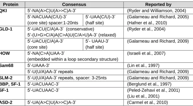
I.2.2.1 The STAR protein family ...39
I.2.2.1.1 Functions of STAR proteins in post-transcriptional regulation ...42
I.2.2.1.2 Functions of STAR proteins in development ...44
I.2.2.1.2.1 STAR proteins in cardiovascular development ...44
I.2.2.1.2.2 STAR proteins in mesoderm and muscle development ...46
xii
CHAPTER II – The RNA binding protein Quaking A is involved in the
establishment of internal organ laterality ... 51
II.1 Introduction ... 53
II.2 Results ... 56
II.2.1 qkia knockdown leads to defects in posterior body morphology ... 56
II.2.2 qkia is involved in Left-Right patterning ... 59
II.2.3 cdh11 - a candidate target of Qkia-mediated post-transcriptional regulation ... 63
II.2.4 cdh11 knockdown leads to Left-Right patterning defects ... 67
II.3 Discussion ... 72
II.4 Materials and methods ... 76
II.4.1 Zebrafish lines ... 76
II.4.2 Morpholino oligonucleotides ... 77
II.4.3 Cloning and site directed mutagenesis ... 79
II.4.3.1 qkia constructs ... 79
II.4.3.2 cdh11 constructs ... 80
II.4.4 In vitro transcription and mRNA microinjections ... 83
II.4.4.1 qkiaATG-MO rescue and qkia overexpression ... 83
II.4.4.2 Fluorescent reporters ... 84
II.4.5 Fluorescent reporter assays ... 85
II.4.6 Whole-mount in situ hybridization and histology ... 85
II.4.7 Fluorescence activated cell sorting and cdh11 detection ... 87
CHAPTER III - Fine-tuning of fgf8a expression through alternative polyadenylation has a selective impact on Fgf-associated developmental processes ... 89
III.1 Introduction ... 91
III.2 Results ... 93
III.2.1 fgf8a alt3’UTRs mediate distinct effects on translation efficiency ... 93
III.2.2 Interference with alternative PAS usage potentiates Fgf signalling ... 99
III.2.3 Interference with fgf8a PAS usage selectively affects sensory system development ... 105
xiii
III.4 Materials and Methods ... 114
III.4.1 Zebrafish lines ... 114
III.4.1.1 Transgenic and mutant lines ... 114
III.4.1.2 TALEN and Crispr mutagenesis... 115
III.4.2 Morpholino oligonucleotides ... 117
III.4.3 RT-qPCR ... 117
III.4.4 Fluorescent reporter assays ... 119
III.4.4.1 Cloning and microinjections ... 119
III.4.4.2 Image acquisition and processing ... 121
III.4.5 Whole-mount in situ hybridization and Immunohistochemistry ... 121
III.4.6 Statistical analysis ... 123
III.5 Supplemental Data ... 124
III.5.1 Supplemental Text ... 125
CHAPTER IV - Discussion and Conclusions ... 131
CHAPTER V - References ... 139
xiv
L
IST OFF
IGURESFig. I.1 – Illustration of the early stages of zebrafish embryonic development ... 4 Fig. I.2 – Illustration of the late stages of zebrafish embryonic development ... 11 Fig. I.3 – Gene expression regulation at the post-transcriptional level. ... 30 Fig. I.4 – The 3′ untranslated region (3′UTR) and associated mechanisms of
post-transcriptional regulation. ... 32 Fig. I.5 – The STAR protein family and the STAR domain. ... 40 Fig. II.1 – qkia knockdown affects posterior body morphology. ... 58 Fig. II.2 – qkia loss-of-function and gain-of-function affect organ laterality. ... 61 Fig. II.3 – Expression of Left-Right patterning genes is affected in qkia morphants. ... 63 Fig. II.4 – qkia knockdown and cdh11 knockdown affect otolith formation. ... 64 Fig. II.5 – Effect of qkia knockdown and overexpression on cdh11 3’UTR-mediated
regulation of reporter expression. ... 66 Fig. II.6 – cdh11 morphants, but not cdh11 mutants, display organ laterality defects. ... 68 Fig. II.7 – Expression of Left-Right patterning genes is affected in the LPM of cdh11
morphants. ... 70 Fig. II.8 – cdh11 expression in wildtype embryos and FACS sorted sox17:GFP cells. ... 71 Fig. II.M1 – qkia and cdh11 morpholino oligos and mutant alleles used in this work. ... 78 Fig. III.1 – Alternative fgf8a 3’UTR usage in the developing embryo. ... 94 Fig. III.2 – Alt3’UTRs fgf8aM and fgf8aS mediate different effects on post-transcriptional
regulation. ... 96 Fig. III.3 – The MM is necessary and sufficient to mediate the fgf8aM-associated
post-transcriptional regulation and this regulation is not substantially affected by the
fa3uiMO... 98
Fig. III.4 – fa3uiMO morphants display a shift in PAS usage preferences. ... 101
Fig. III.5 – fa3uiMO morphants display an increase in Fgf signalling. ... 104
Fig. III.6 – Assessment of fgf8a-associated developmental processes in fa3uiMO
morphants. ... 106 Fig. III.7 – Fgf signalling is involved in the early stages of superficial retinal
vascularization. ... 108 Fig. III.M1- Schematic representation of the mutagenesis strategies used in this work. . 116
xv
L
IST OFT
ABLESTable I.1 – Consensus binding sequences of STAR family proteins. ...42 Table II.M1 – Primers used in the qkia and cdh11 cloning procedures. ...82 Table II.M2 – WISH probes used in this study ...87 Table III.M1 - Primers used for RT-qPCR. ... 119 Table III.M2 – Primers and restriction enzymes used for alt3’UTR and MM cloning. ... 120 Table III.M3 – WISH probes used in this study. ... 122 Table III.S1 – Reported alt3’UTRs for the fgf8a gene. ... 124 Parameter Table ... 129
xvi
L
IST OFA
BBREVIATIONS Aace – acerebellar
alt3’UTR – Alternative 3’ untranslated region
APA – Alternative polyadenylation ASE – Asymmetric enhancer
aus – aussicht
B
BMP – Bone morphogenetic protein C
Cdh11 – Cadherin 11 cdh11MO – splice blocking morpholino
CDS – Coding sequence CDS – Coding Sequence Cpsf6 – Cleavage and
Polyadenylation Specificity Factor subunit 6
CRISPR – Clustered regularly
interspaced short palindromic repeats
CtrMO – Standard control morpholino oligo
D
D Loop – Dextral loop E
eGFP – enhanced Green fluorescent protein
ENU – N-ethyl-N-nitrosourea
F
fa3uiMO - fgf8a alt3’UTR interference morpholino oligo
FACS – Fluorescence activated cell sorting
FGF – Fibroblast growth factor FGFR – FGF receptor
G
GLD-1 – Defective in Germ Line Development
GSG – GRP33, SAM68 and GLD-1 H
Her – hairy and enhancer of
split-related
Hes – hairy and enhancer of split
HH – Hedgehog HOW - Held Out Wing hpf – Hours post fertilization hpi – Hours post injection HS-GAG – heparan sulphate glycosaminoglycan chains K
KV – Kupffer’s Vesicle L
L Loop – Left loop
LPM – Lateral plate mesoderm LROs – Left-Right organizers LSE – Left-side enhancer
xvii M
Mbp – Myelin basic protein
MHB – Midbrain-hindbrain boundary miR – microRNA
MM – Minimal motif
mRNA – messenger ribonucleic acid MZT – Maternal to zygotic transition P
p53MO – p53 translation blocking morpholino oligo
PAS – Polyadenylation signal PCR – Polymerase chain reaction PSM – Presomitic mesoderm Q
QKI – Quaking I
qkia – Quaking A
qkiaATG-MO – qkia translation blocking morpholino oligo
QRE – Quaking response element
R
RA – Retinoic acid
RBP – RNA binding protein Rock – Rho kinase
RT-qPCR – Real time quantitative PCR
S
SAG – Statoacoustic ganglion SAV – Superficial annular vessel. SF-1 – Splicing Factor 1
SHH – Sonic Hedgehog Spaw – Southpaw ss – Somite stage
STAR – Signal Transduction and Activation of RNA
T
TALEN – Transcription activator-like effector nucleases
TGF-β – Transforming growth factor beta
U
UTR – Untranslated region V
VEGF – Vascular endothelial growth factor
W
WISH – Whole-mount in situ hybridization
Wnt – Wingless X
xviii
L
IST OFP
UBLICATIONSFernandes, S.F., Fior, R., Pinto, F., Gama-Carvalho, M. and Saúde, L. (2018) ‘Fine-tuning of fgf8a expression through alternative polyadenylation has a selective impact on Fgf-associated developmental processes’, BBA Gene Regulatory Mechanisms, 1861(9), pp. 783-793.
1
I
C
HAPTER
I
3
I.1 E
MBRYONICD
EVELOPMENTThroughout the animal kingdom, there is a remarkable variety in the morphogenetic processes that take place during embryonic development. However, for most species, the patterns of early embryogenesis tend to follow a common sequential thread.
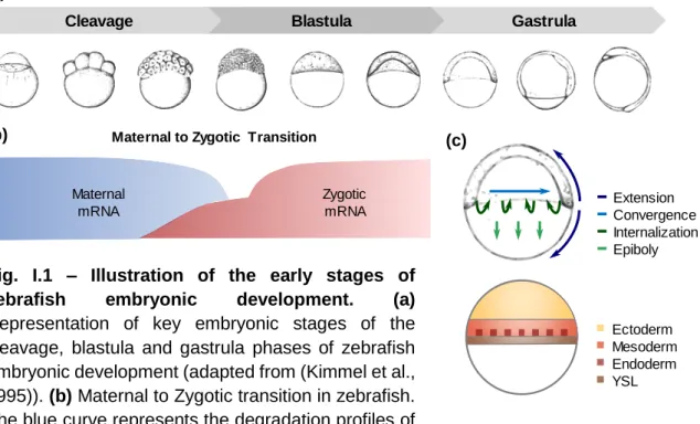
After fertilization, embryonic development begins with the cleavage phase. During this phase, a series of rapid cell divisions takes place giving rise to a substantial increase in cell number. By the end of the cleavage phase these cells, termed blastomeres, are generally arranged in a sphere known as the blastula (Fig. I.1a) (Gilbert, 2003).
The initial stages of animal development occur in the absence of de novo transcription. During this period, the progression of embryogenesis relies entirely on maternally inherited mRNAs and proteins. As early development progresses, maternal mRNAs and proteins are gradually degraded, and zygotic transcription is activated, thus progressively diminishing the maternal influence over embryogenesis. This gradual shift from maternal to zygotic control is known as the Maternal to Zygotic Transition (MZT). The MZT spans the period from the onset of maternal mRNA degradation to the first major developmental requirement for zygotic transcripts. For instance, in the zebrafish the MZT begins at fertilization, spanning the entire cleavage and blastula phases and coming to an end during the gastrula phase (Fig. I.1a,b) (Langley et al., 2014, Tadros and Lipshitz, 2009). The gastrula phase begins after the rate of cell divisions has diminished, and is characterized by extensive cell rearrangements. These highly coordinated cell movements are termed gastrulation. Gastrulation is accompanied by a series of specification and patterning events which enable the establishment of a multi-layered body plan containing three germ layers: the outer ectoderm, the inner endoderm, and the interstitial mesoderm (Fig. I.1c) (Gilbert, 2003, Solnica-Krezel, 2005).
Although the patterns of cell rearrangement during the gastrula phase vary throughout the animal kingdom, there are four evolutionarily conserved gastrulation movements: internalization, epiboly, convergence and extension. Internalization
4
movements carry prospective mesoderm and endoderm cells inward, beneath the prospective ectoderm. Epiboly movements lead to an expansion and thinning of the germ layers. Convergence and extension movements narrow the germ layers medio-laterally and elongate the embryo from head to tail (Fig. I.1c) (Solnica-Krezel, 2005).
In vertebrate embryos the final stages of gastrulation are either accompanied with, or followed by, the onset of neurulation – the formation of the neural tube – and segmentation – the formation of the somites. The neural tube is formed from ectodermal precursors situated above a rod-shaped mesodermal structure termed notochord, which demarcates the anterior-posterior embryonic body axis. The somites are spherical mesodermal structures which form on both sides of the notochord, and contain the precursors of the vertebrae, skeletal muscles, and dermis (Wolpert, 2002) (Fig. I.2a,b).
Embryonic development subsequently progresses to the organogenesis phase, during which, extensive cell rearrangement, differentiation and specialization
Gastrula Blastula Cleavage (a) Maternal mRNA Zygotic mRNA Maternal to Zygotic Transition
(b) (c) Ectoderm Mesoderm Endoderm YSL Extension Convergence Internalization Epiboly
Fig. I.1 – Illustration of the early stages of zebrafish embryonic development. (a)
Representation of key embryonic stages of the cleavage, blastula and gastrula phases of zebrafish embryonic development (adapted from (Kimmel et al., 1995)). (b) Maternal to Zygotic transition in zebrafish. The blue curve represents the degradation profiles of
destabilized maternal transcripts. The red curve illustrates the minor and major waves of zygotic genome activation (adapted from (Tadros and Lipshitz, 2009)). (c) Representation of the gastrulation movements and process of germ layer specification in the zebrafish embryo. YSL, yolk syncytial layer (adapted from (Solnica-Krezel, 2006, Kimelman, 2006)).
5 processes take place to form the different tissues and organs of the embryo (Fig. I.2a). During this phase, the ectoderm will give rise to the epidermis, nervous system and pigmented cells. The endoderm will contribute to the gastrointestinal, urinary and respiratory systems, as well as several endocrine glands. The mesoderm will give rise to the heart, kidneys, gonads, axial skeleton, cartilage, connective tissue, trunk muscles and blood cells. In addition, the formation of various organs will involve interactions between the different germ layers (Gilbert, 2003, Kiecker et al., 2016).
The next subchapter will focus on two critical developmental processes: the elongation and segmentation of the anterior-posterior axis; and the establishment of internal organ asymmetry along the left-right axis. In addition, it will address the specific contributions of the fibroblast growth factor (FGF) signalling pathway to different aspects of embryonic development.
I.1.1 Posterior body elongation and Somitogenesis
The first morphogenetic events that define the shape of the embryonic body are thought to take place during the gastrula phase, as a result of convergence and extension movements. However, this process carries on after gastrulation, with posterior body elongation and segmentation presenting as two major aspects of vertebrate development (McMillen and Holley, 2015, Bénazéraf and Pourquié, 2013).
I.1.1.1 Posterior body elongation
The development and elongation of the posterior body is achieved through the progressive deposition of cells from a posterior growth zone in the embryo. This posterior leading edge of the growing embryo, named tailbud, contains the progenitors of the musculature, axial skeleton, vasculature, spinal cord and blood (McMillen and Holley, 2015, Beck, 2015).
6
Interestingly, studies done in chick and zebrafish embryos have found that posterior body elongation is primarily driven by cell migration rather than cell proliferation, with tailbud musculoskeletal progenitors exhibiting only a modest level of proliferation during posterior body elongation (Bouldin et al., 2014, Kanki and Ho, 1997, Bénazéraf et al., 2010, McMillen and Holley, 2015). In these organisms, instantaneous cell velocities are greater in the posterior tailbud, with posterior growth occurring as these highly motile progenitors lessen their motility and assimilate into more anterior tissues, namely the paraxial mesoderm (Lawton et al., 2013, Dray et al., 2013, Mara et al., 2007, Bénazéraf et al., 2010, Delfini et al., 2005). For instance, during zebrafish posterior body elongation, the progenitor cells of the dorsal medial tailbud dive ventrally as a coherent posterior flow. At the posterior ventral tailbud there is a loss in cell flow coherence which leads to an increase in cell mixing (Lawton et al., 2013, Dray et al., 2013). These mesodermal progenitors subsequently lose velocity as they enter the posterior paraxial mesoderm, concomitantly with the assembly of an extracellular matrix composed primarily of Fibronectin and Laminin (Dray et al., 2013, Latimer and Jessen, 2010, McMillen and Holley, 2015).
The entry of these tailbud cells into the paraxial mesoderm territory appears to include a process of convergence and extension, akin to what is observed during gastrulation, with this process being regarded as an important contributing factor to posterior body elongation (Steventon et al., 2016, Kanki and Ho, 1997).
Furthermore, paraxial mesoderm assembly is accompanied by the formation of the notochord from axial mesoderm precursors. The vacuolation and rearrangement of the notochord cells has also been proposed as a contributing factor to the progression of posterior body elongation (McMillen and Holley, 2015, Kanki and Ho, 1997, Dray et al., 2013).
I.1.1.2 Somitogenesis
As posterior body elongation progresses, the paraxial mesoderm, also known as presomitic mesoderm (PSM), is subdivided into metameric structures, termed somites. In vertebrates, somites form sequentially along the anterior-posterior
7 embryonic axis, budding off in bilateral pairs from the unsegmented PSM. Each somite presents as a spherical cell mass surrounded by an epithelial sheet, and contains the precursors of the vertebrae, skeletal muscles, and dermis (Fig. I.2a,b) (Yabe and Takada, 2016).
The process of somite formation – somitogenesis – is tightly regulated, both spatially and temporally, with the frequency of somite formation and the total number of somites formed being species-specific traits. For instance, in zebrafish a new pair of somites is formed every 25 minutes until a total of approximately 33 somite pairs is reached, whereas in mice a new somite pair is formed every 2 hours resulting in the formation of approximately 65 somite pairs (Yabe and Takada, 2016, Hubaud and Pourquié, 2014).
To account for this spatiotemporal regulation of somitogenesis, a theoretical model termed “Clock and Wavefront model” was proposed. In this model, rhythmic and sequential somite formation is achieved by two regulatory mechanisms: a segmentation clock and a wavefront of differentiation. The cyclic activation of the segmentation clock provides temporal information, which is integrated with the spatial information provided by the continuous regression of the wavefront that results from posterior body elongation. A consequence of this model is that the size of each newly formed somite is fixed by the distance travelled by the wavefront during one period of the segmentation clock (Fig. I.2b) (Cooke and Zeeman, 1976, Yabe and Takada, 2016, Hubaud and Pourquié, 2014).
Since the Clock and Wavefront model was proposed, several genes have been associated with the establishment of the segmentation clock and wavefront mechanisms.
I.1.1.2.1 The segmentation clock
Regarding the segmentation clock, the first gene to be implicated in this mechanism was the chicken HAIRY1. In the chick PSM, HAIRY1 is expressed cyclically in the PSM, with a frequency of expression that is consistent with the frequency of chick somite formation. This gene belongs to the hairy and enhancer of split (Hes)/
HES-8
related (her) family of transcription factors that act mainly as Notch pathway effectors (Palmeirim et al., 1997, Cooke, 1998).
Subsequent studies have implicated multiple members of the Notch, Wingless (Wnt) and FGF pathways in the segmentation clock (Krol et al., 2011, Dequéant et al., 2006, Hubaud and Pourquié, 2014). However, of all the gene families identified to date in connection with the clock, the Hes/her family appears to be the most conserved contributor, with Hes/her cyclic genes having been identified in mouse, chick, zebrafish, medaka and Xenopus (Krol et al., 2011, Dequéant et al., 2006, Elmasri et al., 2004, Li et al., 2003a).
In line with this, the segmentation clock has been proposed to rely heavily on a
Hes/her-based negative-feedback loop. This loop is thought to drive gene
expression oscillations via a mechanism of delayed transcriptional repression (Bessho et al., 2003, Lewis, 2003). For instance, the her1 and her7 genes are widely regarded as the pacemakers of the zebrafish segmentation clock (Henry et al., 2002, Holley et al., 2002, Oates and Ho, 2002, Gajewski et al., 2003). Her1 and Her7 have been shown to act as transcriptional repressors, inhibiting their own transcription, and that of the Notch ligand DeltaC, in the posterior PSM (Giudicelli et al., 2007). Mathematical modelling has shown that this Her1/Her7 autoinhibition has the potential to generate a delayed negative feedback loop, which could underlie the oscillating expression of these genes. The concomitant cyclical inhibition of DeltaC is thought to coordinate gene expression oscillations between neighbouring cells (Lewis, 2003). It follows from this model that this negative-feedback driven gene expression oscillation frequency would provide the temporal information required to set the pace of the segmentation clock (Fig. I.2b) (Pais-de-Azevedo et al., 2018, Hubaud and Pourquié, 2014).
Interestingly, this model postulates that the production of stable oscillations in gene expression is predicated on several conditions, one of which being the instability of the her7, her1 and deltaC mRNAs (Lewis, 2003). This instability was further confirmed by in situ hybridisation and fluorescent reporter experiments, which revealed that the mRNAs of these genes have very short half-lives, specifically 6.1-8.1 minutes (Giudicelli et al., 2007, Gajewski et al., 2003). Regarding the mechanisms that mediate this instability, recent studies conducted in zebrafish,
9 mouse and chick suggest that a Pnrc2-Upf1 complex and the microRNA mir-125a-5p operate as negative regulators of the stability of the cyclic her1 and lunatic fringe mRNAs, respectively (Gallagher et al., 2017, Riley et al., 2013, Wahi et al., 2017).
I.1.1.2.2 The wavefront
The wavefront was originally defined by Cooke and Zeeman as a front of rapid cell change moving slowly in a posterior direction along the axis of the embryo (Cooke and Zeeman, 1976). Subsequent studies have identified the position of this conceptual wavefront (also known as the determination front) as the virtual frontier between the posterior PSM – where the paraxial mesoderm cells have yet to acquire their somitic identity – and the anterior PSM – where cells are already committed to their somitic fate. Furthermore, the clock and wavefront model proposes that the wavefront corresponds to the level at which PSM cells become responsive to a signal from the segmentation clock that potentiates the definition of the future segmental domain, and thus, the size of the formed somites (Fig. I.2b) (Hubaud and Pourquié, 2014, Yabe and Takada, 2016, Cooke and Zeeman, 1976, Dequéant and Pourquié, 2008).
Three major signalling gradients have been implicated in defining the position of the wavefront: a posterior-to-anterior FGF gradient, a posterior-to-anterior Wnt gradient, and an anterior-to-posterior Retinoic Acid (RA) gradient (Fig. I.2b).
Studies done in chick, zebrafish and mouse have shown that both upregulation and downregulation of FGF signalling in the PSM leads to a disruption of somitogenesis, specifically regarding somite boundary positioning (Dubrulle et al., 2001, Sawada et al., 2001, Wahl et al., 2007, Naiche et al., 2011). This wavefront activity appears to be primarily mediated by the fgf8a gene in zebrafish embryos, whereas mouse embryos appear to rely on both FGF8 and FGF4 ligands for this process (Akiyama et al., 2014, Naiche et al., 2011).
Interestingly, Dubrulle and Pourquié reported that in the chick and mouse PSM, Fgf8 transcription is restricted to the growing posterior tip of the embryo. As posterior body elongation progresses, Fgf8 mRNA is gradually degraded in the newly formed tissues leading to the establishment of the observed posterior-to-anterior Fgf8
10
mRNA gradient. Considering that the process of posterior body elongation is relatively slow, these results indicate that a certain degree of Fgf8 mRNA stability must be present to enable FGF8 gradient formation, and consequently, wavefront establishment (Dubrulle and Pourquié, 2004).
The role of the Wnt gradient in wavefront establishment was first identified in mouse, where an upregulation of Wnt signalling in the PSM lead to a disruption of paraxial mesoderm maturation and somite boundary positioning (Aulehla et al., 2008, Dunty et al., 2008). Evidence for the conservation of this function comes from studies done in zebrafish, where temporally-controlled modulations of Wnt signalling led to alterations in somite size (Bajard et al., 2014).
In contrast to the FGF and Wnt gradients, which display higher morphogen concentrations at the posterior tip of the embryo, the RA gradient displays higher concentration levels in the somites and anterior PSM (Rossant et al., 1991, Shimozono et al., 2013). In line with this, the RA gradient was proposed to function as an antagonist of the FGF signalling gradient, with FGF8 and RA contributing to wavefront position establishment through a mechanism of mutual inhibition (Diez del Corral et al., 2003, Vermot et al., 2005, Moreno and Kintner, 2004).
In addition, RA has also been implicated in the maintenance of the lateral symmetry of the somites. In this context, it has been proposed that during the period of development when the asymmetric position of internal organs such as the heart, liver and pancreas is being established, RA functions as a buffer in the somites, ensuring that somitogenesis remains refractory to asymmetry-inducing mechanisms (Kawakami et al., 2005, Vermot and Pourquié, 2005, Sirbu and Duester, 2006).
11
Fig. I.2 – Illustration of the late stages of zebrafish embryonic development.
(a) Representation of key embryonic stages of late zebrafish embryogenesis, specifically,
the segmentation, pharyngula and hatching periods (adapted from (Kimmel et al., 1995)).
(b) The somitogenesis process in zebrafish. Representation of the Clock and Wavefront
model, according to which, two mechanisms control the activation of the somitogenesis programme: the Clock (right) and the Wavefront (left). The frequency of the Clock is thought to rely on a delayed negative feedback loop established by the her1 and her7 genes. The position of the wavefront is defined by three gradients: A Retinoic Acid gradient, a FGF signalling gradient and a Wnt signalling gradient. According to this model, the frequency of somite formation and the size of the formed somites are determined by the interplay between these two mechanisms (partially adapted from (Giudicelli et al., 2007)). (c) Internal organ laterality establishment in zebrafish. The process of Left-Right patterning is initiated by motile cilia present in the KV, which rotate in a counter-clockwise manner creating a leftward extracellular fluid flow. This flow induces the establishment of the first asymmetric cues which, in zebrafish, include the right-side specific expression of the spaw inhibitor dand5. Asymmetric gene expression in the KV functions as a laterality signal which is transmitted to the LPM and triggers asymmetric gene expression in the left LPM, namely the nodal-lefty-pitx2 cascade. This in turn determines the correct lateral positioning of the visceral and cardiac organs (partially adapted from (Wang et al., 2012)).
Hatching Pharyngula Segmentation (a) (b) (c) Lateral Plate Mesoderm Kupffer’s Vesicle Notochord spaw lefty pitx2 dand5 spaw spaw R L L R Clock Somites Wavefront Presomitic mesoderm delay her1/her7 delay deltaC Retinoic Acid Fgf and Wnt
12
I.1.2 Left-Right organ asymmetry
Embryonic morphogenesis takes place along three orthogonal axes: The Anterior-Posterior axis, the Dorsal-Ventral axis and the Left-Right axis. When it comes to the Left-Right axis, most vertebrates have a largely symmetrical body-plan, with this symmetry being broken by the asymmetric placement of several internal organs, such as the heart, gut, liver, spleen and stomach. Furthermore, paired organs such as the lungs and brain tend to develop asymmetrically, presenting morphological and/or functional differences between the left and right sides (Grimes and Burdine, 2017).
I.1.2.1 The Left-Right organizer
In vertebrates, the establishment of Left-Right asymmetry is widely believed to begin in transient midline structures, which appear at the posterior end of the notochord during early somitogenesis stages and are known as Left-Right organizers (LROs) (Fig. I.2c) (Grimes and Burdine, 2017, Amack, 2014).
In mouse the LRO is termed Node, in zebrafish and medaka the LRO is known as the Kupffer’s Vesicle (KV), the Xenopus LRO is the Gastrocoel Roof Plate and the rabbit LRO is the posterior notochord. Studies conducted in these organisms have shown that a largely conserved feature of the LRO is the presence of motile cilia which display a posterior tilt and rotate in a clockwise manner, when observed ventrally (Okada et al., 2005, Nonaka et al., 2005, Kramer-Zucker et al., 2005, Okabe et al., 2008, Schweickert et al., 2007). These motility features allow the cilia to generate an extracellular unidirectional leftward fluid flow within the organizer, with this flow being perceived as a crucial aspect of Left-Right asymmetry establishment (Fig. I.2c) (Cartwright et al., 2004, Okada et al., 2005, Kramer-Zucker et al., 2005, Essner et al., 2005, Hojo et al., 2007, Schweickert et al., 2007, Blum et al., 2009).
There are many factors known to influence the LRO fluid flow, one of which is the cellular architecture and morphology of the LRO. Features such as the size and shape of the LRO, as well as the number of ciliated cells and their spatial
13 organization within the organizer, vary significantly between vertebrate species (Amack, 2014, Blum et al., 2009, Lee and Anderson, 2008, Shook et al., 2004, Wang et al., 2011). However, studies in mouse and zebrafish have shown that the disruption of these specie-specific LRO architectures has a detrimental effect on Left-Right asymmetry establishment (Beckers et al., 2007, Lee et al., 2010, Pulina et al., 2011, Sutherland et al., 2013, Arrington et al., 2013, Oteiza et al., 2010, Wang et al., 2011, Wang et al., 2012, Ablooglu et al., 2010, Matsui et al., 2011).
For instance, the zebrafish Kupffer’s Vesicle is a spherical structure, with a higher concentration of ciliated epithelial cells in the anterior side of the dorsal surface of its lumen (Fig. I.2c) (Kreiling et al., 2007). This dorsal anterior cluster of motile cilia is formed through a process called KV remodelling, whereby the most anterior cells become elongated with tight apical surfaces, and the most posterior cells adopt a cuboid shape with larger apical surfaces (Wang et al., 2012). This process appears to be regulated by the Rho kinase (Rock) 2b-Myosin pathway, with disruptions of this pathway leading to alterations in KV morphology, fluid flow, and ultimately internal organ laterality (Wang et al., 2011, Wang et al., 2012). In mouse the RHO family member RAC1 has also been implicated in LRO morphogenesis, and in
Xenopus, rock2 knockdown has been linked to Left-Right patterning defects, thus
raising the possibility of a conserved role for this pathway in LRO architecture establishment (Migeotte et al., 2011, Fakhro et al., 2011).
Additional proteins with an apparent involvement in mouse LRO architecture establishment, and consequently Left-Right organ asymmetry, include the transcription factors NOTO and ZIC3, the cytoskeletal-associated protein EPB4.1l5, the extracellular matrix component Fibronectin and its receptor Integrin α5β1 (Beckers et al., 2007, Lee et al., 2010, Pulina et al., 2011, Sutherland et al., 2013). The latter has also been implicated in Left-Right organ asymmetry establishment in the zebrafish (Pulina et al., 2011). Furthermore, several genes and signalling pathways have been implicated in different aspects of zebrafish KV morphogenesis, and consequently internal organ laterality establishment. These include Wnt11- and Prickle1a-mediated planar cell polarity signalling, the Integrin subunits αV and β1b, the adhesion molecule Cadherin1, and the transmembrane heparan sulfate proteoglycan Syndecan 2 (Sdc2), with the last two exerting their functions through
14
interactions with the FGF signalling pathway (Arrington et al., 2013, Oteiza et al., 2010, Ablooglu et al., 2010, Matsui et al., 2011).
I.1.2.2 Asymmetric gene expression in the LRO
In most vertebrate species, the leftward fluid flow generated in the LRO precedes and is widely believed to induce asymmetric gene expression (Fig. I.2c). Despite the extensive work that has been conducted in the field, and the many conserved aspects of Left-Right asymmetry establishment, the mechanisms that effectively detect and translate the LRO fluid flow into an asymmetric signalling pathway remain unknown (Grimes and Burdine, 2017).
The most widely accepted models to address the nature of these mechanisms are the morphogen model and the two-cilia model. The morphogen model proposes that a morphogen, or a series of vesicular parcels (termed “nodal vesicular parcels”) containing morphogens such as Sonic Hedgehog or Retinoic Acid, are transported to the left side of the LRO by the fluid flow. Once these morphogens reach the left LRO, they induce a release of Ca2+, which in turn triggers left side specific gene
expression (Nonaka et al., 1998, Tanaka et al., 2005). The two-cilia model proposes that the LRO contains two different populations of cilia: motile cilia and immotile sensory cilia. According to this model, while the motile cilia generate the fluid flow, the immotile cilia sense the flow on the left side of the LRO and trigger the release of Ca2+, which in turn induces left side specific gene expression (McGrath et al.,
2003). Note that these models are not mutually exclusive, thus both mechanisms can exist simultaneously in the LRO.
Left side specific gene expression has been identified in several vertebrate organisms, with the major players in asymmetric signalling belonging to the Nodal pathway. The Nodal genes are members of the Transforming Growth Factor beta (TGFβ) superfamily and have a highly conserved role as left side determinants. While humans, mice and chick have a single Nodal gene, Xenopus have five NODAL-related proteins (Xnr1, 2, 4, 5 and 6) and zebrafish have three (Cyclops, Squint and Southpaw (Spaw)) (Shen, 2007, Schier, 2009).
15 In mouse, Nodal expression is transiently enhanced on the left side of the LRO at the 4-5 somite stage (ss), whereas in zebrafish nodal expression is always symmetric in the LRO (Collignon et al., 1996, Long et al., 2003). However, studies done in mouse have shown that NODAL activity is higher on the left side of the LRO, even during stages when Nodal expression is symmetric. This was proposed to arise from the asymmetric expression of the NODAL inhibitor CERL2 on the right side of the LRO (Kawasumi et al., 2011). A similar mechanism is thought to be present in the zebrafish, with the Nodal inhibitor Dand5 also displaying an asymmetric expression pattern favouring the right side of the LRO (Fig. I.2c) (Lopes et al., 2010).
CERL2 and Dand5 are members of the DAN family of cysteine-rich extracellular proteins that can block Nodal signalling by interacting directly with the NODAL proteins (Shen, 2007, Schier, 2009). In the mouse, CERL2 expression is initially symmetric in the LRO, becoming asymmetric at the onset of LRO ciliary flow (Pearce et al., 1999, Marques et al., 2004). This shift from symmetric to asymmetric expression was proposed to rely on the targeted degradation of Cerl2 mRNA in the apical and left-sided region of the LRO (Nakamura et al., 2012). Regarding zebrafish, dand5 expression is also initially symmetrical in the LRO and at the 8 somite stage, with the onset of ciliary flow, becomes asymmetrically positioned on the right side of the LRO, although the mechanisms that regulate this shift in zebrafish are currently unknown (Lopes et al., 2010).
I.1.2.3 Asymmetric gene expression in the LPM
After the first asymmetry cues are established in the LRO, in the form of an asymmetric activation of the Nodal pathway, these cues are transmitted to the left Lateral Plate Mesoderm (LPM) (Fig. I.2c). The process through which left side specific NODAL activity in the LRO translates into left side specific Nodal expression in the LPM is still not fully understood. However, several lines of evidence suggest that NODAL exhibits a long-range activity, traveling directly from the left side of the LRO towards the left lateral plate mesoderm, through an intra-embryonic route. The efficiency of this transport appears to rely on interactions between NODAL and
16
Sulfated Glycosaminoglycans (Oki et al., 2007, Marjoram and Wright, 2011, Shiratori and Hamada, 2014).
Once the NODAL signal reaches the left LPM, it triggers the activation of the
Nodal-Lefty-Pitx2 gene expression cassette. This cassette contains three remarkably
conserved Nodal pathway target genes: Nodal itself, the Nodal Inhibitor Lefty, and
Pitx2 (Fig. I.2c) (Shen, 2007, Schier, 2009).
Evidence supporting the auto-activation of Nodal came from studies done in mouse, where two NODAL-responsive enhancers have been identified in the Nodal gene, the left-side enhancer (LSE) and the asymmetric enhancer (ASE). The combined action of these two enhancers is thought to drive Nodal expression in the left LPM (Adachi et al., 1999, Norris and Robertson, 1999, Saijoh et al., 2000, Saijoh et al., 2005). This auto-activation of Nodal results in the rapid spread of Nodal expression throughout the left LPM, as well as the induction of Lefty and Pitx2 expression. Lefty proteins are Nodal target genes which establish a negative feedback loop with Nodal. Most vertebrates have only one Lefty protein with the exception of mouse and zebrafish, which have two, LEFTY1 and LEFTY2 (Schier, 2009, Shiratori and Hamada, 2014). Lefty2 expression is activated by NODAL in the left LPM, where it downregulates NODAL activity thus regulating the spread of Nodal expression. Nodal signalling also activates Lefty1 expression in the axial midline, LEFTY1 is therefore thought to function as a molecular barrier that prevents leakage of the Nodal signal from the left to the right side. This mechanism of self-enhancement and lateral inhibition has been proposed to ensure the propagation of NODAL signals throughout the left LPM, while simultaneously inhibiting their activation on the right LPM (Schier, 2009, Shiratori and Hamada, 2014, Nakamura et al., 2006, Saijoh et al., 2000, Meno et al., 1998, Yamamoto et al., 2003).
Much like Nodal, Pitx2 is a highly conserved left-side specific gene, being expressed in the left LPM of all the vertebrate species studied to date (Shiratori and Hamada, 2014, Burdine and Schier, 2000). In addition, Pitx2 also possesses a left-side specific ASE enhancer, which is responsive to NODAL and required for Pitx2 expression in the left LPM. In this context, NODAL appears to induce Pitx2 expression in the left LPM, through the ASE enhancer, with the maintenance of LPM
17 transcription factor NKX-2.5 (Shiratori et al., 2001). Pitx2 expression in the left LPM therefore functions as a readout of Nodal signalling and is thought to contribute to the following stage of Left-Right patterning during which positional information is transferred to the developing internal organs.
I.1.2.4 Asymmetric internal organ placement
There are several lines of evidence supporting the theory that asymmetric gene expression in the LPM conditions the asymmetric placement of internal organs such as the gut, liver, pancreas (also known as visceral organs) and the heart (Fig. I.2c). However, the specific requirements for Nodal-Pitx2 asymmetric signalling and the precise contribution of this pathway to organ laterality establishment require further elucidation.
During gut development, the first break from symmetry occurs when portions of the gut are displaced laterally from the midline, in a process termed gut looping. In zebrafish gut looping occurs when the region that will give rise to the liver and intestinal bulb moves to the left of the midline. The mechanisms that drive gut looping appear to be largely reliant on neighbouring tissues. Specifically, in zebrafish gut asymmetries are driven by the asymmetric migration of the LPM, and in amniotes the initial chirality of gut looping relies on asymmetries in the cellular architecture of the associated dorsal mesentery (Horne-Badovinac et al., 2003, Davis et al., 2008, Kurpios et al., 2008). Importantly, Nodal signalling has been identified as an upstream regulator of asymmetric LPM migration in zebrafish and left-sided Nodal-Pitx2 expression was shown to instruct asymmetric cellular architecture establishment in the amniote dorsal mesentery (Grimes and Burdine, 2017, Horne-Badovinac et al., 2003, Davis et al., 2008, Kurpios et al., 2008). Consistent with this is the observation that mouse mutants lacking left-sided Pitx2 expression exhibit laterality defects in most visceral organs (Shiratori et al., 2006). Furthermore, in zebrafish nodal mutants the lateral positions of the visceral organs are randomized (Noël et al., 2013). However, zebrafish pitx2 mutants do not present laterality defects in the visceral organs, raising the possibility that additional Nodal
18
signalling effectors can contribute to the establishment of visceral organ laterality in the fish (Ji et al., 2016).
In zebrafish, cardiac symmetry breaking can be divided into two steps: a Jogging step and a Looping step. The Jogging step is characterized by a leftwards and cranial displacement of atrial cardiomyocytes and simultaneous involution of ventricular myocardial cells, which generates a leftward pointing cardiac tube. The Looping step involves the repositioning of the atrium in a caudal direction and the repositioning of the ventricle in an anterior direction, which generates a coiled cardiac tube with well-defined inner and outer curvatures. In wildtype conditions the direction of heart tube Jogging (Left jog) prefigures the direction of cardiac Looping (Dextral loop) (Campione and Franco, 2016).
Noticeably, in zebrafish nodal mutant embryos, normal Dextral looping is still observable in approximately 70% of the mutant population (Noël et al., 2013). Furthermore, heart tubes isolated from these embryos and cultivated in vitro, retain the capacity to undergo Dextral looping (Noël et al., 2013). These results raise the possibility that nascent cardiomyocytes possess an intrinsic laterality bias, with robust cardiac asymmetry establishment likely resulting from the integration of this intrinsic program with the laterality signals provided by the Nodal pathway.
Furthermore, Pitx2 has been implicated in several aspects of asymmetric cardiac morphogenesis, with Pitx2 loss-of-function experiments leading to atrial isomerism, impaired atrioventricular remodelling, atrial and ventricular septal defects and morphological defects arising from an impairment of the alignment and rotation of the outflow tract relative to the ventricles (Campione and Franco, 2016). However, while PITX2 seems to be required to establish cardiac looping directionality in the chick, this requirement appears to be absent in zebrafish and mouse (Shiratori et al., 2006, Lu et al., 1999, Gage et al., 1999, Kitamura et al., 1999, Lin et al., 1999, Ji et al., 2016, Yu et al., 2001, Campione and Franco, 2016). Further reinforcing the possibility that additional mechanisms, outside the scope of the Nodal pathway, contribute to the establishment of cardiac laterality.
19
I.1.3 FGF signalling in embryonic development
Embryonic development relies on short- and long-distance cellular communication, with this communication often involving the secretion of signalling molecules and the activation of signalling pathways in response to these secreted signals. Extensive research into the major morphogenetic events that take place during embryogenesis has revealed that a surprisingly small number of signalling pathways appear to regulate the vast majority of developmental programs. One of these major regulators of embryonic development is the FGF signalling pathway (Perrimon et al., 2012).
Fgf ligands are small secreted polypeptides with a partially conserved core of 120– 130 amino acids. The majority of Fgf ligands operate as paracrine signalling molecules, forming a tripartite complex with Fgf receptors (Fgfrs) and heparan sulphate glycosaminoglycan chains (HS-GAG). Fgfrs are receptor tyrosine kinases which are activated by Fgf/HS-GAG binding. Fgfr activation results in receptor dimerization and triggers the activation of intracellular signal transduction pathways, including the RAS-MAPK (Ras – Mitogen activated protein kinase), PI3K-AKT (Phosphoinositide 3 kinase - Protein kinase B), PLC𝛾 (phospholipase-Cγ), and STAT (Signal transducer and activator of transcription) pathways. In most cases, the activation of these pathways ultimately affects the transcriptional program of the cell, with genes which are differentially expressed in response to FGF signalling being commonly referred to as Fgf target genes (Pownall and Isaacs, 2010, Ornitz and Itoh, 2015).
During embryonic development, several Fgf ligands and receptors have been implicated in embryonic patterning, progenitor cell maintenance, growth, differentiation and survival. Furthermore, FGF signalling appears to influence embryogenesis from its earliest stages and throughout the entire organogenesis phase (Pownall and Isaacs, 2010, Ornitz and Itoh, 2015). The next sections will focus on highlighting some of the key functions of the FGF signalling pathway during embryonic development, with a special emphasis on the developmental roles of the zebrafish ligand fgf8a and its orthologues – the Fgf8 genes.