UNIVERSIDADE NOVA DE LISBOA
INSTITUTO DE HIGIENE E MEDICINA TROPICAL
The antimycobacterial activity of thioridazine derivatives against drug
resistant Mycobacterium tuberculosis:
In vitro, ex vivo and in vivo studies
Marta Sofia Lopes Martins
Thesis research submitted to the Instituto de Higiene e Medicina Tropical, Universidade Nova de Lisboa in partial fulfilment of the requirements for the granting of the degree of Doctor of Philosophy with specialisation in the Biomedical Science of Microbiology.
The Thesis research to be described was conducted at the Unit of Mycobacteriology, UPMM at the Instituto de Higiene e Medicina Tropical/Universidade Nova de Lisboa (IHMT/UNL) and supported by grant SFRH/BD/14319/2003 provided by Fundação para a Ciência e a Tecnologia (FCT) of Portugal.
SUPERVISOR:
Professor Doutor Leonard Amaral
Professor Catedrático Convidado e Director da Unidade de Micobacterias Unidade de Micobacterias, UPMM
Instituto de Higiene e Medicina Tropical Universidade Nova de Lisboa, Lisboa
TUTORIAL COMMISSION:
Professora Doutora Filomena Pereira
Professora Associada e Directora da Unidade de Doenças Sexualmente Transmissíveis Instituto de Higiene e Medicina Tropical
Universidade Nova de Lisboa, Lisboa
Professor Doutor Miguel Viveiros Professor Auxiliar de Bacteriologia Unidade de Micobacterias
Instituto de Higiene e Medicina Tropical Universidade Nova de Lisboa, Lisboa
Professor Doutor Leonard Amaral
Professor Catedrático Convidado e Director da Unidade de Micobacterias Unidade de Micobacterias, UPMM
Instituto de Higiene e Medicina Tropical Universidade Nova de Lisboa, Lisboa
Na presente dissertação incluem-se resultados que foram ou estão a ser alvo de publicação em co-autoria. Os artigos publicados ou submetidos para publicação serão integralmente apresentados em Anexo. Para efeitos do disposto no nº1 do Despacho nº2303/2000 do Regulamento de Programas de Doutoramento do Instituto de Higiene e Medicina Tropical, Universidade Nova de Lisboa (Diário da República, 2ª série, nº 23, de 28 de Janeiro de 2000), o autor da dissertação declara que interveio na concepção e execução do trabalho experimental, na interpretação dos resultados e na redacção dos manuscritos publicados, submetidos ou que aguardam submissão.
Lisboa, 31 de Janeiro de 2008
“There is at bottom only one genuine treatment for all diseases,… to stimulate the phagocytes. Drugs are a delusion. … (when) the phagocytes are stimulated; they
devour the disease…”
ACKNOWLEDGEMENTS
This work wouldn’t be possible without the help and support of the following persons. I would like to thank:
Professor Doutor Leonard Amaral, my supervisor, for the opportunity to realise my aspiration to learn more about that dreadful infection of pulmonary tuberculosis and to contribute to the manner by which extensively-drug resistant Mycobacterium tuberculosis and multi-drug resistance M. tuberculosis may be successfully treated. Thank you for all the scientific discussions, daily stimulus and support and for all the help in making me a more conscientious scientist;
Professor Doutor Miguel Viveiros, for all the support, encouragement, scientific discussions, advice and critical review of my work; also for the constant good-humour, patience during all these years and especially, for your friendship, life is so much better when we have good friends like you;
Professora Doutora Isabel Couto, my friend, for always being there and never forgetting that despite all, we are human; and for all the scientific discussions, valuable critique, infinite patience, and good advice. Thank you for the “post-office journeys” and all the small things that made a big difference every day;
Mestre Clara Leandro, for all the good-times spent and much needed assistance during the initial period of my PhD. Thank you for your friendship and for teaching me that we should always pursue our dreams: the proof is Mariana and Gonçalo, a big kiss to you all;
All my colleagues of the Unit of Mycobacteriology for that environment which facilitated my studies and assistance given during these years; special thanks to graduate students Liliana Rodrigues, Diana Machado and Ana Martins for all the patience, support and for listening. Sofia, Catarina, Laura, Jorge and Nadia thank you for keeping me updated in the news from the (un)real world!
D. Josefina and D. Rosário, for all the knowledge and technical assistance given, especially in animal studies. Thank you for continuing to believe in the young;
D. Mariana for the precious help in the laboratory and for making my days so much easier. D. Catarina and D. Cidália for all the technical support along these years;
Zsuzsanna Schelz, my Hungarian friend, thank you for all the help and for the lovely times spent in Szeged. You are truly a very special person, Köszönöm;
My colleagues, Sofia Martins and Pedro Fernandes; Sofia for always being there and making me laugh during difficult times and continuing to believe in my skills and Pedro, thank you for all the support and ideas;
All the Marta’s in my life: Marta Costa (Martinha), Marta Agostinho and Marta Pingarilho. Thank you for all the support and strength. Martinha, thank you for your shoulder during those difficult moments; i hope that Maria brings you a whole sea of happiness;
Professores Doutores João Piedade, Ricardo Parreira and Aida Esteves, from the Unit of Virology of IHMT, for all the help and the precious coffee moments, so vital for maintaining sanity during times of stress. Thank you for all the support and advice. To D. Teresa Veneno for all the technical support and Daniela, for all the conversations and good mude;
The Sexually Transmitted Diseases Unit from IHMT for all the help. Special thanks to Professora Doutora Filomena Pereira for the revisions of my reports and to Filipa for the support, long conversations and availability when needed;
Drª. Teresa Pacheco and colleagues from the Laboratório de Microbiologia, Hospital Egas Moniz, Lisboa, for the valuable contributions to this work;
Professora Doutora Hermínia de Lencastre from the Instituto de Tecnologia Química e Biológica, Universidade Nova de Lisboa, for the gently donation of the MRSA COL and HPV107 strains;
The President of the CC of IHMT Professora Doutora Amélia Grácio and all the members of the CC who created the environment much needed for the development of a graduate student who must compete with others in Europe;
Fundação para a Ciência e a Tecnologia (FCT) for providing me the PhD grant SFRH/BD/14319/2003 that allowed me to develope this work and also for the project grants EU-FSE/FEDER-POCTI-37579/FCB/2001 and EU-FSE/FEDER-POCI/SAU-MMO/59370/2004;
To the following colleagues from Europe and the USA:
Doutora Diane Ordway, for bringing me to the Immunology world and giving me the opportunity to work at the Unit of Mycobacteriology with such wonderful colleagues, who are now friends;
Professor Wilfrid Bleiss and his team from the Department of Molecular Parasitology, Institut für Biologie, Humboldt-Universität zu Berlin, Berlin, Germany, for all the electron microscopy work that was so important for helping us to “see” inside the macrophage;
Professor Sujata G. Dastidar from the Division of Microbiology, Department of Pharmaceutical Technology, Jadavpur University, Calcutta, India for all the cooperation with our team in the studies involving antibiotic resistance and novel non-antibiotics;
Professor Seamus Fanning from the Centre for Food Safety, School of Agriculture, Food Science and Veterinary Medicine, University College Dublin, Belfield, Dublin, Ireland; for the wonderful lectures and discussions and the interest demonstrated in all of our work;
Professor George Hajös from the Institute of Chemistry, Chemical Research Center, Budapest, Hungary; for the synthesis of the thioridazine derivatives;
Professor Peter Henderson from the Astbury Centre for Structural Molecular Biology, Faculty of Biological Sciences, University of Leeds, for his gracious hosting during my visit to this University, UK and for his wonderful sense of humor during the 7th ECC in Budapest;
Professor Winfried V. Kern and his team from the Center for Infectious Diseases and Travel Medicine, University Hospital, Freiburg, Germany; for cooperating with our team;
Doctor Jette E. Kristiansen from the Department of Clinical Microbiology, Sønderborg, Southern Danish University, Sønderborg, Denmark; for all the work developed with thioridazine;
Professor Joseph Molnar, and his team, from the Institute of Medical Microbiology, Albert-Szent Gyorgyi School of Medicine, Szeged, Hungary; for all the scientific discussions and the special moments spent in Hungary. Thank you for the concerts, the strudel and the palacsintas (I will never forget those days);
Professor Jean-Marie Pagès from the Département EA2197 - Enveloppe Bactérienne, Perméabilité et Antibiotiques, Université de la Méditerranée, UFR/ Institut Faculté de Médecine; Marseille, France; for the fruitful discussions and suggestions and the constant enthusiasm demonstrated in science;
Doctor Andras Varga from the Institute of Molecular Parasitology, Humboldt University, Berlin, Germany for the synthesis of SILA compounds that have huge potential for the therapy of MDR-TB/XDR-TB;
Professor Mattias Winterhalter from Jacobs University, Bremen, Germany, for his gracious invitation to spend a valuable week at his workshop that defined the “State of the Art” of research in the field of efflux mediated MDR of bacteria;
My friends, the best in the all world; special thanks to Perpétua, Rita and Susana for always being there during the good but especially in the less good moments of my life;
Paula, my big sister for all the support and for remind me that life is just three days and we should enjoy it as much as we can. And for Ema, the new love of my life;
All of my family, for the laughs, support and the big lunches! To my grand-father and especially to my grand-mother, Maria; despite the fact that you don’t recognize me anymore, thank you for your sweet eyes and for continuing to give me your smile every day;
My parents, the anchors of my life; words cannot express what you both have given me other than life itself. Thank you for being the underpinnings of my existence, the support and for believing in me when at times I did not. I am really blessed for having such a wonderful parents like you. I love you both.
To all who have contributed to these studies, thank you for your generosity and for the help in revealing the secrets of the microcosm in which the macrophage lives and protects us from infectious disease.
xvii
ABSTRACT
The main objective of this Thesis was to evaluate thioridazine (TZ) and chemically derived derivatives for anti-Mycobacterium tuberculosis activity in particular against multi-drug resistant (MDR) M. tuberculosis. Twenty-two TZ derivatives were obtained and screened for toxicity and mutagenicity by the trypan blue exclusion assay in human lymphocytes and the Ames test, respectively. The derivatives that were devoid of any toxicity and mutagenicity were then tested against Methicillin-resistant Staphylococcus aureus (MRSA) used as a model during the entire work, and against antibiotic resistant M. tuberculosis (MDR-TB) strains in vitro and subsequently in the macrophage that has phagocytosed the organism. Since tuberculosis is an infection of the alveolar macrophage it is important to evaluate the activity of these compounds inside the phagocytic cell where the mycobacteria are to be found. Thioridazine was also tested for its ability to cure the mouse of infection by M. tuberculosis. Therefore, animal studies using Balb/C mice infected with M. tuberculosis H37Rv ATCC27294 strain were initiated to parameterize the route of infection, dose of compound to be administered, among others. The results obtained showed that from the twenty-two TZ derivates none was toxic or mutagenic under the conditions tested. Therefore, all the twenty-two derivatives were selected for in vitro evaluation against S. aureus and M. tuberculosis strains. From the in vitro results six derivatives showed greater activity than TZ and were selected for ex vivo studies. Three of these derivatives were shown to enhance the macrophage killing activity and one of the derivatives was even more active than TZ. From the animal studies it was possible to select the conditions to apply in further studies with the most active derivatives. From all the data obtained during this Thesis it was possible to develop a model based on the macrophage interaction with the bacteria and the subsequent action of the compounds. The macrophage model can elucidate what takes place inside the human macrophage when these compounds are added to the medium.
Due to the uniqueness of the approaches developed during the Thesis research, other potential sources of anti-tubercular compounds were explored: plants and organosilicon compounds (SILA). These studies demonstrated that extracts from the nuisance plant Carpobrotus edulis could enhance the killing of intracellular bacteria such as MDR-TB and MRSA. In addition, the extract was shown to modulate the immune system thereby
xviii
indicating its potential use for cellular immune deficient disorder as well as render MDR mouse lymphoma cells carrying the human mdr1 gene completely susceptible to cytotoxic drugs to which they were initially resistant. The results of this component of my research have resulted in the design of new experiments which are now being carried out by another Ph.D. student in our Unit and at the Medical University of Szeged, Hungary.
In conclusion, the results obtrained from my Thesis research has paved the way for clinical trial consideration of the many compounds studied shown to have significant intracellular activity against XDR-TB/MDR-TB at concentrations that are non-toxic and that can be readily achieved in the infected human.
xix
RESUMO
O objectivo principal desta tese foi o de avaliar a acção da tioridazina (TZ), bem como de compostos derivados desta, obtidos por manipulação química, como agentes com actividade contra Mycobacterium tuberculosis, em particular contra M. tuberculosis multi-resistente (MDR-TB). Desta forma foram obtidos vinte e dois derivados e efectuados testes de toxicidade e mutagenicidade pelo método de exclusão com azul de trypan (realizado em linfócitos humanos) e o teste de Ames, respectivamente. Todos os derivados não tóxicos e não mutagénicos foram testados in vitro contra estirpes de Staphylococcus aureus resistente à meticilina (MRSA), que foi o microrganismo modelo utilizado durante todo o trabalho, e estirpes de MDR-TB. Visto que a tuberculose é uma infecção do macrófago alveolar, estes estudos foram subsequentemente aplicados a macrófagos infectados. Desta forma, é importante analisar a actividade que estes compostos apresentam dentro do macrófago, local onde normalmente a micobactéria se encontra. Iniciaram-se estudos animais com a TZ, de forma a verificar a eficácia deste composto em curar murganhos Balb/C infectados com M. tuberculosis H37Rv ATCC27294. Desta forma foi possível optimizar parâmetros, tais como, a via de infecção, a concentração de composto a administrar, entre outros. Os resultados obtidos demonstraram que dos vinte e dois derivados nenhum apresentava toxicidade ou efeitos mutagénicos, nas condições testadas. Desta forma, os vinte e dois derivados foram seleccionados para estudos in vitro contra estirpes de S. aureus e de M. tuberculosis. Dos estudos in vitro foi possível verificar que seis derivados apresentaram uma maior actividade do que a TZ e desta forma foram seleccionados para os estudos ex vivo. Quando testados em macrófagos infectados três derivados demonstraram um efeito marcado na activação das células fagocitárias (“enhancement of the killing activity”), sendo um dos derivados ainda mais activo do que a TZ. Dos estudos em animais, foi possível seleccionar as condições a serem implementadas em estudos futuros com os derivados mais activos. De todos os resultados obtidos durante esta tese foi possível desenvolver um modelo baseado na interacção do macrófago com a bactéria e a subsequente acção destes compostos. O modelo desenvolvido (“macrophage model”) pode assim contribuir para clarificar o que ocorre a nível intracelular aquando da adição dos compostos ao meio de cultura.
xx
Devido à inovação das abordagens desenvolvidas durante esta tese, outras potenciais fontes de compostos tuberculostáticos foram exploradas, tais como, plantas e compostos SILA. Os estudos desenvolvidos demonstraram que extractos da planta Carpobrotus edulis podem apresentar actividade antibacteriana contra bactérias intracelulares, tais como, MDR-TB e MRSA. Adicionalmente, foi demonstrado que este extracto é um imuno-modulador do sistema imunitário, o que reflecte a sua potencial utilização em disfunções imunes verificadas a nível celular. Outra das potenciais aplicações é a sua utilização em linhas celulares murinas de linfoma multi-resistentes (que apresentam o gene mdr1), tornando-as completamente susceptíveis a compostos citotóxicos, aos quais estas células eram inicialmente resistentes. Os resultados desta componente da minha tese contribuíram para o “desenho” de novas experiências que se encontram de moemento a ser desenvolvidas por outra aluna de doutoramento, na Unidade de Micobacterias e na Universidade Médica de Szeged, Hungria.
Em conclusão, o trabalho desenvolvido durante esta tese veio abrir caminho para a aplicação dos vários compostos analisados em ensaios clínicos, dado que estes demonstraram ter actividade intracelular contra estirpes de XDR-TB/MDR-TB a concentrações não tóxicas e que podem ser facilmente atingidas num indivíduo infectado.
xxi
PUBLICATIONS
Publications that have resulted from this thesis and which are presented in the text are listed below. Copies of these publications as well others which resulted from the Thesis research are collated in the Appendix which due to the largesse of the text, is separately bound.
In International Scientific Journals
1. Martins, M., M. Viveiros, and L. Amaral. 2008. Inhibitors of Ca2+ and K+ transport enhance intracellular killing of M. tuberculosis by non-killing macrophages. In Vivo 22: In press.
2. Martins, M., M. Viveiros,and L. Amaral. 2008. The TB Laboratory of the Future:
The role of the macrophage in the selection of agents that can be used for the successful therapy of an XDR-TB infection. Future Medicine: In press.
3. Martins M., S. G. Dastidar, S. Fanning, J. E. Kristiansen, J. Molnar, J. M. Pagès, Z. Schelz, G. Spengler, M. Viveiros, and L. Amaral. 2008. Potential role of
non-antibiotics (helper compounds) in the treatment of multidrug-resistant Gram-negative infections: mechanisms for their direct and indirect activities. Int. J. Antimicrob. Agents 31:198–208.
4. Martins, M., M. Viveiros, and L. Amaral. 2008. Enhanced killing of intracellular
pathogenic bacteria by phenothiazines and the role of K+ efflux pumps of the bacterium and the killing macrophage. Anti-Infective Agents In Medicinal Chemistry 7:63–72.
5. Amaral, L., M. Martins, and M. Viveiros. 2007. Phenothiazines as
Anti-Multi-Drug Resistant Tubercular Agents. Infect. Disord. Anti-Multi-Drug Targets 7:257–265.
6. Amaral, L., M. Martins, and M. Viveiros. 2007. Enhanced killing of intracellular
multi-drug resistant Mycobacterium tuberculosis by compounds that affect the activity of MDR efflux pumps: a review. J. Antimicrob. Chemother. 59:1237–1246.
xxii
7. Martins, M., M. Viveiros, J. E. Kristiansen, J. Molnar, and L, Amaral. 2007.
The curative activity of thioridazine on mice infected with Mycobacterium tuberculosis. In Vivo 21:771–776.
8. Martins, M., Z. Schelz, A. Martins, J. Molnar, G. Hajös, Z. Riedl, M. Viveiros, I. Yalcin, and L. Amaral. 2007. In vitro and ex vivo activity of thioridazine derivatives
against Mycobacterium tuberculosis. Int. J. Antimicrob. Agents 29:338–340.
9. Martins, M., M. Viveiros, D. Ordway, J. E. Kristiansen, J. Molnar, and L. Amaral. 2006. Reserpine, ouabain and the calcium channel blocker verapamil, cause
intracellular killing of Staphylococcus aureus. Research J. Microbiology 1:203–209.
10.Martins, M., B. Santos, A. Martins, M. Viveiros, I. Couto, A. Cruz, J. M. Pagès, J. Molnar, S. Fanning, L. Amaral, and Management Committee Members of COST B16; European Commission/European Science Foundation. 2006. An
instrument-free method for the demonstration of efflux pump activity of bacteria. In Vivo 20:657–664.
11.Martins, M., D. Ordway, M. Kristiansen, M. Viveiros, C. Leandro, J. Molnar, and L. Amaral. 2005. Inhibition of the Carpobrotus edulis methanol extract on the
growth of phagocytosed multidrug-resistant Mycobacterium tuberculosis and methicillin-resistant Staphylococcus aureus. Fitoterapia 76:96–99.
12.Viveiros, M., M. Martins, I. Couto, J. E. Kristiansen, J. Molnár, and L. Amaral. 2005. The in vitro activity of phenothiazines against Mycobacterium avium:
potential of thioridazine for therapy of the co-infected AIDS patient. In Vivo 19:733– 736.
13. Martins, M., W. Bleiss, A. Marko, D. Ordway, M. Viveiros, C. Leandro, J.
Molnar, J. E. Kristiansen, J. Wecke, and L. Amaral. 2004. Clinical concentrations of
thioridazine enhance the killing of intracellular methicillin-resistant Staphylococcus aureus: an in vivo, ex vivo and electron microscopy study. In Vivo 18:787–794.
xxiii
14. Ordway, D., M. Viveiros, C. Leandro, R. Bettencourt, J. Almeida, M. Martins,
J. E. Kristiansen, J. Molnar, and L. Amaral. 2003. Clinical concentrations of
thioridazine kill intracellular multidrug-resistant Mycobacterium tuberculosis. Antimicrob. Agents Chemother. 47:917–922.
Articles Submitted
• Martins, M., M. Viveiros, J. Ramos, I. Couto, J. Molnar, and L. Amaral. 2008.
SILA compound 421, an inhibitor of efflux pumps of cancer cells, enhances the killing of intracellular MDR-TB. Submitted to J. Antimicrob. Chemother.
xxv TABLE OF CONTENTS Page ABSTRACT……… xvii RESUMO……… xix PUBLICATIONS………... xxi TABLE OF CONTENTS………... xxv
INDEX OF FIGURES……… xxxii
INDEX OF TABLES ………. xxxvi
LIST OF ABREVIATIONS………... xxxviii
LIST OF UNITS………. xlii
INTRODUCTION………... 1
THESIS OUTLINE AND OBJECTIVES……… 3
CHAPTER I. State of the art……… 7
I.1 History and Epidemiology of Tuberculosis (TB)………... 9 I.1.1 Evolution of TB in Europe and diagnosis of infection……… 11 I.1.2 Approaches developed for treating TB, its control and prevention………. 12
I.1.3 Evolution of the Therapy of TB……….. 13
I.1.4 BCG vaccination………... 14
I.1.5 Infection: processes and sites………... 15
I.1.6 Global TB today………... 15
I.2 Multi-Drug Resistance (MDR)……… 16
I.2.1 Extensively Drug Resistance TB (XDR-TB)………... 20
I.3 TB in Portugal………... 23
I.4 TB diagnosis………... 26
I.4.1 Smear microscopy……….... 27
I.4.2 Culture………... 27
I.4.3 Biochemical methods………... 28
I.4.4 Chromatographic methods………... 29
I.4.5 Phage systems………... 30
I.4.6 Nucleic acid – based methods………... 31
xxvi
I.4.6.2 Commercial available tests………... 31
I.4.6.3 Genotype® Mycobacterium CM and Genotype® Mycobacterium AS……… 33
I.4.6.4 INNO-LIPA Rif. TB………. 34
I.4.7 The TB-Task Force – The Lisbon experience………. 35
I.4.7.1 The contribution of the Unit of Mycobacteriology to the control of TB in the
Greater Lisbon Area………. 37
I.4.8 Tests to detect latent infection………. 37
I.4.8.1 Tuberculin skin test (TST)……… 37
I.4.8.2 Interferon-gamma (IFN-γ) detection assays………... 39
I.4.8.3 Antibody detection tests……… 41
I.5 Antimycobacterial agents………. 41
I.5.1 First-line and second-line anti-TB drugs………. 42
I.5.2. Phenothiazines……… 44
I.5.2.1 Antimicrobial activity……….. 46
I.5.2.2 Mode of action in prokaryotic and eukaryotic cells………... 47
I.5.2.3 Secondary side-effects………... 48
I.5.2.4 The use of phenothiazines to treat intracellular infections………... 49
I.5.3 Other inhibitors of efflux pumps………... 50
I.5.4 Limitations of drugs………. 53
I.6 Immunology of TB……… 54
I.6.1 Mycobacterial interaction with the macrophage………... 54 I.6.1.1 Macrophage receptors for M. tuberculosis………... 54
I.6.1.2 Mycobacterial phagosome……… 56
I.6.1.3 Modulation of the macrophage trafficking events by M. tuberculosis………. 56 I.6.1.4 Mycobacterial components modulation of the phagosome maturation……… 58 I.6.2 The role of Ca2+ signalling in the macrophage……… 60 I.6.3 Cellular immune responses to M. tuberculosis……… 62
I.6.3.1 Experimental models of TB infection………... 63
I.6.4 Cytokines and chemokines in M. tuberculosis infection………. 65 I.6.4.1 The immune system as the means for predicting who is to progress to active
xxvii
CHAPTER II. Materials, Methods and Instrumentation………... 71
II.1. Determination of Toxicity of compounds to be evaluated for activity
against bacteria under study………. 73
II.1.1 Materials………. 73
II.1.2 Carpobrotus edulis………. 77
II.1.3 Instrumentation………... 77
II.1.4 Quality control……… 78
II.1.5 Method……… 78
II.1.5.1 QC for method………. 79
II.2. Mutagenicity of compounds………... 80
II.2.1 Bacteria……….. 80
II.2.2 Materials………. 80
II.2.3 Instrumentation……….. 81
II.2.4 Quality control……… 81
II.2.5 Method……… 81
II.2.5.1 QC for method………. 81
II.3. Evaluation of in vitro activity of compounds studied against
Staphylococcus aureus strains………... 82
II.3.1 Microplate Microdilution Method………... 82
II.3.1.1 Bacteria……… 82
II.3.1.2 Materials………... 83
II.3.1.3 Instrumentation……… 83
II.3.1.4 Quality control………. 83
II.3.1.5 Method by MIC and its principle……… 83
II.3.1.5.1 QC for method………... 84
II.3.2 Kirby-Bauer……… 84
II.3.2.1 Bacteria……… 84
II.3.2.2 Materials………... 84
II.3.2.3 Instrumentation……… 84
II.3.2.4 Quality control………. 84
II.3.2.5 Method and its principle………... 85
xxviii
II.4. Ethidium bromide (EB)-agar method for the demonstration of efflux
pump activity of bacteria………... 86
II.4.1.1 Bacteria……… 86
II.4.1.2 Materials………... 87
II.4.1.3 Instrumentation……….... 87
II.4.1.4 Quality control………. 87
II.4.1.5 Method and its principle………... 87
II.4.1.5.1 QC for method………... 88
II.5. Evaluation of in vitro activity of compounds studied against
Mycobacterium tuberculosis strains………... 88
II.5.1 BACTEC 460-TB method………... 90
II.5.1.1 Bacteria……… 90
II.5.1.2 Materials………... 90
II.5.1.3 Instrumentation……… 90
II.5.1.4 Quality control………. 91
II.5.1.5 Method………. 92
II.5.1.5.1 QC for method………... 93
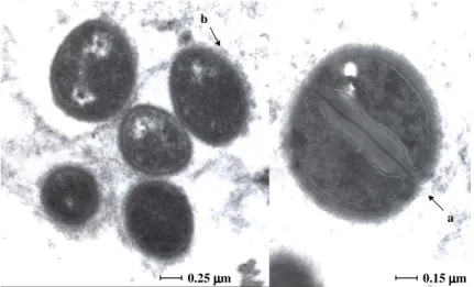
II.6. Electron microscopy (in vitro)……….. 94
II.6.1 Bacteria………... 94
II.6.2 Materials………. 94
II.6.3 Instrumentation………... 95
II.6.4 Quality control……… 95
II.6.5 Method……… 95
II.7. Evaluation of ex vivo activity of compounds studied against
Staphylococcus aureus……… 96
II.7.1 Bacteria……….. 96
II.7.2 Materials………. 96
II.7.3 Instrumentation………... 96
II.7.4 Quality control……… 96
II.7.5 Method……… 96
II.7.5.1 Isolation of Human monocyte-derived macrophages………... 96
II.7.5.2 Phagocytosis assay………... 97
xxix
II.8. Evaluation of ex vivo activity of compounds studied against
Mycobacterium tuberculosis strains……….. 98
II.8.1 Bacteria……….. 98
II.8.2 Materials………. 99
II.8.3 Instrumentation……….. 99
II.8.4 Quality control……… 99
II.8.5 Method……… 99
II.8.5.1 Isolation of Human monocyte-derived macrophages………... 99 II.8.5.2 Phagocytosis and killing activities of M. tuberculosis strains by peripheral
blood monocyte-derived macrophages (PBMDMs) ………... 100
II.8.5.3 QC for method………. 101
II.9. Electron microscopy (ex vivo)……….. 101
II.9.1 Bacteria………... 101
II.9.2 Materials………. 101
II.9.3 Instrumentation………... 102
II.9.4 Method……… 102
II.10. Evaluation of in vivo activity of TZ against Balb/C mice infected with
Mycobacterium tuberculosis………... 103
II.10.1 Bacteria………. 103
II.10.2 Animals………. 103
II.10.2.1 Animal and Human Safety conditions……….. 104
II.10.3 Materials……….. 104
II.10.4 Instrumentation………. 104
II.10.5 Quality control………... 104
II.10.6 Method………... 105
II.10.6.1 Quarantine………. 105
II.10.6.2 Toxicity assays………... 105
II.10.6.3 Infection studies……… 105
Chapter III. Clinical Concentrations of Thioridazine Enhance the Killing of Intracellular Methicillin-resistant Staphylococcus aureus: an In Vivo, Ex Vivo
xxx
Chapter IV. In vitro and ex vivo activity of TZ and its derivatives against
Mycobacterium tuberculosis and M. avium………... 119
IV.1 Clinical concentrations of thioridazine kill intracellular multidrug-resistant
Mycobacterium tuberculosis……… 122
IV.2 In vitro and ex vivo activity of thioridazine derivatives against Mycobacterium
tuberculosis……….. 129
IV.3 The in vitro activity of phenothiazines against Mycobacterium avium:
potential of thioridazine for therapy of the co-infected AIDS patient………. 132
Chapter V. The Curative Activity of Thioridazine on Mice Infected With
Mycobacterium tuberculosis………... 137
Chapter VI. Effect of Carpobrotus edulis, reserpine, ouabain and verapamil on the growth of phagocytosed multidrug-resistant Mycobacterium tuberculosis
and methicillin-resistant Staphylococcus aureus………. 143 VI.1 Inhibition of the Carpobrotus edulis methanol extract on the growth of
phagocytosed multidrug-resistant Mycobacterium tuberculosis and
methicillin-resistant Staphylococcus aureus………... 146
VI.2 Reserpine, ouabain and the calcium channel blocker verapamil, cause
intracellular killing of Staphylococcus aureus………. 148
VI.3 Inhibitors of Ca2+ and K+ Transport Enhance Intracellular Killing of M.
tuberculosis by Non-Killing Macrophages………... 153
Chapter VII. The effect of efflux pumps inhibitors, such as SILA compounds, on macrophages infected with MDR M. tuberculosis. New methods for the
screening of efflux pump activity and efflux pump inhibitors………... 159 VII.1 SILA compound 421, an inhibitor of efflux pumps of cancer cells, enhances
the killing of intracellular MDR-TB……… 162
VII.2 An instrument-free method for the demonstration of efflux pump activity of
xxxi
Chapter VIII. The future of chemotherapy... 177 VIII.1 The future of chemotherapy: From where will new antimicrobial agents
come from?... 180 VIII.2 MDR- and XDR-TB; Rationale for alternative therapy; Recommended
therapy based on the results obtained from Thesis Research………... 190
Chapter IX. The Tuberculosis laboratory of the Future: The role of the macrophage in the selection of agents that can be used for the successful
therapy of an XDR-TB infection………... 197
Chapter X. The Macrophage Model………. 211
X.1 Phenothiazines as Anti-Multi-Drug Resistant Tubercular Agents……… 214 X.2 Enhanced killing of intracellular pathogenic bacteria by phenothiazines and the
role of K+ efflux pumps of the bacterium and the killing macrophage……… 222
Chapter XI. Concluding Remarks and Future Perspectives……….. 235
xxxii
INDEX OF FIGURES
Chapter I. State of the Art
I.1 History and Epidemiology of Tuberculosis (TB)
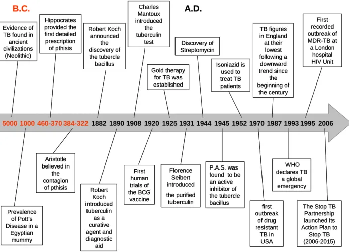
Figure 1. Time-line of Tuberculosis………... 10
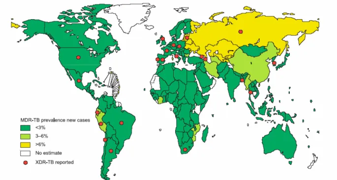
Figure 2. Tuberculosis notification rates (2005)………. 16 Figure 3. Global distribution of MDR-TB rates. Prevalence among new TB cases
(1994-2002) (A); (2004) (B)………... 19
Figure 4. Global distribution of MDR-TB among new cases and countries that
reported XDR-TB………... 21
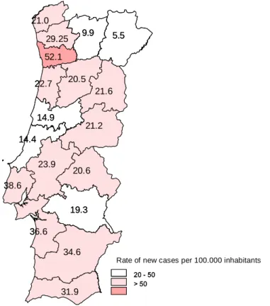
Figure 5. Rate of new cases of TB per district of Portugal………. 24 Figure 6. Molecular structures of phenothiazines………... 45
Chapter III. Clinical Concentrations of Thioridazine Enhance the Killing of Intracellular Methicillin-resistant Staphylococcus aureus: an In Vivo, Ex Vivo and Electron Microscopy Study
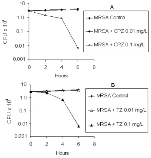
Figure 1. The average effect of chlorpromazine (CPZ) and thioridazine (TZ) on intracellular growth of the 3 MRSA strains tested……….. 113 Figure 2. The in vitro effect of TZ on the ultrastructure of MRSA after 18 hours of
culture……….. 114
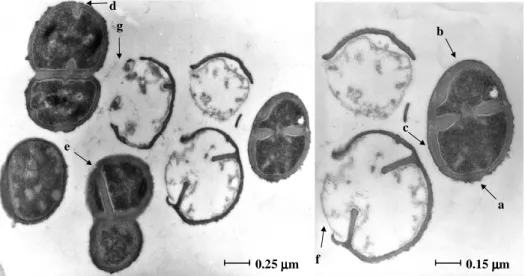
Figure 3. The ultrastructure of MRSA 6 hours after it has been phagocytosed by the
human macrophage (Controls)……… 115
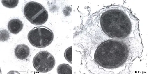
Figure 4. The ultrastructure of MRSA 4 hours after it has been phagocytosed by the human macrophage that had been previously exposed to a concentration of TZ of 0.1
mg/L……… 116
Figure 5. The ultrastructure of MRSA 6 hours after it has been phagocytosed by the human macrophage that had been previously exposed to a concentration of TZ of 0.1
mg/L……… 116
Chapter IV. In vitro and ex vivo activity of TZ and its derivatives against
Mycobacterium tuberculosis and M. avium
IV.1 Clinical concentrations of thioridazine kill intracellular multidrug-resistant Mycobacterium tuberculosis
xxxiii Figure 1. Effects of CPZ and TZ on percent annexin V binding by monocyte subsets
of Ficoll preparations of PBMCs……… 124
Figure 2. Average killing activities of human PBMDMs against M. tuberculosis ATCC 27294 (ATCC), two antibiotic-susceptible strains (strains A and B [TB]), and three MDR M. tuberculosis strains (strains C, D, and E; resistant at least to RIF and
INH [MDR-TB])………. 125
Figure 3. Average killing activities of human PBMDMs against three MDR M. tuberculosis strains (resistant to RIF and INH) (MDR-TB) and three antibiotic-susceptible M. tuberculosis strains (TB) including strain ATCC 27294 when either CPZ (A) or TZ (B) was present in the medium at concentrations of 0.01 and 0.1
mg/L……… 126
IV.2 In vitro and ex vivo activity of thioridazine derivatives against Mycobacterium tuberculosis
Figure 1. Effect of TZ and its derivatives in the killing activity of macrophages
infected with M. tuberculosis H37Rv ATCC27294. ……….. 131
IV.3 The in vitro activity of phenothiazines against Mycobacterium avium: potential of thioridazine for therapy of the co-infected AIDS patient
Figure 1. The in vitro activity of chlorpromazine (CPZ), thioridazine (TZ), promazine (PMZ) and desipramine (DSP) against Mycobacterium avium……… 133
Chapter V. The Curative Activity of Thioridazine on Mice Infected With
Mycobacterium tuberculosis
Figure 1. Effect of daily TZ treatment of mice infected with M. tuberculosis……….. 140
Chapter VI. Effect of Carpobrotus edulis, reserpine, ouabain and verapamil on the growth of phagocytosed multidrug-resistant Mycobacterium tuberculosis and methicillin-resistant Staphylococcus aureus
VI.2 Reserpine, ouabain and the calcium channel blocker verapamil, cause intracellular killing of Staphylococcus aureus
Figure 1. The effect of reserpine, ouabain and verapamil (A) and chlorpromazine and thioridazine (B) on Staphylococcus aureus that have been phagocytosed by human
xxxiv
Figure 2. Model suggested for the killing of intracellular Staphylococcus aureus by
ouabain, reserpine and verapamil……… 152
VI.3 Inhibitors of Ca2+ and K+ Transport Enhance Intracellular Killing of M. tuberculosis by Non-Killing Macrophages
Figure 1. Effect of TZ and other efflux pumps in the killing activity of macrophages
infected with M. tuberculosis……….. 155
Chapter VII. The effect of efflux pumps inhibitors, such as SILA compounds, on macrophages infected with MDR M. tuberculosis. New methods for the screening of efflux pump activity and efflux pump inhibitors
VII.1 SILA compound 421, an inhibitor of efflux pumps of cancer cells, enhances the killing of intracellular MDR-TB
Figure 1. Ex vivo effect of SILA compounds 421 and 409 in human macrophages
infected with XDR-TB……… 163
VII.2 An instrument-free method for the demonstration of efflux pump activity of bacteria
Figure 1. Fluorescence associated with isolated S. aureus colonies grown on agar
containing increasing concentrations of EB……… 168
Figure 2. The minimal concentration of EB that produces fluorescence of S. aureus
ATCC, MRSA COL and MRSA HPV107 MDR phenotype……….. 169
Figure 3. The effect of temperature on the minimal concentration of EB that produced fluorescence of S. aureus ATCC, MRSA COL and MRSA HPV107……… 170 Figure 4. The use of the EB-agar method for the demonstration of different strains of E. coli that differ with respect to the absence and over-expressed AcrAB efflux pump 172 Figure 5. The ability of the method to distinguish the antibiotic-susceptible S. aureus ATCC25923 strain from the antibiotic-resistant MRSA HPV107 strain………... 173
Chapter IX. The Tuberculosis laboratory of the Future: The role of the macrophage in the selection of agents that can be used for the successful therapy of an XDR-TB infection
Demonstration of degrees of killing activity promoted by drugs against S. aureus and M. tuberculosis - Thioridazine: the positive control for the demonstration of enhanced killing of intracellular bacteria
xxxv Figure 1. In vitro effect of TZ on the ultra-structure of MRSA after 16 hours of
culture………. 206
Figure 2. Ex vivo effect of TZ (0.1 mg/L) in phagocytosed S. aureus exposed for 6
hours……… 207
Figure 3. Effect of TZ in the killing activity of macrophages infected with MRSA
(A) and MDR-TB (B)………. 208
Figure 4. Effect of TZ treatment of Balb/C mice infected with M. tuberculosis……… 209
Chapter X. The Macrophage Model
X.2 Enhanced killing of intracellular pathogenic bacteria by phenothiazines and the role of K+ efflux pumps of the bacterium and the killing macrophage
How does a Phenothiazine enhance the killing of intracellular bacteria including mycobacteria?
Figure 1. The ex-vivo effects of Thioridazine on the ultrastructure of Staphylococcus aureus phagocytosed by monocyte derived macrophages……….. 229 The requirement of K+ for intracellular killing
Figure 2. Effects of ouabain, verapamil and reserpine in the killing activity of human
monocyte-derived macrophages………. 230
Figure 3. Hypothetical model suggested for the enhancement of killing of human monocyte-derived macrophages by phenothiazines and other efflux pumps. (A) Infected and untreated macrophage and (B) Infected and treated macrophage……….. 232
xxxvi
INDEX OF TABLES
Chapter II. Materials, Methods and Instrumentation
II.1. Determination of Toxicity of compounds to be evaluated for activity against bacteria under study
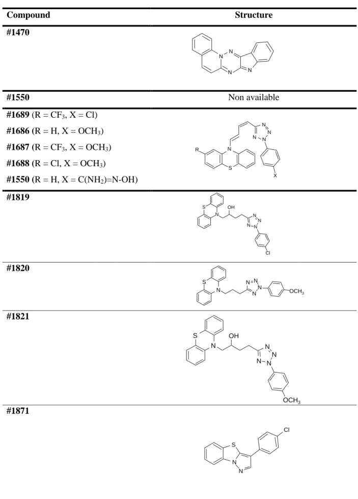
Table 1. Structure of TZ derivatives……….. 74-75
Table 2. Structures of other efflux pump inhibitors………... 76
Chapter III. Clinical Concentrations of Thioridazine Enhance the Killing of Intracellular Methicillin-resistant Staphylococcus aureus: an In Vivo, Ex Vivo and Electron Microscopy Study
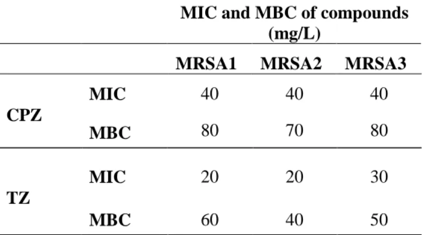
Table 1. MIC and MBC of CPZ and TZ against methicillin-resistant S. aureus
(MRSA) strains……….. 112
Chapter IV. In vitro and ex vivo activity of TZ and its derivatives against
Mycobacterium tuberculosis and M. avium
IV.1 Clinical concentrations of thioridazine kill intracellular multidrug-resistant Mycobacterium tuberculosis
Table 1. MICs and MBCs of CPZ and TZ for antibiotic-sensitive and MDR strains
of M. tuberculosis……….. 123
IV.2 In vitro and ex vivo activity of thioridazine derivatives against Mycobacterium tuberculosis
Table 1. Minimum inbibitory concentration of TZ and its derivatives against
Mycobacterium tuberculosis……….. 129
Table 2. Percent toxicity of TZ and TZ derivatives on human lymphocytes…………. 130
Chapter VI. Effect of Carpobrotus edulis, reserpine, ouabain and verapamil on the growth of phagocytosed multidrug-resistant Mycobacterium tuberculosis and methicillin-resistant Staphylococcus aureus
VI.1 Inhibition of the Carpobrotus edulis methanol extract on the growth of phagocytosed multidrug-resistant Mycobacterium tuberculosis and methicillin-resistant Staphylococcus aureus
xxxvii Table 1. Effects of C. edulis leaves methanolic extract (CELME) on phagocytosed S.
aureus………. 147
Table 2. Effects of C. edulis leaves methanolic extract (CELME) on phagocytosed
M. tuberculosis……….. 147
VI.3 Inhibitors of Ca2+ and K+ Transport Enhance Intracellular Killing of M. tuberculosis by Non-Killing Macrophages
Table 1. Minimum inhibitory concentration of TZ and other efflux pumps inhibitors
against M. tuberculosis H37Rv ATCC27294……… 154
Table 2. Percent toxicity of reserpine, ouabain and verapamil on human
lymphocytes………... 154
Chapter VII. The effect of efflux pumps inhibitors, such as SILA compounds, on macrophages infected with MDR M. tuberculosis. New methods for the screening of efflux pump activity and efflux pump inhibitors
VII.1 SILA compound 421, an inhibitor of efflux pumps of cancer cells, enhances the killing of intracellular MDR-TB
Table 1. The in vitro activity of TZ and SILA compounds 409 and 421 against M.
tuberculosis strains………. 163
VII.2 An instrument-free method for the demonstration of efflux pump activity of bacteria
Table 1. The minimum inhibitory concentration (MIC) of EB against
bacteria………... 167 Table 2. Lowest concentration of EB that produced fluorescence associated with
bacterial cell mass after 24 hours at 37ºC, after 48 hours at 37ºC and after transfer
xxxviii
LIST OF ABREVIATIONS
AFB Acid-fast-bacillus
AhpC Alkyl hydroperoxide reductase subunit C
AIDS Acquired Immune Deficiency Syndrome
AMK Amikacin
AMPLICOR MTB Roche AMPLICOR® Mycobacterium tuberculosis
AS Additional Species
ATCC American Type Cell Culture
ATP Adenosine triphosphate
ATS American Thoracic Society
B.C. Before Christ
BCG Bacille Calmette-Guérin
CaM Calmodulin
CaMKII CaM-dependent protein kinase II
CCCP Carbonyl cyanide m-chlorophenylhydrazone
CDC Centers for Disease Control and Prevention
CELME Carpobrotus edulis leaves methanolic extract
CFP-10 Culture Filtrate Protein-10
CFU Colony Forming Units
CIP Ciprofloxacin
CLSI Clinical Laboratory Standards Institute
CM Common Mycobacteria
CPZ Chlorpromazine
CR Complement Receptors
DGS Direcção Geral de Saúde
DNA Desoxirribonucleic Acid
DOT Directly Observed Treatment
DTH Delayed-Type Hypersensitivity
EB Ethidium Bromide
EEA1 Early Endosome Autoantigen 1
ELISA Enzyme-Linked Immunosorbent Assay
xxxix
EMB Ethambutol
EPIs Efflux Pump Inhibitors
ESAT-6 Early Secretory Antigenic Target-6
EU European Union
Fc Fragment, crystallizable
FDA Food and Drug Administration
G.I. Growth Index
GLC Gas Liquid Chromatography
GTP Guanosine-5'-triphosphate
HBSS Hank’s Balanced Salt Solution
his Histidine
HIV Human Immunodeficiency Virus
HPLC High Performance Liquid Chromatography
hVPS34 PI-specific PI 3-kinase
i.p. Intraperitoneally
i.v. Intravenous
ICL Isocitrate Lyase
IFN Interferon
Ig Immunoglubolin
IL Interleukin
INH Isoniazid
IUATLD International Union Against Tuberculosis and Lung Disease
KAN Kanamycin
kb Kilo-base
LAM Lipoarabinomannan
LPS Lipopolysaccharides
LZD Linezolid
ManLAM Glycosylated phosphatidylinositol LAM
MAP Mitogen-Activated Protein
MATE Multidrug And Toxic Compound Extrusion
MBC Minimum Bactericidal Concentration
MBP Mannose Binding Protein
xl
MDM Monocyte Derived Macrophages
MDR Multi-Drug Resistance
MDR-TB Multi-Drug Resistant Tuberculosis
MH Mueller-Hinton
MHA Mueller-Hinton Agar
MHC Major Histocompatibility Complex
MIC Minimum Inhibitory Concentration
MOTT Mycobacteria Other Than Tuberculosis
MRSA Methicillin-Resistant Staphylococcus aureus
MTD Gen-Probe AMPLIFIEDTM Mycobacterium tuberculosis Direct
NADH Nicotinamide Adenine Dinucleotide (reduced form)
NICE National Institute for Clinical Excellence
NK Natural Killer
NMR Nuclear Magnetic Resonance
NO Nitric Oxide
NOS2 Nitric Oxide Synthase 2
NTM Non-Tuberculous Mycobacteria
O.D. Optical Density
OFX Ofloxacin
OHS Office of Health and Safety
OXA Oxacillin
PAβN β-naphthylamide
PANTA Antimicrobial mixture that contains Polymyxin B, Amphotericin B, Nalidixic acid, Trimethropim and Azlocillin
PAS Para-amino Salicylic acid
PBMC Peripheral Blood Mononuclear Cells
PBMDMs Peripheral Blood Monocyte-Derived Macrophages
PBS Phosphate Buffered Saline
PCR Polymerase Chain Reaction
PI Phosphatidylinositol
PI3P Phosphatidylinositol 3-phosphate
PIM Phosphatidylinositol mannoside
xli
POES Polyoxyethylene stearate
PPD Purified Protein Derivative
PZA Pyrazinamide
QFT QuantiFERON®-TB test
QFT-G QuantiFERON®-TB Gold Test
r.p.m. Rotations per minute
RANTES Regulated upon Activation, Normal T-cell Expressed, and Secreted
RD Region of Difference
RIF Rifampin
RNA Ribonucleic acid
RND Resistance-nodulation-cell division
RNI Reactive Nitrogen Intermediates
RPMI Roswell Park Memorial Institute
rRNA Ribosomal RNA
SapM Secreted acid phosphatase of M. tuberculosis
SDS Sodium Dodecyl Sulfate
SHP-1 Tyrosine phosphatase
SILA Organosilicon compounds
SILA 409 1,3-Dimethyl-1,3-bis(4-fluorophenyl)-1,3-bis(3-morpholino-propyl)-disiloxan-dihydrochlorid
SILA 421 1,3-Dimethyl-1,3-bis(4-fluorophenyl)-1,3-bis{3-[1(4-buthyl-piperazinyl)]-propyl}-disiloxan-tetrahydrochlorid
SNARE Soluble N-ethylmaleimide-sensitive factor-attachment protein receptor
SP-A Surfactant Protein A
STR Streptomycin
TACO Tryptophan aspartate rich coat protein
TB Tuberculosis
TCH Thiophene-2-carboxylic acid hidrazide
TEM Transmission electron microscopy
TET Tetracycline
TGFβ Transforming growth factor beta
xlii
TLRs Toll-like receptors
TNF Tumour necrosis factor
TSA Trypticase Soy Agar
TSB Tryptic Soy Broth
TST Tuberculin skin test
TZ Thioridazine
UNICEF United Nations Children's Fund
US United States
UV Ultraviolet
V-ATPase Vacuolar-type proton ATPase
WHO World Health Organization
XDR-TB Extensively-Drug Resistant Tuberculosis XXDR-TB Extremely-Drug Resistant Tuberculosis
γδ T cells Gamma-delta T cells
LIST OF UNITS
G Gravitacional force
Kg Kilograms; g - grams (10-3Kg)
L Liter; mL - mililiter (10-3L), µL - microliter (10-6L)
3
THESIS OUTLINE AND OBJECTIVES
This Thesis includes the in vitro, ex vivo and in vivo studies of TZ against M. tuberculosis strains resistant to antibiotics. It is the intent to provide data about new anti-tubercular compounds and novel approaches that could be used for the treatment of MDR-TB and XDR-TB infections. The general aims of this thesis are:
• Screening of thioridazine derivatives obtained by chemical manipulation for toxicity and mutagenicity;
• Determination of the activity of the selected derivatives on antibiotic resistant and susceptible M. tuberculosis in vitro;
• Determination of the activity of derivatives against M. tuberculosis that has been phagocytosed by the human macrophage, the amount of derivative that is concentrated by the phagocytic cell and if it is altered by the human macrophage;
• Determination of selected thioridazine derivatives to clear the mouse pulmonary tree from TB infection and to prevent the infection of the mouse by MDR-TB.
The organization of this Thesis is in accordance to the Bologna Agreement Guidelines which employs as a basis the publications that have resulted from the Thesis research. The sections of the Thesis have been organized so that each one can stand on its own as a full report that refers to the methods used in that section, the results, its discussion and conclusions as well as all references cited. The sequence of Result sections has been designed so that the reader can be led from discovery to discovery in a developmental manner. However, this sequence does not reflect the chronological order of the work done since many of the experiments were conducted while some other experiments were “cooking” (mycobacteria do grow rather slowly).
Introduction
4
This thesis is divided into 11 chapters, as follows:
Chapter I. State of the Art. This section provides a review of the current knowledge on
Tuberculosis and the emergence of MDR and XDR resistance. The issues focused on this Thesis were put into context to highlight the relevance of the studies presented.
Chapter II. Materials and Methods. This section describes all the methodology used
during the work performed in these studies.
Chapter III. The in vitro and ex vivo effects of clinical concentrations of TZ against
Methicillin-resistant Staphylococcus aureus are described. In vitro, ex vivo and electron microscopy studies were conducted.
Chapter IV. This section presents all the results obtained from the in vitro and ex vivo
activity of TZ and its derivatives against Mycobacterium tuberculosis. It also describes the in vitro effects of several phenothiazines tested against M. avium.
Chapter V. Describes the results of the curative activity of TZ on mice infected with
Mycobacterium tuberculosis. Animal studies were conducted using TZ for the treatment of the infection.
Chapter VI. This chapter focuses on the effect that other efflux pumps inhibitors such
as, the methanolic extract of Carpobrotus edulis, reserpine, ouabain and verapamil have on the growth of phagocytosed multidrug-resistant M. tuberculosis and methicillin-resistant S. aureus.
Chapter VII. This chapter provides data on new patented and synthesized SILA
compounds and their effect on infected human monocyte-derived macrophage, as a special example relationship between efflux inhibitors and its effect on macrophage infected with MDR M. tuberculosis strain. In this chapter a new and innovative method used to screen efflux pump activity and efflux pumps inhibitors is also described.
5
Chapter VIII. The future of chemotherapy is the main subject presented. The discovery
of new anti-microbial agents that can be used in the therapy of MDR bacteria as well as a new approach that uses these agents in conjunction with the classical antibiotics will be analysed and discussed.
Chapter IX. The Tuberculosis laboratory of the Future. In this section it will be
discussed the role of the macrophage in the selection of agents that can be employed in the therapy of an MDR infection.
Chapter X. From all the studies performed a hypothetical model (macrophage model)
was developed in order to explain the enhancement of the killing activity of the infected macrophages when TZ, its derivatives or other inhibitors of Ca2+ and K+ transport are added to the cell cultures. The Macrophage Model will be described in detail to clarify the intracellular processes that take place inside the infected and treated macrophage.
Chapter XI. Concluding Remarks and Future Perspectives. This section highlights the
main conclusions obtained in each of the previous chapters as well as future recommendations and guidelines to be developed and implemented in order to treat MDR- and XDR-TB.
CHAPTER I.
9
I.1 History and Epidemiology of Tuberculosis (TB)
Tuberculosis (TB) continues to be considered the major cause for worldwide morbidity and mortality caused by an infective organism. This disease is caused by the steadfast human pathogen Mycobacterium tuberculosis, the bacterium known to infect the man since the birth of civilization (Gernaey et al., 2001; Morell, 1994). The M. tuberculosis complex presently includes: M. tuberculosis, M. africanum, M. bovis, M. microti, M. pinnipedii, M. caprae, and M. canettii (Ernst et al., 2007). However, in some studies, M. canettii is not considered in this complex (Gutierrez et al., 2005). This difference is due to the molecular approaches employed in the strains identification (difference in the number of genes analysed, etc.). The development of new molecular genetics tools and the sequencing of the genome of several M. tuberculosis strains allow a more accurate estimation of the time of origin of mycobacteria (Gutierrez et al., 2005). It is expected that all the members of the M. tuberculosis complex had a common African ancestor about 35,000-15,000 years ago (Daniel, 2006), since an early progenitor of M. tuberculosis was discovered to be present in East Africa as early as 3 million years ago (Gutierrez MC et al., 2005).
M. tuberculosis infections can involve any organ or tissue of the human body (Hopewell, 1995; Lillebaek et al., 2002). Because of this extensive involvement, it has been possible to determine that this infection has been with man since the dawn of civilisation (Zink et al., 2003) as evident from typical skeletal abnormalities produced by TB that have been found in Egyptian mummies. The frequency of skeletons with apparent tubercular deformities and use of DNA amplification of M. tuberculosis obtained from tissues suggests that the disease was common among that population (Nerlich et al., 1997). In recorded history, Assyrian clay tablets describe patients coughing blood in the 7th century B.C. In classical Greece, Hippocrates writes of patients with phthisis (the Greek term for TB), which means consumption, i.e., wasting away associated with chest pain and coughing, frequently with blood in the sputum. Hippocrates thought the disease was largely inherited, while Aristotle (4th century B.C.) stressed its contagious nature, as did Galen, in the 2nd century A.D. (Sakula, 1983). By this time, the frequency of descriptions of patients with TB-like symptoms indicates that the disease was already well entrenched. Figure 1 describes some of the more representative events that took place during the history of TB.
Chapter I
10
Figure 1. Time-line of TB. Some of the most important events that occurred in the history of TB until the present time (sources: Daniel, 2006; Di Perri and Bonora, 2004; Dietrich et al., 2006; Murray, 2004; Myers, 1967; Nerlich et al., 1997; Sakula, 1983) .
In the 19th century TB was well established in East Africa by the time Europeans reached that area. Analysis of human phenotypes, like lactose tolerance, that is associated with the raising of cattle and selection for the ability to utilize milk, as well as the resulting exposure to M. tuberculosis suggests that Indo-Europeans cattle herders that entered East Africa spread the disease to Europe and Asia during their migrations into these regions (Smith, 2003). In studies recently published (Ernst et al., 2007), this theory is not supported and the transmission of the disease from cattle to human is considered to be impossible. However, this is not a consensual subject since some studies based on genetic analysis of the strains refute this belief. The actual representatives of the common ancestor of the M. tuberculosis complex include the ancestral and modern strains of M. tuberculosis. The latter are characterized by the deletion of a 2.1-kb fragment termed TbD1, account for most current TB cases, and include the Beijing family of strains that are prevalent in Asia and have caused
5000 1000 460-370 384-322 1882 1890 1908 B.C. A.D. 1920 1925 1931 Evidence of TB found in ancient civilizations (Neolithic) Prevalence of Pott’s Disease in a Egyptian mummy Robert Koch announced the discovery of the tubercle bacillus Hippocrates provided the first detailed prescription of pthisis 1944 1945 1952 First human trials of the BCG vaccine Florence Seibert introduced the purified tuberculin P.A.S. was found to be an active inhibitor of the tubercle bacillus Aristotle believed in the contagion of pthisis Robert Koch introduced tuberculin as a curative agent and diagnostic aid Charles Mantoux introduced the tuberculin test Gold therapy for TB was established Discovery of Streptomycin Isoniazid is used to treat TB patients 1970 first outbreak of drug resistant TB in USA 1987 TB figures in England at their lowest following a downward trend since the beginning of the century 1993 WHO declares TB a global emergency 1995 First recorded outbreak of MDR-TB at a London hospital HIV Unit 2006 The Stop TB Partnership launched its Action Plan to Stop TB (2006-2015) 5000 1000 460-370 384-322 1882 1890 1908 B.C. A.D. 1920 1925 1931 Evidence of TB found in ancient civilizations (Neolithic) Prevalence of Pott’s Disease in a Egyptian mummy Robert Koch announced the discovery of the tubercle bacillus Hippocrates provided the first detailed prescription of pthisis 1944 1945 1952 First human trials of the BCG vaccine Florence Seibert introduced the purified tuberculin P.A.S. was found to be an active inhibitor of the tubercle bacillus Aristotle believed in the contagion of pthisis Robert Koch introduced tuberculin as a curative agent and diagnostic aid Charles Mantoux introduced the tuberculin test Gold therapy for TB was established Discovery of Streptomycin Isoniazid is used to treat TB patients 1970 first outbreak of drug resistant TB in USA 1987 TB figures in England at their lowest following a downward trend since the beginning of the century 1993 WHO declares TB a global emergency 1995 First recorded outbreak of MDR-TB at a London hospital HIV Unit 2006 The Stop TB Partnership launched its Action Plan to Stop TB (2006-2015)
11 outbreaks worldwide. A separate lineage, marked by deletion of region of difference 9 (RD9), diverged from an ancestral M. tuberculosis and sequentially gave rise to M. africanum, M. microti, M. pinnipedii, M. caprae, and M. bovis. It is thought that M. bovis, which has the greatest number of deletions, is the most recent member of this lineage. Based on these results the popular belief that M. tuberculosis evolved from M. bovis through the adaptation of a bovine strain to the human host is clearly refuted. Since M. tuberculosis contains chromosomal segments that have been deleted from M. bovis, M. tuberculosis could not have evolved from M. bovis (Ernst et al., 2007). Based on these new studies and genetic approaches the manner of transmission between humans and cattle is still not completely clarified.
I.1.1 Evolution of TB in Europe and diagnosis of infection
As Europe entered the middle ages, the written record of TB is almost non-existent. However, this does not mean that the disease was not present. In modern times, TB reached epidemic proportions in Europe and North America during the 18th and 19th centuries. The understanding of the pathogenesis of TB began with the work of Theóphile Laennec in the 19th century. He clearly elucidated the pathogenesis of TB and unified the concept of this disease. At that time, TB surged across Europe with death rates in London, Stockholm and Hamburg that approached 800-1000 cases/100,000 inhabitants/year (Daniel, 2006). The demonstration of the transmissibility of M. tuberculosis infection was accomplished by Jean-Antoine Villemin in 1865. He inoculated a rabbit with a purulent liquid obtained from an individual who had died of TB (Murray, 2004). Although the animal remained healthy, it was found to have extensive TB when sacrificed and autopsied (Daniel, 2006). This was an important achievement; however, the history of TB was markedly changed in 1882, with Robert Koch's discovery of the tubercle bacillus. In his presentation “Die Aetiologie der Tuberculose”, Koch identified the tubercle bacillus as the etiologic agent and presented his famous postulates, which continue to set the standard for the demonstration of infectious etiology (Kaufmann and Schaible, 2005). In 1890, Koch started the isolation of tuberculin (Myers, 1967). The tuberculin skin test was developed in 1907 by Clemens von Pirquet and 3 years later used to demonstrate latent infection in asymptomatic children. Charles Mantoux introduced the use of a cannulated needle and syringe to inject tuberculin intra-cutaneously in 1908, and Florence Seibert developed