Full paper published online: February 28, 2010 ISSN 1678-9199.
Hemolytic toxin from the soft coral Sarcophyton trocheliophorum: isolation
and physiological characterization
Karthikayalu S (1, 2, 3), Rama V (2), Kirubagaran R (1), Venkatesan R (1)
(1) National Institute of Ocean Technology, Chennai, Tamil Nadu, India; (2) Department of Industrial Biotechnology, Dr. M.G.R. University, Maduravoyal, Chennai, Tamil Nadu, India; (3) AxioGen Biotech Private Limited, Pondicherry, India.
ABSTRACT: The unifying characteristic of cnidarians is the production of protein and polypeptide toxins. The present study describes the identification of a hemolytic toxin from the soft coral Sarcophyton trocheliophorum. The crude extract was highly cytotoxic (EC50 = 50 ng/mL) against human erythrocytes. It was also tested for
hemolytic activity by the blood agar plate method, resulting in a hemolytic halo of 12 mm with 50 µg of protein. The stability of the venom under different physiological conditions was analyzed. The venom hemolytic activity was augmented by alkaline and neutral pH whereas it was reduced in acidic pH. The activity was stable up to 60°C. The hemolytic activity was completely abolished by the addition of serum and reduced significantly during frequent freezing-thawing cycles. Toxin purification was performed by ammonium sulfate precipitation and subsequently desalted by dialysis against 10 mM sodium phosphate buffer (pH 7.2), followed by anion exchange chromatography on DEAE cellulose column and gel filtration chromatography using Sephadex G-50 matrix. The purified active fractions possessed a prominent protein of approximately 45 kDa, as revealed by SDS-PAGE.
KEY WORDS: Sarcophyton trocheliophorum, cnidarian toxin, cytolysin.
CONFLICTS OF INTEREST: There is no conflict.
FINANCIAL SOURCE: Ministry of Earth Sciences (MoES), Government of India.
CORRESPONDENCE TO:
KARTHIKAYALU SUBBRAYALU, Department of Industrial Biotechnology, Dr. M.G.R.
Educational and Research Institute, Dr. M.G.R. University, Maduraoyal, Chennai
600095, India. Phone: +91 44 23782176. Fax: +91 44 23783165. Email:
INTRODUCTION
The phylum Cnidaria includes the following benthic and pelagic animal classes:
anthozoa, scyphozoa, cubozoa and hydrozoa. They are diploblastic (ectoderm and
endoderm) in their cellular organization with a homogeneous elastic material
(mesoglea) between these two layers. Soft corals (anthozoa) are found worldwide in
tropical environments. They are sessile, colonial forms having soft, fleshy and fragile
tissues without any physical defense capability against their potential predators.
However, they can survive in such competitive environments because of their
chemical defense strategy (1).
The soft coral Sarcophyton trocheliophorum (common name: toadstool coral or
leather coral) resembles a toadstool and presents a fine carpet of polyps, a
light-brown color and is found predominantly in the shallow tropical waters. Its buccal
polyps are covered by a ring of tentacles. The tentacles are armed with nematocysts.
S. trocheliophorum was also covered with a layer of mucus that functions as an
energy carrier (2). When stressed the coral release toxins that get trapped in the
mucus and function as first line of defense. Moreover, toxin production, a common
feature of cnidarians, allows them to produce a variety of peptides and proteins that
act as either neurotoxins or cytolysins (3-6). Toxins are produced by specialized
stinging cells, the nematocysts. These nematocysts contain fine harpoon-like
microscopic structures (cnida) that penetrate the surface layer of the victim and
deliver a mixture of highly toxic substances. The role of toxins includes the capturing
and killing of prey as well as digestion and protection from predators.
The toxins from sea anemones are better characterized than the other groups of
cnidarians. Despite the belief that all cnidarians are venomous, the extant
biochemical and functional studies have focused only on a few groups and very little
research has been done on the toxicology of soft corals. But soft coral stinging
nematocysts contain active proteinaceous venoms, while the extra-nematocyst
tissues possess many other biologically and pharmacologically active metabolites
(1-7). Hence, the present study was undertaken to identify the cytolytic toxins from the
soft coral Sarcophyton trocheliophorum. Furthermore, the study expands on the
MATERIALS AND METHODS
Collection of Soft Coral Sarcophyton trocheliophorum
Soft coral S. trocheliophorum was collected from Chidiatappu (latitude N: 11o29’493”;
longitude E: 92o42’483”), South Andaman region of Andaman Islands, India. The soft
coral was collected by scuba diving at a depth of 5 m.
Toxin Extraction
The live coral was subjected to osmotic thermal stress, by spraying warm distilled
water at 45°C, to induce the secretion of toxin (8). The soft coral was always covered
with thin layer of mucus. Hence, the secreted toxin and the mucus mixed together to
form a thick slimy layer on the coral surface. The mucus layer was filtered through a
sieve and stored in liquid nitrogen during transportation and at –70°C in the
laboratory. When required, the aliquots were thawed and concentrated by
lyophilization and reconstituted in phosphate buffered saline (PBS, pH 7.4).
Protein Assay
Concentrations of the proteins were determined by the method of Lowry et al. (9)
using bovine serum albumin (BSA) as standard. The concentrations of proteins
during purification studies were measured at 280 nm. Purified fractions of 100 μL
were read at 280 nm using a microtiter plate reader (Molecular Devices, USA).
Ammonium Sulfate Precipitation
The crude extract was treated with ammonium sulfate (Hi-media Labs, India) to 100%
saturation as per Rosenberg Table (10). The mixture was stirred for 30 minutes at
4°C and later centrifuged at 10,000 x g for ten minutes at 4°C. The precipitate was
resuspended in 10 mM sodium phosphate buffer (pH 7.2) and desalted by dialysis
through a dialysing tube with a cut-off at 12,000-14,000 Da (Hi-media Labs, India)
against 10 mM sodium phosphate buffer at pH 7.2.
Hemolytic Assay
Hemolytic activity was measured as the attenuation of human red blood cells at
ambient room temperature using a microtiter plate reader (Molecular Devices, USA).
Freshly collected human blood with heparin was centrifuged to remove the buffy
and stored at 4°C. Desired concentrations of protein solution were added in the first
well to erythrocyte buffer (140 mM NaCl, 10 mM Tris-HCl, pH 7.4), and then serially
diluted 2-fold with erythrocyte buffer. RBCs (100 μL; D630 = 0.5) in erythrocyte buffer
were added to the protein solution. The final volume in all wells was 200 μL.
Hemolysis was monitored by measuring attenuation at 630 nm for 20 minutes at
room temperature. The percentage of hemolysis was determined at the end of the
assay using the following equation of Maček et al. (11):
Hemolysis (%) = (Dmax – Dobs)/(Dmax – Dmin) x 100
in which Dobs is the attenuation measured in the well after 20 minutes and Dmax is the
maximum attenuation by distilled water and Dmin is the minimal attenuation by
erythrocyte buffer.
Hemolytic Activity on Human Blood Agar Plate
Human blood agar plate was prepared by adding 5 mL of human blood to 95 mL of
sterile nutrient agar aseptically, with the result poured immediately onto the Petri
dishes. After solidification, wells were cut into the agar plate-using a corkscrew borer
(8 mm diameter). Wells were loaded with 50 μL (1 mg/mL) of protein samples. The
plates were observed for hemolysis after overnight incubation at room temperature.
Characterization of the Hemolytic Fraction
The crude extract was characterized with respect to thermal and pH stability, stability
during freezing-thawing cycles and treatment with human serum.
Effect of temperature
In order to study the effect of temperature on hemolytic activity, the extracted
proteins were exposed to various heat treatments: 25°C (room temperature), 37°C
for one hour, 60°C for one hour, 80°C for one hour, 100°C for one hour and by
autoclaving at 121°C and 15 psi for 15 minutes.
Effect of freezing-thawing procedure
The extract was frozen at –80°C and then thawed quickly to 37°C. The
freezing-thawing procedure was repeated three times and the hemolytic activity was
Effect of pH
The extract buffer was adjusted to pH 3, 4, 5, 6, 7, 8 and 9 with hydrochloric acid
(HCl), incubated for 1 h at room temperature and assayed.
Effect of human serum
Human serum was added to the extract at a final concentration of 1% and incubated
at room temperature for one hour. The mixtures were assessed for hemolytic activity
against human RBCs, with serum-free extract as the control.
Purification of Hemolytic Proteins by Ion Exchange Chromatography
The dialyzed fractions were purified by ion exchange chromatography.
Anion-exchange chromatography was performed using DEAE cellulose column (1 x 5 cm)
at a flow rate of 60 mL/hour (10). The column-stabilizing buffer was 10mM sodium
phosphate, pH 7. One milliliter of extracted proteins in the stabilizing buffer was
loaded on the column (10 mg/mL). Elution of the bound proteins was done by using a
linear gradient of sodium chloride (0.1 M, 0.25 M, 0.5 M, 1 M and 1.25 M) in
stabilizing buffer. The protein concentrations in the collected fractions were
measured at 280 nm by using a microtiter plate reader. All the fractions were tested
for hemolytic activity and fractions showing hemolytic activity were pooled together,
lyophilized and dialyzed overnight against 10 mM Tris buffer, pH 7.0 at 4°C.
Gel Filtration Chromatography
The dialyzed fractions were subjected to gel filtration chromatography in a Sephadex
G-50 column (1.5 x 60 cm) pre-equilibrated with 10 mM Tris-HCl; pH 7.0 at a flow
rate of 40 mL/hour (10). One milliliter (25 mg/mL) of anion-exchange chromatography
proteins purified in the running buffer was loaded on the column. Chromatography
was performed at 4°C. The fractions collected were tested for hemolytic activity and
the fractions showing activity were pooled, lyophilized and analyzed by SDS-PAGE.
SDS-PAGE
Purified protein was analyzed by SDS-PAGE (12). SDS-PAGE was performed using
5% stacking gel and 10% resolving gels. Samples were denatured by boiling in
Following electrophoresis at 30 mA for four hours, gels were stained by silver
staining (10).
RESULTS
Toxin production by cnidarians is an important aspect of their defense. In the present
investigation we report the presence of hemolytic toxin for the first time from the soft
coral S. trocheliophorum. The live coral was stressed to secrete toxin and the
secreted toxin got trapped in the mucus. Moreover, corals slough off large amounts
of mucus when stressed (13). Then 450 mL of mucus containing the toxin was
collected. The protein concentration was 0.46 mg/mL in the extracted fraction and 30
mg/mL in the ammonium sulfate-precipitated fraction.
Hemolytic activity of crude extract is shown in Figure 1. The hemolysis induced by
hemolysin in red blood cells was concentration-dependent and 50% hemolysis was
observed at a concentration of ~50 ng/mL (Figure 1). The hemolytic activity was
further confirmed on blood agar plate, while 50 µg of crude extract produced a
hemolytic halo 12 mm in diameter (Figure 2).
Figure 1. Titration of hemolytic activity. Hemolysis was measured turbidimetrically at
room temperature using a microplate reader. The percentage of hemolysis was
calculated as described in the Materials and Methods section. Data points shown are
Figure 2. Hemolytic activity of the crude extract on blood agar plate.
C: control (PBS buffer, pH 7.4), T: crude extract.
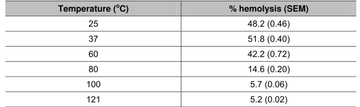
Although the hemolytic assay of the extract provided an excellent response, it was
dependent on temperature (Table 1) and pH (Table 2). Hemolytic activity peaked at
room temperature (25°C) and 37°C. The activity was partially reduced at 60°C and
no activity was observed when the crude extract was treated at 80°C, 100°C and
autoclaved (Table 1), indicating that the toxin responsible for hemolytic effect is heat
labile. Hemolysis was favored by alkaline and neutral pH (Table 2) whereas this
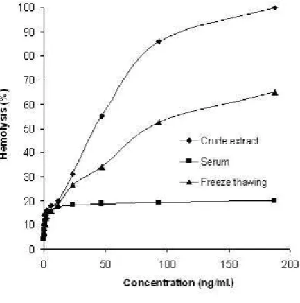
activity was reduced in acidic medium (Table 2). When 1% human serum was added
to the extract, the hemolytic activity on human RBCs was neutralized (Figure 3).
Activity was reduced by one-third during frequent freezing-thawing cycles, indicating
the unstable nature of the toxin (Figure 3).
Table 1. Effect of temperature on the hemolytic activity of the crude extract
Temperature (oC) % hemolysis (SEM)
25 48.2 (0.46)
37 51.8 (0.40)
60 42.2 (0.72)
80 14.6 (0.20)
100 5.7 (0.06)
121 5.2 (0.02)
Table 2. Effect of pH on the hemolytic activity of the crude extract
pH % hemolysis (SEM)
3 –
4 35.2 (1.90)
5 46.1 (0.55)
6 52.6 (0.20)
7 58.4 (0.15)
8 58.8 (0.10)
9 58.6 (0.17)
At pH 3 RBCs were lysed due to very low pH, hence effects of pH cannot be calculated at pH 3. Data points shown are means of three independent experiments (triplicates).
Figure 3. Effects of serum and freezing-thawing on hemolytic activity.
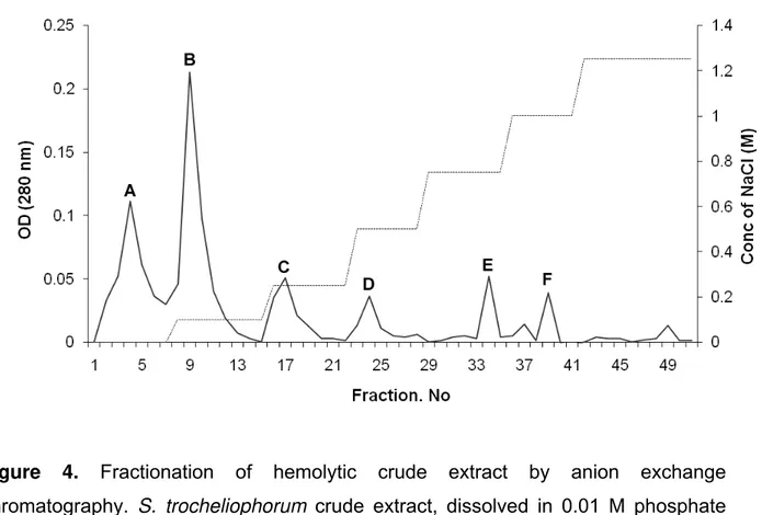
The crude extract of S. trocheliophorum was fractionated by anion exchange
chromatography. Six peaks, labeled A, B, C, D, E and F were obtained by
DEAE-Cellulose column chromatography (Figure 4). Peak A exhibited hemolytic activity and
the fractions under peak A were pooled, lyophilized and dialyzed against 10mM
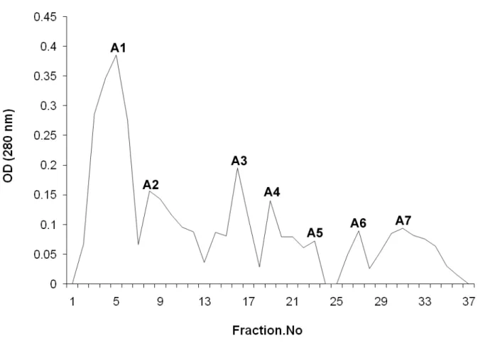
using Sephadex G-50 produced seven peaks (A1, A2, A3, A4, A5, A6 and A7). The
highest peak, A1, showed hemolytic activity (Figure 5). SDS-PAGE analysis of the
fractions under the peak A1 displayed a prominent protein band with a molecular
weight of approximately 45 kDa (Figure 6). Due to the low yield and unstable nature
of the purified toxin, the activity level (EC50) and yield of the pure protein could not be
calculated.
Figure 4. Fractionation of hemolytic crude extract by anion exchange
chromatography. S. trocheliophorum crude extract, dissolved in 0.01 M phosphate
buffer (pH 7.0), was fractionated by DEAE cellulose anion-exchange chromatography
using a flow rate of 1 mL/minute. Five-milliliter fractions were collected and fractions
Figure 5. The fractions under A in the anion-exchange chromatography were pooled
and fractionated by gel filtration chromatography at a flow rate of 40 mL/hour.
Five-millimeter fractions were collected. Fractions under peak A1 exhibited hemolytic
Figure 6. SDS-PAGE analysis of purified fractions.
M: standard protein molecular weight marker (Fermentas, Canada). Lane 1: pooled active fractions
from gel filtration chromatography.
DISCUSSION
Cnidarians, in general, are venomous and contain one or more cytolytic and
neurotoxic peptides or proteins. Cytolysins, ranging in size from short peptides (5-8
kDa) to larger proteins (98 kDa), were reported from different species of cnidarians
(5). Based on the size, 30-45 kDa toxins were classified into a group of cytolysins (5)
and were detected in toxin preparations obtained from sea anemones, jelly fishes,
soft corals, sea fans and sea pens (5, 14-17). Hemolytic toxins of 30, 31, 32.5 and 35
kDa were detected from the different species of the fire coral Millepora (class:
Hydrozoa) (18-20). Venoms isolated from the red sea soft corals Nephthea sp.,
toxins (7). Nematocyst venom with phospholipase A2 activity was reported from the
tissue homogenates of the soft corals Alcyonium digitatum, Sinularia flexibilis,
Sarcophyton elegans and Dendronephthya sp. (17). The hemolytic toxin from the sea
anemone Aiptasia pallida exists in two isoforms α and β of 45 and 43 kDa,
respectively (15, 16). In this study, SDS-PAGE analysis of the purified fractions
showed a prominent protein band of ~45 kDa. Based on this weight it is believed that
this toxin may also belong to the group of cytolysins of 30 to 45 kDa.
This paper reports the presence of hemolytic toxin from the soft coral S.
trocheliophorum, and is the first work to identify peptide toxin from this species. The
cytolytic effect has been assessed using human erythrocytes. Interestingly, human
serum exerts a total inhibitory effect on the cytolytic activity of the toxin. The
elimination of hemolytic activity by the serum is probably due to the antagonistic
effects of serum proteins. Significant reduction in the hemolytic activity was observed
during storage even at –70°C, thus indicating the unstable nature of the hemolytic
protein. The result of freezing-thawing experiments also confirms the unstable nature
of the toxin. Based on the experiment data it is evident that the soft coral S.
trocheliophorum secretes cytolytic toxin when stressed. The toxin is cytolytic in
nature and is used for the host defense. Moreover, mucus secretion, a characteristic
of corals, forms a coating over the polyps and is implicated in a number of
physiological functions. Given that coral mucus serves as an energy carrier, it is rich
in nutrients and diverse in bacterial populations. It has been hypothesized that the
microbial communities found on the coral surface may also play a role in coral
defense. Hence, further investigation on the identification and characterization of the
mucus-associated bacteria would be helpful in understanding the role of the toxins in
REFERENCES
1. Changyun W, Haiyan L, Changlun S, Yanan W, Liang L, Huashi G. Chemical
defensive substances of soft corals and gorgonians. Acta Ecol Sin.
2008;28(5):2320-8.
2. Wild C, Huettel M, Klueter A, Kremb SG, Rasheed MY, Jørgensen BB. Coral
mucus functions as an energy carrier and particle trap in the reef ecosystem. Nature.
2004;428(6978):66-70.
3. Turk T. Cytolytic toxins from sea anemones. Toxin Rev. 1991;10(3):223-62.
4. Maček P. Polypeptide cytolytic toxins from sea anemones (Actiniaria). FEMS
Microbiol Immunol. 1992;5(1-3):121-30.
5. Anderluh G, Maček P. Cytolytic peptide and protein toxins from sea anemones
(Anthozoa: Actiniaria). Toxicon. 2002;40(2):111-24.
6. Honma T, Shiomi K. Peptide toxins in sea anemones: structural and functional
aspects. Mar Biotechnol. 2006;8(1):1-10.
7. Radwan FFY, Aboul-Dahab HM, Burnett JW. Some toxicological characteristics of
three venomous soft corals from the Red Sea. Comp Biochem Physiol C.
2002;132(1):25-35.
8. Wang Y, Chua KL, Khoo HE. A new cytolysin from the sea anemone Heteractis
magnifica: isolation, cDNA cloning and functional expression. Biochim Biophys Acta.
2000;1478(1):9-18.
9. Lowry OH, Rosebrough NJ, Farr AL, Randall RJ. Protein measurement with the
folin phenol reagent. J Biol Chem. 1951;193(1):265-75.
10. Bollag DM, Rozycki MD, Edelstein SJ. Protein methods. New York: Willey-Liss
Inc.; 1996. p. 72-7.
11. Maček P, Malovrh P, Barlic A, Podlesek Z, Menestrina G, Anderluh G. Structure:
function studies of tryptophan mutants of equinatoxin II, a sea anemone pore-forming
protein. Biochem J. 2000;346(1):223-32.
12. Laemmli UK. Cleavage of structural proteins during the assembly of the head of
bacteriophage T4. Nature. 1970;227(5259):680-85.
13. Castro P, Huber M. Coral reefs. In: Marine Biology. 5th ed. Boston: McGraw-Hill
14. Cline EI, Weibe LI, Young JD, George R, Samuel D. Isolation and
characterization of a novel cardiac stimulatory and haemolytic protein from the sea
anemone Urticina piscivora (Sebens and Laakso). Pharm Sci. 1995;1:155-62.
15. Grotendorst GR, Hessinger DA. Purification and partial characterization of the
phospholipase A2 and co-lytic factor from sea anemone (Aiptasia pallida) nematocyst
venom. Toxicon. 1999;37(12):1779-96.
16. Grotendorst GR, Hessinger DA. Enzymatic characterization of the major
phoshoplipase A2 component of sea anemone (Aiptasia pallida) nematocyst venom.
Toxicon. 2000;38(7):931-43.
17. Nevalainen TJ, Peuravuori HJ, Quinn RJ, Llewellyn LE, Benzie JA, Fenner PJ,
Winkel KD. Phospholipase A2 in Cnidaria. Comp Biochem Physiol Part B.
2004;139(4):731-5.
18. Radwan FFY. Comparative toxinological and immunological studies on the
nematocyst venoms of the Red Sea fire corals Millepora dichotoma and M.
platyphylla. Comp Biochem Physiol C Toxicol Pharmacol. 2002;131(3):323-34.
19. Radwan FFY, Aboul-Dahab HM. Milleporin-1, a new phospholipase A2 active
protein from the fire coral Millepora platyphylla nematocysts. Comp Biochem Physiol
C Toxicol Pharmacol. 2005;139(4):267-72.
20. Ibarra-Alvarado C, Alejandro García J, Aguilar MB, Rojas A, Falcón A, Heimer de
la Cotera EP. Biochemical and pharmacological characterization of toxins obtained
from the fire coral Millepora complanata. Comp Biochem Physiol C Toxicol
Pharmacol. 2007;146(4):511-8.