R E S E A R C H
Open Access
Cytolytic and systemic toxic effects induced
by the aqueous extract of the fire coral
Millepora
alcicornis
collected in the Mexican Caribbean
and detection of two types of cytolisins
Rosalina Hernández-Matehuala
1,2, Alejandra Rojas-Molina
2*, Alma Angelica Vuelvas-Solórzano
1,2,
Alejandro Garcia-Arredondo
2, Cesar Ibarra Alvarado
2, Norma Olguín-López
2and Manuel Aguilar
3Abstract
Background:Millepora alcicornisis a branching hydrocoral common throughout the Caribbean Sea. Like other members of this genus, this species is capable of inducing skin eruptions and blisters with severe pain after contact. In the present study, we investigated the toxicity of theM. alcicornisaqueous extract on several animal models. Considering that some cnidarian hemolysins have been associated to local tissue damage, since they also induce lysis of other cell types, we also made a partial characterization of the hemolytic activity ofM. alcicornisaqueous extract. This information is important for understanding the defense mechanisms of the“fire corals”.
Methods:The effects of pH, temperature, and some divalent cations on the hemolytic activity of the extract were assayed, followed by a zymogram analysis to detect the cytolysins and determine their approximate molecular weight. The toxicity of the aqueous extract was assayed in mice, by intravenous administration, and histopathological changes on several tissues were analyzed by light microscopy. The toxicity of the extract was also tested inArtemia salina nauplii, and the damages caused on the crustaceans were analyzed by transmission and scanning electron microscopy. Results:The hemolytic activity of the hydrocoral extract was enhanced in the presence of Ca2+ (≥2 mM), Mg2+(≥6 mM), and Ba2+(≥0.1 mM); however, it was reduced in the presence of Cu2+(≥0.1 mM), Zn2+(≥6 mM), and EDTA (≥0.34 mM). Differences in the pH did not affect the hemolytic activity, but it was temperature-sensitive, since preincubation at≥50 °C sharply reduced hemolysis. The zymogram showed the presence of two types of hemolysins: ~ 28–30 kDa proteins with phospholipase A2activity and ~ 200 kDa proteins that do not elicit enzymatic activity. The aqueous extract of this cnidarian was lethal to mice (LD50= 17μg protein/g), and induced kidney, liver, and lung damages. Under denaturing conditions, the aqueous extract completely lost its toxic and hemolytic activities. Conclusions:The results showed that theM. alcicornisaqueous extract contains two types of thermolabile hemolysins: proteins of approximately 28–30 kDa with PLA2activity, while the others are larger proteins of approximately 200 kDa, which do not possess PLA2activity. Those thermolabile cytolysins, which are stable to pH changes and whose activity is calcium dependent, are capable of inducing damage in lung, kidney and liver tissues, resulting in a slow death of mice.M. alcicorniscytolysins also provoke tissue dissociation inArtemia salinanauplii that might be attributed to pore forming mechanisms.
Keywords:Fire coral,Millepora alcicornis, Hemolysis, Systemic toxicity, Electronic microscopy
* Correspondence:[email protected]
2Laboratorio de Investigación Química y Farmacológica de Productos
Naturales, Facultad de Química, Universidad Autónoma de Querétaro, Centro Universitario, Querétaro 76010, Mexico
Full list of author information is available at the end of the article
Background
Mexican marine biodiversity is extremely rich and the coral reefs of the Mexican Caribbean own a particularly high number of species; these include hydrozoans with calcareous exoskeleton of the genus Millepora (phylum Cnidaria; class Hydrozoa), which are the second-most abundant reef-forming organisms in coral reef ecosys-tems [1]. Like all cnidarians,Milleporaspecies are armed with stinging cells, cnidocytes, that hold nematocysts, which consist of a capsule filled with venom that is injected through a tubule when a physical or chemical stimulus is detected [2–4].
Millepora spp. are commonly known as “fire corals” since they can puncture the human skin and produce le-sions similar to burns. These injuries provoke irritation, burning or stinging pain, erythematous and edematous dermatitis, pruritus, hives, and skin necrosis. Systemic symptoms consist of malaise, nausea, vomiting, abdom-inal pain, muscle spasm, diarrhea, respiratory difficulty, tachycardia, hypotension, and fever [1, 5]. A clinical case of severe systemic toxicity due to Millepora spp. en-venomation has been reported, including nephrotic syn-drome, acute renal failure, and pulmonary edema [6].
Despite their ecological and toxicological importance, few studies have been conducted on Millepora species. Early investigations of M. tenera demonstrated that the crude extracts of these fire corals exhibited hemolytic and dermonecrotic properties, and are highly toxic in mice when intravenously administered, inducing dys-pnea, convulsions, and death [7, 8]. Later, it was found that M. plathyphylla and M. dichotoma venoms were lethal to mice and had hemolytic, dermonecrotic, vasopermeable, and antigenic properties [9, 10]. In another study, Iguchi et al. [11] isolated a 18-kDa cytotoxin from nematocysts of M. dichotoma var. tenera. This cytotoxin, named MCTx-1, was toxic to L1210 mouse leukemia cells and was lethal to cryfish. The primary structure of MCTx-1 was deduced from the corresponding cDNA [11].
Studies carried out by our research group showed that the venom of M. complanata provokes hemolysis, dis-plays PLA2 activity, and contains proteins that induce
calcium-dependent contractions in guinea pig ileum and in rat aorta [12, 13]. Hemolytic and PLA2activities were
abolished when M. complanata crude venom extract was incubated in a boiling water bath for 20 min. However, although contractile effects on intestinal and arterial smooth muscle were reduced, they were not completely blocked [14].
ConcerningM. alcicornis, an extract obtained by soak-ing specimens from Florida in Sörensen buffer caused extensive hemolysis and death in mice after several hours [7]. Chromatography on DEAE-cellulose and Sephadex G-100 of the extract led to the partial purification of a
protein (approximately 100 kDa) with a 14-fold increased toxicity [15]. Recently, our group demonstrated that the aqueous extract ofM. alcicornis induced hemolysis in rat erythrocytes, exhibited PLA2 activity and elicited a
concentration-dependent contraction of isolated rat aortic rings [14]. Although the toxic and lethal effects ofM. alci-cornishave been previously reported, to date the identity of the toxins, which cause the systemic toxicity and the key affected tissues are unknown.
In the present study, we assessed the lethality, systemic toxicity and histopathological effects induced in mice by the aqueous extract of M. alcicornis collected in the Mexican Caribbean. We also evaluated the toxicity of this extract to brine shrimpArtemia salinanauplii. The damages caused by the extract on crustacean and erythrocyte membranes were analyzed by scanning elec-tron microscopy (SEM) and transmission elecelec-tron mi-croscopy (TEM). In addition, the molecular weights of the toxins responsible for the hemolytic and PLA2
activ-ities are reported for the first time.
Methods Materials
Citric acid, sodium citrate, andp-bromophenacyl brom-ide (p-BPB) were obtained from Sigma, USA. Salts and other reagents were obtained from J.T. Baker, USA. The reagents used in the determination of protein concentra-tion and electrophoresis were from Bio-Rad, USA.
Sample collection and aqueous extract preparation
Fragments ofM. alcicornis were collected by scuba div-ing from coral reefs in the area known as “La Bocana Chica” that belongs to the Parque Nacional Arrecifes de Puerto Morelos (Quintana Roo, Mexico) in November 2008 at depths of 4 to 10 m. The fragments were imme-diately frozen in dry ice and transported to our labora-tory where extraction was carried out.
Nematocyst discharge was accomplished by stirring the hydrozoan fragments in deionized water (pH 7) at 4 °C for 18 h. The extract obtained was centrifuged at 2060g for 15 min at 4 °C. This procedure was repeated twice, and the supernatant was lyophilized and stored at −70 °C. The lyophilized supernatant was dissolved in deionized water at 150 mg/mL, and was centri-fuged at 2060g for 15 min at 4 °C. The supernatant was filtered through a 0.45 μm pore filter (Millipore). The filtrate was stored at −20 °C until its use in the
bioassays. Protein content was determined according to the method of Bradford, using a standard curve prepared with lyophilized bovine serum albumin [16].
Hemolytic activity
described with some modifications [17]. Briefly, samples for the assay contained a mixture (1 mL) of Alsever’s so-lution (120 mM D-glucose, 30 mM sodium citrate, 7 mM NaCl, and 2 mM citric acid; pH 7.4) with 50 μL of a 1 % suspension of erythrocytes (from rat, rabbit, hu-man, chicken, mouse, or guinea pig) and the required volume of the extract. These samples were incubated at 37 °C for 30 min. After centrifugation at 1430g for 4 min at 4 °C, the A415nm of the supernatant fluid
con-taining the hemoglobin released from lysed erythrocytes was measured in a spectrophotometer (Lambda Bio, Perkin Elmer, USA). Each experiment was normalized with respect to complete hemolysis, which was mea-sured by diluting the erythrocyte sample in deionized water instead of Alsever’s buffer. One hemolytic unit (HU50) was defined as the amount of protein sample
re-quired to cause 50 % hemolysis.
Effect of temperature on the hemolytic activity
In order to analyze the effect of temperature on the hemolytic activity of the aqueous extract, the hemolysis assay was performed at various incubation temperatures (0 to 100 °C) for 30 min. For thermal stability analysis, samples of the aqueous extract were preincubated at dif-ferent temperatures (0 to 100 °C) for 60 min.
Effect of pH on the hemolytic activity
Samples of the lyophilized extract were incubated for 24 h at 4 °C with several buffers at a pH range from 0 to 13, namely 0.2 M KCl-HCl, 0.02 M acetate–acetic acid, 0.02 M Tris–HCl, 0.02 M sodium borate–boric acid, and 0.025 M NaHCO3-NaOH. After incubation, the
hemolytic effect of the extract was assayed (n= 3).
Effect of different divalent cations on the hemolytic activity
Different divalent cations (Ca2+, Mg2+, Ba2+, Cu2+, and Zn2+) and ethylenediaminetetraacetic acid (EDTA) were added to the Alsever’s solution and the hemolytic effect of the extract was assayed (n= 3).
Effect of a PLA2inhibitor on the hemolytic activity Inhibition of hemolysis was evaluated after incubating the extract with 0.3, 1 or 3.3 mMp-BPB in the Alsever solution for 22 h at 4 °C.
Electrophoresis and zimography
Sodium dodecyl sulfate polyacrylamide gel electrophor-esis (SDS-PAGE) was carried out as previously described [18]. Samples of 10 μg protein were run on 12 % poly-acrylamide gels at 90 V for 30 min and then at 120 V for 2 h at 4 °C, with tris-glycine as buffer under reducing and non-reducing conditions. Protein bands were visual-ized with Coomassie blue. Molecular masses were
determined against broad-range and low-range polypep-tide standards (Bio-Rad, USA).
Proteins with PLA2 activity were identified and their
molecular weights were determined using a zymography technique with some modifications [19]: the aqueous ex-tract (50 and 100 μg protein/mL) was run on a 12 % polyacrylamide gel at 90 V at 4 °C under non-reducing conditions. Then, the gel was washed with 100 mM Tris–HCl, pH 7.4 (with 1.0 % Triton X-100), during 1 h to remove traces of sodium dodecyl sulfate and it was incubated for 15 h at room temperature with 50 mM Tris–HCl, pH 7.4 (with 140 mM NaCl and 2.5 mM CaCl2), over a 2 % agarose gel prepared with 50 mM
Tris–HCl and 6 % egg yolk. PLA2 from honey bee
venom (Apis mellifera) was used as a positive control. The proteins which displayed hemolytic activity were detected and their molecular weights were determined using the modified zymography technique. The gel was electrophoresed with the extract at 90 V at 4 °C under non-reducing conditions and after that it was washed with 100 mM Tris–HCl for 1 h. Thereafter, the gel was incubated for 4 h at room temperature over a 1.5 % agar gel; which was prepared by adding egg yolk (5 %) at 60 °C, and washed erythrocytes (3 %) added at 40 °C, on phos-phate buffered saline (PBS) (pH 7.4) supplemented with CaCl210 mM. The presence of a clear band in the agar
gel indicated the presence of hemolysins.
Caseinolytic activity
Caseinolytic activity was assayed according to the method proposed by Murata et al. [20]. Briefly, 0.4 mL of casein (2 % in 0.2 M Tris–HCl buffer pH 8.5) was in-cubated in the presence of the extract at 20, 40, 60, 80, and 100 μg protein at 37 °C for 2 h. The reaction was stopped by adding 1.5 mL of 0.44 M trichloroacetic acid. The mixture was allowed to stand for 30 min, and then centrifuged at 1500g for 15 min. An aliquot (1.0 mL) was mixed with 2.5 mL of 0.4 M sodium carbonate and 0.5 mL of 1:2 diluted Folin’s reagent and the color devel-oped was read at 660 nm. One unit of the enzymatic activity was defined as the amount of enzyme required to increase the absorbance by 0.01 at 600 nm/h at 37 °C. Activity was expressed as units/h at 37 °C.
Lethality assay in mice
Additionally, the protein contained in the aqueous extract was denatured by incubation in a boiling water bath for 20 min and injected employing the same concen-tration range. Deaths occurring within 24 h were re-corded and the mean lethal dose (LD50) was estimated
by a Probit analysis. Control mice were treated with 200 μL of saline solution. All experiments were per-formed in accordance with the Mexican Official Standard NOM-062-ZOO-1999 for the production, care and use of laboratory animals.
Histopathological analyses
Mice that survived the intravenous administration of the intact and denatured (extract incubated in a boiling water bath for 20 min) aqueous extracts ofM. alcicornis, including the control group, were sacrificed by cervical dislocation at the end of the experimental period (24 h after the extract administration). Lung, kidney, liver, heart, skeletal muscle and brain samples were dissected out and fixed in 4 % formaldehyde and dehydrated in an increasing alcohol series, cleared in xylene series, and embedded in paraffin blocks. These blocks were cut into 5–6 μm thick sections, using a microtome Ecoshel 335, which were prepared and stained with hematoxylin-eosin for routine histological examination by light mi-croscopy (LM) with a Leica DM500 microscope.
Lethality test inArtemia salina
M. alcicornis extract toxicity was evaluated for lethality in brine shrimp larvae according to a procedure previ-ously described [21]. Briefly, 25 mg of dried brine shrimp eggs were placed in saline medium (Instant Ocean, USA). After 48 h, the hatched shrimps were ready for testing. One-day-old larvae (ten per vial) were transferred into 5-mL vials containing the aqueous ex-tract in saline solution. The exex-tract was tested by tripli-cate at 0.01, 0.1, 1, 10, 100, and 316 μg protein/mL. After 24 h, the number of dead shrimps was counted and the percentage of mortality was calculated. From these data, the LD50 (μg of protein/mL) was calculated by a Probit analysis.
Analysis by electron microscopy techniques
Samples of A. salina nauplii—employed in the lethality test—and samples of rat erythrocytes—obtained from the hemolysis assay—were examined for morphological alterations induced by the aqueous extract of M. alci-cornis. Some samples were dyed with starch blue and observed directly through light microscopy. For obser-vations by SEM and TEM, samples were fixed in fil-tered sea water or isotonic saline solution containing 3 % glutaraldehyde and 0.1 M of sodium cacodylate, postfixed in 2 % OsO4 in cacodylate buffer, and
dehy-drated in an ethanol series. Then, for TEM analysis,
samples were embedded in Epon epoxy resin and the blocks obtained were sliced (60 nm) in an ultramicro-tome (Mtx RMC Boeckler Instruments, USA) and contrasted with uranyl acetate and lead citrate. These sections were observed in an electron microscope (JEM 1010, JEOL, USA) operated at 80 kV. For SEM analysis, the samples were dried in a critical-point dryer (Polaron E5000, Quorum Technologies, UK), covered with carbon in an evaporator (JEE4X, JEOL, USA) and with a thin sheet of gold in an ion sput-terer (Polaron 11-HD, Quorum Technologies, UK), and finally observed with a scanning electron micro-scope (DMS 950, Zeiss International) at an accelerat-ing voltage of 20 to 25 kV.
Data analysis and statistics
Results are expressed as the mean ± S.E.M. from three experiments. The LD50was calculated by Probit analysis.
For hemolytic activity, concentration-response curves (CRC) were repeated three times employing different an-imals including human samples. CRC were plotted and fitted to the Boltzmann equation using the data analysis and graphics program Prism (Graph Pad Software, USA). The values of the mean hemolytic concentrations (HU50) were obtained from the CRC. Statistical
evalu-ation of the data was performed using one-way ANOVA, followed by a Tukey’s multiple comparison test. In all cases statistical significance is indicated byp< 0.05.
Results
Hemolytic activity
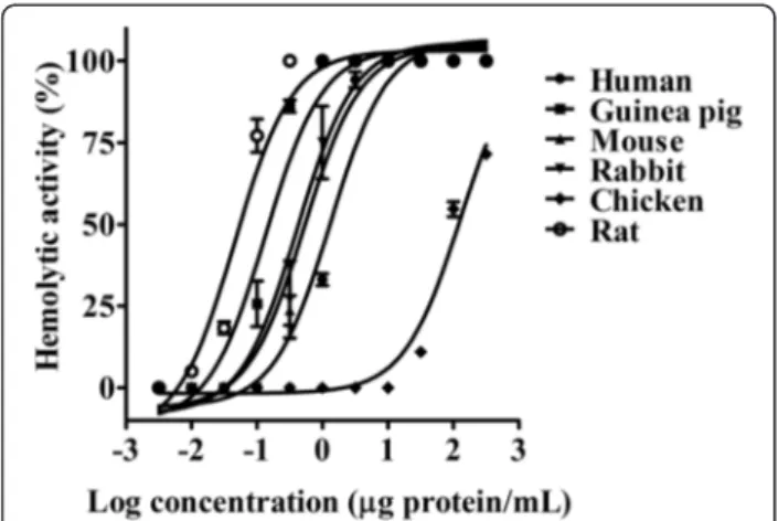
TheM. alcicornisaqueous extract (0.0001 to 150μg/mL) produced a concentration-dependent hemolysis on erythrocytes (Fig. 1) that was completely abolished after incubation in boiling water bath for 20 min. Chicken erythrocytes were markedly more resistant to the hemolytic effect of the M. alcicornis extract than the erythrocytes from other species (Table 1). In the present study rat erythrocytes were used to characterize the hemolytic activity of the extract. The hydrocoral extract (HU50= 0.042 ± 0.005 μg protein/mL) was approximately 2200-fold more potent than ionomycin (HU50= 92.57 ±
1.013μg/mL), used as a control.
Characterization of the hemolytic activity
inactivated at temperatures higher than 60 °C, while at preincubation temperatures lower than 45 °C, the hemolytic activity of the extract was conserved (Fig. 2c).
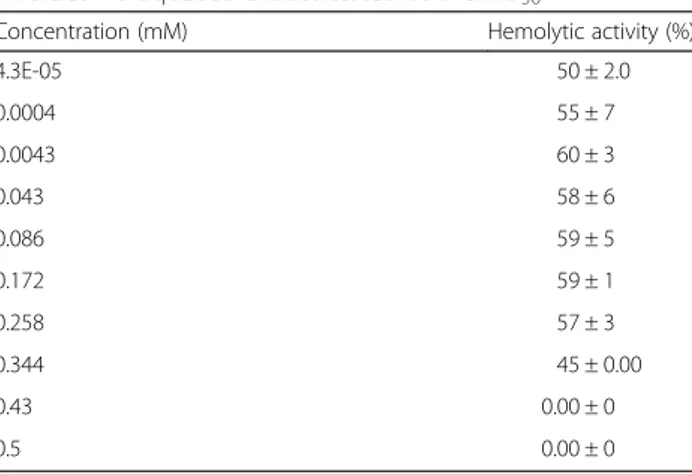
The hemolysis caused by the hydrocoral extract was assessed in the presence of divalent cations such as Ca2+, Mg2+, Cu2+, Zn2+, or Ba2+. These experiments showed that the hemolytic activity was depressed by Cu2+0.1 mM or Zn2+ 6 mM, however it was increased by addition of Ca2+and Mg2+. In the case of Ba2+, concentrations lower than 6 mM increased the hemolytic activity of the extract, whereas concentrations higher than 8 mM induced in-hibition of this activity (Table 2). Hemoysis produced by the extract was reduced after incubation with EDTA 0.34 mM and sharply disappeared at EDTA 0.43 mM (Table 3).
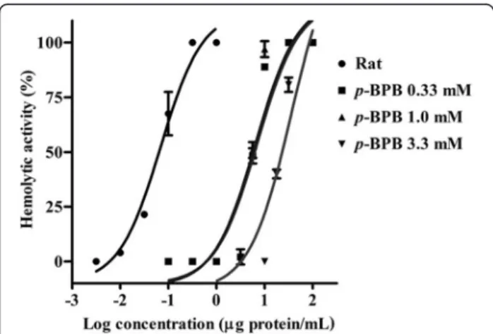
The hemolytic activity of theM. alcicornisaqueous ex-tract was significantly reduced after incubation with the PLA2inhibitorp-BPB. In these experiments, the
concen-tration response curves were rightward shifted depend-ing on the concentration of the inhibitor. In the absence of p-BPB the HU50 was 0.069 ± 0.081 μg protein/mL,
after incubation with 0.33 mM p-BPB the HU50 was
7.42 ± 0.13 μg protein/mL, with 1.0 mM p-BPB the
HU50 was 6.51 ± 0.14μg protein/mL. Finally, at 3.3 mM p-BPB a greater inhibitory effect was observed and the value of the HU50 increased up to 35.89 ± 0.16 μg
protein/mL (Fig. 3).
SDS-PAGE electrophoresis and zymography
TheM. alcicornisaqueous extract contains proteins with a broad range of molecular weights, between 8 and 200 kDa and under reducing conditions, the electro-phoretic profile was altered (Fig. 4a). Two main hemolytic zones were identified by zymography, one of which corresponded to a band of approximately 28– 30 kDa, and the other one was related to a broad band of 200 kDa, approximately (Fig. 4b). Under reducing conditions, the 200 kDa band disappeared, which sug-gested that it consists of two or more dimers. The zymo-graphy analysis showed that the 28 kDa band possessed PLA2activity, while the 200 kDa did not elicit this
en-zymatic activity (Fig. 4c).
Lethality assay in mice and systemic effects
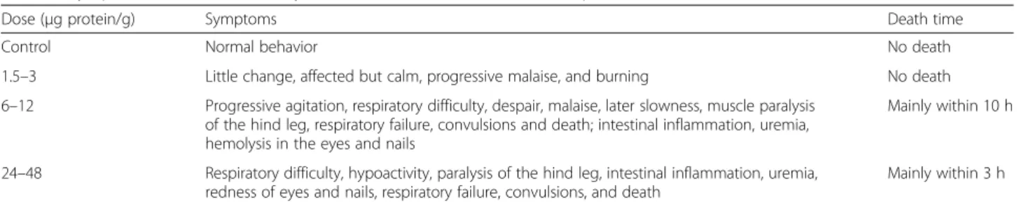
When intravenously injected, the aqueous extract of M. alcicornis was lethal to mice with a LD50 of 17 μg protein/g of body weight (Table 4). At doses lower than the LD50 (1.5 and 3.0 μg protein/g) the extract induced forced respiration immediately after the adminis-tration, but mice recovered within a few minutes. Mice also showed a progressive depressed responsiveness, which was usually pain related. However, they progres-sively recovered their normal behavior. At higher doses (6 and 12 μg protein/g), the aqueous extract also in-duced respiratory difficulty, hypoactivity, and other symptoms such as grooming, paralysis of the hind legs, intestinal inflammation, hemoglobinuria, and after a while, complete paralysis. In some cases, respiratory failure and convulsions preceded death. Doses higher than the LD50 (24 and 48 μg protein/g) induced symp-toms similar to those of the lower doses, although in most cases, deaths occurred in a shorter time. The de-natured aqueous extract of M. alcicornis did not have any effect in mice.
Histopathological analysis
LM images showed histopathological changes in kidney, lung and liver tissues but not in heart, brain or skeletal muscle. In kidney sections, glomerular capillaries ap-peared congested with proteinaceous material within the tubules. In some cases, acute tubular necrosis (Fig. 5a and b) was observed. Lung sections showed severe al-veolar epithelial cells degeneration and desquamation, damage to capillaries, and marked infiltration of erythro-cytes and proteinaceous material within alveoli (Fig. 5c and d). Liver sections showed varying degrees of patho-logical lesions, with acute vascular congestion and areas Fig. 1Concentration-response curves showing the hemolytic
activity of theM. alcicornisaqueous extract on erythrocytes from various species
Table 1Hemolytic activity of theM. alcicornisextract on several types of erythrocytes
Erythrocytes type Hemolytic unit (HU50)
Rat 0.042 ± 0.005
Guinea pig 0.29 ± 0.15
Mouse 0.41 ± 0.015
Rabbit 0.63 ± 0.35
Human 1.0 ± 0.31
Chicken 129 ± 8.2*
with fibrinoid material mixed with neutrophils and cell apoptosis, which indicated the development of moderate inflammatory reactions and activation of the comple-ment system (Fig. 5e and f ). Some bloods samples were taken from mice 4 h after the administration of the ex-tract for quantification of alanine aminotransferase and aspartate aminotransferase. Unfortunately, these experi-ments failed since all blood samples were completely hemolysed. Injection of the denatured extract did not cause any histopathological damage in the tissues examined.
Lethality assay inArtemia salina
The aqueous extract ofM. alcicorniswas toxic toA. sal-ina with a LD50= 70.71 μg protein/mL. It is important to point out that crude extracts are considered toxic when their LD50is lower than 1000μg/mL [20], and the denatured aqueous extract had a LD50close to this value
(LD50= 960.28μg protein/mL).
Analysis by electron microscopy techniques
Analysis by SEM revealed that theM. alcicornisaqueous extract broke the erythrocyte membranes, which pro-voked release of intracellular material from the cells Fig. 2Effect of (a) pH and (b) incubation temperature on the hemolytic activity of the aqueous extract ofM. alcicornis.cStability of the
hemolytic activity of the extract after its incubation at different temperatures
Table 2Effects of divalent cations on the hemolytic activity of M. alcicornisaqueous extract tested at the HU50
Concentration (mM) Hemolytic activity (%)
Ca2+ Mg2+ Cu2+ Zn2+ Ba2+
0.001 48 ± 9 54 ± 4 49 ± 4 66 ± 2 51 ± 6 0.01 49 ± 05 55 ± 1 40 ± 2 68 ± 2 54 ± 5 0.1 52 ± 1 48 ± 3 26 ± 3 65 ± 1 59 ± 6 1 51 ± 4 54 ± 4 29 ± 3 62 ± 0.0 79 ± 2 2 65 ± 4 61 ± 3 7 ± 4 46 ± 1 84 ± 4 4 76 ± 1 57 ± 6 0 ± 0 36 ± 1 81 ± 4
6 98 ± 2 63 ± 3 2 ± 6 83 ± 2
8 100 ± 0.0 73 ± 3 0 ± 0 1 ± 0.0
10 100 ± 0.0 87 ± 2 0 ± 0 0 ± 0.0
12 100 ± 0.00
Table 3Effect of EDTA on the hemolytic activity of the M. alcicornis aqueous extract tested at the HU50
Concentration (mM) Hemolytic activity (%)
4.3E-05 50 ± 2.0
0.0004 55 ± 7
0.0043 60 ± 3
0.043 58 ± 6
0.086 59 ± 5
0.172 59 ± 1
0.258 57 ± 3
0.344 45 ± 0.00
0.43 0.00 ± 0
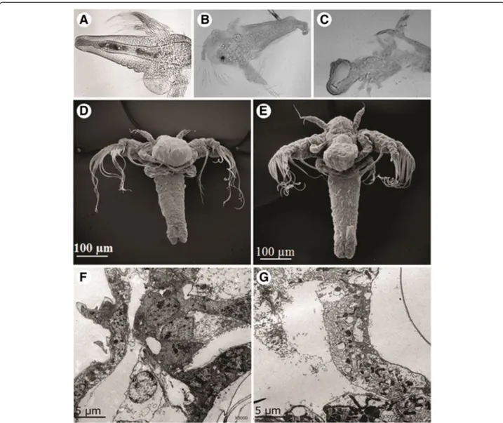
(Fig. 6). However, control erythrocytes remained viable and intact following 24 h of incubation. Microscopic analysis by optic microscopy showed that control brine shrimps had a normal cuticle and their visceral mass ap-peared well defined (Fig. 7a). In contrast, the visceral mass of the main body cavity of the A. salina nauplii was disturbed and their cuticle was damaged in the pres-ence of the aqueous extract (Fig. 7b and c). Microscopic analysis by SEM (Fig. 7d and e) and ultrathin longitu-dinal sections, analyzed by TEM (Fig. 7f and g) showed cell disorganization in treated nauplii exposed to the M. alcicornis aqueous extract.
Discussion
Millepora species are recognized for the painful stings they can inflict on human skin. However, up to now, the mechanism of the envenomation caused by these species
has not been completely defined. Hemolysis is a well-known effect induced by cnidarian cytolysins, including those produced by hydrocorals. These cytolysins have also the capacity to affect other cell types and provoke damage such as inflammation and dermonecrosis [22]. In the present study, the hemolysis caused by the aque-ous extract of the“fire coral”M. alcicorniswas tested on rat, human, mouse, chicken, guinea pig, and rabbit erythrocytes. Our results showed that rodent erythro-cytes were the most sensitive to the hydrocoral aqueous extract, whereas chicken erythrocytes were significantly less susceptible. It is very likely that differences observed in the erythrocytes susceptibility to lysis depend on their lipid membrane composition. In this regard, studies on the mechanism of insertion of cnidarian pore-forming cytolysins have shown that actinoporins (20 kDa proteins) irreversibly bind with high affinity to mem-branes containing sphingomyelin, whereas other types of cytolytic peptides (5–8 kDa) show a preference for phosphatidylcholine-containing membranes [23]. A var-iety of novel pore-forming cytolysins have been isolated from cnidarians and their activity strongly depends on the lipid membrane composition [22, 24, 25].
Concerning cytolysins with PLA2 activity, kinetic
studies have revealed that these enzymes can display dif-ferent affinities for membranes depending on the phospholipid composition [26]. In a previous study, re-garding characterization of the hemolytic activity of the aqueous extract of M. complanata collected in the Mexican Caribbean, we found a similar behavior on the erythrocytes susceptibility to lysis [27], which suggests that cytolysins produced by both Caribbean hydrocorals have a high similarity in the membrane-binding do-mains. These results are in contrast with those found by Shiomi et al. [9], who tested the hemolytic activity of Fig. 3Concentration-response curves showing the effect ofp-BPB,
a PLA2inhibitor, on the hemolytic activity of theM. alcicornis
aqueous extract
Fig. 4aElectrophoretic profile under non-reducing (Mad) and reducing (Madr) conditions. The protein bands were visualized by the Coomassie blue staining method.bHemolysis zymogram on a 1.5 % agar gel supplemented with 5 % egg yolk and 3 % of washed erythrocytes, two hemolytic zones were observed in the gel corresponding to ~28-30 and ~200 kDa.cPLA2zymogram on a 2 % agarose gel supplemented with
M. dichotoma and M. platyphylla venoms on cow, horse, sheep, rabbit, guinea pig, mouse, and chicken erythrocytes. These researchers observed that horse erythrocytes were highly sensitive to the M. dichot-oma venom, while they were much less sensitive to theM. plathyphyllavenom. Conversely, chicken erythro-cytes were more sensitive to the M. plathyphyllavenom than to that ofM. dichotoma.
In this study we also found that the hemolytic activity induced by the extract ofM. alcicorniswas progressively
reduced when it was preincubated at temperatures above 45 °C and was completely lost after preincubation at temperatures over 70 °C. These results indicate that the hemolysins contained in this extract are mainly thermo-labile proteins. These hemolysins are relatively stable in a range of incubation temperatures from 30 to 43 °C. At temperatures lower than 30 °C, the hemolytic activity of the aqueous extract was reduced, which indicates that warm temperatures stimulate the hemolytic reaction. Furthermore, the M. alcicornis hemolysins also showed
Table 4Symptoms in mice induced by intravenous administration of the aqueous extract ofM. alcicornis
Dose (μg protein/g) Symptoms Death time
Control Normal behavior No death
1.5–3 Little change, affected but calm, progressive malaise, and burning No death 6–12 Progressive agitation, respiratory difficulty, despair, malaise, later slowness, muscle paralysis
of the hind leg, respiratory failure, convulsions and death; intestinal inflammation, uremia, hemolysis in the eyes and nails
Mainly within 10 h
24–48 Respiratory difficulty, hypoactivity, paralysis of the hind leg, intestinal inflammation, uremia, redness of eyes and nails, respiratory failure, convulsions, and death
Mainly within 3 h
high stability to pH variation, since their activity was preserved in the range from 2 to 12. These results differ from those observed in a study performed with an ex-tract prepared from M. alcicornis collected on the bea-ches in Miami [15]. In that study, it was found that the extract preserved its activity in a pH range of 5.5 to 8.7, but lost its activity at a pH of 4.8. These differences con-firm that toxin synthesis highly depends on the environ-mental conditions.
It has also been reported that hemolytic activity of cni-darian venoms exhibits a differentiated dependence on divalent cations, and particularly, cnidarian phospholi-pases A2have a Ca2+binding loop, which is highly
con-served [28]. Regarding cnidarian pore-forming toxins, it has been demonstrated that Ca2+ is necessary for the dimerization of the toxin within the membrane [29], while other cnidarian pore-forming toxins can be inhib-ited by Zn2+ [30]. In this study, we found that the hemolytic activity of the aqueous extract ofM. alcicornis was increased in the presence of≥0.01 mM (Ca2+, Mg2+,
or Ba2+) and was reduced in the presence of Cu2+ (≥0.1 mM), Zn2+ (≥6 mM), and EDTA (≥0.43 mM).
These results indicate that divalent cations modify the hemolytic activity induced by the aqueous extract of this
“fire coral”. In the case of the inhibition by EDTA, this effect might be attributed to the chelation of Ca2+ and Mg2+. These findings differ from those obtained in our previous study, in which we examined the effect of Ca2+ and Mg2+on the hemolytic activity induced by the aque-ous extract of M. complanata[27], which suggest struc-tural differences between the Ca2+ binding loop of the
cytolysins produced by these Millepora species. It is worth mentioning that inhibition of the cytolytic activity by Cu2+, Zn2+, and EDTA could be useful for the
devel-opment of adequate treatments for stings caused by the “fire corals”. Previous studies have shown that zinc glu-conate inhibits potassium efflux from erythrocytes ex-posed to venom or purified hemolysin from Chironex fleckeriand prolongs survival time in mice injected with the venom. Therefore, administration of this ion has been considered useful in the treatment of victims of C. fleckeri stings [30].
Up to now, the hemolytic mechanisms of Millepora venoms have not been completely characterized, but previous studies suggest that PLA2proteins significantly
contribute to this effect. In this context, it has been re-ported that Milleporaspecies possesses higher PLA2
ac-tivity levels than other cnidarians [31]. In a previous Fig. 6aControl erythrocytes preincubated with Alsever’s solution.bandcErythrocytes preincubated with an HU50of theM. alcicornisaqueous
study, we found that the hemolytic activity of the aque-ous extract of M. complanata significantly decreased after incubation with the PLA2inhibitorp-bromophenacyl
bromide, and we detected a hemolysin with a molecular weight similar to that of milleporin-1, a PLA2isolated in
the venom ofM. platyphylla[32]. We also found that the aqueous extract ofM. complanata has PLA2activity, but
not protease activity [33] Accordingly, the results obtained in this study showed that the hemolysis produced by the aqueous extract of M. alcicornis was significantly inhib-ited, but not completely blocked byp-bromophenacyl and it does not contain proteases. These findings indicate that toxins with PLA2 activity significantly contribute to the
overall hemolysis induced by this hydrocoral extract. However, it is very likely that this extract also contains
pore-forming toxins, since the hemolytic activity of the extract was not completely suppressed with the PLA2
inhibitor.
Electrophoretic analysis revealed that theM. alcicornis extract contains proteins with a broad range of mo-lecular weights. When the electrophoresis gel, run under non-reduction conditions, was incubated on blood agar gel, two bands displayed hemolytic activity: an approximately 28–30 kDa-band and an approxi-mately 200 kDa-band. The last one was not observed when electrophoresis was run under reducing condi-tions, which suggests that these high molecular weight proteins could be polymers. The PLA2
zymo-grams showed that the hemolytic 28–30 kDa-band also induces PLA2 activity, whereas the 200-kDa band
Fig. 7aArtemia salinabrine shrimp used as control, after 24-h incubation in seawater.bandcBrine shrimps after 24 h of incubation with 10-fold the LD50of theM. alcicornisaqueous extract; membranes are partially disintegrated. Light microscope, 400× magnification.dControl and (e) treated brine
region did not have enzymatic activity, suggesting the presence of pore-forming polymeric cytolysins. This type of toxin has been described in venoms of cnidar-ians of the class Hydrozoa. For example, physalitoxin is a potent hemolysin of 240 kDa detected in the Physalia physalis venom [34].
In another set of experiments, the systemic toxicity of the extract was assessed in mice by intravenous adminis-tration. Previous studies showed that the venom of M. alcicornis caused extensive hemolysis and death in mice after several hours [15]. In the case of this study, similar symptoms were observed, which con-sisted in generalized hemolysis, forced respiration, convulsions, and death; depending on the dosage. However, the lethality of the extract prepared from M. alcicornisspecimens recently collected in the Mexican Caribbean was lower (LD50= 17 μg protein/g) than that produced by the specimens previously studied (LD50= 0.55 and 1.136 μg protein/g). These differ-ences might be attributed to environmental changes, since the specimens were collected in different zones and times, which results in variations in toxins pro-duction. Interestingly, important differences were found between the toxicity induced by theM. alcicornisaqueous extract and that provoked by the M. complanata extract [33], both species collected in the Mexican Caribbean. In our previous study, we observed that the aqueous extract of M. complanata induced two types of death in mice after its intravenous administration: at doses higher than the LD50(LD50= 4.62 μg protein/g) the extract produced
violent convulsions and death within 1 min; however, lower doses induced a slow death similar to that provoked by theM. alcicornisextract. After denaturation by heating, theM. complanata extract completely lost its capacity to induce the slow death, but conserved its immediate lethal effect [33]. In contrast, the denatured extract of M. alci-cornis did not induce any response in mice, which sug-gests that the compounds responsible for the systemic toxicity induced by the M. alcicornis extract are heat-labile proteins, whereas the M. complanata extract also contains thermo-stable toxins that are capable of inducing a rapid death in mice.
Histophatological analysis of tissues dissected from mice administrated with the aqueous extract of M. alci-cornisrevealed important damage to the lung, liver and kidneys. Similar effects have been observed in mice after intravenous administration of a tentacle extract from the jellyfish Cyanea capillata. At high doses, mice deaths occurred within 2 h probably due to acute heart-or-ner-vous-related toxicities. At lower doses, deaths occurred between 2 and 48 h with acute liver and renal failure. That toxin-induced multiple organ dysfunction was defined as the“delayed jellyfish envenomation syndrome”, whose eti-ology has not been completely elucidated, but it has been
related to the synergy of various toxic mechanisms caused by a variety of toxins contained in the jellyfish venom [35]. In the present research, it was found that the aqueous extract of M. alcicornis induced a delayed envenom-ation syndrome similar to those elicited by the venom of C. capillata and the aqueous extract of M. complanata [33]. It is likely that these toxic effects are caused mainly by cytolysins that disrupt several cell types, including those of epithelial tissues. It has been observed that epi-thelial cells of nephrons can be markedly affected by cyto-lysins [36], since kidneys represent the main route of elimination of toxins. Therefore, the hemoglobinuria caused by theM. alcicornisextract in the mice could be a consequence of both renal damage and hemolysis. More-over, we have previously found that the aqueous extracts ofM. alcicornisandM. complanataprovoke vasoconstric-tion on isolated rat aortic rings [14]. This vasoconstrictor effect could also trigger multiple organ injuries as a conse-quence of compensatory responses due to ischemia or hypoxia. These systemic toxic effects observed in mice could explain the fact that human contact with these hydrocorals can result in renal disease [5].
A cytotoxicity assay usingA. salina was carried out to further analyze the toxicity of the aqueous extract of M. alcicornis. This extract was lethal to A. salina nauplii (LD50= 70.71 μg protein/mL), which is not surprising since small crustaceans are part of the diet of Millepora hydrocorals [1]. The denatured extract was significantly less lethal (LD50= 960.28μg protein/mL), in-dicating that the cytotoxic compounds are mainly thermo-labile proteins. Microscopic analysis of brine shrimps incubated in the presence of the extract showed tissue dis-sociation. This effect is usually attributed to pore-forming cytolysins, which form pores in the membranes of these crustaceans, lysing the cells and disintegrating tissues in order to facilitate phagocytosis and the subsequent diges-tion of the prey [37].
Conclusions
The results of the present study demonstrate that the aqueous extract ofM. alcicorniscontains hemolytic pro-teins that are thermolabile, stable to pH changes, and whose activity is calcium-dependent. This study also evi-denced the presence of two types of cytolysins in this ex-tract: some of them consist of proteins of approximately 28–30 kDa with PLA2activity, while the others are
lar-ger proteins of approximately 200 kDa, which do not possess PLA2activity and possibly act by a pore-forming
Ethics committee approval
All experiments were performed in accordance with the Mexican Official Standard NOM-062-ZOO-1999 for the production, care and use of laboratory animals. Care and use of animals was approved by the Bioethics Committee of the School of Medicine, UAQ.
Competing interests
The authors declare that they have no competing interests.
Authors’contributions
RH-M wrote the manuscript, performed the systemic toxicity assays, and characterized the hemolytic activity of theM. alcicornisaqueous extract as part of her doctoral degree thesis. AR-M designed this project, coordinated all the activities, and contributed with the preparation of the manuscript. AAVS and AG-A collaborated in the toxicity assays. CI-A supervised the electrophoresis and zimography methods. NO-L helped in carrying out the zimography experiments, and MA suggested some improvements in the manuscript. All authors read and approved the final manuscript.
Acknowledgments
This work was supported by Grants CB-2009-01 (Project 133785) and INFR-2014-01 (Project 226186) to A. Rojas from the Consejo Nacional de Ciencia y Tecnología (CONACYT). R. Hernández-Matehuala acknowledges a scholarship from CONACYT. The authors thank Armando Zepeda-Rodríguez (Facultad de Medicina) and Lourdes Palma (Instituto de Neurobiología) from Universidad Nacional Autónoma de México for their technical support in the electron microscopy. Thanks are also due to Marco Alonso Gallegos Corona and Carolina Gutiérrez from the Universidad Autónoma de Querétaro for their assistance in the histopatological studies and hemolysis assays, respectively.
Author details
1Posgrado en Ciencias del Mar y Limnología, Universidad Nacional
Autónoma de México, Mexico City, Mexico.2Laboratorio de Investigación
Química y Farmacológica de Productos Naturales, Facultad de Química, Universidad Autónoma de Querétaro, Centro Universitario, Querétaro 76010, Mexico.3Instituto de Neurobiología, Universidad Nacional Autónoma de
México, Campus Juriquilla, Querétaro 76201, Mexico.
Received: 1 January 2015 Accepted: 14 September 2015
References
1. Lewis JB. Biology and ecology of the hydrocoralMilleporaon coral reefs. Adv Mar Biol. 2006;50:1–55.
2. García-Arredondo A, Rojas A, Iglesias-Prieto R, Zepeda-Rodriguez A, Palma-Tirado L. Structure of nematocysts isolated from the fire corals
Millepora alcicornisandMillepora complanata(Cnidaria: Hydrozoa). J Venom Anim Toxins incl Trop Dis. 2012;18(1):109–15.
3. Ozbek S, Balasubramanian PG, Holstein TW. Cnidocyst structure and the biomechanics of discharge. Toxicon. 2009;54(8):1038–45.
4. Rojas-Molina A, García-Arredondo A, Ibarra-Alvarado C, Bah M.Millepora
(“fire corals“) species: toxinological studies untill 2011. In: Daniels JA, editor. Advances in environmental research, vol. 26. New York: Nova; 2012. p. 133–48.
5. Moats WE. Fire coral envenomation. J Wilderness Med. 1992;3(3):284–7. 6. Prasad R, Vincent L, Hamilton R, Lim K. Minimal change disease in
association with fire coral (Milleporaspecies) exposure. Am J Kidney Dis. 2005;47(1):e15–16.
7. Wittle LW, Wheeler CA. Toxic and immunological properties of stinging coral toxin. Toxicon. 1974;12(5):487–93. doi:10.1016/0041-0101(74)90038-5. 8. Middlebrook RE, Wittle LW, Scura ED, Lane CE. Isolation and partial purification
of a toxin fromMillepora dichotoma. Toxicon. 1971;9(4):333–6.
9. Shiomi K, Hosaka M, Yanaike N, Yamanaka H, Kikuchi T. Partial characterization of venoms from two species of fire coralsMillepora platyphyllaandMillepora dichotoma. Nippon Suisan Gakk. 1989;55(2):357–62.
10. Radwan FFY. Comparative toxinological and immunological studies on the nematocyst venoms of the Red Sea fire coralsMillepora dichotomaandM. platyphylla. Comp Biochem Physiol C Toxicol Pharmacol. 2002;131(3):323–34.
11. Iguchi A, Iwanaga S, Nagai H. Isolation and characterization of a novel protein toxin from fire coral. Biochem Biophys Res Commun. 2008;365(1):107–12.
12. Ibarra-Alvarado C, García JA, Aguilar MB, Rojas A, Falcón A, Heimer de la Cotera EP. Biochemical and pharmacological characterization of toxins obtained from the fire coralMillepora complanata. Comp Biochem Physiol C Toxicol Pharmacol. 2007;146(4):511–8.
13. Rojas A, Torres M, Rojas JI, Feregrino A, Heimer-de la Cotera EP. Calcium-dependent smooth muscle excitatory effect elicited by the venom of the hydrocoralMillepora complanata. Toxicon. 2002;40(6):777–85.
14. García-Arredondo A, Rojas-Molina A, Ibarra-Alvarado C, Iglesias-Prieto R. Effects of bleaching on the pharmacological and toxicological activities elicited by the aqueous extracts prepared from two“fire corals“collected in the Mexican Caribbean. J Exp Mar Biol Ecol. 2011;396(2):171–6.
15. Wittle LW, Middlebrook R, Lane C. Isolation and partial purification of a toxin fromMillepora alcicornis. Toxicon. 1971;9(4):327–31.
16. Bradford MM. A rapid and sensitive method for the quantitation of microgram quantities of protein utilizing the principle of protein-dye binding. Anal Biochem. 1976;72:248–54.
17. Torres M, Aguilar MB, Falcón A, Sánchez L, Radwan FF, Burnett JW, et al. Electrophysiological and hemolytic activity elicited by the venom of jellyfish
Cassiopea xamachana. Toxicon. 2001;39(9):1297–307.
18. Laemmli UK. Cleavage of structural proteins during the assembly of the head of bacteriophage T4. Nature. 1970;227(5259):680–5.
19. Campos LB, Pucca MB, Roncolato EC, Netto JC, Barbosa JE. Analysis of phospholipase A2, L-amino acid oxidase, and proteinase enzymatic activities of theLachesis mutarhombeata venom. J Biochem Mol Toxicol. 2012;26(8):308–14.
20. Murata Y, Satake M, Suzuki T. Studies on snake venom XII. Distribution of proteinase activities among Japanese and formosan snake venoms. J Biochem. 1963;53(6):431–43.
21. Déciga-Campos M, Rivero-Cruz I, Arriaga-Alba M, Castañeda-Corral G, Ángeles-López GE, Navarrete A, et al. Acute toxicity and mutagenic activity of Mexican plants used in traditional medicine. J Ethnopharmacol. 2007;110(2):334–42.
22. Mariottini GL, Pane L. Cytotoxic and citolytic cnidarian venoms. A review on health implications and possible therapeutic applications. Toxins. 2014;6(1):108–51.
23. Anderluh G, Macek P. Cytolytic peptide and protein toxins from sea anemones (Anthozoa: Actiniaria). Toxicon. 2002;40(2):111–24.
24. Razpotnik A, Krizaj I, Kem WR, Macek P, Turk T. A new cytolytic protein from the sea anemoneUrticina crassicornisthat binds to cholesterol- and sphingomyelin-rich membranes. Toxicon. 2009;53(7–8):762–9. 25. Brinkman DL, Burnell JN. Biochemical and molecular characterization of
cubozoan protein toxins. Toxicon. 2009;54(8):1162–73.
26. Saikia D, Bordoloi NK, Chattopadhyay P, Choklingam S, Ghosh SS, Mukherjee AK. Differential mode attack on membrane phospholipids by an acidic phospholipase A2(RVVA-PLA2-I) fromDaboia russellivenom.
Biochim Biophys Acta. 2012;1818(12):3149–57.
27. García-Arredondo A, Murillo-Esquivel LJ, Rojas A, Sanchez-Rodriguez J. Characteristics of hemolytic activity induced by the aqueous extract of the Mexican fire coralMillepora complanata. J Venom Anim Toxins incl Trop Dis. 2014;20:49.
28. Sher D, Knebel A, Bsor T, Nesher N, Tal T, Morgensten D, et al. Toxic polypeptides of the hydra–a bioinformatic approach to cnidarian allomones. Toxicon. 2005;45(7):865–79.
29. Gusmani L, Avian M, Galil B, Patriarca P, Rottini G. Biologically active polypeptides in the venom of the jellyfishRhopilema nomadica. Toxicon. 1997;35(5):637–48.
30. Yanagihara AA, Shohet RV. Cubozoan venom-induced cardiovascular collapse is caused by hyperkalemia and prevented by zinc gluconate in mice. PLoS One. 2012;7(12):e51368.
31. Nevalainen TJ, Peuravuori HJ, Quinn RJ, Llewellyn LE, Benzie JAH, Fenner PJ, et al. Phospholipase A2 in cnidaria. Comp Biochem Physiol B Biochem Mol Biol. 2004;139(4):731–5.
32. Radwan FFY, Aboul-Dahab HM. Milleporin-1, a new phospholipase A2active
protein from the fire coralMillepora platyphyllanematocysts. Comp Biochem Physiol C Toxicol Pharmacol. 2004;139(4):267–72.
partial purification of thermostable neurotoxins with lethal effects in mice. Comp Biochem Physiol C Toxicol Pharmacol. 2015;169:55–64. 34. Tamkun MM, Hessinger DA. Isolation and partial characterization of a
hemolytic and toxic protein from the nematocyst venom of the Portuguese Man-of-War. Physalia physalis Biochim Biophys Acta. 1981;667(1):87–98. 35. Wang B, Zhang L, Zheng J, Wang Q, Wang T, Lu J, et al. Multiple organ
dysfunction: a delayed envenomation syndrome caused by tentacle extract from the jellyfishCyanea capillata. Toxicon. 2013;61:54–61.
36. Ali SA, Alam JM, Abbasi A, Zaidi ZH, Stoeva S, Voelter W. Sea snake
Hydrophis cyanocinctusvenom. II. Histopathological changes, induced by a myotoxic phospholipase A2 (PLA2-H1). Toxicon. 2000;38(5):687–705. 37. Sher D, Fishman Y, Melame-Book N, Zhang M, Zlotkin E. Osmotically driven
prey disintegration in the gastrovascular cavity of the green hydra by a pore-forming protein. FASEB J. 2008;22(1):207–14.
Submit your next manuscript to BioMed Central and take full advantage of:
• Convenient online submission
• Thorough peer review
• No space constraints or color figure charges
• Immediate publication on acceptance
• Inclusion in PubMed, CAS, Scopus and Google Scholar
• Research which is freely available for redistribution