Ide ntificatio n o f he m o lytic and
ne uro active fractio ns in the ve no m
o f the se a ane m o ne
Bunod osom a
cangicum
1Departamento de Farmacologia, Faculdade de Medicina, and 2Laboratório de Neuroquímica, Departamento de Psicobiologia,
Faculdade de Filosofia, Ciências e Letras de Ribeirão Preto, Universidade de São Paulo, Ribeirão Preto, SP, Brasil
3Laboratório de Peptideos e Proteinas, Instituto de Investigaciones Biológicas Clemente Estable, Montevideo, Uruguay
4Departamento de Fisiologia Geral, Instituto de Biociências,
e Instituto de Biologia Marinha, Universidade de São Paulo, São Paulo, SP, Brasil
P. Lagos1, R. Duran3, C. Cerveñansky3, J.C. Freitas4 and R. Silveira2
Abstract
Sea anemones are a rich source of biologically active substances.In crayfish muscle fibers, Bunodosoma cangicum whole venom selec-tively blocks the IK(Ca) currents. In the present study, we report for the first time powerful hemolytic and neuroactive effects present in two different fractions obtained by gel-filtration chromatography from whole venom of B. cangicum. A cytolytic fraction (Bcg-2) with components of molecular mass ranging from 8 to 18 kDa elicited hemolysis of mouse erythrocytes with an EC50 = 14 µg/ml and a maximum dose of 22 µg/ml. The effects of the neuroactive fraction, Bcg-3 (2 to 5 kDa), were studied on isolated crab nerves. This fraction prolonged the compound action potentials by increasing their duration and rise time in a dose-dependent manner. This effect was evident after the washout of the preparation, suggesting the existence of a reversible substance that was initially masking the effects of an irreversible one. In order to elucidate the target of Bcg-3 action, the fraction was applied to a tetraethylammonium-pretreated preparation. An additional increase in action potential duration was observed, suggesting a blockade of a different population of K+ channels or of tetraethylammonium-insensitive channels. Also, tetrodotoxin could not block the action potentials in a Bcg-3-pretreated preparation, suggesting a possible interaction of Bcg-3 with Na+ channels. The present data suggest that B. cangicum venom contains at least two bioactive fractions whose activity on cell membranes seems to differ from the IK(Ca) blockade described previously.
Co rre spo nde nce
P. Lagos
Departamento de Farmacologia FMRP, USP
Av. Bandeirantes, 3900 14049-900 Ribeirão Preto, SP Brasil
Fax: + 55-16-633-2301 E-mail: lagos@ usp.br
Research partially supported by PEDECIBA (Uruguay) and CO NICYT Grant No. 033/94 (Uruguay). Publication supported by FAPESP.
Received June 27, 2000 Accepted March 27, 2001
Ke y wo rds
·Bunodosom a cangicum
·Sea anemone
·Hemolytic activity
·Neurotoxins
Intro ductio n
Sea anemones contain a variety of bio-logically active substances including poly-peptide toxins which affect sodium and po-tassium channels (1,2). Cytolysins that act on cell membranes (pore-forming toxins) have also been described (3,4). All these types of peptides have been isolated from different species such as Anemonia sulcata (5), Stoichactis helianthus (6), Actinia equina
(7), Bunodosoma granulifera (2,8,9) and
Bunodosoma caissarum (10,11). Sea anemone neurotoxins that affect sodium channels slow down the inactivation phase of the currents without affecting the activa-tion process, so that the channels remain open for a longer period of time leading to a prolongation of the action potential duration (1,3). In contrast, neurotoxins that act on potassium channels behave as blockers of voltage-sensitive channels, similar to den-drotoxins (DTX) or to mast cell degranulat-ing peptide isolated from mamba snakes (12,13) and bee venoms (14), respectively.
B. cangicum is a common sea anemone found along the Uruguayan and Brazilian seashores. Its venom selectively blocks the Ca2+-dependent K+ current (I
K(Ca)) present in
crayfish muscle fibers in a reversible manner without affecting voltage-gated Ca2+ or K+ currents (15). Furthermore, the venom re-duces IK(Ca) in chromaffin cells without modi-fying voltage-gated Na+, Ca2+ or K+ currents (15). The venom also inhibits the binding of radiolabeled DTX to synaptosomal mem-branes (13). Although these studies (13,15) described interesting neuroactive effects of whole venom, no attempt was made to purify the toxin(s) responsible for such actions on ionic channels. Furthermore, the presence of cytolytic toxins in the venom has not been explored previously.
The aim of the present study was to de-scribe the hemolytic and neuroactive proper-ties of isolated fractions obtained after gel-filtration chromatography from the whole
venom of B. cangicum, using different in vitro approaches.
Mate rial and Me tho ds
Ve no m e xtractio n
The extraction method was a modifica-tion of that reported by Malpezzi and Freitas (10). B. cangicum specimens were collected on Cabo Polonio rocky shores (Rocha, Uru-guay) and kept alive in laboratory aquaria for milking for several months. The isolation of the venom (probably including some other substances from the animal body) was per-formed by electrical stimulation of 20 ani-mals immersed in 50 ml of 0.10 M ammo-nium acetate, pH 7.0. Each animal received an electric discharge (100 V, 10 ms and 20 Hz for 20 s) using two carbon electrodes, as described in the original method. This pro-cedure allows most animals to recover and to be re-used to obtain more venom or to be returned to the sea. The solution obtained was freeze-dried and stored at -70oC.
Gel-filtration chromatography. The freeze-dried venom was dissolved in 10-12 ml of 0.10 M ammonium acetate, pH 7.0, and centrifuged at 2000 g for 15 min and the supernatant was submitted to gel filtration on a Sephadex G-50 column (1.9 x 131 cm; Pharmacia-LKB Biotechnology, Uppsala, Sweden) and eluted with the same solution. Fractions of 5 ml were collected at a flow rate of 20 ml/h, absorbance was measured at 280 nm and the effluent was then pooled as indicated in Figure 1. The molecular mass range of the components present in each group of fractions was estimated by the elu-tion of a mixture of four standard proteins: bovine albumin (67 kDa), chymotrypsino-gen (25 kDa), cytochrome c (12.5 kDa) and bacitracin (1.4 kDa) under the same elution conditions as used for the venom.
he-molytic activity in the whole venom and fractions obtained by gel filtration, a qualita-tive test was performed using multiwell plates. Each well received 50 µl of the test substance, plus 50 µl of a 4% mouse erythro-cyte suspension in 0.85% saline, containing 10 mM CaCl2. After gentle shaking at room temperature for 30-40 min, the lytic effect (positive) was detected visually and com-pared to the effect of Triton X-100 deter-gent. The fractions showing hemolytic prop-erties were assayed quantitatively (10). Briefly, blood was diluted with 30 volumes of Krebs-Henseleit solution, pH 7.4, aerated with 95% O2 and 5% CO2 and maintained under conditions of constant gentle shaking without the addition of anticoagulant for 15 min. To reduce plasma contamination of the erythrocytes the blood suspension was washed three times by successive centrifu-gations (3000 g/10 min) and a 0.5% final dilution of the erythrocytes (v/v) was pre-pared. After incubation for 1 h at room tem-perature and centrifugation at 3000 g for 5 min, the percent hemolysis was estimated from the absorbance at 540 nm of the hemo-globin released. In both hemolytic assays, total hemolysis (100%) was obtained with Triton X-100 detergent.
Nerve sucrose gap assay. The nerve preparation was obtained from the crab leg sensory nerve. A walking leg was isolated from an adult blue Callinectes danae crab and its nerve exposed by cutting the mem-branes and articulations of the leg as de-scribed by Malpezzi et al. (11). The seg-ments were then removed and the remaining nerve was placed in a groove of a Lucite chamber across eight interconnected com-partments, each one isolated with vaseline plugs. The electrodes for stimulation (plati-num-iridium) were connected to compart-ments 1 (positive) and 2 (negative), and the recording electrode (silver chloride) to com-partments 5 and 8. Comcom-partments 1-5 con-tained physiological solution for C. danae, 6 and 7 contained 1 M sucrose, and 8
con-tained isosmotic KCl (0.46 M). The test substance was added to compartment 5, which contained 100 µl of physiological solution. Compound action potentials were evoked by single supramaximal stimuli (20 V) at 0.1 Hz and lasting 0.05 ms (Grass SD-9 Stimulator). The resting membrane poten-tial and the action potenpoten-tials of the nerve were amplified with a DC pre-amplifier (NF-1, Bioelectrics Instruments, Hastings-on-Hudson, NY, USA) (cut-off frequency 0 to 10 kHz), displayed on an oscilloscope screen (Tektronix model 5103) and recorded with a polygraph (Beckman R411). Also, they were recorded and saved on a computer hard disk using an appropriate software (Whole Cell Electrophysiology Program (WCP), version 1.2) controlling an A/D board (Digidata 1200, Axon Instruments, Union City, CA, USA). The sampling interval was 0.2 ms and the record size was 1024 samples within each record. The following parameters were ana-lyzed on the recorded samples: action poten-tial duration (ms), postpotenpoten-tial duration (ms) and rise time (time taken for the signal to rise
A
bs
or
ba
nc
e
at
2
80
n
m
0.8
0.6
0.4
0.2
0
20 40 60 80 100
Fraction number 1
2
3 4
5
6
from 10 to 90% of peak amplitude, in ms). Action potentials measured before each treat-ment were used as controls. The effect of 15 mM tetraethylammonium (TEA) and 1 µM tetrodotoxin (TTX) on action potentials were studied in combination with the fractions in order to clarify their mechanism of action. The composition of the C. danae physiologi-cal solution was as follows: 470.4 mM NaCl,
8.0 mM KCl, 18.0 mM CaCl2, 31.5 mM
MgCl2, 6.0 mM NaCO3, and 5.6 mM
glu-cose.
Statistical analysis
The results are reported as the mean ± SEM. Statistical significance (P<0.05) was assessed by one-way analysis of variance (ANOVA) followed by the Dunnett test.
Re sults
In the extraction procedure, 0.6 to 2 g of venom (dry weight) could be obtained from 20 electrically stimulated specimens.
Gel filtration of B. cangicum whole ven-om on Sephadex G-50 yielded six fractions (Figure 1) named Bcg-1 to Bcg-6. The esti-mated molecular mass for the components of each fraction were: 30 to 60 kDa for Bcg-1, 8 to 18 kDa for Bcg-2, 2 to 5 kDa for Bcg-3, and 1 to 2 kDa for Bcg-4. Fractions Bcg-5 and Bcg-6 contained components with mo-lecular mass below 1.4 kDa.
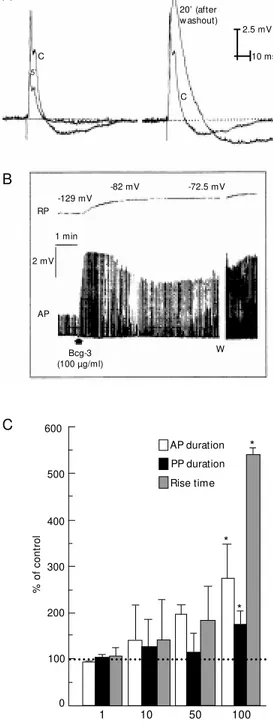
A mild cytolytic activity on mouse eryth-rocytes was obtained with whole venom and some of the fractions (data not shown). One of the fractions, Bcg-2, produced the most potent effects and when tested in the qualita-tive hemolytic assay it elicited a dose-de-pendent effect. The data obtained were fitted to a sigmoid dose-response curve, the EC50 value obtained was 14 µg/ml and the maxi-mum dose of Bcg-2 was 22 µg/ml (Figure 2). All gel-filtration fractions were tested on the isolated crab leg sensory nerve using the sucrose-gap method. The Bcg-3 fraction elic-ited the most interesting neuroactive effects. First at all, it produced a rapid decrease in the amplitude of the action potentials (Figure 3A), as well as depolarization of the resting membrane potential (Figure 3B). After wash-out of the preparation, the amplitude in-creased and there was an increase of the action potential and postpotential duration with a concomitant increase in the rise time of the signal (Figure 3A and C). The effects that appeared after washout were irrevers-ible (no alteration was obtained after a sec-ond washout) and dose-dependent, and be-came significant with 100 µg/ml of Bcg-3 (Figure 3C).
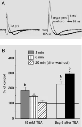
To clarify the mechanism of action of Bcg-3 on this nerve preparation, we com-bined the effects produced by TEA or TTX with those produced by Bcg-3. TEA (15 mM) application to the nerve produced a rapid and significant increase of the action potential duration, which was reversed after washout (Figure 4). When Bcg-3 (100 µg/ ml) was applied to a TEA-pretreated prepa-ration, an additional and significant increase
0.25 0.50 0.75 1.00 1.25 1.50
Log [Bcg-2] (µg/ml)
Figure 2. Cytolytic activity of fraction Bcg-2 on mouse erythrocytes. The data w ere fitted to a sigmoid dose-response curve (EC50 = 14 µg/ml; maximum dose = 22 µg/ml). Percent hemolysis w as measured at 540 nm and compared w ith the hemolytic effect of Triton X-100. Data are reported as means ± SEM of percent hemolysis (N = 3).
H
em
ol
ys
is
(%
)
100
75
50
25
of the action potential duration was observed (Figure 4A,B), lasting as long as 1 h after washout of the preparation.
The blockade of Na+ channels by TTX (1 µM) after Bcg-3 (100 µg/ml) application and washout of the preparation produced a de-crease in the action potential amplitude (80%) that did not block nerve conduction. The increase of the duration of the action poten-tial persisted, as observed before TTX (Fig-ure 5). When the effects of TTX were re-versed after a second washout, the effects of Bcg-3 soon appeared (data not shown).
D iscussio n
The present results show the presence of powerful hemolytic and neuroactive sub-stances in two fractions obtained by gel-filtration chromatography of B. cangicum whole venom.
The extraction method was very simple, with the venom being less contaminated with other compounds from the sea anemone body, with consequent simplification of the purifi-cation procedures. Also, it is important to note that the animals stay alive and can therefore be re-used to obtain more venom or be returned to the sea.
Hemolytic activity on mouse erythrocytes was found in whole venom and some of its fractions. The Bcg-2 fraction elicited the most potent and marked hemolysis in a dose-dependent manner, with an EC50 of 14 µg/ml and a maximum dose of 22 µg/ml. The cy-tolytic toxins isolated from sea anemones had a molecular mass between 16 and 21 kDa (3,17). The components of the Bcg-2 fraction are in the same range, with a molec-ular mass between 8 and 18 kDa, and there-fore may probably contain one or more cy-tolytic substances. Further purification will be necessary in order to test this hypothesis and to clarify the mechanism of action of the cytolytic components on cell membranes. It is interesting to note that Malpezzi and Freitas (10) isolated a hemolysin of 14 kDa from an
equivalent G-50 fraction from B. caissarum, a sea anemone phylogenetically related to B. cangicum.
Bcg-3 elicited highly interesting neuro-active effects when tested on the isolated crab leg sensory nerve by the sucrose-gap method. After Bcg-3 was applied to the
prepa-Figure 3. Effects of fraction Bcg-3 (100 µg/ml) on crab nerve. A, Representative traces show ing control (C) action potential (AP) and traces obtained 5 min after Bcg-3 (left side traces). Right side traces compare control AP (C) and AP 20 min after Bcg-3 (after w ashout). B, Effects of Bcg-3 on resting membrane po-tential (RP) and AP duration; the effects w ere not w ashed out. W = w ashout. The value of RP ob-tained is indicated. C, Dose-re-sponse effect of Bcg-3 on AP, postpotential (PP) duration and rise time after w ashout of the preparation (20 min after the ap-plication). Absolute control val-ues: AP duration = 14.8 ± 0.3 ms; PP duration = 63.2 ± 6.7 ms; rise time = 2.5 ± 1.2 ms (N = 3). * P<0.01 (ANOVA-Dunnett test).
C
5’
20’ (after w ashout)
2.5 mV
10 ms
C
-129 mV
-82 mV -72.5 mV
RP
1 min
2 mV
AP
Bcg-3 (100 µg/ml)
W A
B
C
%
o
f
co
nt
ro
l
600
500
400
300
200
100
0
AP duration PP duration Rise time
1 10 50 100
Bcg-3 (µg/ml)
*
*
*
effect of one substance on another is also proposed for a unique substance like guani-dine, which elicited two different effects before and after the washout of the prepara-tion (18).
Since the effects produced by Bcg-3 on the duration of the action potential could be related to a blockade of the inactivation phase of Na+ currents, or a blockade of K+ currents (19), we studied the combined effects of Bcg-3 with those of the known toxins TEA and TTX.
When Bcg-3 (100 µg/ml) was added to a TEA-pretreated preparation, a synergistic effect on action potential duration appeared. This fact suggests an additional blockade of TEA-sensitive K+ channels, or a blockade of a population of TEA-insensitive ones or a modulation of the Na+ channels. The sugges-tion that Bcg-3 could be blocking a popula-tion of TEA-insensitive K+ channels is prob-ably valid since Bcg-3 application to this nerve preparation produced depolarization of the resting membrane potential, an effect that was not observed with TEA in this prepa-ration (Figure 3B).
The hypothesis that another Bcg-3 com-ponent was probably modulating Na+ chan-nels is supported by the fact that 1 µM TTX failed to block completely the nerve conduc-tion in a Bcg-3-pretreated and washed prepa-ration. The neurotoxic effects of TTX may be reduced by the concentration of Na+ ions in the solution of the Bcg-3 fraction obtained by gel filtration, thus preventing TTX bind-ing to Na+ channels. On the other hand, a possible interaction of Bcg-3 with these chan-nels may prevent TTX binding to its site of action. The above results confirm the irre-versible interaction of the component(s) of Bcg-3 with the nerve membrane, that also occurs in the presence of TTX, a fact that has been consistently observed with neuroactive substances isolated from sea anemones (20, 21).
Thus, the use of specific Na+- and K+ -blocking agents in combination with Bcg-3 Figure 5. Effect of 1 µM
tetrodo-toxin (TTX) on action potential in a Bcg-3 (100 µg/ml)-pretreated nerve. C = control conditions (before TTX); 25' = 25 min after Bcg-3 application and w ashout of the preparation (and after TTX). 20 ms 5 mV 25’ C TTX Figure 4. Effects of
tetraethyl-ammonium (TEA, 15 mM ) and TEA plus Bcg-3 (100 µg/ml) on crab nerves. A, Left side traces
show control (C) action potential (AP) and AP obtained 3 min after TEA applicat ion. Right side traces compare the effects of TEA (TEA, 3') w ith those elicited by Bcg-3 in a TEA-pretreated preparation (after w ashout). B, Left bars represent the effects of AP duration obtained at differ-ent times after TEA application. Right bars represent the effect of Bcg-3 on a TEA-pretreated preparation (open bar = 1 min after Bcg-3; black bar = 14 min after Bcg-3). The significance of the data as compared w ith their ow n controls are indicated by a and b (a = P<0.05, b = P<0.01); the significance obtained w hen comparing the tw o types of ex-periments at the same time (3, 6 and 20 min) is expressed as * P<0.05 and +P<0.01 (N = 4; ANOVA-Dunnett test). TEA (3’) TEA (3’) Bcg-3 (after w ashout) 5 mV 20 ms C A 12345 12345 12345 12345 12345 12345 12345 12345 123456 123456 123456 123456 123456 123456 123456 123456 123456 123456 123456 123456 123456 123456 1234 1234 1234 123 123 123 400 300 200 100 0 % o f co nt ro l 3 min 6 min
20 min (after w ashout)
b a b * b +
15 mM TEA Bcg-3 after TEA
B
may suggest the co-existence of two types of neuroactive substances in this fraction, one modulating Na+ channels and the other block-ing K+ channels, with an overall effect on the prolongation of the nerve action potential. Since the estimated molecular weight of Bcg-3 is of the same order as other Na+ and K+ channel toxins isolated from sea anemones, the co-existence of these two types of toxins cannot be ruled out. Data reported by Araque et al. (15) demonstrated that the venom ob-tained from the same anemone includes a toxin that selectively blocks the Ca2+ -de-pendent K+ currents in a reversible manner without modifying the Na+ currents. More-over, Harvey et al. (13) demonstrated that the venom of the same anemone blocks the binding of labeled DTX (a known K+ chan-nel blocker) to synaptosomal preparations.
Similar pharmacological activities have been observed in other species of the same genus. BgK, isolated from B. granulifera, displaces DTX binding from rat brain synap-tosomes and suppresses K+ currents in cul-tured ganglion cells (8). Also, two neurotox-ins, one of which interacts with Na+ chan-nels and the other with K+ channels, have been isolated from B. caissarum, a co-habi-tant of B. cangicum along the Brazilian sea-shore (22).
It has been shown before that many sea anemone toxins that interact with Na+ chan-nels have a very small activity toward mam-mals but a very high activity toward crusta-cean (23,24). Araque et al. (15), using mam-malian chromaffin cells, demonstrated that the whole venom of B. cangicum had no
effects on Na+ or K+ currents. However, our results clearly show that the Bcg-3 fraction modified these currents in an invertebrate nerve preparation. These differences may be related to the species-specific activity of the sea anemone venoms described above.
The above data raise a number of ques-tions about the role of different toxins found in the same organism. From a biological point of view, it makes sense that sea anemo-nes should produce a diverse spectrum of toxins more oriented toward ionic channels normally found in their preys (fish, crusta-cean, jellyfish, etc.), a fact that gives preda-tors a distinct evolutionary advantage. The mixture of cytotoxic and ion channel toxins found in sea anemone venom comprising pore-forming toxins (3,6,25), Na+ channel toxins tending to activate these channels (1,5,23) and K+ channel toxins tending to block K+ currents (2,8,9,24), is expected to have devastating neurotoxic effects by pro-ducing a massive release of neurotransmit-ters and exerting a potent effect on heart, muscle and endocrine cells. As the number of anemone toxins continues to grow, more variability within a given species will un-doubtedly be discovered.
We demonstrated the presence of power-ful hemolytic and neuroactive substances in two fractions obtained by gel filtration of B. cangicum whole venom. Further purifica-tion of their components will permit us to clarify their mechanism of action on cell membranes and should provide interesting pharmacological tools isolated from marine organisms.
Re fe re nce s
1. Norton RS (1991). Structure and struc-ture-function relationships of the sea anemone proteins that interact w ith the sodium channel. Toxicon, 29: 1051-1084.
2. Karlsson E, Adem A, Aneiros A, Castañeda O, Harvey A, Jolkkonen M & Sotolongo V (1991). New toxins from marine organ-isms. Toxicon,29: 1168 (Abstract).
3. Kem WR (1988). Sea anemone toxins: structure and action. In: Hessinger DA & Lenhoff HM (Editors), The Biology of the Nematocysts. Academic Press, Inc., New York.
4. M acek P (1992). Polypeptide cytolytic tox-ins from sea anem ones (Actiniaria).
FEM S, M icrobiology and Immunology,
105: 121-130.
5. Beress L, Beress R & Wunderer G (1975). Isolation and characterization of three polypeptides w ith neurotoxic activity from
Anemonia sulcata. FEBS Letters, 50: 311-314.
channels formed in planar lipid bilayer membranes by the cytolytic toxin from the sea anemone, Stoichactis helianthus. Journal of M embrane Biology,55: 203-211.
7. M acek P & Lebez D (1988). Isolation and characterization of three lethal and hemo-lytic toxins from the sea anemone Actinia equina. Toxicon,26: 441-451.
8. Aneiros A, Garcia I, M artinez JR, Harvey A, Anderson AJ, M arshall DL, Engström Å, Hellman U & Karlsson E (1993). A po-tassium channel toxin from the secretion of the sea anemone Bunodosoma granuli-fera. Isolation, amino acid sequence and
biological activity. Biochimica et Biophy-sica Acta,1157: 86-92.
9. Santana ANC, Leite AB, França M SF, França L, Vale OC, Cunha RB, Ricart CAO, Sousa M V & Carvalho KM (1998). Partial sequence and toxic effects of granulo-toxin, a neurotoxic peptide from the sea anemone Bunodosoma granulifera. Bra-zilian Journal of M edical and Biological Research,31: 1335-1338.
10. M alpezzi ELA & Freitas JC (1991). Hemo-lytic activity of the nematocyst venom from the sea anemone Bunodosoma cais-sarum. Brazilian Journal of M edical and Biological Research,24: 1245-1249. 11. M alpezzi ELA, Freitas JC, M uramoto K &
Kamiya H (1993). Characterization of pep-tides in sea anemone venom collected by a novel procedure. Toxicon,31: 853-864. 12. Harvey AL, Anderson AJ, Braga M FM , M arshall DL, Row an EG, Vatanpour H, Castañeda O & Karlsson E (1992). Toxins affecting neuronal ion channels. In: Gopa-lakrishakone P & Tan CK (Editors), Recent
Advances in Toxicology Research.Vol. 1. National University of Singapore, Sin-gapore.
13. Harvey A, Row an EG, Vatanpour H, Young LC, Castañeda O, M ebs D, Cerveñansky C & Karlsson E (1996). Potassium channel toxins from sea anemones. In: Lazarovici P (Editor), Biochemical Aspects of M arine Pharmacology. Allaken Inc., Fort Collins. 14. Dolly JO, Stanfeld CE, Breeze AL,
Pel-chen-M atthew s A, M arsh SJ & Brow n DA (1987). Neuronal acceptor sub-types for dendrotoxin and their relation to K+. In: Jenner PG (Editor), Neurotoxins and their Pharm acological Im plicat ions. Raven
Press, New York.
15. Araque A, Urbano FJ, Cerveñansky C, Gandia L & Buño W (1995). Selective block of Ca2+-dependent K+ current in crayfish neuromuscular system and chro-maffin cells by sea anemone Bunodo-soma cangicum venom. Journal of Neuro-science Research,42: 539-546.
16. Galletis P & Norton RS (1990). Biochemi-cal and pharmacologiBiochemi-cal studies of the mechanism of action of tenebrosin-C, a cardiac stimulatory and hemolytic protein from the sea anemone Actinia tenebrosa. Toxicon,28: 695-706.
17. Harvey AL (1990). Cytolytic toxins. In: Shier WT & M ebs D (Editors), Handbook of Toxicology. M arcel Dekker Inc., New York and Basel.
18. Rodrigues-Simioni L, Silva-Carvalho I, Heluany NF, Leite GB, Prado-Franceschi J, Cruz-Höfling M A, Ballejo G & Corrado AP (1997). Novel effects of guanidine on the neuromuscular junction. General Phar-macology, 28: 599-605.
19. Hille B (1992). Ionic Channels of Excitable M embranes. 2nd edn. Sinauer Associates Inc., Sunderland, M A.
20. Vincent JP, Balerna M , Barhanin J, Fosset M & Lazdunski M (1980). Binding of sea anemone toxin to receptor sites associ-ated w ith gating system of sodium chan-nel in synaptic nerve endings in vitro. Pro-ceedings of the National Academy of Sci-ences, USA,77: 1646-1650.
21. Salgado VL & Kem WR (1992). Actions of three structurally distinct sea anemone toxins on crustacean and insect sodium channels. Toxicon,30: 1365-1381.
22. M alpezzi ELA (1996). Estudo das toxinas de nematocistos da anêmona de mar Bu-nodosom a caissarum Corrêa 1964
(Cnidaria, Anthozoa, Actiinidae). Doctoral thesis. IBUSP, Universidade de São Paulo, São Paulo, Brazil.
23. Lazdunski M , Frelin C, Barhanin J, Lombet A, M eiri H, Pauron D, Romey G, Schmid A, Schw eitz H, Vigne P & Vijverberg HPM (1987). Polypeptide toxins as tools to study voltage-sensitive Na+ channels.
An-nals of the New York Academy of Sci-ences,479: 204-220.
24. Schw eitz H, Bruhn T, Guillemare E, M oinier D, Lancelin J-M , Beress L & Lazdunski M (1995). Kalicludines and kaliseptine. Tw o different classes of sea anemone toxins for voltage-sensitive K+ channels. Journal of Biological Chemistry, 270: 25121-25126.