Full paper published online: May 30, 2009 ISSN 1678-9199.
EXPRESSION OF SCHISTOSOMAL CATHEPSIN L1 IN Escherichia coli AND EVALUATION OF ITS PROTECTIVE CAPACITY IN AN ANIMAL CHALLENGE
Miyasato PA (1), Ramos CRR (2), Abreu PAE (3), Dias WO (2), Nascimento C (1), Ho PL (2), Kawano T (1)
(1) Department of Parasitology, Butantan Institute, São Paulo, São Paulo State, Brazil; (2) Biotechnology Center, Butantan Institute, São Paulo, São Paulo State, Brazil; (3) Laboratory of Bacteriology, Butantan Institute, São Paulo, São Paulo State, Brazil.
ABSTRACT: Schistosomes utilize proteinases to accomplish several activities such as tissue penetration, tissue digestion and evasion of host immune responses. Cathepsin L is a cysteine proteinase of the papain superfamily detected in their gut lumen which indicates that this enzyme contributes to the proteolysis of ingested hemoglobin. Due to the roles played in the schistosome biology, proteolytic enzymes are considered potential targets for developing and guiding antischistosomal therapies. In the present work, the cathepsin L1 cDNA coding of Schistosoma mansoni was cloned into the pAE vector that provides high-level expression of heterologous proteins in Escherichia coli. The recombinant protein was expressed as inclusion bodies, purified under denaturing conditions through nickel-charged chromatography and used for experimental animal vaccination. ELISA was performed with the pooled sera. Although this protein was shown to be immunogenic, mice immunized with three doses of recombinant protein plus aluminum hydroxide as adjuvant were not protected against S. mansoni infection.
KEY WORDS: Schistosoma mansoni, cysteine proteinase, cathepsin L1, vaccine,
immunogenicity.
CONFLICTS OF INTEREST: There is no conflict.
FINANCIAL SOURCE: Butantan Foundation.
CORRESPONDENCE TO:
PAULO LEE HO, Centro de Biotecnologia, Instituto Butantan, Av. Vital Brasil, 1500,
São Paulo, SP, 05503-900, Brasil. Phone: +55 11 3726 7222, ext. 2244. Fax: +55 11
INTRODUCTION
Schistosomiasis remains a major public health problem in developing countries. It is
estimated that 200 million people are infected by it and six hundred million live in risk
areas (1). Despite two decades of widespread chemotherapy with safe and effective
drugs, the number of individuals with schistosomiasis remains high (2). The
construction of water resources and agricultural requirements for human
development have led to increasing transmission of schistosomiasis (3). Control of its
transmission can be achieved by elimination of intermediary hosts, use of drugs like
oxamniquine and praziquantel, besides providing adequate socioeconomic
conditions and sanitary education for the population. However, the risk of
reintroduction of the disease into a treated area, particularly where there are irrigation
projects and population migration, must be taken into account (4, 5).
Reinfection demands repeated treatment and continued administration of medication,
which can favor the emergence of drug-resistant parasites (6). Moreover, supplying
efficient treatment that covers all parts of an endemic area can make chemotherapy
an expensive and impractical alternative (7).Thus, vaccination would be a necessary
approach to complement chemotherapy (8).
Several factors make schistosomiasis vaccine development an advantageous option: • protective mechanisms have been demonstrated in experimental animals, • rapid reinfection demands continuing treatment,
• the risk of drug-resistance caused by expanded chemotherapy programs, • acquired partial immunity reduces the infection intensity as observed in
adolescent and older age groups compared to children.
Control based on chemotherapy followed by vaccination would result in a short-term
effect with long-term protection (2, 9). Thus, a definitive vaccine must be elaborated
from diverse recombinant antigens and from different parasite stages to improve the
possibilities for synergistic action by involving separate defense mechanisms (9).
Secreted or surface proteins are crucial to maintaining the pathway of parasites
through their molecular interaction with host cells. Since these molecules are
exposed to the host immune system, they become candidate targets for vaccine
Many histochemical and immunolocalizational studies report that cathepsin L (EC
3.4.22.15) is packed into the lysosomal-like vesicles of gastrodermal cells of
helminthes and plays an important role in nutrient acquisition (11). Its strong
involvement in hemoglobin digestion indicates that this cysteine proteinase is
important to Schistosoma mansoni (12). Cathepsin L1 was first isolated by Smith et al. (13) whereas its presence was detected in gastrodermal cells lining the gut and in excretory/secretory (ES) products (14), as later confirmed by immunolocalization
studies (15). Michel et al. (16) constructed an expression plasmid in E. coli to produce recombinant schistosome cathepsin L. The utilization of purified cathepsins
L1 and L2 from Fasciola hepatica for vaccination in rats, sheep and cattle has induced protection, ranging from 33 to 80%, in experimental challenge with F. hepatica metacercariae as well as provoked potent anti-embryonation/hatch rate effects (17, 18). However, there are still no studies on recombinant SmCL1
(cathepsin L1 from Schistosoma mansoni) as a vaccine antigen candidate against S. mansoni infection. Therefore, the general objective of the current work was to verify SmCL1 immunogenicity and feasibility as a vaccine antigen against this worm.
MATERIALS AND METHODS Bacterial Strains and Plasmids
Escherichia coli DH5α and BL21-SI strains (Invitrogen, USA) were employed in all
routine cloning and expression experiments. In the BL21-SI strain, the T7 RNA
polymerase expression was controlled by the osmotically inducible promoter proU
(19).
Parasites
Snail intermediate hosts (Biomphalaria glabrata) were infected with S. mansoni BH strain. Cercariae were allowed to shed from infected snails for two hours under a
halogen lamp. Cercarial counts and viability were determined through
stereomicroscopy (20).
Cloning Techniques
Total RNA was isolated from S. mansoni worms with Trizol® reagent (Invitrogen, USA). Two micrograms of total RNA was reverse-transcribed by SuperScript II®
Healthcare, USA). The first cDNA strand was utilized as a reference in the PCR
reaction according to the following sense and antisense primers: forward SmCL1 5’
ATGATATCCATATGCCTGTAAACCTCGAGTAC 3’ and reverse SmCL1 5’
GCACGCGTTAGTCGACGTAGATCATCGCTGACGTAGC 3’. Underlined sequences
indicate NdeI restriction site in the forward primer and MluI and SalI restriction sites in the reverse primers, respectively. The PCR product was purified from agarose gels
after electrophoretic separation using a commercial kit, cloned into pGEM®-T vector
(Promega, USA), and sequenced with the T7 and SP6 promoter primers. The
nucleotide sequence analysis of SmCL1 cDNA was performed on the ABI PRISM®
377 sequencer (Applied Biosystems, USA) using the ABI PRISM Big Dye®
Terminator cycle sequencing kit. The cDNA sequence was subcloned into the
expression vector based on pAE plasmid, modified to allow the expression of the
recombinant protein with a His6 tag fusion in the C-terminal end at the NdeI and SalI sites, resulting in the pAE SmCL1 plasmid (21).
Expression and Purification of the Recombinant SmCL1 Protein
The pAE SmCL1 plasmid was employed to transform Escherichia coli BL21 SI strain. A half liter of LBON (Luria-Bertani medium without NaCl) was inoculated with 15 mL
of the overnight bacterial culture from single colonies and grown until the optical
density at 600 nm reached 0.8. The expression of recombinant protein was induced
with 200 mM NaCl, overnight at 30°C. The cells were harvested by centrifugation and
the bacterial cell pellet was resuspended in a solution containing 20 mM Tris HCl (pH
8.0), 0.5 M NaCl and lysed in a French pressure cell.
The inclusion bodies containing the recombinant protein were isolated from bacterial
lysates by centrifugation, and solubilized with 50 mL of denaturation buffer (8 M urea,
20 mM Tris HCl, pH 8.0, 0.5 M NaCl, 5 mM imidazole and 1 mM β-mercaptoethanol)
and stirred for 16 hours at room temperature. The recombinant protein present in the
suspension was purified by metal-affinity chromatography in Chelating-Sepharose®
Fast Flow resin (GE Healthcare, USA), previously charged with 300 mM NiSO4 and
equilibrated with denaturation buffer. The column was washed with ten volumes of
denaturation buffer (8 M urea, 20 mM Tris HCl, pH 8.0, 0.5 M NaCl, 5 mM imidazole
and 1 mM β-mercaptoethanol). This procedure was followed by three additional
M urea and 20 mM imidazole in the first wash; ten volumes of the same buffer
containing 4 M urea and 40 mM imidazole in the second wash; and ten volumes of
the same buffer containing 3 M urea and 60 mM imidazole in the third wash.
The recombinant protein was eluted with five volumes of the elution buffer containing
20 mM Tris HCl, 0.5 M Na Cl (pH 8.0), 3 M urea and 1 M imidazole. Several steps of
dialysis were also performed: first against a solution containing 20 mM Tris HCl, 0.5
M NaCl (pH 8.0), 2 M urea, 0.1% glycine, 10 mM EDTA and 0.5 M imidazole; a
second step, against a solution containing 20 mM Tris HCl, 0.5 M NaCl, (pH 8.0), 2 M
urea, 0.1% glycine and 10 mM EDTA. The last dialysis was against
phosphate-buffered saline solution (PBS), 0.1% glycine and 2 M urea. Purified protein samples
were analyzed by 12% sodium dodecyl sulfate polyacrylamide gel electrophoresis
(SDS-PAGE).
Immunization of Swiss Mice and Challenge with S. mansoniCercariae
Two independent experiments were conducted in accordance with Brazilian animal
protection and welfare laws and the research project was approved by the Ethics
Committee on the Use of Animals (protocol number 160/2004), Butantan Institute.
Ten outbred female Swiss mice (4 to 6 weeks old, Butantan Institute) received
footpad immunization with three doses of 20 μg of recombinant protein in PBS
emulsified in aluminum hydroxide as adjuvant at intervals of seven days. The
recombinant protein was adsorbed onto 10% (vol/vol) of Alhydrogel (2% Al(OH)3
Biosector, formerly Superfos Biosector, Denmark). At this concentration, the protein
adsorption capacity of the adjuvant is 2.3 +/- 0.5 mg per mL, which is within the range
of protein concentration used. The adjuvant was provided by the Vaccine Quality
Control Section of Butantan Institute and we specifically followed the procedures
routinely used in the section.
Control mice were injected with PBS in aluminum hydroxide. One day before each
immunization, mice were bled from the retro-orbital plexus, and enzyme-linked
immunosorbent assay (ELISA) was performed with pooled sera. Sixty days after the
last immunization, animals were challenged by subcutaneous injection with 100
cercariae (each mouse) and perfused 45 days later. Protection was calculated by the
equation [(C – V)/C] x 100, where C is the average number of worms in control
animals and V is the average number of worms in vaccinated animals, expressed as
ELISA
ELISA was carried out according to Sato et al. (22). Plates were coated overnight at
4°C with rSmCL1 (10 μg/mL), and blocked with 10% nonfat dry milk in PBS-T (PBS
with 0.05% Tween-20) at 37°C for one hour. Subsequently, plates were washed
three times with PBS-T. After washes, serial dilutions with immunized animal sera
were performed at 37°C, for one hour each dilution. The reaction with goat
anti-mouse total immunoglobulin G (IgG) conjugated to horseradish peroxidase (HRP)
(dilution 1:15,000 – Sigma, USA) was performed in PBS-1% nonfat dry milk at 37°C,
for one hour and then washed with PBS-T. For HRP substrate, we added θ
-phenylenediamine (0.04%) in citrate phosphate buffer (pH 5.0) plus 0.01% H2O2 and
the reaction was stopped by the addition of 50 μL of 4 M H2SO4. The absorbance
was measured at 492 nm. The antibody titer was considered the last dilution of sera
that registered an optical density of 0.1.
RESULTS
Cloning of SmCL1 cDNA
cDNA was obtained from RNA isolated from adult worms of the Brazilian endemic BH
S.mansoni strain. The cDNA sequence of recombinant protein SmCL1 was amplified from two independent RT-PCRs. The amplified segments were cloned into pGEM-T
and five independent clones were sequenced. DNA sequence analysis indicated that
SmCL1 cDNA sequences were the same found by Smith et al. (13).
Expression and Purification of Recombinant SmCL1
The expected band of recombinant SmCL1 protein (36.95 kDa) was observed, by
SDS-12% PAGE, in bacterial cell lysates from induced cultures (Figure 1, lane 3).
The recombinant SmCL1 was present in the pellet of bacterial cell lysates in an
insoluble form as inclusion bodies, as revealed by 12% SDS-PAGE (Figure 1, lane
4). Recombinant proteins present in inclusion bodies were isolated, solubilized and
purified (Figure 1, lane 6) under denaturing conditions through nickel charged
chromatography, as previously described. The purified recombinant protein yield was
Figure 1. Analysis of the recombinant SmCL1expressed from NaCl-induced E. coli BL21-SI by SDS-12% PAGE. Bands were visualized by Coomassie staining. Lane 1:
molecular mass protein markers; lane 2: non-inducedculture; lane 3: induced culture;
lane 4: inclusion body pellet after centrifugation (insoluble fraction); lane 5:
supernatant after centrifugation (soluble fraction); lane 6: protein purified from
insoluble fraction by Ni-charged resin beads.
Antibody Response and Protection against Challenge with S. mansoni
Cercariae in Mice Immunized with the Recombinant SmCL1
Swiss mice were immunized with three 20-μg doses of purified recombinant SmCL1
according to Materials and Methods. Negative control mice were injected with PBS in
the presence of the adjuvant. Sera from the immunized animals were analyzed by
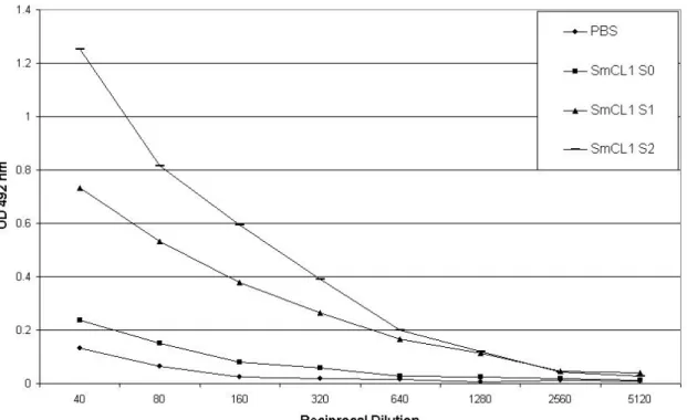
ELISA using recombinant SmCL1 as coating antigen. Experimental group showed
higher titer of anti-SmCL1 antibodies (1/1 280) than the control group (Figure 2).
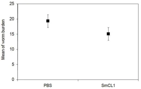
Animals were challenged with S. mansoni cercariae and the protection level was calculated by comparing the number of adult worms recovered from the experimental
vaccinated group with the number from the control group. Mice immunized with
recombinant SmCL1 showed a decrease of 21.8% in worm burden (Figure 3).
However, the difference in the number of worms recovered from SmCL1 group and
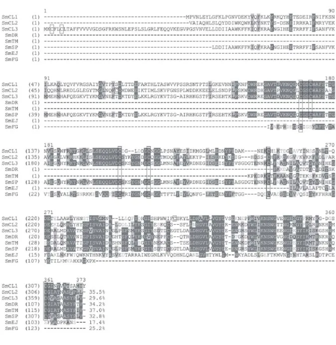
from the control group was not statistically significant. Recently, a SmCL1 related
n. EU022371) (Figure 4). We do not know if anti-SmCL1 also cross reacts with
SmCL3.
Figure 2. Antibody responses induced by recombinant SmCL1. Swiss mice received
footpad immunization, in intervals of seven days, that comprised three 20-μg doses
of recombinant protein (SmCL1) in PBS adsorbed in aluminum hydroxide. Control
mice were injected with PBS in aluminum hydroxide. One day before the first and the
last immunization, as well as before challenge, mice were bled from the retro-orbital
plexus and pooled sera were analyzed by ELISA. PBS: sera collected from mice
injected with PBS; SmCL1 S0: sera collected from mice before the first immunization;
SmCL1 S1: sera collected from immunized mice before the last immunization;
Figure 3. Animal protection assay against challenge with Schistosoma mansoni
cercariae. Swiss mice were immunized with three doses of 20 µg of recombinant
SmCL1 adsorbed to aluminum hydroxide in intervals of seven days. The control
group was immunized with PBS adsorbed in aluminum hydroxide. All mice were
challenged subcutaneously 60 days after the last immunization with 100
cercariae/mouse and perfused 45 days later. The mean worm burden was calculated
using results from two experiments. Percentage of protection in the two independent
experiments was calculated by comparing their results to the results obtained from
the control groups (not shown). Statistical analysis was performed with Student’s
Figure 4. Alignment of amino acid sequences of new potential cathepsin L family
sequences (http://verjo18.iq.usp.br/schisto/) from its transcriptome (30). Dark grey
background and white letters: conserved residues; light grey background and black
letters: block of similar amino acid residues; and dark grey letters: weakly similar.
The Cys amino acid residues were indicated by box. The alignment was made by
Program Align X, a component of Vector NTI® suite, version 9.0 (Invitrogen, USA).
DISCUSSION
The results presented herein showed the expression of the recombinant SmCL1
protein in Escherichia coli BL21 SI strain, with high final yield of purified protein. The expressed protein displayed six histidine residues at the carboxy terminus of the
recombinant protein, which allowed its purification through affinity chromatography on
nickel-charged beads. The recombinant protein was expressed as insoluble inclusion
bodies, thus a solubilization step was necessary. In the final purification step, urea
concentration in the buffer was maintained at 2 M to avoid protein precipitation, as
observed in lower concentrations of urea.
The purified recombinant SmCL1 was not able to hydrolyze hemoglobin (results not
shown), which indicates that the purified enzyme was inactive. However, Verity et al. (23) carried out immunization assays with two forms of recombinant cathepsin D of
Schistosoma japonicum, one purified from Escherichia coli that was enzymatically
inactive and the other, from insect cells, that was enzymatically active. The highest
level of protection was achieved in animals immunized with the purified protein from E. coli, suggesting that even the inactive form of the protein can provide significant
epitopes that contribute for protection. Therefore, immunizations were carried out
with the inactive form of the recombinant SmCL1 expressed in E. coli to verify its immunoprophylactic potential.
The SmCL1 antigen showed to be immunogenic, being able to induce a more
significant titer of anti-SmCL1 antibodies (1/1,280) than the control group.
Nevertheless, mice immunized with recombinant SmCL1 showed only a 21.8%
decrease in worm burden, below of the goal recommended by WHO (40%). In
contrast, homologous cathepsin L1 of Fasciola hepatica revealed positive correlation between antibody titer and protection against challenge with Fasciola hepatica metacercariae (24). Moreover, in a recent study, Kesik et al. (18) obtained 78 to 80% of protection against challenge with metacercariae, using recombinant cathepsin L of
At least three reasons could explain the lack of protection of the recombinant SmCL1
in our conditions. First, there is the possibility that this protein do not provide a
complete protection. In this case, it would be necessary to co-administer this antigen
with other antigens or adjuvants.
The second factor that could have influenced the protection response of recombinant
SmCL1 was the lack of its correct folding, since it was not able to hydrolyze
hemoglobin (results not shown). This consequence is distinct from that obtained for
the recombinant inactive form of cathepsin D of Schistosoma japonicum, which showed significant protective activity, slightly higher than the recombinant active form
produced in insect cells (23).
In our conditions, protective epitopes may not be presented to the immunized
animals. In this regard, it would be interesting to express this antigen in another
bacterial expression system, for instance in fusion with maltose binding protein in an
attempt to express the protein in a soluble form as used for Schistosoma mansoni tegumental Sm13 antigen (25) or using a protocol able to produce functional and
correctly folded F. hepatica cathepsin L in E. coli (18). On the other hand, expression systems like yeast or insect cells can also be explored in order to obtain functional
SmCL1 and to evaluate if properly folded, the recombinant protein is able to improve
the level of protection observed in unfolded proteins expressed in E. coli, as described herein. Interestingly, it was observed that even small differences in
recombinant proteins may interfere in protection ability. For instance, vaccination and
challenge experiments in rats with Fasciola hepatica procathepsin L3 protein, produced in yeasts or insect cells, showed that only the protein expressed in insect
cells was able to induce protection, in contrast to the yeast derived antigen, although
both elicited similar antibody responses (26).
The third reason that could elucidate the shortage of protection of the recombinant
SmCL1 in the present study is the occurrence of positive diversification of cysteine
proteinase genes in helminthes. The available data suggests that parasites possess
an ample and complex panel of proteinases (11). Possibly, they take advantage of
such multiple proteinases for immune evasion. The activity of several proteases –
other than SmCL1 – that could participate in hemoglobin proteolysis, like cathepsin
L2 or cathepsins B, C and D, could have contributed to the lack of protection of
SmCL1. Each of these enzymes has an optimum pH for activity; consequently, the
schistosome gut, provides adequate conditions for each enzyme to degrade
hemoglobins systematically. Probably, they act synergistically or possess activity
redundancies to ensure the hemoglobin catabolism (27, 28). In this regard, it is
expected that immunization with a combination of such proteases would improve the
protection response. Moreover, the presence of paralogs in the same organisms
would also assure this redundant effect in order to avoid function loss of SmCL1.
The analysis of Schistosoma mansoni transcriptome provides an important support for the comprehension of molecular mechanisms involved in the nutrition and
metabolism of this parasite; the host-dependent development and maturation; the
immune evasion and evolution mechanisms; as well as new potential vaccine
candidates and drug targets (29). On this matter, S. mansoni transcriptome provided us an opportunity to search for potential SmCL1 paralogs (30). Some new sequences
found in “Schistosoma mansoni EST Genome Project” – assembled in S. mansoni EST sequences (SmAEs) and transcriptome analysis in Services Mirror – IQ USP
(http://verjo18.iq.usp.br/schisto/) – presented similarity with other cathepsins L of the
following organisms: Sarcophaga peregrina (SmSP), Fasciola gigantica (SmFG), Engraulis japonicus (SmEJ), Delia radicum (SmDR) and Tenebrio molitor (SmTM), showing that Schistosoma mansoni possesses more than the three full length
cathepsins L-like proteins characterized so far (SmCL1, SmCL2 and SmCL3).
By alignment of the amino acid sequences from these cathepsins (Figure 4), we can
observe variable identities of 17.4 to 37% among all the Schistosoma mansoni cathepsins L-like sequences with SmCL1. However, it was evident that the
transcribed sequence uncovered by transcriptomic studies that present the best hit
against Sarcophaga peregrina cathepsin L protein (SmSP) is, in fact, identical or close related to SmCL3 sequence recently deposited in the databanks (unpublished
results, accession n. EU022371). The multiplicity of cathepsin L-like isoforms in
Schistosoma mansoni may allow the parasite to escape from the immune response elicited by the vaccination with recombinant SmCL1. In this case, the use of
associated paralogs may avoid the evasion mechanism and improve protection
against infections. Indeed, immunization with F. hepatica cathepsin L1 combined with cathepsin L2 provides a higher level of protection than with the use of each antigen
alone (31).
The development of an anti-schistosomiasis vaccine is an enormous challenge, due
possible that the lack of the protection observed in our experimental conditions are
the combined results of all factors discussed before. Further studies must be carried
out to clarify these points and demonstrate the feasibility of S. mansoni cathepsin L1 as a vaccine antigen.
The current Schistosoma vaccine candidates are not very effective. It is important to identify new antigens and to explore vaccination strategies to improve vaccine
efficacy. Functional annotation of S. mansoni transcriptome and the public accessibility to its database (http://verjo18.iq.usp.br/schisto/), in combination with
postgenomic technologies, provide the potential to identify a new generation of
potential vaccine target molecules (7).
ACKNOWLEDGEMENTS
The present study was supported by the Butantan Foundation.
REFERENCES
1. World Health Organization. Prevention and control of schistosomiasis and
soil-transmitted helminthiasis. Report of the Joint WHO Expert Committees. WHO technical
report series: 912. Geneva: World Health Organization. Available from:
http://whqlibdoc.who.int/trs/WHO_TRS_912.pdf.
2. Bergquist, NR. Schistosomiasis vaccine development: progress and prospects. Mem
Inst Oswaldo Cruz. 1998;93(Suppl I):95-101.
3. Chitsulo L, Engels D, Montresor A, Savioli L. The global status of schistosomiasis and
its control. Acta Trop. 2000;77(1):41-51.
4. Al-Sherbiny M, Osman A, Barakat R, El Morshedy H, Bergquist NR, Olds R. In vitro
cellular and humoral responses to Schistosoma mansoni vaccine candidate antigens.
Acta Trop. 2003;88(2):117-30.
5. Engels D, Chitsulo L, Montresor A, Savioli L. The global epidemiological situation of
schistosomiasis and new approaches to control and research. Acta Trop.
2002;82(2):139-46.
6. Morgan JA, Dejong RJ, Snyder SD, Mkoji GM, Loker ES. Schistosoma mansoni and
Biomphalaria: past history and future trends. Parasitology. 2001;123:S211-28.
7. McManus DP, Loukas A. Current status of vaccines for schistosomiasis. Clin Microbiol
8. Bergquist NR, Al-Sherbiny M, Barakat R, Olds R. Blueprint for schistosomiasis
vaccine development. Acta Trop. 2002;82(2):183-92.
9. Bergquist NR. Schistosomiasis vaccine development: approaches and prospects.
Mem Inst Oswaldo Cruz. 1995;90(2):221-27.
10. Dalton JP, Brindley PJ, Knox DP, Brady CP, Hotez PJ, Donnelly S, O'Neill SM,
Mulcahy G, Loukas A. Helminth vaccines: from mining genomic information for vaccine
targets to systems used for protein expression. Int J Parasitol. 2003;33(5-6):621-40.
11. Tort J, Brindley PJ, Knox D, Wolfe KH, Dalton JP. Proteinases and associated genes
of parasitic helminths. Adv Parasitol. 1999;43:161-266.
12. Halton DW. Nutritional adaptations to parasitism within the platyhelminthes. Int J
Parasitol. 1997;27(6):693-704.
13. Smith AM, Dalton JP, Clough KA, Kilbane CL, Harrop SA, Hole N, Brindley PJ. Adult
Schistosoma mansoni express cathepsin L proteinase activity. Mol Biochem Parasitol.
1994;67(1):11-9.
14. Brady CP, Dowd AJ, Brindley PJ, Ryan T, Day SR, Dalton JP. Recombinant
expression and localization of Schistosoma mansoni cathepsin L1 support its role in the
degradation of host hemoglobin. Infect Immun. 1999;67(1):368-74.
15. Bogitsh BJ, Dalton JP, Brady CP, Brindley PJ. Gut-associated immunolocalization of
the Schistosoma mansoni cysteine proteases, SmCL1 and SmCL2. J Parasitol.
2001;87(2):237-41.
16. Michel A, Ghoneim H, Resto M, Klinkert MG, Kunz W. Sequence, characterization
and localization of a cysteine proteinase cathepsin L in Schistosoma mansoni. Mol
Biochem Parasitol. 1995;73(1-2):7-18.
17. Dalton JP, Neill SO, Stack C, Collins P, Walshe A, Sekiya M et al. Fasciola hepatica
cathepsin L-like proteases: biology, function, and potential in the development of first
generation liver fluke vaccines. Int J Parasitol. 2003;33(11): 1173-81.
18. Kesik M, Jedlina-Panasiuk L, Kozak-Cieszczyk M, Plucienniczak A, Wedrychowicz H.
Enteral vaccination of rats against Fasciola hepatica using recombinant cysteine
proteinase (cathepsin L1). Vaccine. 2007;25(18):3619-28.
19. Bhandari P, Gowrishankar J. An Escherichia coli host strain useful for efficient
overproduction of cloned gene products with NaCl as the inducer. J Bacteriol. 1997;
179(13):4403-6.
20. Tendler M, Brito CA, Vilar MM, Serra-Freire N, Diogo CM, Almeida MS, et al. A
Schistosoma mansoni fatty acid-binding protein, Sm14, is the potential basis of a
21. Ramos CRR, Abreu PAE, Nascimento ALTO, Ho PL. A high-copy T7 Escherichia coli
expression vector for the production of recombinant proteins with a minimal N-terminal
His-tagged fusion peptide. Braz J Med Biol Res. 2004;37(8):1103-9.
22. Sato Y, Cowell JL, Sato H, Burstyn DG, Manclarck CR. Separation and purification of
the hemagglutinins from Bordetella pertussis. Infect Immun. 1983; 41(1):313-20.
23. Verity CK, McManus DP, Brindley PJ. Vaccine efficacy of recombinant cathepsin D
aspartic protease from Schistosoma japonicum. Parasite Immunol. 2001;23(3):153-62.
24. Mulcahy G, O’Connor F, McGonigle S, Dowd A, Clery DG, Andrews SJ, Dalton JP.
Correlation of specific antibody titre and avidity with protection in cattle immunized
against Fasciola hepatica. Vaccine. 1998;16(9-10):932-9.
25. Chaves AC, Silveira E, Bezerra RP, Moreira KA, Lucena-Silva NLC, Abath FGC et al.
Influence of partition parameters on a recombinant antigen of Schistosoma mansoni
expressed on E. coli using poly(ethylene glycol)-hydroxypropyl starch aqueous
two-phase system. World J Microbiol Biotechnol. 2002;18(7):645-8.
26. Reszka N, Cornelissen JB, Harmsen MM, Biénkowska-Szewczyk K, de Bree J,
Boersma WJ, Rijsewijk FA. Fasciola hepatica procathepsin L3 protein expressed by a
baculovirus recombinant can partly protect rats against fasciolosis. Vaccine.
2005;23(23):2987-93
27. Brindley PJ, Kalinna BH, Dalton JP, Day SR, Wong JY, Smythe ML, MacManus DP.
Proteolytic degradation of host hemoglobin by schistosomes. Mol Biochem
Parasitol.1997;89(1):1-9.
28. Dalton JP, Clough KA, Jones MK, Brindley PJ. Characterization of the cathepsin-like
cysteine proteinases of Schistosoma mansoni. Infect Immun. 1996;64(4):1328-34.
29. Hu W, Brindley PJ, McManus DP, Feng Z, Han ZG. Schistosome transcriptomes:
new insights into the parasite and schistosomiasis. Trends Mol Med. 2004;10(5):217-25.
30. Verjovski-Almeida S, De Marco R, Martins EA, Guimarães PE, Ojopi EP, Paquola,
AC et al. Transcriptome analysis of the acoelomate human parasite Schistosoma
mansoni. Nat Genet. 2003;35(2):148-57.
31. Piacenza L, Acosta D, Basmadjian I, Dalton JP, Carmona C. Vaccination with
cathepsin L proteinases and with leucine aminopeptidase induces high levels of