Associations among body composition, inflammatory profile
and disease extent in ulcerative colitis patients
ANA PAULA SIGNORI URBANO1, LIGIA YUKIE SASSAKI1, MARIANADE SOUZA DORNA1, PAULA TORRES PRESTI1,
MARIA ANTONIETADE BARROS LEITE CARVALHAES2, LIGIA ARAÚJO MARTINI3, ANA LUCIA ANJOS FERREIRA1*
1Department of Internal Medicine, Universidade Estadual Paulista (Unesp), Faculdade de Medicina, Botucatu, SP, Brazil 2Nursing Department, Universidade Estadual Paulista (Unesp), Faculdade de Medicina, Botucatu, SP, Brazil
3Associate Professor, Department of Nutrition, Faculdade de Saúde Pública, Universidade de São Paulo (FSP-USP), São Paulo, SP, Brazil
S
UMMARYStudy conducted at Universidade
Estadual Paulista (Unesp), Faculdade de Medicina, Botucatu, SP, Brazil
Article received: 7/24/2017
Accepted for publication: 8/6/2017
*Correspondence:
Departamento de Clínica
Médica, Faculdade de Medicina de Botucatu, Unesp
Address: Distrito de Rubião Junior, s/n Botucatu, SP – Brasil
Postal code: 18618-970 [email protected]
http://dx.doi.org/10.1590/1806-9282.64.02.133
Objective: The aim of our study was to assess body composition status and its association with inflammatory profile and extent of intestinal damage in ulcerative colitis patients during clinical remission.
Method: This is a cross-sectional study in which body composition data (phase
angle [PhA], fat mass [FM], triceps skin fold thickness [TSFt], mid-arm circumference [MAC], mid-arm muscle circumference [MAMC], adductor pollicis muscle thickness [APMt]), inflammatory profile (C-reactive protein [CRP], α1-acid glycoprotein, erythrocyte sedimentation rate [ESR]) and disease extent were recorded.
Results: The mean age of the 59 patients was 48.1 years; 53.3% were women. Most patients were in clinical remission (94.9%) and 3.4% was malnourished according to body mass index. PhA was inversely correlated with inflammatory markers such as CRP (R=-0.59; p<0.001) and ESR (R=-0.46; p<0.001) and directly correlated with lean mass: MAMC (R=0.31; p=0.01) and APMt (R=0.47; p<0.001). Lean mass was inversely correlated with non-specific inflammation marker (APMt vs. ESR) and directly correlated with hemoglobin values (MAMC vs. hemoglobin). Logistic regression analysis revealed that body cell mass was associated with disease extent (OR 0.92; 95CI 0.87-0.97; p<0.01).
Conclusion: PhA was inversely correlated with inflammatory markers and directly
correlated with lean mass. Acute inflammatory markers were correlated with disease extent. Body cell mass was associated with disease extent.
Keywords: Body Composition. Ulcerative Colitis. C-Reactive Protein. Biomarkers.
Severity of Illness Index.
I
NTRODUCTIONUlcerative colitis (UC) is one of the main forms of inflam-matory bowel disease (IBD) characterized by chronic in-flammation of the gastrointestinal tract. It represents an important public health problem, for it begins in young adulthood, lasts throughout life and may affect education, working ability, long-term productivity, social life and the quality of life of the patients.1Studies showed
in-creased incidence of IBD in developing countries, includ-ing Brazil.2-6 Although its pathogenesis remains unknown,
genetic, immunological and environmental factors have been associated with UC.7,8
Follow-up of inflammatory biomarkers has been considered useful to measure disease activity and sever-ity.9,10 It is also known that nutritional status has strong
correlation with disease severity.11 On the other hand,
the nutritional status of patients during clinical remis-sion still remains to be clarified.12 It is plausible that the
intes-tinal damage in UC patients during clinical remission or mild disease activity.
M
ETHOD PatientsA total of 61 UC patients treated at the IBD outpatient care of our hospital from March 2009 to March 2010 were prospectively evaluated. Inclusion criteria were patients of both genders in clinical remission or with mild disease activity according to clinical and laboratory findings. Exclusion criteria were individuals with moderate or severe disease, those who had partial or total resection of the colon and those with severe disease that led to decreased food intake compared with the usual food intake. Disease activity was assessed according to the Truelove and Witts criteria.13 Patients were evaluated according to the clinical
course of disease, body composition and inflammatory profile. To determine disease extent (distal colitis, left-sided colitis or pancolitis), data at diagnostic were used. Time of diagnosis, time of disease remission and drugs being used were also recorded. The study was approved by the Research Ethics Committee of the São Paulo State University (Unesp), Medical School, Botucatu (protocol #3190/2009). An informed consent form was signed by all participants.
Body composition assessment
Body composition was evaluated using anthropometry and bioelectrical impedance analysis (BIA).
Anthropometric measurements
Body height and weight were measured and used to cal-culate body mass index (BMI).14 Mid-arm circumference
(MAC) was measured using a measuring tape, as previ-ously described.15 Triceps skin fold thickness (TSFt) was
measured according to previous standardization.16
Mid-arm circumference (MAC) was measured at the midpoint between the acromioclavicular joint and olecranon process. Mid-arm muscle circumference (MAMC) was obtained from the following respective formulas: MAMC=MAC-(pxTSFt).17 The adductor pollicis muscle thickness (APMt)
was assessed as previous study.18 Nutritional status was
classified using BMI, MAC and TSFt variables. The strat-ification for BMI (kg/m2) depended on the age. Subjects
< 60 years: malnutrition (< 18.4), eutrophic (18.5-24.9), overweight (25.0-29.9), obese (> 30).14 Subjects ≥ 60 years:
low weight (≤ 22.0), eutrophic (22.1-26.9) and overweight (≥ 27.0).19 The classification for MAC and TSFt occurred
according to percentiles as follow: malnourished (< 10th),
eutrophic (10th-90th), obese (> 90th).20
Bioelectrical impedance analysis (BIA)
BIA was performed using a tetrapolar single-frequency apparatus (50 kHz and 0.8 mA; Biodynamic-450, Biody-namics Corporation, USA) applied to the skin using ad-hesive electrodes. Phase angle (PhA) derived from the BIA was determined as previously described21 and its values
were calculated as follows: PhA=arc tangent reactance/ resistance*(180°/π). Body cell mass (BCM) and fat mass (FM) were recorded according to the parameters given by the device. Measurements were taken in patients after 12-hour overnight fast, voiding the urine bladder and laying in the supine position for 15 minutes.12
Inflammatory profile
A venous blood sample (50 mL) was obtained from patients after overnight fasting and analyzed in the routine labo-ratory for hemoglobin, C-reactive protein (CRP), α1-acid glycoprotein and erythrocyte sedimentation rate (ESR). All determinations were made by means of standardized laboratory techniques.
Statistical analysis
Statistical analysis was performed using SAS, version 9.2.3 (SAS Institute Inc., Cary, NC, USA). p<0.05 was considered statistically significant. Results were expressed in medians (percentile range 25-75th). The significance of differences
among groups was calculated by Kruskal-Wallis’s test complemented by Tukey’s test. For correlation analysis, Pearson correlations were computed in each case. In order to verify the possible association between disease extent and body composition variables, logistic regression was used for ordinal data with adjustment for proportional hazards model.
R
ESULTSSixty-one (61) consecutive patients were evaluated and two patients were excluded (one pregnancy, one non-re-liable data). A total of 59 patients (32 women and 27 men; 48.14±13.9 years) were studied. Twenty-six (26, 44.1%) patients had distal colitis, 11 (18.6%) had left-sided colitis and 22 (37.3%) had pancolitis. The time (median [percen-tile range 25th-75th] months) of diagnose among the three
[4.0-24.0] > left-sided colitis: 3.00 [0.5-18.0]; p<0.001, by Kruskal-Wallis’s test complemented by Tukey’s test). Fifty-three (53, 89.8%) patients used daily medication, most commonly aminosalicylates (mesalamine and sul-fasalazine), and five (8.5%) used low-dose corticosteroids. Most patients were classified as eutrophic according to MAC (84.70%) and TSFt (83%). Few (3.40%) patients were stratified as malnourished, 40.60% as eutrophic and 56% as overweigh/obese, according to BMI.
The values (median [percentile range 25th-75th]) of
hemoglobin (13.85 [13.10-14.90] g/dL), albumin (4.40 [3.90-4.50] g/dL), CRP (5.00 [3.00-11.00] mg/L), α1-acid glycoprotein (103.00 [79.00-124.00] mg/dL) and ESR (21.00 [14.00-39.00] mm h-1) were normal according to
reference ranges.
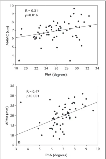
Phase angle was directly correlated with hemoglobin values and inversely correlated with acute (CRP) and non-specific (ESR) inflammatory markers (Table 1). Phase angle also was positively correlated with lean mass indica-tors (MAMC and APMt) (Figures 1A and 1B). Fat mass (FM and TSFt) was directly correlated with acute-phase protein (α1-acid glycoprotein), while lean mass (APMt) was inversely correlated with non-specific inflammation marker (ESR). Additionally, lean mass (MAMC) also was directly correlated with hemoglobin values (Table 1).
Furthermore, acute inflammatory markers were as-sociated with duration of clinical remission and extension of disease. Alfa-1-acid glycoprotein was inversely corre-lated with remission time (R=-0.23; p<0.05). Subjects with more extensive injury (pancolitis) displayed higher CRP levels (median [percentile range 25th-75th] mg/dL) than
those with less extensive lesion (distal colitis) (0.8 [0.4-1.9] > 0.3 [0.2-0.6]; p=0.02, by Kruskal-Wallis’s test comple-mented by Tukey’s test).
Logistic regression was used to assess possible asso-ciation between disease extent and body composition variables. After adjustment for energy, gender, age and CRP, logistic regression analysis showed that BCM was associated with disease extent. There was no association with other parameters (Table 2).
D
ISCUSSIONThe results revealed that body composition parameters, inflammatory markers and disease extent were associ-ated in UC patients. Phase angle was inversely correlassoci-ated with inflammatory markers such as CRP and ERS and directly correlated with hemoglobin values and lean mass indicators (MAMC and APMt). Logistic regression anal-ysis revealed that body cell mass was associated with disease extent. This is the first time that association
FIGURE 1 Pearson’s correlation between phase angle and anthropometric variables. A. PhA versus MAMC. B. PhA versus APMt.
R: Pearson’s correlation coefficient; MAMC: mid-arm muscle circumference; APMt: adductor pollicis muscle thickness; PhA: phase angle.
35
30
25
20
15
10
5
10
3 4 5 6 7 8 9
APMt (mm)
PhA (degrees) B
R = 0.47 p<0.001 10
9
8
7
6
5
4
3
18 20 22 24 26 28 30 32 34
MAMC (cm)
PhA (degrees) A
R = 0.31 p=0.016
TABLE 1 Correlation between biochemical and body composition parameters in patients with ulcerative colitis.
BMI FM TSFt MAC MAMC APMt PhA
Hb 0.44 0.01 -0.10 0.21 0.303 0.23 0.491
CRP -0.008 0.01 0.08 0.90 -0.09 -0.16 -0.591
ESR 0.02 0.12 0.07 0.49 -0.18 -0.402 -0.461
α1-AG 0.361 0.262 0.292 0.253 0.03 -0.02 -0.233
Values represent Pearson’s correlation coefficient (R); Hb: hemoglobin; CRP: C-reactive pro-tein; ESR: erythrocyte sedimentation rate; α1-AG: α1-acid glycoprotein; BMI: body mass in-dex; FM: fat mass; TSFt: triceps skin fold thickness; MAC: arm circumference; MAMC: mid--arm muscle circumference; APMt: adductor pollicis muscle thickness; PhA: phase angle;
gastroin-testinal tract and its effects on food intake and absorp-tion.4,22-24 Malnutrition is especially common in active UC
patients after long-term hospitalization;11,25 however, few
studies have evaluated the nutritional status in patients with UC during remission.4,12,26,27 Only one trial found a
more compromised nutritional status (body weight and BMI) in UC patients during remission than in a control group.28 In our study, we identified a low incidence of
calorie malnutrition. Most patients had normal levels of fat mass and muscle mass, according to the MAC and TSFt parameters, respectively. Besides, 56% were overweight/ obese, according to BMI. The low rate of underweight identified (3.4%) can be partly explained by the remission status of patients, and confirms data shown by other authors.11,12,28 Most of the patients presented distal colitis
in our study; this is not reflective of the general UC pop-ulation and can be explained by the exclusion of those with moderate-severe disease.
BIA is a method frequently used for body composition measurements and offers advantages such as simplicity, portability, cost and absence of radiation exposure.29 It
is known that PhA and body cell mass (BCM) are good prognosis indicators in several clinical situations.12,21,30,31
Although the biological meaning is not completely un-derstood, PhA is applied as a surrogate marker for qual-ity of lean body mass32 and is considered a marker of cell
health since high PhA values reflect a strong cell function.33
Previous studies have shown an association between low values of PhA and BCM with prognosis worsening in patients with hepatitis C and hemodialysis patients.30,34
However, few studies have approached PhA and BCM in UC.12,32 It is important to emphasize that the gold standard
method for assessing body composition is dual-energy X-ray absorptiometry (DEXA), which allows direct and non-invasive measurement of bone mass, fat-free mass
and fat mass. However, DEXA requires skilled personnel, has low affordability, high radiation exposure and is con-sidered a costly examination.35,36 In addition, studies show
good correlation between the parameters of body com-position assessed using either BIA or DEXA.37
Our study showed that anthropometric and biochem-ical parameters were correlated with PhA. The lean body mass anthropometric variable (MAMC) currently corre-lated with PhA has been reported in hemodialysis pa-tients.34 A direct association between PhA and lean body
mass parameter has also been suggested by the positive correlation found between grip strength and PhA in IBD children with mild activity.32 In addition to the
anthro-pometric indicators,Pearson’s correlation analysis revealed that PhA presented inverse correlation with inflamma-tory markers such as CRP and ESR and direct correlation with hemoglobin, although these markers were in the normal range. Modest CRP changes have been observed in the active UC38 and the utility of α1-acid glycoprotein,
ESR39 and hemoglobin have been questioned in UC. The
lack of studies analyzing the relation between PhA and those markers in UC patients prevents further comparisons. In the present study, we found that lean mass was inversely correlated with the non-specific inflammation marker (APMt vs.ESR) and directly correlated with he-moglobin values (MAMC vs. hehe-moglobin). The results indicate a role of inflammatory reactants in the body composition of UC patients. There are few studies evalu-ating body composition in UC patients12,28,39-41 and some
of them12,40 included inflammatory variables in the
anal-ysis. Examining inactive UC patients, absence of associa-tions was found between inflammatory and body com-position variables in two studies40,42 and decreased BCM
in subjects with supranormal CRP values (≥ 8 mg/dL) was identified in one.12
The inverse correlation found between α1-acid gly-coprotein and remission time has not been reported in UC patients. High serum concentration of α1-acid glyco-protein was associated with high susceptibility to induce colitis in rodents43 andpositively correlated with the risk
of relapse in UC patients.44
Although the acute phase protein, CRP, has been positively correlated with IBD activity,10 its association
with disease extent in UC patients in clinical remission was presently identified. Remission time was lower in patients with pancolitis (12 months) compared to those with distal colitis (24 months). Therefore higher CRP levels were found in this group. Even though CRP short half-life of 19 h could not support such explanation, a recent study showed that a mean CRP levels of 5.4 mg/L TABLE 2 Association between body composition variables
and disease extent in patients with ulcerative colitis.
Variables OR CI p-value
BMI 0.94 0.84-1.04 0.24
MAC 0.95 0.83-1.08 0.42
TSFt 0.99 0.94-1.05 0.93
MAMC 0.94 0.81-1.09 0.43
APMt 1.01 0.92-1.10 0.74
PhA 1.02 0.65-1.62 0.90
BCM 0.92 0.87-0.97 <0.01
(normal range) was associated with high histological inflammation scores in patients with ulcerative colitis during clinical remission.45 Such results raise awareness
to the fact that patients in clinical remission from UC still may have inflammation.
It is valid to evaluate the inflammatory profile of IBD patients, even in clinical remission. But it is impor-tant to bear in mind that the classification of the disease activity was based on clinical criteria (Truelove and Witts13). The best classification of clinical remission is
based on a combination of clinical parameters (stool frequency ≤ 3/day with no bleeding) and absence of mu-cosal lesions at endoscopy.46 This was not performed in
our study, which is a limitation. Because of that, patients could be classified as clinical remission and still present active inflammatory process with changes in inflamma-tory markers and intestinal inflammation. Besides, in-flammatory markers such as CRP and ESR are not spe-cific47 and may not reflect the intestinal inflammation
accurately. On the other hand, fecal markers such as calprotectin and lactoferrin can be considered accurate markers of colonic inflammation.47
We also evaluated possible associations between dis-ease extent and body composition variables. High BCM levels were associated with better prognosis and quality of life12,31 and low BCM levels were associated with
supra-normal CRP values in UC patients,12 but studies
approach-ing BCM and disease extent are lackapproach-ing. After adjustment for confounders (energy, gender, age and CRP), logistic regression analysis revealed that body cell mass was as-sociated with extension of the disease. BCM is a marker for combined visceral and somatic protein deposits. It represents the most metabolically active compartment of the body and catabolic conditions may lead to its reduc-tion.17 It is plausible that more extensive UC is associated
with less protein deposits, as seen in other diseases,45 and/
or related with high catabolic conditions.17 Considering
the results found in our population we might suggest that patients with better lean body mass had a lower sus-ceptibility to the development of more extensive injury.
It is important to emphasize that these patients can-not be considered representative of the population of ulcerative colitis, since they derive from a hospital for individuals in the range of low income and low education. UC disease activity was assessed based on Truelove and Witts criteria, although the Mayo score is preferable. No endoscopic data were available at the time of the study to ascertain disease activity, and disease remission was based on clinical aspects. We do not have fecal calprotectin test available. Other limiting factors should also be mentioned,
namely sample size and study design (cross-sectional), as well as the absence of a control group for comparison.
In spite of the limitations above, the outcomes of our study contribute for the expansion of knowledge about the associations of body composition, inflammatory sta-tus and disease extension in clinical remission or mild disease activity in patients with ulcerative colitis. These findings are of great importance for clinical practice, since bioelectrical impedance analysis can be considered a simple examination, noninvasive and rapidly implement-ed. Additionally, the results indicate that some factors could be modified to prevent inflammation and extension of ulcerative colitis. The factors deserving attention in-clude free fat and body cell masses.
C
ONCLUSIONPatients with ulcerative colitis in clinical remission do not present impairment of nutritional status according to BMI and BIA parameters. There were associations be-tween body composition, inflammatory profile and disease extent; acute inflammatory markers were inversely cor-related with phase angle, duration of remission and di-rectly correlated with fat mass anthropometric indicators and also disease extent. Furthermore, body cell mass was associated with disease extent.
A
CKNOWLEDGMENTSWe thank the São Paulo Research Foundation (FAPESP) (grant # 2009/03449-3 [Master’s Scholarship] and grant #2007/07455-2 [Research Financial Support]) and Re-search Support Group at São Paulo State University (Un-esp), Medical School, Botucatu, which helped with the statistical analysis.
C
ONFLICT OF INTERESTThe authors declare no conflict of interest.
R
ESUMOAssociações entre composição corporal, perfil inflamatório e extensão da doença em pacientes com retocolite ulcerativa
Objetivo:Avaliar a composição corporal de pacientes portadores de retocolite ulcerativa em remissão clínica e sua associação com o perfil inflamatório e a extensão da lesão intestinal.
braço (CMB) e espessura do músculo adutor do polegar (EMAP). O perfil inflamatório foi avaliado através da do-sagem da proteína-C reativa (PCR), α1-glicoproteína ácida e velocidade de hemossedimentação (VHS) e a extensão da doença foi avaliada de acordo com o exame endoscópico.
Resultados: Foram avaliados 59 pacientes. A média de
idade foi de 48,1 anos e 53,3% eram mulheres. A maioria dos pacientes (94,9%) estava em remissão clínica da doen-ça e 3,4% foi classificada como desnutrida de acordo com o IMC. Observou-se uma correlação inversa entre AF e marcadores inflamatórios como a PCR (R=-0,59; p<0,001) e VHS (R=-0,46; p<0,001) e uma correlação direta entre AF e os indicadores de massa magra como CMB (R=0,31; p=0,01) e EMAP (R=0,47; p<0,001). A massa magra foi inversamente correlacionada com marcadores inflamatórios não específicos, como a VHS, e diretamente correlacionada com a hemoglobina. De acordo com a análise de regressão logística, a massa celular corporal foi associada com exten-são da leexten-são intestinal (OR 0,92; IC95% 0,87-0,97; p<0,01).
Conclusão: AF foi inversamente correlacionado com
marcadores inflamatórios e diretamente correlacionado com a massa magra. Marcadores inflamatórios de fase aguda e massa celular corporal foram correlacionados com extensão da lesão intestinal.
Palavras-chave: Composição Corporal. Colite Ulcerativa.
Proteína C-Reativa. Biomarcadores. Índice de Gravidade de Doença.
R
EFERENCES1. Mowat C, Cole A, Windsor A, Ahmad T, Arnott I, Driscoll R, et al. Guidelines for the management of inflammatory bowel disease in adults. Gut. 2011; 60(5):571-607.
2. Gaburri PD, Chebli JM, Castro LE, Ferreira JO, Lopes MH, Ribeiro AM, et al. [Epidemiology, clinical features and clinical course of Crohn’s disease: a study of 60 cases]. Arq Gastroenterol. 1998; 35(4):240-6.
3. Souza MH, Troncon LE, Rodrigues CM, Viana CF, Onofre PH, Monteiro RA, et al. [Trends in the occurrence (1980-1999) and clinical features of Crohn’s disease and ulcerative colitis in a university hospital in Southeastern Brazil]. Arq Gastroenterol. 2002; 39(2):98-105.
4. Elia PP, Fogaça HS, Barros RG, Zaltman C, Elia CS. [Descriptive analysis of the social, clinical, laboratorial and anthropometric profiles of inflammatory bowel disease inwards patients from the “Clementino Fraga Filho” University Hospital, Rio de Janeiro, RJ, Brazil]. Arq Gastroenterol. 2007; 44(4):332-9. 5. Cosnes J, Gower-Rousseau C, Seksik P, Cortot A. Epidemiology and natural
his-tory of inflammahis-tory bowel diseases. Gastroenterology. 2011; 140(6):1785-94. 6. Victoria CR, Sassaki LY, Nunes HRC. Incidence and prevalence rates of
inflammatory bowel diseases, in Midwestern of São Paulo State, Brazil. Arq Gastroenterol. 2009; 46(1):20-5.
7. Wild GE, Drozdowski L, Tartaglia C, Clandinin MT, Thomson AB. Nutritional modulation of the inflammatory response in inflammatory bowel disease: from the molecular to the integrative to the clinical. World J Gastroenterol. 2007; 13(1):1-7.
8. Cho JH, Brant SR. Recent insights into the genetics of inflammatory bowel disease. Gastroenterology. 2011; 140(6):1704-12.
9. Masoodi I, Kochhar R, Dutta U, Vaishnavi C, Prasad KK, Vaiphei K, et al. Fecal lactoferrin, myeloperoxidase and serum C-reactive are effective biomarkers
in the assessment of disease activity and severity in patients with idiopathic ulcerative colitis. J Gastroenterol Hepatol. 2009; 24(11):1768-74.
10. Lewis JD. The utility of biomarkers in the diagnosis and therapy of inflammatory bowel disease. Gastroenterology. 2011; 140(6):1817-26. 11. Rocha R, Santana GO, Almeida N, Lyra AC. Analysis of fat and muscle mass
in patients with inflammatory bowel disease during remission and active phase. Br J Nutr. 2009; 101(5):676-9.
12. Valentini L, Schaper L, Buning C, Hengstermann S, Koernicke T, Tillinger W, et al. Malnutrition and impaired muscle strength in patients with Crohn’s disease and ulcerative colitis in remission. Nutrition. 2008; 24(7-8):694-702. 13. Truelove SC, Witts LJ. Cortisone in ulcerative colitis: final report on a
therapeutic trial. Br Med J. 1955; 2(4947):1041-8.
14. World Health Organization. Obesity: preventing and managing the global epidemic. Report on a WHO consultation (WHO Publication WHO/NUT/ NCD/98.1). Geneva: World Health Organization; 1997.
15. Callaway CW, Chumlea WC, Bouchard C, Himes JH, Lohman TG, Martin AD. Circumferences. In: Lohman TG, Roche AF, Martorell R, editors. Anthropometric standardizing reference manual. Champaign: Human Kinetics Books; 1991. p. 39-54.
16. Harrison GG, Buskirk EK, Carter JEL, Ohmston JFE, Lohman TG, Pollock ML. Skinfold thicknesses and measurements technique. In: Lohman TG, Roche AF, Martorell R, eds. Anthropometric standardizing reference manual. Champaign: Human Kinetics Books; 1991. p. 55-80.
17. Frisancho AR. Anthropometric standards for the assessment of growth and nutritional status. Michigan: The University of Michigan Press; 1990. 18. Andrade FN, Lameu EB, Luiz RR. Musculatura adutora do polegar: um novo
índice prognóstico em cirurgia cardíaca valvar. Rev SOCERJ. 2005; 18(5):384-91. 19. Lipschitz DA. Screening for nutritional status in the elderly. Prim Care.
1994; 21(1):55-67.
20. Frisancho AR. New standards of weight and body composition by frame size and height for assessment of nutritional status of adults and the elderly. Am J Clin Nutr. 1984; 40(4):808-19.
21. Kyle UG, Bosaeus I, De Lorenzo AD, Deurenberg P, Elia M, Manuel Gómez J, et al. Bioelectrical impedance analysis-part II: utilization in clinical practice. Clin Nutr. 2004; 23(6):1430-53.
22. Weisshof R, Chermesh I. Micronutrient deficiencies in inflammatory bowel disease. Curr Opin Clin Nutr Metab Care. 2015; 18(6):576-81.
23. Csontos ÁA, Molnár A, Piri Z, Pálfi E, Miheller P. Malnutrition risk questionnaire combined with body composition measurement in malnutrition screening in inflammatory bowel disease. Rev Esp Enferm Dig. 2017; 109(1):26-32. 24. Thangarajah D, Hyde MJ, Konteti VK, Santhakumaran S, Frost G, Fell JM.
Systematic review: body composition in children with inflammatory bowel disease. Aliment Pharmacol Ther. 2015; 42(2):142-57.
25. Geerling BJ, Badart-Smook A, Stockbrugger RW, Brummer RJ. Comprehensive nutritional status in recently diagnosed patients with inflammatory bowel disease compared with population controls. Eur J Clin Nutr. 2000; 54(6):514-21. 26. Ulivieri FM, Piodi LP, Taioli E, Lisciandrano D, Ranzi T, Vezzoli M, et al.
Bone mineral density and body composition in ulcerative colitis: a six-year follow-up. Osteoporos Int. 2001; 12(5):343-8.
27. Ripoli J, Miszputen SJ, Ambrogini Jr O, Carvalho L. Nutritional follow-up of patients with ulcerative colitis during periods of intestinal inflammatory activity and remission. Arq Gastroenterol. 2010; 47(1):49-55.
28. Jahnsen J, Falch JA, Mowinckel P, Aadland E. Body composition in patients with inflammatory bowel disease: a population-based study. Am J Gastroenterol. 2003; 98(7):1556-62.
29. Ling CH, de Craen AJ, Slagboom PE, Gunn DA, Stokkel MP, Westendorp RG, et al. Accuracy of direct segmental multi-frequency bioimpedance analysis in the assessment of total body and segmental body composition in middle-aged adult population. Clin Nutr. 2011; 30(5):610-5.
30. Kahraman A, Hilsenbeck J, Nyga M, Ertle J, Wree A, Plauth M, et al. Bioelectrical impedance analysis in clinical practice: implications for hepatitis C therapy BIA and hepatitis C. Virol J. 2010; 7:191.
31. Werkstetter KJ, Ullrich J, Schatz SB, Prell C, Koletzko B, Koletzko S. Lean body mass, physical activity and quality of life in paediatric patients with inflammatory bowel disease and in healthy controls. J Crohns Colitis. 2012; 6(6):665-73. 32. Kyle UG, Soundar EP, Genton L, Pichard C. Can phase angle determined
by bioelectrical impedance analysis assess nutritional risk? A comparison between healthy and hospitalized subjects. Clin Nutr. 2012; 31(6):875-81. 33. Wirth R, Volkert D, Rösler A, Sieber CC, Bauer JM. Bioelectric impedance
34. Oliveira CM, Kubrusly M, Mota RS, Silva CA, Choukroun G, Oliveira VN. The phase angle and mass body cell as markers of nutritional status in hemodialysis patients. J Ren Nutr. 2010; 20(5):314-20.
35. Haderslev KV, Haderslev PH, Staun M. Accuracy of body composition measurements by dual energy X-ray absorptiometry in underweight patients with chronic intestinal disease and in lean subjects. Dyn Med. 2005; 4(1):1. 36. Kohrt WM. Preliminary evidence that DEXA provides accurate assessment
of body composition. J Appl Physiol. 1998; 84(1):372-7.
37. Thibault R, Genton L, Pichard C. Body composition: why, when and for who? Clin Nutr. 2012; 31(4):435-47.
38. Vilela EG, Torres HO, Martins FP, Ferrari ML, Andrade MM, Cunha AS. Evaluation of inflammatory activity in Crohn’s disease and ulcerative colitis. World J Gastroenterol. 2012; 18(9):872-81.
39. Capristo E, Mingrone G, Addolorato G, Greco AV, Gasbarrini G. Metabolic features of inflammatory bowel disease in a remission phase of the disease activity. J Inter Med. 1998; 243(5):339-47.
40. Capristo E, De Gaetano A, Mingrone G, Addolorato G, Greco AV, Castagneto M, et al. Multivariate identification of metabolic features in inflammatory bowel disease. Metabolism. 1999; 48(8):952-6.
41. Capristo E, Mingrone G, Addolorato G, Greco AV, Gasbarrini G. Glucose metabolism and insulin sensitivity in inactive inflammatory bowel disease. Aliment Pharmacol Ther. 1999; 13(2):209-17.
42. Reimund JM, Arondel Y, Escalin G, Finck G, Baumann R, Duclos B. Immune activation and nutritional status in adult Crohn’s disease patients. Dig Liver Dis. 2005; 37(6):424-31.
43. Hochepied T, Wullaert A, Berger FG, Baumann H, Brouckaert P, Steidler L, et al. Overexpression of α1-acid glycoprotein in transgenic mice leads to sensitisation to acute colitis. Gut. 2002; 51(3):398-404.
44. Vermeire S, Van Assche G, Rutgeerts P. Laboratory markers in IBD: useful, magic, or unnecessary toys? Gut. 2006; 55(3):426-31.
45. Rosenberg L, Nanda KS, Zenlea T, Gifford A, Lawlor GO, Falchuk KR, et al. Histologic markers of inflammation in patients with ulcerative colitis in clinical remission. Clin Gastroenterol Hepatol. 2013; 11(8):991-6. 46. Travis SP, Dinesen L. Remission in trials of ulcerative colitis: what does it
mean? Pract Gastroenterol. 2010; 30:17-20.
47. Magro F, Gionchetti P, Eliakim R, Ardizzone S, Armuzzi A, Barreiro-de Acosta M, et al. Third European Evidence-based Consensus on Diagnosis and Management of Ulcerative Colitis. Part 1: definitions, diagnosis, extra-intestinal manifestations, pregnancy, cancer surveillance, surgery, and ileo-anal pouch disorders. J Crohns Colitis. 2017; 11(6):649-70.