ARTIGO DE REVISÃO ARTIGO ACEITE P ARA PUBLICAÇÃO DISPONÍVEL EM WWW .ACT AMEDICAPORTUGUESA.COM
Uterine Compression Sutures in Controlling Postpartum
Haemorrhage: A Narrative Review
Suturas Uterinas de Compressão no Controlo da
Hemorragia Pós-Parto: Uma Revisão Narrativa
Maria Lúcia MOLEIRO1, Jorge BRAGA1, Maria João MACHADO2, Luís GUEDES-MARTINS1,3,4
Acta Med Port 2020 xxx;33(AOP):xxx-xxx ▪ https://doi.org/10.20344/amp.11987
ABSTRACT
Introduction: Postpartum haemorrhage is still the main cause of maternal morbidity and mortality. Many treatments are available, but they may threaten fertility potential. As a uterine sparing procedure, we aimed to review uterine compression sutures in order to better understand when they should represent an appropriate option.
Material and Methods: A comprehensive search in MEDLINE and PubMed databases including the terms ‘postpartum haemorrhage’ and ‘uterine compression sutures’ was performed. Results were revised and articles reviewing or presenting case reports of uterine compression sutures to treat postpartum haemorrhage were included.
Results: The first description of uterine compression sutures to control postpartum haemorrhage was published in 1997, by B-Lynch et
al. After this publication, many others have reported successful management of postpartum haemorrhage with different suturing
tech-niques. Most of them describe success rates above 75% and the possibility of fertility preservation, with cases of uneventful pregnancy after uterine compression sutures already published. Complications associated with each technique are rare.
Discussion: Reports of use of uterine compression sutures include small series of cases or even single case reports which limits the quality of existing evidence to support one technique over another. Nevertheless, uterine compression sutures are recognized as an effective surgical conservative strategy to control postpartum haemorrhage due to uterine atony and its use is recommended, if pos-sible, prior to hysterectomy.
Conclusion: Uterine compression sutures are effective, safe and simple to perform in an emergent situation and preserve fertility potential in cases of postpartum haemorrhage.
Keywords: Hysterectomy; Obstetric Labour Complications; Postpartum Hemorrhage; Sutures RESUMO
Introdução: A hemorragia pós-parto é a principal causa de morbimortalidade materna. Apesar dos tratamentos disponíveis, o po-tencial fértil da mulher pode ser colocado em causa. As suturas uterinas de compressão representam uma terapêutica conservadora do útero. Assim, revimos os tipos de suturas uterinas de compressão para compreender quando devem ser uma opção terapêutica. Material e Métodos: Foi realizada pesquisa na MEDLINE e PubMed com os termos ‘postpartum haemorrhage’ e ‘uterine compression sutures’ separados e em conjunto. Os resultados foram revistos e os artigos de revisão ou descrevendo casos clínicos de suturas uterinas de compressão foram selecionados.
Resultados: Em 1997, B-Lynch et al descreveu pela primeira vez as suturas uterinas de compressão para tratamento da hemorragia pós-parto. Desde aí, publicações de diferentes tipos de suturas uterinas de compressão, com registo de casos bem-sucedidos, têm sido publicadas. A maioria reporta taxas de sucesso acima de 75%, com preservação da fertilidade, existindo vários casos de bom desfecho obstétrico posteriormente descritos. As complicações associadas são raras.
Discussão: A evidência acerca do uso de suturas uterinas de compressão é limitada pela qualidade dos artigos existentes que incluem apenas pequenas séries de casos ou descrições de casos isolados. Apesar disso, tem sido reconhecido o seu potencial enquanto estratégia conservadora no controlo da hemorragia pós-parto devido a atonia uterina, sendo recomendado o seu uso, se possível, antes de realizar histerectomia.
Conclusão: Em situações de hemorragia pós-parto, as suturas uterinas de compressão são eficazes, seguras e simples de realizar, preservando o potencial reprodutivo.
Palavras-chave: Complicações do Trabalho de Parto; Hemorragia Pós-Parto; Histerectomia; Suturas
INTRODUCTION
Postpartum haemorrhage is an obstetrical emergency and the major cause of maternal morbidity and mortality.1–4 It is
challenging: it can occur during the first twenty-four hours until twelve weeks after delivery, and estimating blood loss is difficult, as bleeding is not always visible.2,5 However, its recognition is vital as it requires immediate action.4,6–9
Uterine atony is the most frequent cause, despite prophylactic measures during the third stage of labour.8 Other causes are
vaginal or uterine trauma, retained placental fragments or coagulopathies.1,3 Medical treatment is the first approach, but
in many situations other interventions are needed, namely when the haemorrhage is severe, requires blood transfusion or causes haemodynamic instability.4,6,9–14 A surgical approach to uterine atony depends on its cause and may ultimately lead 1. Departamento da Mulher e da Medicina Reprodutiva. Centro Materno Infantil do Norte. Centro Hospitalar Universitário do Porto. Porto. Portugal.
2. Serviço de Neurocirurgia. Hospital de Braga. Braga. Portugal.
3. Unidade de Investigação e Formação. Centro Materno Infantil do Norte. Centro Hospitalar Universitário do Porto. Porto. Portugal. 4. Instituto de Investigação e Inovação em Saúde. Universidade do Porto. Porto. Portugal.
Autor correspondente: Maria Lúcia Moleiro. lucia.moleiro@gmail.com
ARTIGO DE REVISÃO ARTIGO ACEITE P ARA PUBLICAÇÃO DISPONÍVEL EM WWW .ACT AMEDICAPORTUGUESA.COM
to hysterectomy. Conservative techniques are first attempted to control postpartum haemorrhage and avoid hysterectomy: balloon tamponade, uterine compression sutures, uterine artery ligation, hypogastric artery ligation, and selective radiologi-cal arterial embolization.6,14–16 Despite many pros and cons of these techniques, Doumouchtsis et al6 points that so far there
is no evidence to suggest that any method is better than another.
Nevertheless, uterine compression sutures (UCS) are a conservative option and their results in controlling postpartum haemorrhage are encouraging given efficacy and potential to preserve fertility.6,9–11,13,14,17 Since the first cases presented by
B-Lynch et al,18 many others have published results on uterine compression sutures’ efficacy in postpartum haemorrhage
due to uterine atony. These reports describe different suture techniques that may be used to treat uterine atony or haemor-rhage due to placenta praevia or accreta.16,18–31
Given the importance of uterine compression sutures to preserve fertility potential, we aimed to present the different types of sutures, how they are performed, their complications and impact on fertility. Our final goal was to understand when and how these techniques should be chosen over more interventional surgical procedures and which one is more suitable for each situation.
MATERIAL AND METHODS
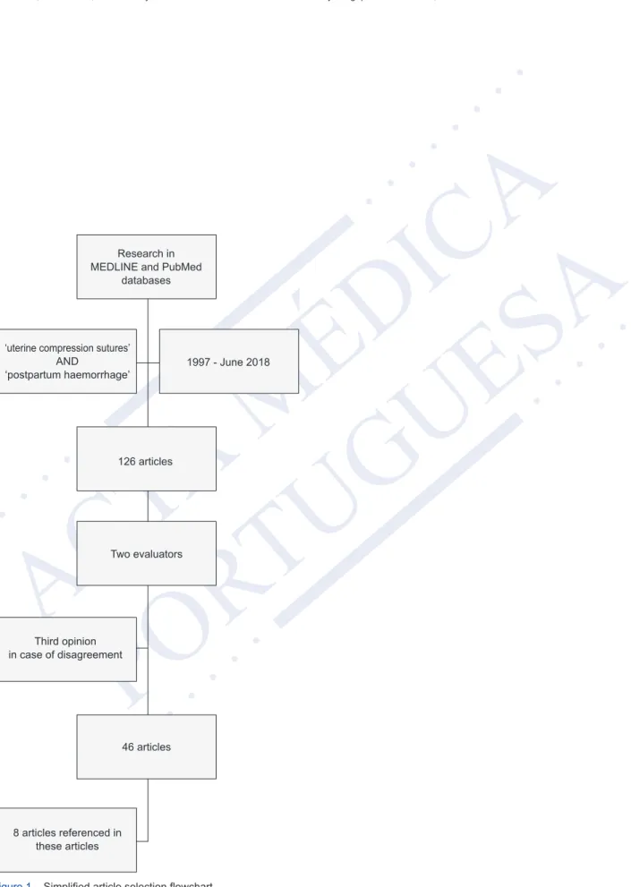
We have performed a comprehensive search in the MEDLINE and PubMed databases including articles published between 1997 and June 2018, with the terms ‘uterine compression sutures’ and ‘postpartum haemorrhage’ combined. Results included 126 articles whose abstracts were analysed by two authors (Fig. 1). Articles reviewing postpartum atony treatments (including UCS), presenting series of cases or case reports of UCS and/or techniques of UCS, and those pre-senting results or reviews of uterine compression sutures’ results and follow-up were included. In case of disagreement between authors, a third opinion was sought. In some cases, in order to better understand and strengthen the evidence, articles referenced in the selected articles were also included in the final review.
RESULTS
Types of uterine compression sutures
B-Lynch et al18 were the first to report five women who were successfully treated with uterine sutures after
life-threat-ening postpartum haemorrhage. Regardless of whether the delivery was vaginal or by caesarean section, a Pfannenstiel incision of the abdomen and a lower segment incision of the uterus are needed. The uterus must be exteriorised to assess the bleeding cause, and if uterine atony is the most likely diagnosis, bimanual uterine compression should be performed to verify the technique’s potential success.18,25 The suture begins below the right inferior limit of the uterine incision, inserting
the needle through the anterior uterine wall. The needle is exteriorized again in the anterior uterine wall above the superior border of the incision. The thread then goes to the uterine posterior wall, passing above the fundus, and then the needle is inserted through the posterior wall in a point corresponding to the thread’s anterior exit. Afterwards, the needle should be inserted a few centimetres laterally to the entry point, and the thread is then passed longitudinally through the fundus to the anterior wall. The needle is then inserted above the left superior margin of the uterine incision through the uterus wall and exits on the inferior margin (parallel to the first stitches). An assistant should compress the uterus while the surgeon pulls the thread firmly and applies a strong knot (Fig. 2). Finally, the uterine incision should be closed.18,25 B-Lynch et al18 also
advocate UCS in cases of placenta praevia, performing an independent figure-eight suture anteriorly, posteriorly, or both, prior to the application of their suturing technique.
Most of the techniques reported after B-Lynch’s work present subtle variations. Bhal et al26 have described two sutures
which start by inserting the needle below the uterine section and passing it through the uterine wall until it reaches the posterior side of the uterus. Then, the thread passes above the uterine fundus exteriorly until the anterior side of the uterus, where the needle is inserted above the uterine section and crosses the anterior wall, exiting below the section in a point superior to the initial inserting needle spot. The procedure was repeated on the other side, and two knots were tied in the anterior-inferior margin of the lower uterine segment.26 Nelson and Birch,32 bearing in mind the risk of lateral thread slipping
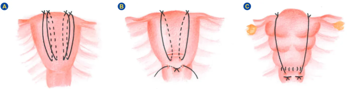
above the fundus during uterine involution, have proposed a suture initiated like in the technique by B-Lynch, but when the needle comes out anteriorly above the hysterotomy site, the thread is not passed above the uterine fundus. Instead, the needle is introduced through the uterine fundus until its posterior aspect. Then, the thread runs along the posterior wall and the needle is inserted again in the lower segment of the uterus through the uterine cavity until the anterior wall, exiting below the uterine incision (Fig. 3A). The same steps are repeated on the other side, and knots are tied tightly on the anterior aspect of the uterus.32 Marasinghe et al31,33 have also claimed to avoid the sutures slip off the uterine fundus,
using two sutures beginning anteriorly below the uterine section. The needle is passed through the uterine cavity until its posterior side, where the thread is passed over the fundus until the anterior wall; there, the needle is inserted below the fundus, passing the uterine cavity until its posterior wall; the thread is then passed over the fundus, reaching the anterior aspect, where a tight knot is performed (Fig. 3B). The procedure is repeated on the other side.31,33
More recently, the use of a double B-Lynch suture was described as successful in 14 cases where only one suture was unable to control haemorrhage. Authors performed the original B-Lynch technique and the second suture 0.5 cm from the
ARTIGO DE REVISÃO ARTIGO ACEITE P ARA PUBLICAÇÃO DISPONÍVEL EM WWW .ACT AMEDICAPORTUGUESA.COM
lateral side of it.34 Another modification of the technique was performed in 19 women in whom Mansoura-VV sutures were
applied. They use two longitudinal sutures that pass through the uterine cavity below the hysterotomy incision (one left and one right). The knots are tied in the uterine fundus as the thread is divided in two, creating a V-shaped suture (Fig. 3C).21,35
El-Sokkary et al22 have published another variation: it begins on the anterior aspect of the uterus below the hysterotomy,
but the thread, after exiting above the hysterotomy, crosses the uterine fundus on the contralateral side and is inserted in the posterior wall on that same side. On the posterior aspect of the uterus the suture is performed as B-Lynch description but on the anterior wall the thread crosses again creating an X-shaped suture.22
The association of B-Lynch suture with other postpartum haemorrhage control techniques have also been described. Nelson and O’Brien29 have reported five cases in which B-Lynch’s compression sutures were performed but were not
suc-cessful, so they’ve applied an intrauterine Bakri balloon – ‘uterine sandwich technique’. According to the authors, after plac-ing the balloon, the hysterotomy incision is closed and the balloon is filled with normal saline while observplac-ing the uterine response to tamponade.29 Similar descriptions also support the success of these combined strategies.36–38
Cho et al19 have published their experience in controlling postpartum haemorrhage due to uterine atony, but also
ab-normal placentation, after caesarean delivery. This technique uses multiple square sutures to get anterior and posterior uterine walls closer so that no space is left between them and haemorrhage is controlled. It is performed in the bleeding area, where the anterior wall is punctured until the posterior aspect of the uterus. There, 2-3 cm laterally, the uterine wall is punctured again until the anterior wall, where 2-3 cm below or above a new point of the wall is punctured until the posterior side. Again, the needle is inserted until the anterior aspect of the uterus so that a square/rectangle is formed after the knot. This suture is repeated four to five times from the uterine fundus until its lower segment in cases of uterine atony. If placenta accreta is the bleeding cause, two to three areas in it are selected, and the square suture is performed; in cases of placenta praevia, the sites with heavier bleeding are selected, and sutures are performed.19 Two years after Cho’s publication,
Hay-man and colleagues24 have described a technique in which two to four longitudinal compression sutures are placed from
the anterior to the posterior uterine wall (Fig. 4). A straight needle enters the anterior wall below the uterine incision until the posterior wall, and a knot is made on the uterine fundus. This procedure can be performed on the other side so that two or more longitudinal sutures wrap the uterus and control haemorrhage. If an incision was not performed, the needle is inserted in the place where it should be. Hayman et al24 also highlight the possibility of a transverse suture near the cervix
when haemorrhage originates at that level, namely, in cases of placenta praevia: the needle passes through the anterior wall approximately 3 cm below the inferior caesarean section and 2 cm from the lateral border; on the posterior wall, the needle should then be inserted 1 cm medially on the same exit level passing through the uterine walls until the anterior one; the same procedure is repeated on the contralateral side.24
Ouahba et al28 have published a technique with four sutures to control postpartum haemorrhage in a series of 20 cases
of uterine atony. The procedure consists of four uterine sutures inserted from the anterior to posterior wall; the needle is again inserted until the anterior wall approximately 8 cm from the initial point, and a tight double knot is tied on the anterior aspect of the uterus. Two of the sutures are transverse (one in the middle of the fundus and one in the lower segment) and two are oblique and placed 2-3 cm medial from the uterine horns.28 Hackethal et al30 have also described a technique where
the suture enters the uterine cavity from the anterior to the posterior wall. The needle passes 2 to 4 cm from the initial point through the uterine cavity until it reaches the anterior aspect of the uterus, where a double knot is performed, resulting in a U-transverse suture. The number of sutures used depends on the uterine size and the persistence of the bleeding.30
Being different from the ones already described, Pereira’s suture includes longitudinal and transverse sutures around the uterus that do not cross the uterine wall, being inserted only superficially in the serous membrane and the subserous myometrium (Fig. 5).27 Two to three transverse sutures should be performed first, starting from the superior one: it starts
anteriorly by inserting the needle in the subserous myometrium and passing it through the broad ligament to the posterior side of the uterus; then, some bites of the subserous myometrium should be taken, and the needle crosses the broad liga-ment again to the anterior wall, where the knot is performed. The number of bites depends on uterine size and tension that is perceived as necessary to compress the uterus. The most inferior transverse suture is used as an anchor to the knots of the longitudinal sutures, which should start on the posterior wall. After performing a knot to the lowest transverse suture, several bites of the subserous myometrium are taken until the anterior side of the uterus, where it should end. This proce-dure can then be repeated two to three times.27 A modification of Pereira’s technique has been recently described in two
patients: longitudinal sutures were performed according to B-Lynch’s technique, but additional transverse sutures included superficial bites involving only the serous membrane and the subserous myometrium like those used by Pereira.23
Effectiveness of the different techniques
Kayem et al17 reported the efficacy of UCS after reviewing 199 deliveries which required UCS in the United Kingdom
between September 2007 and March 2009. The most frequent technique chosen was the B-Lynch suture, and a modified version of it was the second most frequent option. The overall failure rate was 25%, and in these cases, a hysterectomy was necessary; the rate of failure did not differ significantly according to the suturing method. A higher risk of hysterectomy was observed when there was a prolonged delay of 2 to 6 hours between delivery and UCS, and when there was a vaginal
ARTIGO DE REVISÃO ARTIGO ACEITE P ARA PUBLICAÇÃO DISPONÍVEL EM WWW .ACT AMEDICAPORTUGUESA.COM
delivery, maternal age was 35 years or older or when the woman was multiparous.17
A systematic review in 20076 analysed the success rates of conservative techniques to control postpartum
haemor-rhage, and a success rate of 91.7% for UCS was estimated. It was noted that there was no statistically significant difference in success rates between conservative procedures, and therefore, the authors advised clinicians to use the least invasive, easiest and quickest approach, namely, balloon tamponade.6 These results have been recently corroborated by a Çetin et
al39 when comparing Hayman suture and Bakri balloon tamponade.
Ghezzi et al40 reported eleven cases of massive postpartum haemorrhage in which ten were successfully controlled
with UCS, and only one patient ultimately required a hysterectomy. More recent articles reviewed comparative studies and case series and estimated a success rate of UCS of 71% to 75%.14,41 Variations of B-Lynch suture have also been reported
as effective or even more effective than the original technique.22,34,35
Although a randomized control trial comparing the different types of UCS’ effectiveness is lacking, the results published so far point to a success rate between 36% and 98% (the majority of them above 75%).14,34,35,38 The technique chosen may
not be the only contributor to different results. The time between the beginning of the haemorrhage, its diagnosis, previous medical treatment and the time by which the UCS are applied may be vital to success.14,34,35,38
Complications related to uterine compression sutures
From the beginning, the importance of ensuring that compression of the uterus controls active haemorrhage before performing the suture has been outlined.18,25 Otherwise, the uterine suture may not be effective.
The cases described by Nelson and O’Brien,29 which involved the uterine sandwich technique, were all successful in
controlling postpartum haemorrhage, but the authors outlined one patient who developed endomyometritis and another with postpartum oliguria. The risk of endometritis has also been reported by Suzuki et al42 when evaluating perioperative
complications of UCS in their hospital.
As the thread is passed through the uterine cavity in Hayman’s and Cho’s procedure, there is at least, a theoretical risk of blood trapping within it.25,43 Cho’s suture could also interfere with physiologic uterine involution, and it was already
reported to be associated with pyometra and uterine cavity synechiae.25,43–45 Also, a higher risk of uterine ischaemia and
necrosis seemed to be associated with UCS combined with vessel ligation, so long-term follow up in these women is advis-able.16,46,47 Pereira’s technique does not involve suturing the anterior and posterior wall together, and the needle only enters
the subserous myometrium without penetrating the uterine cavity. Therefore, the aforementioned risks are overcome and the risk of infection is reduced.27,43 Given the small size of the bites, it is believed that the risk of bowel or omentum
trap-ping in the suture is low. This is a minimal risk even with other techniques due to the applied compression and the fact that suturing materials are absorbed in a few weeks.43
Given the absence of larger trials and the reporting biases regarding complications, it is important to bear in mind a possible higher risk of complications than what is reported in literature.
Results for future fertility and pregnancy outcome
Data regarding menstrual cycles and fertility post-UCS are scarce since follow-up is mostly short and only a few cases of long follow-ups have been reported. A 2013 review48 about fertility rates and pregnancy outcomes after postpartum
haemorrhage treated with UCS estimated a fertility rate ranging from 10% to 100%, noting lower performance with B-Lynch’s and Hayman’s modified procedures. Authors have explained this wide range with the information described in case reports, case series or even studies performed by surgeons who first described the techniques. Despite these important biases, they stated that the majority of pregnancies were mostly uneventful, but the most frequent mode of delivery was an elective caesarean section. It seems safer and ensures the quick management of postpartum haemorrhage complications since recurrence is not negligible.48
These results are comparable to those obtained by Doumouchtsis et al.49 By analysing six studies assessing menstrual
and fertility data after UCS, they estimated that more than 90% of women resumed normal menstruation. Between women who revealed a desire for a subsequent pregnancy, approximately 85% achieved conception. They noted that this desire may have been inadequately reported and that many women did not want another pregnancy after a life-threatening event.49 Another review in 201350 evaluated the outcomes of subsequent pregnancies after UCS – Hayman’s suture and the
multiple square suture technique – at their hospital. They compared 42 women treated with UCS with 139 untreated women during their previous delivery by caesarean section. They concluded that all women treated with UCS who got pregnant conceived naturally and that pregnancy outcomes were similar (namely prematurity rates, estimated blood loss and rate of UCS in subsequent deliveries). It is important to point out that pelvic adhesions in a subsequent caesarean delivery were more prevalent in women who were treated with UCS. Despite the relevant number of women evaluated, this study only compared outcomes in pregnant women and did not assess the potential effect of UCS on fertility.50
Cowan et al51 compared adverse outcomes in subsequent pregnancies in women with postpartum haemorrhage
treat-ed with a B-Lynch suture versus women treattreat-ed with another method (artery ligation and uterine artery embolization). They found no significant difference between groups, and they also found no association between the use of B-Lynch
compres-ARTIGO DE REVISÃO ARTIGO ACEITE P ARA PUBLICAÇÃO DISPONÍVEL EM WWW .ACT AMEDICAPORTUGUESA.COM
sion sutures and adverse outcomes in future pregnancies.51 Recent series of cases also support the possibility of
success-ful pregnancy after UCS.34
The overall conclusion from small reports to bigger reviews is that most women resumed normal menses and that those women who desired another pregnancy were able to conceive naturally with comparable outcomes to other women who did not undergo UCS on their previous caesarean delivery.
DISCUSSION
The choice of an optimal uterine suture technique
Although it exists, the evidence supporting UCS has poor quality, which has been a main conclusion of many studies. Nevertheless, almost every article and recent review about the conservative management of postpartum haemorrhage agrees with the relevant success rate of UCS and the simplicity of performing them under an emergent situation, even by obstetricians with less training.9,12,16,20,25,26,28,32,34,40,43,48–50
Despite many similarities, these techniques have some specific characteristics that can make them more suitable in some situations. When choosing a UCS, at first, it is important to determine whether the uterine incision is already closed or was not performed (cases of postpartum haemorrhage after vaginal delivery in which a laparotomy was considered necessary). In these situations, Pereira’s or Hayman’s sutures are possible options24,27(Fig. 6).
Equally important is the perceived cause of haemorrhage, since all UCS may be applied in cases of uterine atony, which implies the exclusion of other causes of postpartum haemorrhage. On the other hand, Hayman’s technique24
in-cludes a possible isthmic-cervical suture to control haemorrhage due to placenta praevia and Cho’s suture19 can also
control postpartum haemorrhage due not only to placenta praevia but also placenta accreta (Fig. 6).
Ultimately, the surgeon’s experience and knowledge about each technique will define the best approach since there are no randomized or controlled trials to compare the different types of UCS. It’s also important to note that the option of a UCS should be considered as soon as possible when pharmacological measures fail to control postpartum haemorrhage and a surgical option is considered.6,7,10,12–14,16,20,41,43,50,52,53
CONCLUSION
Uterine compression sutures are a conservative treatment option to control postpartum haemorrhage that should be used when first line medical treatment isn’t enough. They can be performed either after vaginal and operative delivery, do not require interventional radiology support and may be performed shortly after the onset of haemorrhage. The main rea-sons favouring this conservative surgical technique are its effectiveness but also its simplicity, while preserving women’s fertility potential. The data published so far suggests that, after UCS almost all women resumed normal menses and could conceive spontaneously. Subsequent pregnancies tended to be uneventful. Also, complications described after UCS were rare.
Since there are no randomized control trials comparing UCS with other surgical conservative procedures or even com-paring different types of sutures, critical judgement is vital. Nevertheless, the UCS success rates and its simplicity should lead to its inclusion in PPH treatment protocols before more complicated or aggressive surgical approaches like hysterec-tomy. It is vital to teach UCS in order to prepare every obstetrician to know when and how to perform each technique.
CONFLICTS OF INTEREST
The authors report no conflict of interest.
FUNDING SOURCES
This research received no specific grant from any funding agency in the public, commercial, or not-for-profit sectors.
REFERENCES
1. Carroli G, Cuesta C, Abalos E, Gulmezoglu AM. Epidemiology of postpartum haemorrhage: a systematic review. Best Pract Res Clin Obstet Gynaecol. 2008;22:999-1012.
2. Schorn MN. Measurement of blood loss: review of the literature. J Midwifery Womens Health. 2010;55:20–7.
3. Say L, Chou D, Gemmill A, Tunçalp Ö, Moller AB, Daniels J, et al. Global causes of maternal death: a WHO systematic analysis. Lancet Glob Heal. 2014;2:323-33.
4. Kramer MS, Berg C, Abenhaim H, Dahhou M, Rouleau J, Mehrabadi A, et al. Incidence, risk factors, and temporal trends in severe postpartum hemorrhage. Am J Obstet Gynecol. 2013;209:449.e1–7.
5. Cunningham FG, Leveno KJ, Bloom SL, Spong CY, Dashe JS, Hoffman BL, et al. Williams obstetrics. New York : McGraw-Hill Education/Medical; 2014. 6. Doumouchtsis SK, Papageorghiou AT. Systematic review of conservative management of postpartum hemorrhage: what to do when medical treatment
fails. Obstet Gynecol Surv. 2007;62:540-7.
7. Leduc D, Senikas V, Lalonde AB, Ballerman C, Biringer A, Delaney M, et al. Active management of the third stage of labour: prevention and treatment of postpartum hemorrhage. J Obstet Gynaecol Can. 2009;31:980-93.
8. World Health Organization. Department of Reprodutive Health and Research. WHO recommendations for the prevention and treatment of postpartum haemorrhage. WHO. 2012:41.
9. Abdul-Kadir R, McLintock C, Ducloy AS, El-Refaey H, England A, Federici AB, et al. Evaluation and management of postpartum hemorrhage: consensus from an international expert panel. Transfusion. 2014;54:1756-68.
ARTIGO DE REVISÃO ARTIGO ACEITE P ARA PUBLICAÇÃO DISPONÍVEL EM WWW .ACT AMEDICAPORTUGUESA.COM
10. Tuncalp O, Souza JP, Gulmezoglu M. New WHO recommendations on prevention and treatment of postpartum hemorrhage. Int J Gynaecol Obs. 2013;34:254-6.
11. Mousa HA, Blum J, Abou El Senoun G, Shakur H, Alfirevic Z. Treatment for primary postpartum haemorrhage. Cochrane Database Syst Rev. 2014;2:CD003249.
12. Cekmez Y, Ozkaya E, Öcal FD, Küçüközkan T. Experience with different techniques for the management of postpartum hemorrhage due to uterine atony: compression sutures, artery ligation and Bakri balloon. Ir J Med Sci. 2015;182:399-402.
13. Weeks A. The prevention and treatment of postpartum haemorrhage: What do we know, and where do we go to next? Br J Obstet Gynaecol. 2015;122:202-10.
14. Sathe NA, Likis FE, Young JL, Morgans A, Carlson-Bremer D, Andrews J. Procedures and uterine-sparing surgeries for managing postpartum hemorrhage: a systematic review. Obstet Gynecol Surv. 2016;71:99-113.
15. Shah M, Wright JD. Surgical intervention in the management of postpartum hemorrhage. Semin Perinatol. 2009;33:109-15.
16. Palacios-Jaraquemada JM. Efficacy of surgical techniques to control obstetric hemorrhage: analysis of 539 cases. Acta Obstet Gynecol Scand. 2011;90:1036-42.
17. Kayem G, Kurinczuk JJ, Alfirevic Z, Spark P, Brocklehurst P, Knight M. Specific second-line therapies for postpartum haemorrhage: a national cohort study. Br J Obstet Gynaecol. 2011;118:856-64.
18. B-Lynch C, Coker A, Lawal A, Abu J, Cowen M. The B-Lynch surgical technique for the control of massive postpartum haemorrhage: an alternative to hysterectomy? Five cases reported. Br J Obstet Gynaecol. 1997;104:372-5.
19. Cho JH, Jun HS, Lee CN. Hemostatic suturing technique for uterine bleeding during cesarean delivery. Obstet Gynecol. 2000;96:129-31.
20. Kayem G, Kurinczuk JJ, Alfirevic Z, Spark P. Uterine compression sutures for the management of severe postpartum. Obstet Gynecol. 2011;117:14-20. 21. El-Refaeey AA, Gibreel A, Fawzy M. Novel modification of B-lynch uterine compression sutures for management of atonic postpartum hemorrhage: VV
uterine compression sutures. J Obstet Gynaecol Res. 2014;40:387-91.
22. El-Sokkary M, Wahba K, El-Shahawy Y. Uterine salvage management for atonic postpartum hemorrhage using “modified lynch suture.” BMC Pregnancy Childbirth. 2016;16:1-5.
23. Moleiro ML, Guedes-Martins L, Mendes A, Marques C, Braga J. Modified Pereira suture as an effective option to treat postpartum hemorrhage due to uterine atony. Rev Bras Ginecol Obs. 2018;40:92-5.
24. Hayman RG, Arulkumaran S, Steer PJ. Uterine compression sutures: surgical management of postpartum hemorrhage. Obstet Gynecol. 2002;99:502-6. 25. Allam MS, B-Lynch C. The B-Lynch and other uterine compression suture techniques. Int J Gynecol Obstet. 2005;89:236-41.
26. Bhal K, Bhal N, Mulik V, Shankar L. The uterine compression suture - a valuable approach to control major haemorrhage at lower segment caesarean section. J Obstet Gynaecol. 2005;25:10-4.
27. Pereira A, Nunes F, Meirinho M. Compressive uterine sutures to treat postpartum bleeding secondary to uterine atony. Obstet Gynecol. 2005;106:5-8. 28. Ouahba J, Piketty M, Huel C, Azarian M, Feraud O, Luton D, et al. Uterine compression sutures for postpartum bleeding with uterine atony. BJOG.
2007;114:619-22.
29. Nelson WL, O’Brien JM. The uterine sandwich for persistent uterine atony: combining the B-Lynch compression suture and an intrauterine Bakri balloon. Am J Obstet Gynecol. 2007;196:2006-7.
30. Hackethal A, Brueggmann D, Oehmke F, Tinneberg HR, Zygmunt MT, Muenstedt K. Uterine compression U-sutures in primary postpartum hemorrhage after Cesarean section: Fertility preservation with a simple and effective technique. Hum Reprod. 2008;23:74-9.
31. Marasinghe JP, Condous G. Uterine compression sutures for post-partum bleeding with atony; modification of the B-Lynch suture. Aust New Zeal J Obstet Gynaecol. 2009;49:67-70.
32. Nelson GS, Birch C. Compression sutures for uterine atony and hemorrhage following cesarean delivery. Int J Gynecol Obstet. 2006;92:248-50. 33. Marasinghe JP, Condous G, Seneviratne HR, Marasinghe U. Modified anchored B-Lynch uterine compression suture for post partum bleeding with
uterine atony. Acta Obstet Gynecol Scand. 2011;90:280-3.
34. Sahin H, Karapinar OS, Sahin EA, Dolapçioglu K, Baloglu A. The effectiveness of the double B-lynch suture as a modification in the treatment of intractable postpartum haemorrhage. J Obstet Gynaecol. 2018;38:796-9.
35. El-Refaeey AE, Abdelfattah H, Mosbah A, Gamal AM, Fayla E, Refaie W, et al. Is early intervention using Mansoura-VV uterine compression sutures an effective procedure in the management of primary atonic postpartum hemorrhage?: A prospective study. BMC Pregnancy Childbirth. 2017;17:1-6. 36. Danso D, Reginald P. Combined B-lynch suture with intrauterine balloon catheter triumphs over massive postpartum haemorrhage. Br J Obstet Gynaecol.
2002;109:963.
37. Price N, Whitelaw N, B-Lynch C. Application of the B-Lynch brace suture with associated intrauterine balloon catheter for massive haemorrhage due to placenta accreta following a second-trimester miscarriage. J Obstet Gynaecol. 2006;26:267-8.
38. Mohamed MA, Mohamed AH. Parallel vertical compression sutures to control bleeding in cases of placenta previa and accreta. J Matern Neonatal Med. 2017:1-5.
39. Çetin BA, Mathyk BA, Aydin AA, Koroglu N, Bahat PY, Yuksel IT, et al. Comparing success rates of the Hayman compression suture and the Bakri baloon tamponade. J Matern Neonatal Med. 2018:1-5.
40. Ghezzi F, Cromi A, Uccella S, Raio L, Bolis P, Surbek D. The Hayman technique: a simple method to treat post partum haemorrhage. BJOG. 2007;114:362-5.
41. Chan LL, Lo TK, Lau WL, Lau S, Law B, Tsang HH, et al. Use of second-line therapies for management of massive primary postpartum hemorrhage. Int J Gynecol Obstet. 2013;122:238-43.
42. Suzuki Y, Matsuzaki S, Mimura K, Kumasawa K, Tomimatsu T, Endo M, et al. Investigation of perioperative complications associated with use of uterine compression sutures. Int J Gynecol Obstet. 2017;139:28-33.
43. Mallappa Saroja CS, Nankani A, El-Hamamy E. Uterine compression sutures, an update: Review of efficacy, safety and complications of B-Lynch suture and other uterine compression techniques for postpartum haemorrhage. Arch Gynecol Obstet. 2010;281:581-8.
44. Ochoa M, Allaire A, Stitely M. Pyometra after hemostatic square suture technique. Obstet Gynecol. 2002;99:506-9. 45. Wu H, Yeh G. Uterine cavity synechiae after hemostatic square suturing technique. Obstet Gynecol. 2005;105:1176-8.
46. Riyami NA, Hui D, Herer E, Nevo O. Uterine compression sutures as an effective treatment for postpartum hemorrhage : case series. AJP Rep. 2011;1:47-51.
47. Fuglsang J. Later reproductive health after B-Lynch sutures: a follow-up study after 10 years’ clinical use of the B Lynch suture. Fertil Steril. 2014;101:1194. 48. Gizzo S, Saccardi C, Patrelli TS, Di Gangi S, Breda E, Fagherazzi S, et al. Fertility rate and subsequent pregnancy outcomes after conservative surgical
techniques in postpartum hemorrhage: 15 years of literature. Fertil Steril. 2013;99:2097-107.
49. Doumouchtsis S, Nikolopoulos K, Talaulikar V, Krishna A, Arulkumaran S. Menstrual and fertility outcomes following the surgical management of postpartum haemorrhage: a systematic review. BJOG. 2013:1-7.
50. An GH, Ryu HM, Kim MY. Outcomes of subsequent pregnancies after uterine compression sutures for postpartum hemorrhage. Obstet Gynecol. 2013;122:565-70.
51. Cowan AD, Miller ES, Grobman WA. Subsequent pregnancy outcome after B-Lynch suture placement. Obstet Gynecol. 2014;124:558-61.
52. El-Hamamy E, B-Lynch C. A worldwide review of the uses of the uterine compression suture techniques as alternative to hysterectomy in the management of severe post-partum haemorrhage. J Obstet Gynaecol. 2005;25:143-9.
ARTIGO DE REVISÃO ARTIGO ACEITE P ARA PUBLICAÇÃO DISPONÍVEL EM WWW .ACT AMEDICAPORTUGUESA.COM
54. Koh E, Devendra K, Tan LK. B-Lynch suture for the treatment of uterine atony. Singapore Med J. 2009;50:693-7.
Figure 1 – Simplified article selection flowchart Research in MEDLINE and PubMed
databases
‘uterine compression sutures’ AND
‘postpartum haemorrhage’ 1997 - June 2018
126 articles 46 articles 8 articles referenced in these articles Two evaluators Third opinion in case of disagreement
ARTIGO DE REVISÃO ARTIGO ACEITE P ARA PUBLICAÇÃO DISPONÍVEL EM WWW .ACT AMEDICAPORTUGUESA.COM
Figure 2 – B-Lynch suture.54 (A) Anterior view of the uterus before performing the knot, showing how the thread passes through the uterine
wall; (B) Posterior aspect of the uterus before performing the knot; (C) Anterior view of the compressed uterus after performing the knot and the suture of the hysterotomy.
Dashed lines represent the suture on the posterior aspect of the uterus.
A
B
C
Figure 3 – Longitudinal sutures variations. (A) Nelson and Birch’s description of uterine compression sutures.32; (B) Marasinghe et al
technique to avoid thread slipping over the uterine fundus.33; (C) Mansoura VV-suture.21,35
Dashed lines represent the suture on the posterior aspect of the uterus.
ARTIGO DE REVISÃO ARTIGO ACEITE P ARA PUBLICAÇÃO DISPONÍVEL EM WWW .ACT AMEDICAPORTUGUESA.COM
Figure 4 – Scheme of Hayman’s et al24 suture technique. (A) Anterior aspect of the uterus after a vaginal delivery, when no hysterotomy
was needed.24; (B) and (C) Anterior aspect of the uterine wall illustrating the transverse sutures also described by Hayman et al24;
(B) Aspect before the suture of the uterine incision; (C) Final aspect after performing the knots of both transverse sutures and closure of the hysterotomy.
Dashed lines represent the suture on the posterior aspect of the uterus.
A B C
Figure 5 – Pereira’s suture27. (A) Posterior view; (B) Anterior view.
ARTIGO DE REVISÃO ARTIGO ACEITE P ARA PUBLICAÇÃO DISPONÍVEL EM WWW .ACT AMEDICAPORTUGUESA.COM
Figure 6 – Simplified flowchart on how to choose a uterine compression suture technique Cause of postpartum
haemorrhage
Uterine atony
Uterine incision closed / not performed
Uterine incision open / during caesarean section Hayman’s isthmic-cervical suture Pereira’s suture Hayman’s suture
All types of UCS are suitable
Cho’s suture
Cho’s suture Placenta praevia