“DIVERSITY AND SPECIFICITY OF THE MARINE SPONGE MICROBIOME AS INSPECTED BY NEXT GENERATION SEQUENCING”
André Rodrigues Soares
Mestrado em Biologia Molecular e Microbiana
Trabalho efetuado sob a orientação de:
Dr. Rodrigo da Silva Costa
Prof.ª Dr.ª Maria Margarida dos Prazeres Reis
Acknowledgements
For the completion of the present work, the support of the Microbial Ecology and Evolution (MicroEcoEvo) research group at the CCMAR was crucial. Firstly, I thank Rodrigo Costa, my supervisor, who drew me into the field of microbial ecology of marine sponges and posed me the great challenge of tackling the EMP dataset. Furthermore, I am thankful for all the enthusiasm and opportunities provided and for the immense patience! Gianmaria Califano, now in Jena, Austria, and Asunción Lago-Lestón dispensed precious time and patience in the starting phase of this thesis. I further thank Elham Karimi for the help in the laboratory and for our relaxing coffee talks! Tina Keller-Costa, Telma Franco and more recently Miguel Ramos, along with the abovementioned, are all part of the MicroEcoEvo team, to whom I thank for a wonderful first medium-term experience in a laboratory!
I admittedly started my Biology Bsc. at the University of Algarve not being fond of any biological entity smaller than 2cm. It was Prof. Margarida Reis (Microbial and Molecular Ecology Laboratory, CIMA, UAlg) who showed me that ‘microbes can do anything’ and went on to accepting the challenge of supervising my Bsc. Technical and Scientific Project. I therefore thank her for introducing me to Microbiology in the best way possible.
For the completeness of this work, Lucas Moitinho-Silva (currently at the University of South Wales, Australia), was essential in introducing me to R scripting whilst finishing his PhD at Wüezburg University, Germany. I still cannot imagine how one is able to answer basic R questions the night before defending his PhD thesis, but Lucas did, and I thank him for his incredible availability and kindness throughout this year. Cymon Cox, head of the Plant Systematics and Bioinformatics group in CCMAR, very kindly introduced me to python scripting and handling of the Linux OS. Thoughtfully, Cole Easson and Robert Thacker from Stony Brook University, USA, let me know of various tools for 16S dataset analysis. All the help received from Pat Schloss and Sarah Wescott, mothur software developers at the University of Michigan was precious.
Out of the academic scope, ‘Associação Geonauta’, ‘my’ caving association in the Algarve has, for the last 4 years, given me the opportunity to discover the subterranean wonders of Algarve. I have learned greatly from all, but mainly from Cristiano Cavaco, Carlos Oliveira and Friedrich Zabel. Prof. Cristina Veiga-Pires, whom I met in a cave, has been a great supporter of our work in Associação Geonauta and a friend.I also want to thank all those I knew during my experience at the Biology Students Association at
the University of Algarve (NEBUA) and at the National Board of Biology Students (ANEBio). They are too many to list, but they know who they are!
Personally, I thank Andreia Nunes and Catarina Diniz, my friends and colleagues in the Molecular and Microbial Biology Msc., who accompanied me throughout all kinds of hurdles and coffee breaks during this thesis. Jessica Fróis and Beatriz Cruz are further credited for frequently giving out valuable wise advices which truly helped me for the last 4 years. Starting my Bsc. in 2009, I met Nadja Velez, who is to this day one of my best friends. Throughout these 6 years, she showed me how to get excited with Science and accompanied me in all sorts of events.
The support of my family has been crucial throughout my life, but mostly during its academic component and all external events occurring during that time. I would therefore like to dedicate this thesis to my late grandfather Fernando Rodrigues, who used to say that nothing in life is impossible if one works for it. He always supported my choices, and will always be a reference and source of inspiration to me regarding my personal or working future life. My mother Isabel and my sister Carolina have been essential throughout my life and both are therefore credited for having raised me to be who I am today.
Finally, I thank Sónia Serrão, who has been for the last three years my source of inspiration and joy. A lot is to come, but we will get through it all!
Abstract
Sponges are early-branching metazoans whose hosted microbial communities are currently seen as highly fruitful sources of microbial ecological, evolutionary and metabolic novelties.
This study aims to determine the composition and structure of the prokaryotic communities found in four marine sponges found off the coast of Algarve in 2012. These comprised 26 specimens belonging to the species Phorbas fictitius (n=12),
Dysidea fragilis (n=3), Cliona viridis (n=4) and C. celata (n=7). Prior to this thesis, the
latter specimens were subjected to DNA extraction, 16S rRNA gene-directed PCR, Illumina HiSeq sequencing and sequence pre-processing according to Earth Microbiome Project (EMP) standards.
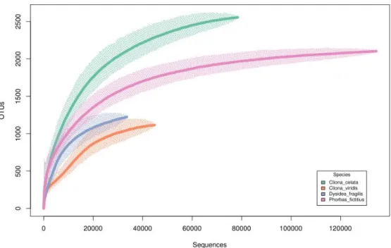
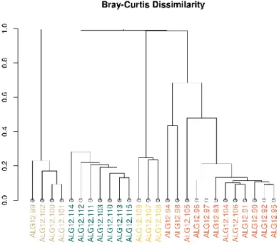
A total of 3,215 prokaryotic Operational Taxonomic Units (OTUs), defined at 97% similarity, were identified from 291,278 sequence reads considered in the normalized dataset, which was rarefied for 11,203 sequences per sample, for statistical purposes. Rarefaction curves revealed that, in spite of the high sequencing effort employed, all sponge species, except C. viridis, would require further sequencing depth for complete coverage of their associated prokaryotic communities. Bray-Curtis dissimilarity-based non-metric multidimensional scaling (nMDS) showed distinct and host-specific communities that did not follow host phylogeny. High proteobacterial dominance (mostly α- and γ-Proteobacteria) was observed across all sponge-associated prokaryotic consortia, except in the case of C. celata, where unclassified bacteria prevailed. Mostly species-specific OTUs classified as α-, γ- and unclassified Proteobacteria were seen to predominate the core of highly abundant phylotypes, as seen by heatmap plotting of decreasing abundances. Further, using stringent phylogenetic assessments, it was possible to re-classify abundant bacterial phylotypes previously regarded as “unclassified” based on database-dependent taxonomic assignments.
Here, host-specific prokaryomes were found, meaning that other factors than host phylogeny must drive sponge prokaryome structure and composition. Moreover, striking prokaryotic diversity was noted within the surveyed sponge hosts, suggesting that further metagenome mining of these prokaryomes may unveil further novelties.
Resumo
As esponjas marinhas (filo Porifera) vivem em simbiose com microrganismos que frequentemente apresentam alto interesse ecológico, evolutivo e metabólico. A descoberta da presença de procariotas simbiontes em esponjas marinhas ocorreu nos anos 1970. No entanto, foi nos anos 2000 que o surgimento das tecnologias de sequenciação de última geração permitiu conhecer mais a fundo as interações simbionte-hospedeiro.
Atualmente, estas tecnologias são amplamente aplicadas e com grande êxito na exploração molecular das simbioses que ocorrem entre micróbios e esponjas. Ademais, por via destas, tem sido possível desvendar o nível de ‘intimidade’ molecular a que vivem estes organismos. Sabe-se hoje em dia que as comunidades microbianas simbiontes de esponjas marinhas contribuem imensamente para o ‘bem-estar’ do seu hospedeiro por via da produção de moléculas antimicrobianas, por exemplo. Por outro lado, por meio do hospedeiro são ‘providenciados’ abrigo, produtos finais metabólicos como amónia, e produtos orgânicos que derivam da predação por filtração de plâncton unicelular. Todos os anteriores fatores contribuem de forma ainda não verdadeiramente quantificada para o estabelecimento e estruturação do microbioma destes metazoários. Neste estudo, foi abordado o procarioma (isto é, o consórcio de todas as bactérias e arqueias -procariotas- presentes num dado ambiente) de quatro esponjas marinhas. Estas foram amostradas ao largo da costa do Algarve e a baixa profundidade em 2012. No total, 26 espécimes pertencentes às espécies Phorbas fictitius (n=12), Dysidea fragilis (n=3), Cliona viridis (n=4) and C. celata (n=7) foram recolhidos. Todas as amostras foram processadas no sentido de isolar o endossoma de cada indivíduo, isto é, o seu interior, coberto pelo ectossoma. Todas as amostras (indivíduos) foram ademais sujeitas à extração de ADN segundo protocolos padronizados pelo ‘Earth Microbiome Project’.
Posteriormente, o ARN ribossomal 16S destas amostras foi amplificado por via de PCR (reação de polimerase em cadeia) e sequenciado por via de tecnologia Illumina HiSeq na sede do Earth Microbiome Project, nos EUA. Os produtos de sequenciação foram então pré-processados por software específico no sentido de eliminar ou corrigir possíveis erros de diversas origens, criar unidades taxonómicas operacionais (OTUs) a um índice de similaridade de 97% e classificá-las de acordo com bases de dados taxonómicas de referência. Esta tese teve como objetivo inferir características ecológicas diversas dos procariomas encontrados em esponjas marinhas, focando
adicionalmente a componente rara destes: o “singletoma” (‘singletome’, na tradução em inglês). Para isto, foram utilizados diversos software e mesmo a linha de comandos do sistema operativo Ubuntu. Foi ademais caracterizada a filogenia das esponjas em estudo com base nos perfis de similaridade do gene da subunidade 1 da proteína citocromo oxidase (cox1). Assim, extraiu-se ADN de amostras conservadas das esponjas em estudo, ao que se amplificou, sequenciou e analisou o gene cox1.
Após filtração das amostras relativas a este estudo, seguiu-se a normalização das amostras, aplicada em função do menor número de sequências encontradas numa dada amostra. Assim, o limiar de normalização foi definido em 11.203 sequências por indivíduo, associadas a um total geral de 3.215 OTUs. Por via de análise de rarefação, foi possível estimar que apenas o procarioma de C. viridis foi sequenciado a uma ‘profundidade’ tomada como verdadeiramente representativa da comunidade procariótica da esponja (no sentido da sequenciação de todos os produtos de amplificação na amostra). Deste modo, o acesso a uma base de dados verdadeiramente representativa dos procariomas nestas esponjas dependerá de futuras rondas de sequenciação a maior profundidade. Ordenação da similaridade entre procariomas revelou perfis de grande intraespecificidade e notória diferenciação entre amostras correspondentes a diferentes esponjas (inter-especificidade).
No geral, α-, γ-, bem como clades não classificadas de Proteobacteria dominaram todos os procariomas, com a exceção de C. celata, em que dominaram OTUs bacterianas não classificadas (segundo classificação atribuída durante o pré-processamento no âmbito do EMP em 2013). Independentemente da filogenia dos hospedeiros, estes procariomas mostraram-se específicos de cada esponja, enquanto que os das esponjas do género Cliona se revelaram os mais dissimilares. Nos procariomas das restantes esponjas, P. fictitius e D. fragilis, notaram-se padrões de alguma similaridade, apesar de estas pertencerem a duas sub-classes diferentes (respetivamente Heteroscleromorpha sensu Cárdenas et al., 2012 e Keratosa, classe Demospongiae). Por meio de visualização das OTUs mais abundantes, observou-se também que cada esponja albergava um conjunto de dois a cinco filótipos específicos. Em C. viridis, cinco destas, muito abundantes, definiam o maior ‘núcleo’ de simbiontes específicos encontrados, possivelmente influenciando por demais os valores de equitabilidade atribuídos. D.
fragilis revelou também um núcleo dominante, ainda que composto por OTUs menos
Por outro lado, a componente rara deste procarioma (‘singletons’, ou OTUs com apenas uma sequência atribuída) revelou diversidade maioritariamente conhecida. Ao contrário do esperado, o ‘singletoma’, isto é, o conjunto de todos os singletons numa base de dados, foi na sua maioria eficazmente identificado. Além disso, não foi encontrada qualquer tendência para padrões de representação específica de singletons de determinado filo ao longo das esponjas marinhas amostradas.
Dada a vincada expressão de OTUs de taxonomia bacteriana não identificada, uma base de dados relativa apenas às 20 mais abundantes foi criada e seguidamente sujeita a reclassificação por meio da mais recente base de dados de referência taxonómica SILVA (versão 123, Julho 2015). Com esta abordagem, foi possível a reclassificação de todos os filótipos analisados em filos conhecidos. Nomeadamente, a segunda OTU mais abundante da base de dados, também a dominante no procarioma de C. celata, foi reclassificada como pertencendo a uma divisão parafilética da classe Clostridia, filo Firmicutes. Foi também reclassificado um filótipo como membro do filo candidato Nitrospinae.
Foi deste modo possível inferir que os presentes procariomas apresentam-se como específicos relativamente ao hospedeiro correspondente, independentemente do grau de parentesco filogenético entre hospedeiros. Tal implica que fatores ambientais ou características morfológicas, de estilo de vida ou metabólicas específicas de cada esponja desempenhem um papel decisivo relativamente à composição e estrutura destes procariomas. Assim, é possível que a natureza do substrato que sustenta a esponja, o seu estilo de vida (ereto ou incrustante, por exemplo) e mesmo diferentes ritmos metabólicos definam a composição dos microbiomas presentes. Uma análise da componente rara do procarioma presentemente analisado não revelou diversidade inesperada ou quaisquer padrões de especificidade ao nível de filo ao longo dos espécimes analisados. O isolamento e reclassificação das OTUs bacterianas não classificadas ao nível de filo resultou na identificação eficaz de diversidade previamente desconhecida.
Contents
1. Introduction
1.1. Aims ... 1
1.2. Importance ... 1
1.3. Morphological and phylogenetic diversity within Porifera ... 2
1.4. Sponge aquiferous system and sponge cell types ... 3
1.5. Ecology and lifestyles of poriferans ... 4
1.6. Sponge microbiology ... 5
1.6.1. Host-specificity of sponge microbiomes ... 9
1.6.2. Rare microbiota in sponges ... 11
1.7. Earth Microbiome Project ... 12
1.8. Targeted sponge species ... 13
2. Methodology ... 17
2.1. EMP dataset generation and processing... 17
2.1.1. Sample collection ... 17
2.1.2. DNA extraction, sequencing and primary processing ... 17
2.1.3. Local dataset processing for Algarve samples ... 19
2.2. Ecological analysis ... 20
2.3. Phylogenetic cox1 analysis ... 23
3. Results ... 25
3.1. Ecological analysis of sponge prokaryomes ... 25
3.2. Exploration for prokaryotic ‘microbial dark matter’ ... 43
3.3. Sponge host phylogeny ... 47
4. Discussion ... 49
4.1. Pre-processing methodologies ... 49
4.3. Patterns of OTU presence across prokaryomes ... 54 4.4. Taxonomy across prokaryomes ... 55 4.5. Sponge- and species-specific associations ... 58 4.6. Exploration for the ‘microbial dark matter’ within the sponge prokaryome ... 60 4.7. Phylogenetic inference of abundant but unclassified OTUs ... 62 4.8. Phylogenetic relationships among hosts and prokaryomes ... 64
6. Bibliography ... 68 Annexes
I. General R script for ecological and statistical analysis I
II. R script for generation of an averaged abundance matrix II
III. Ubuntu CLI script for Greengenes taxonomy extraction from EMP “.database”
file III
IV. Mothur script for “singletome” dataset generation IV
V. DNA extraction and cox1 PCR figures V
VI. Non-normalized OTU and sequence numbers table VI
Figure Index
Figure 1.1 – General Leuconoid sponge morphology schematic picture (in Hentschel,
2012 9) ... 4
Figure 1.2 – Worldwide distribution of phylum Porifera based on the World Porifera
Database (http://www.marinespecies.org/porifera/). ... 5
Figure 1.3 – Cliona viridis (A), Cliona celata (B), Phorbas fictitius (C) And Dysidea fragilis (D) pictured in situ, off the Algarve coast.. ... 14 Figure 1.4 – World distribution of the studied sponges, based on the World Porifera
Database (http://www.marinespecies.org/porifera/).. ... 15
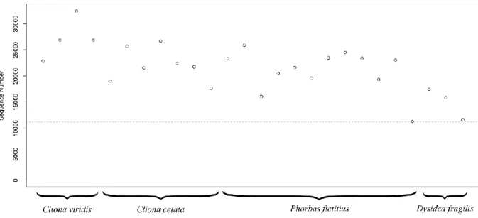
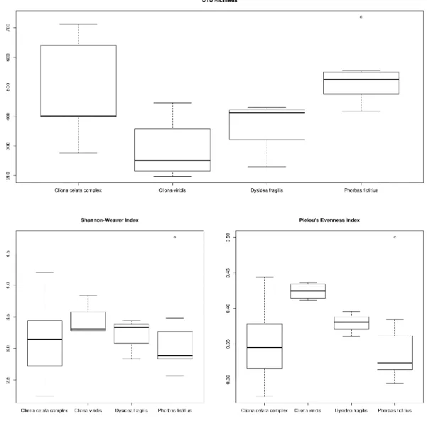
Figure 3.1 - Sequence number for all samples after singleton exclusion.. ... 25 Figure 3.2 - Boxplots depicting OTU richness, Shannon-Wiener diversity and Pielou’s
evenness indices averaged across all samples. ... 26
Figure 3.3 - Rarefaction curves depicting cumulative (across all samples) richness for
the normalized dataset. ... 27
Figure 3.4 - Rarefaction curves depicting averaged (across all samples) richness for the
normalized dataset. ... 28
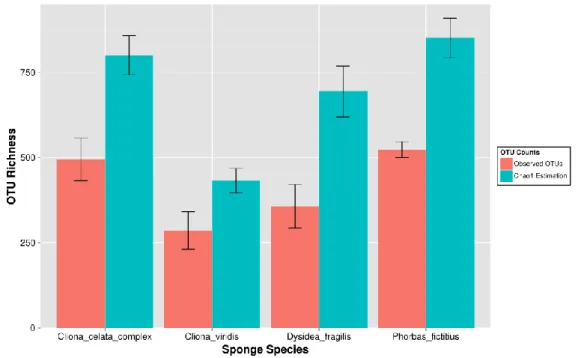
Figure 3.5 -Averaged observed and Chao1 estimated number of OTUs across all sponge
species. ... 29
Figure 3.6 -Rarefaction curves depicting cumulative Chao1 estimated richness for the
normalized dataset.. ... 30
Figure 3.7 - Clustered Bray-Curtis dissimilarity profiles for Algarve samples, coloured
according to sponge species. ... 31
Figure 3.8 - non-metric Multidimensional scaling of Bray-Curtis dissimilarity profiles.
... 32
Figure 3.9 - Shepard’s diagram for Bray-Curtis dissimilarity nMDS scaling, further
showing the linear and non-metric fit for ordination distances. ... 33
Figure 3.10 - Phylum-level relative abundance barchart for sequence libraries
normalized by size (11.203 sequence reads per sample, singletons excluded) ... 34
Figure 3.11 - Class-level relative abundance barchart for sequence libraries normalized
by size (11.203 sequence reads per sample, singletons excluded). ... 37
Figure 3.12 - Order-level relative abundance barchart for sequence libraries normalized
Figure 3.13 - General profiles of OTU abundance set in ascending order across all
samples in a fourth-root-transformed normalized dataset.. ... 39
Figure 3.14 – Heatmap showing the distributions of the 50 most abundant OTUs across
all samples.. ... 40
Figure 3.15 – Venn diagram depicting the cumulative total number of shared and
specific OTUs regarding each sponge species. ... 41
Figure 3.16 - Venn diagram depicting the cumulative number of shared and specific
OTUs with a minimum of 50 assigned sequences regarding each sponge species. 42
Figure 3.17 - Venn diagram depicting the cumulative number of shared and specific
OTUs with a minimum of 100 assigned sequences regarding each sponge species. ... 43
Figure 3.18 - Phylogenetic tree depicting the twenty most abundant OTUs unclassified
at the phylum-level across the dataset, re-aligned using the SILVA 123 "Non-Redundant" (NR) SSU Reference dataset (July 2015). ... 44
Figure 3.19 - Phylum-level barchart depicting singleton-specific taxonomic
assignments across the non-normalized dataset. ... 46
Figure 3.20 - Unrooted phylogenetic tree depicting cox1-nased relationships among the
Acronym list
OTU: operational taxonomic unit bp: base pair
rRNA: ribosomal ribonucleic acid DNA: deoxyribonucleic acid DOM: dissolved organic matter POM: particulate organic matter
WoRMS: World Register of Marine Species AR: ankyrin-repeat proteins
TPR: tetratricopeptide-repeat proteins ELP: eukaryotic-like proteins
HMA: high microbial abundance LMA: low microbial abundance SCC: sponge-specific cluster EMP: Earth Microbiome Project PCR: polymerase chain reaction NGS: next-generation sequencing RDP: Ribosomal Database Project CLI: command-line interface ANOVA: analysis of variance
MRPP: multiple response permutation procedure nMDS: non-metric multidimensional scaling
QIIME: Quantitative Insights Into Microbial Ecology COI/cox1: cytochrome oxidase subunit 1
MB-CD: mean between-cluster dissimilarity W-CD: within-cluster dissimilarity
ANOSIM: analysis of similarities NR: non-redundant
SSU: small subunit
14 1. Introduction
Poriferans (members of the ‘Porifera’ phylum, from the Latin ‘porus’, pore, and
‘ferre’, “to carry, to bear”) are the most ancient multicellular metazoans. They are
therefore a pivotal group in the study of the metazoan transition from unicellularity to multicellularity. This very successful lifestyle encompasses a simple body plan, highly totipotent mobile cells, a characteristic aquiferous system and flexible reproduction strategies. Sponges are and have putatively been since pre-Cambrian times prevalent in oceans worldwide, ranging from coastal shores to abyssal depths and from tropical to polar settings1. Although recent literature points towards there being little evidence for the veracity currently known “sponge” fossils, molecular clocks calculated for the phylogenetic divergence of poriferans and eumetazoans still support a pre-Cambrian origin2,3. Apparently simple in its morphology and physiological traits, this phylum is regarded as an important model for animal phylogenetic, neuronal and immunological evolution, given its early-branching position in the metazoan tree of life.
1.1. Aims
This study aimed to characterize the prokaryotic communities of four species of marine sponge found off the coast of Algarve, southern Portugal. A dataset generated by means of high-throughput sequencing of 16S rRNA genes amplified from marine sponge total community DNA was thoroughly analysed using state-of-the-art bioinformatics and statistical tools.
The hypothesis that the taxonomic composition and structure of prokaryotic symbiont communities correlates with phylogenetic relatedness of their corresponding hosts was addressed.
Further, the diversity and magnitude of the “microbial dark matter” (that is, the pool of low-abundance prokaryotes) present in marine sponges was determined.
1.2. Importance
The marine sponge holobiont, that is, the ensemble of microbial communities inhabiting a marine sponge and their corresponding symbiosis, has now been researched for more than four decades. Recent advances in molecular biology techniques, namely next-generation sequencing (NGS), have allowed for a deeper understanding of the
15 structuring and composition of microbial communities within sponges. Besides the remarkable advances made in microbial ecology in general, studies of the marine sponge microbiome have been relevant from a biotechnological standpoint. It was long thought that sponges were the main producers of molecules of biotechnological interest. It is now widely accepted that microbes populating poriferans are the main producers of such compounds 4.
As such, it is of great interest to direct efforts towards a complete characterization of sponge “prokaryomes”: defined here as the ensemble of all bacterial and archaeal microbes present within a given setting. Previous explorations of poriferan microbiomes have consistently delivered novelties of biotechnological and ecological interest in the last two decades. For instance, new compounds ranging a plethora of bioactivities to previously unknown bacterial phyla have been found.
This study served as an exploratory analysis of the taxonomic and compositional characterization of the prokaryomes within four understudied marine sponges. Importantly, the present work’s data was the first high-throughput sequencing dataset to become available in the scope of molecular ecology of sponge prokaryomes at a worldwide scale. Given the extent of its size and depth, this dataset bore the potential to evaluate with higher confidence the 'microbial dark matter' within these organisms. This component of the prokaryome has recently been given greater attention because of its putative pivotal role in mediating important biochemical processes.
1.3. Morphological and phylogenetic diversity within Porifera
Accounting for all living sponges, it is estimated that up to 81% are taxonomically affiliated to the Demospongiae class 5. This class encompasses sponges whose skeleton, if present, is made up of siliceous or fibrous spicules, or both. When siliceous, spicules are either simple, or monaxonic, or present four axes with a common origin (tetraxonic). Further, the core of the aforementioned spicules, colloquially named axial filaments, are enclosed either in a triangular or in a hexagonal cavity 6.
Not all sponges possess a silica-based structural organization. In fact, some do not present any extracellular structure to provide a ‘skeleton’ of some sort. These sponges are part of a recently described class called Homoscleromorpha 5. This morphological characteristic has been the historical basis for sponge taxonomy and its reliance was
16 recently supported by molecular evidence 5. The remaining classes composing the
phylum Porifera are Calcarea, made up of sponges bearing calcium carbonate spicules, Hexactinellida, generally known as ‘glass sponges’, which encompass siliceous spicules, and Demospongiae, in which spongin fibres, a kind of fibrous spicule, generally act as the main structural component.
Demosponges, the common denomination for members of the Demospongiae class, present a tremendous panoply of shapes, colours and sizes. Generally, sponge morphologies may be divided into massive (erect and large in size), encrusting (growing on top of a surface of either biological or geological origin) or excavating (by active erosion of the substrate on which the sponge lies).
1.4. Sponge aquiferous system and sponge cell types
Most marine sponges are benthic filter-feeders that rely on an aquiferous system in order to filter planktonic microorganisms 7. The external surface, or ectosome, as well as the outer lining of inner channels of poriferans are in general composed of a monolayer of pinacocytes, flattened cells which functionally act as ‘epithelial’. Along the ectosome, channels made up of one or more specialized cells, namely ostia or dermal pores, which allow for the entrance of water (therefore, incurrent channels) into the sponge endosome, that is, all that is surrounded by the ectosome. Seawater is further propelled through the sponge by a simple but effective mechanism 8. The pumping
action of choanocytes, flagellated cells responsible for capture and intake of suspended particles, within choanocyte chambers, causes seawater to be driven further into the sponge by means of a continuous and uncoordinated beating of the flagella. A flow is then set from incurrent channels, connecting the external environment to the choanocyte chambers, leading to a central atrium and back towards the water column through the osculum and excurrent channels (see Figure 1.1) 9,10. The aquiferous system in sponges is seen as early evidence for a metazoan circulatory system 11. It allows, for example, effective gaseous exchanges, excretion of toxic metabolic end-products as ammonia, as well as easy take-up of dissolved and particulate organic matter (DOM, POM), the latter the subject of most attention since it leads to the retrieval of planktonic microorganisms from seawater.
The inner matrix encompassing the sponge ‘tissue’ in between the ectosome and all its inner surfaces is generally named mesohyl, or mesoglea (see Figure 1.1). This complex
17 framework is usually made up of specialized cells and a secreted skeleton of silicate or calcium carbonate spicules or collagen-based fibres, being that exceptions may occur in some sponges. Secretion of the aforementioned structural elements of the sponge occurs by action of sclerocytes, a cell type which biomineralizes the poriferan skeleton.
Figure 1.1 – General leuconoid sponge morphology schematic picture (adapted from
Hentschel, 2012 10)
Within the mesohyl, archaeocytes (see Figure 1.1) are characterized by being highly totipotent ameboid cells which proliferate, carrying out tasks which range from food digestion and transport to asexual reproduction. Archaeocytes are key to food digestion, as these cells are able to digest phagocytised material captured by choanocytes, as well as capture food particles by phagocytosis directly through inner walls of water channels.
1.5. Ecology and lifestyles of poriferans
Being extremely diverse in form, shape and colour, the morphology of sponges is determined by genotype and environmental conditions. Regarding the latter, sponges are mostly affected by underwater current velocity as well as turbidity, this is, the level of suspended planktonic detritus 12.
Poriferans are widespread across all latitudes and longitudes, occurring mostly in seawater, but also in freshwater and in brackish environments (see Figure 1.2).
18
Figure 1.2 – Worldwide distribution of the phylum Porifera based on the World
Porifera Database (http://www.marinespecies.org/porifera/). Warm colours depict high abundances whereas cold colours represent low levels of abundance. Accessed on the 28th of July 2015.
Freshwater sponges, although restricted to a single family formerly known as
Spongillina, currently recognized as Spongillida Manconi & Pronzato, prevail across
inland bodies of water across the world, ranging from Northern America to the Netherlands, India and China (WoRMS, 2015) 5. Brackish sponges are also prevalent
across the world, but remain understudied. The known studied exemplars are still poorly understood at a phylogenetic level, such as the Malawispongiidae and
Metschnikowiidae families 5.
Across oceans worldwide, demosponges populate several ecological niches, ranging from the intertidal regions to abyssal planes and marine caves 13,14. In general, sponges are sessile, benthic filter-feeders, but exceptions occur, as is the case of carnivorous sponges, which are able to predate selectively by means of a “sit-and-wait” strategy in generally oligotrophic and isolated settings such as marine caves 15. These use specialized microscleres, or small spicules, which have evolved to a hook-like shape, for directed predation of microinvertebrates.
1.6. Sponge microbiology
It was in 1977 that Vacelet and Donadey published that demosponges were often densely populated by bacteria 16. At the time, ‘tissue density’ of the hosts were set as defining factors of high and low observable microbial abundance. Although no
19 statistical evidence was provided, a linear relationship (e.g., high microbial abundance correlated to high sponge tissue density) was predicted. Further, it was noted that most of the microbes were present in the extracellular mesohyl matrix of the host animals.
The advent of molecular techniques enabled an in-depth exploration of the underlying diversity and microbial abundance within the sponge host in subsequent studies. By means of cloning-and-sequencing of bacterial 16S rRNA gene fragments amplified from sponge communal DNA, Hentschel and colleagues (2002) investigated marine sponges from the Mediterranean region, Japan and the Palau Republic 17. They found evidence for a core of shared prokaryotic species among poriferans that was not correlated with host biogeography. These so-called “sponge-specific” groups of OTUs were recurrently found in the following years, and the uniqueness of the sponge microbiome was further recognized, usually through comparative analyses with the surrounding seawater microbial communities 18,19. The most iconic sponge-specific taxa described to date are the Poribacteria, candidate bacterial phylum discovered by Hentschel and co-workers in demosponges 20. This candidate phylum was found to be one of the most abundant across several sponge microbiomes, existing only scarcely in seawater 21. Further, the lifestyle of these vertically transmitted symbionts has now began to be uncovered by single-cell genomics and other last generation molecular biology techniques 22. Special attention has been given, for example, to the discovery of genome-encoded eukaryotic-like proteins which could mediate sponge-Poribacteria symbiosis 23. Recent evidence
provided by Taylor et al. in 2013 has shown, however, that sponge-specific taxa may be much more widespread throughout diverse marine environments than previously thought. This study analysed more than 12 million 16S- rRNA gene reads (from the V6 hypervariable region) generated by 454 pyrosequencing in 42 studies worldwide and found that although about 55% of the previously set sponge-specific taxa were found only in sponges, the remaining taxa were noted in other hosts and marine environments in relative abundances between 0 and 1% 10. Poribacteria was among these and was detected in abundances reaching 0.25% in coral hosts, comprising up to 0.19% of other samples, which included seawater sampled near hydrothermal vents, for example.
The previous argument was not a rejection of the sponge-specific taxa theory, but a call for further proof of “true” symbiosis, in the sense that more complete information towards the lifestyle of true sponge symbionts must be provided. Analysing microbial communities from a functional point of view has allowed for valuable insights
20 regarding the metabolic diversity in these, and has further unveiled how complementary host and microbial metabolisms may be. For example, in 2010, Thomas and colleagues used shotgun metagenome sequencing to approach the mechanisms of microbe-host functionality in the light of the holobiont theory of evolution 24. Special relevance was given to ankyrin-repeat (AR) and tetratricopeptide-repeat proteins (TPR), whose expression was found to be significantly augmented in sponges when compared to the surrounding seawater. These eukaryotic-like proteins (ELPs) were known to be present in genomes of prokaryotic symbionts of eukaryotic hosts, and were thought to play critical roles in the establishment of symbioses. Nguyen et al. in 2013 gave further evidence towards processes of colonization of symbionts by escaping digestion by phagocytosis25. Ankyrin-repeat proteins are thought to interfere with phagosome maturation by protein-protein interaction and therefore avoid digestion of bacterial cells by the sponge host, allowing for settlement of the first in the mesohyl of the latter. The finding of ELPs in symbiont genomes could indicate the mechanisms behind primordial settlement of microbes in the sponge mesohyl and symbiosis establishment 22. These putative indicators of prokaryote-eukaryote symbiosis have also been found in intracellular amoebal symbionts 25.
Functional profiling of the sponge microbiome has until now contributed for an understanding of how symbiont prokaryotic species take advantage of sponge excretion products, but also produce secondary metabolites of interest for the sponge, which may, in turn, become a selecting factor for symbiotic settlement 10. Indeed, it has been found
by means of genomic analysis that sponge-associated microorganisms retain genes encoding for several of the carbon and nitrogen metabolism pathways. The assimilation of carbon dioxide via reductive tricarboxylic acid cycle and a modified 3-hydroxypropionate cycle has been reported for bacterial and archaeal sponge symbionts, for example 23,26,27. Further, regarding nitrogen metabolism, current genomic evidence shows a microbial metabolic ensemble directed towards the assimilation of ammonia, naturally excreted by sponges as a metabolic waste product, and nitrite 22–24. This occurs either in archaeal and bacterial symbionts, being that at least in cold water sponges, the first seem to dominate ammonia assimilation either total or partially (3 to 4 orders of magnitude), as communitarian ammonia-monooxygenase activity profiling showed 28.
Aside from primary metabolism, important discoveries were made regarding secondary metabolism, which mainly encompassed vitamin biosynthesis 27. Thought to
21 be intimately associated with microcompartment genesis in Poribacteria, biosynthetic pathways encoding for vitamin B1, B2, B6, B7 and B12 were found through genome mining of these symbionts 10,26.
Although mostly distinct across geography and phylogeny, the sponge microbiome seems to generally converge regarding metabolism 29,30. Indeed, it was shown by means of functional profiling of sponge prokaryotic communities that core metabolic tasks are present across a broad host phylogenetic range 29. This could mean that core prokaryotic functions are transversal to diverse sponge hosts disregarding geography, phylogeny and even the taxonomic composition of their associated microbial communities.
Abundance patterns for microbial communities have been noted since the early works by Vacelet and Donadey 16. The terms ‘high’ and ‘low microbial abundance’ (HMA, LMA) are currently set based on electron microscopy, as they have been since early sponge microbiology studies 31, but flow cytometry and epifluorescence microscopy methods have recently enabled the exact quantification of living prokaryotic cells in solution 31–33. To date, there is no evidence for a correlation between microbial abundance and phylogeny in sponges. Indeed, it is known that hosts belonging to the same sponge taxonomic order, or even family, may harbour microbiomes of contrasting abundances 31.
It is known that HMA sponges tend to harbour microbiomes in abundances surpassing those of seawater by a magnitude of 3 or 4, while LMA tend to reach similar values as to those of the latter environment 32. Further, LMA sponges appear to host less diverse
microbiomes, which often dominated by Proteobacteria, Cyanobacteria and Nitrospira
34,35. Molecular profiling of asympatric sponges led Giles et al. to assert that LMA
microbiomes further lack typical core-HMA lineages, such as Poribacteria 34.
Present-day sequencing technologies fail do discern true microbial abundance, due to known PCR and sequencing biases 36. These techniques are further limited by the essential nature of each analysis, which will not give true cell abundance in a sample. For these reasons, robust efforts are still made towards cataloguing microbial abundance in sponges by means of electron microscopy 31. Hopefully, by relating these with high-throughput sequencing data, it will be possible to generate standard thresholds of microbial abundance which could serve as reference for future descriptions of new sponge microbiomes.
22 With the advent of sequencing technologies in the last decade and a half, knowledge of microbial diversity in marine sponges has exponentially increased 16. By generating
phylogenetic data from 16S-rRNA clone libraries, up to eight bacterial phyla were found to be present in the marine sponge microbiome 17,18,20,34. Further, in-depth analysis of some consistently recovered sequences, transversely present across sponge species and geography, showed how some archaeal and bacterial species could putatively form sponge-specific clusters (SSC) in a phylogenetic tree 37. The advent of pyrosequencing applied to microbiology allowed for deeper insights into the sponge microbiome and is, at the current state-of-the-art, the most utilized NGS in sponge molecular microbiology research. This technique usually unveils from 20 to 28 bacterial phyla as being in association with sponges, commonly obtained from the mesohyl
9,13,38,39.
Characteristically high abundances of proteobacterial phylotypes are frequently noted in sponges, and closer inspection of these assemblages will often reveal varying abundances of each proteobacterial class in different marine sponges. The diversity in proteobacterial lineages is true for either HMA or LMA sponges, although other bacterial lineages, such as Chloroflexi, Acidobacteria, Deferribacteres and Poribacteria, are generally absent or in minimal proportions in the latter poriferans (LMA) 35,40,41. These phyla have thus been selected as “indicator phyla” for HMA sponges, while LMA, as stated before, are frequently dominated by Proteobacteria and Cyanobacteria
35. Contrastingly, the frequently observed cyanobacterial sponge-specific species Synechococcus spongiarum is known to be specifically maintained in LMA sponges
throughout their geographical range 34.
1.6.1. Host-specificity of sponge microbiomes
Ever since Hentschel and colleagues suspected the sponge-associated microbiome to be uniform across geographic boundaries, studies addressing patterns of host-specific microbiome assemblage in these animals have been conducted17. In fact, it was further
noted in 2005 that Chondrilla nucula (Verongimorpha, Chondrillida), a demosponge sampled in The Netherlands, could harbour a sponge-specific and uniform microbiome, following the concepts found in the latter work from Hentschel and colleagues 42. A total of 21 OTUs (e.g., groups of sequences found to share similarities of 97% or more and therefore treated as putative species. If not stated otherwise, all mentioned OTUs
23 are assumed to have been created by sets of sequences with the latter similarity values – 97%) were found to be absent from seawater and highly similar to known sponge-specific microbes found in other studies. This microbiome was therefore considered sponge-specific with regard to the uniformity in the presence of several prokaryotic phyla found by profiling marine sponges across the world. However, no direct comparison was made with other species within the same genus or relatives. It was suggested in 2013 by Blanquer et al. that similarities across microbiomes seemed to be more correlated to the assigned microbial abundance (HMA, LMA) than to host phylogeny 41. The latter study further found that Chloroflexi seemed to be enriched in HMA sponges and that some α-Proteobacteria taxa were often present in LMA sponges.
Analysing 20 species of tropical sponges, Easson and colleagues found host-species specificity relating prokaryomes to sponge phylogeny 43. They found up to 30 OTUs defining dissimilarities across sponge species 43. Likewise, evidence of host species-specificity across geographical borders and among species within a same genus has been given by Erwin and colleagues 44,45. Either sympatrically or across distances up to 800km across the north-west Mediterranean Sea, these sponges maintained stable and defined prokaryomes, whose structure was highly correlated to host phylogeny.
Sampling at a depth gradient and along the eastern and north Atlantic, Reveillaud et
al. did not notice host species-specificity patterns among the microbiomes of seven
species of the Hexadella (Verongimorpha, Verongida) genus and five exemplars of the
Mycale (Heteroscleromorpha, Poecilosclerida) genus 46. Nitrospira (Nitrospirae) was
observed to be one of the most abundant OTUs in the dataset, being present in small amounts in sponges of the Mycale genus and almost absent in seawater. Analysed among other two sponges of the Poecilosclerida order, an exemplar of the Halichondria genus (Heteroscleromorpha, Suberitida), a LMA sponge, was shown to be dominated by several α-proteobacterial OTUs 38. Moitinho-Silva et al. sampled two sponges each
belonging to a microbial abundance category (i.e., LMA and HMA) in the Red Sea 35. Here, it was posed, in accordance with previous studies from the same research group, that contrary to prevailing ideas, LMA sponges seemed to harbour higher abundances of Proteobacteria, Cyanobacteria and Nitrospirae 34. Further, in sponges displaying high microbial abundance, Chloroflexi, Poribacteria and Deferribacteres were found to be the dominating phyla. Controversially, this study presented further evidence that suggested the specificity of the sponge microbiota was defined by the host’s microbial
24 abundance. However, several studies in irciniids (this is, marine sponges affiliated to family Irciniidae, order Dyctioceratida) point towards a phylogeny-dependent microbial community structuring 45,47, and indeed, the inner morphology of both LMA and HMA
sponge categories has already been shown to be significantly different 48. With regard to morphological dependency, Weisz et al. showed not only that HMA and LMA sponges differ in mesohyl densities, but also in retention time of pumped water volume. As such, the first were shown to hold a denser mesohyl and therefore a lower water pumping rate than the latter sponges. Microbial abundance also seemed to be the defining factor with regards to the functionality of the microbiome when these were analysed by means of a Geochip 4.2 gene array, a high-throughput functional gene array, which enables the hybridization of prokaryotic coding sequences to pre-set light-emitting probes 49.
As such, recent evidence seems to point towards host-specificity being less affected by sponge phylogeny but by LMA or HMA status. Further knowledge of the sponge microbiome across geography and sponge phylogeny is necessary before firm conclusions can be drawn.
1.6.2. Rare microbiota in sponges
It was posed in 2009 that the rare biosphere within seawater could be a stable source of sponge symbionts 50. The advent of “ultra-high-throughput” sequencing technologies such as Illumina HiSeq and MiSeq provided further possibilities regarding the depth at which investigation of sponge microbiomes was possible 36. With this, it has been possible to uncover more of the diversity within the rare fraction of the sponge microbiome, and to infer its possible functional roles 35,51. Giles et al. noted how up to 81% of the uncovered phylotypes of five LMA sponges originating from the Caribbean, Red Sea and South Pacific were indeed singleton (occurring only once across a given dataset) OTUs 34.
In 2014, Reveillaud and colleagues provided the first evidence towards host-specificity regarding ‘microbial dark matter’ within the sponge microbiome 46.
Examining into the OTUs present in relative abundances below 0.01% and 0.001% (within a total of 100,000 sequence reads per sample), it was found that indeed their presence, as determined by accumulation of phylotypes of a corresponding phylum, was selective regarding the correspondent sponge host, therefore indicating host-specificity.
25 Further, 33 singletons were uncovered in the Illumina HiSeq-generated dataset, as well as 18 doubletons (OTUs containing only two sequence reads across the entire dataset).
The rare microbiota within sponges has therefore far been under-characterized, but evidence points towards the possibility that with increasing sequencing capabilities, it will be possible to further analyse the diversity and functionality of these so-far elusive but seemingly essential consortia. Currently, it is expected that studies utilizing NGS technologies will be able to uncover highly diverse ‘singletomes’, that is, singleton OTUs consortia, as well as other components of the microbial rare biosphere. As suggested by Reveillaud et al., it could happen that the consortia of low-abundance OTUs in sponge microbiomes are host-specific and may provide important functional input for the holobiont 46.
1.7. Earth Microbiome Project
The Earth Microbiome Project (www.earthmicrobiome.org36) is an international network that aims to catalogue the biodiversity of microorganisms across several biomes on Earth. This project set out to sequence extensively the microbial communities from a multitude of environmental sources worldwide, in a collaborative sampling effort. So far, it has analysed 200,000 samples, being that approximately 30% of those are sponge-related 52.
Within the framework of the Earth Microbiome project, the Consortium for Sponge Microbiology organized a standardized sampling of marine sponges, which took place in every continent worldwide, further collecting sponge-associated marine sediments and seawater, as well as detailed metadata. The contribution of this international project to the sponge microbiology research community is yet to be quantified, since only one scientific article has so far been published 43. It can, however, be expected to propel the development of state-of-the-art approaches towards core questions in this research area, and help set a threshold for low and high microbial abundance or the determination of true sponge-specific microbes worldwide.
This work was developed within the scope of the Earth Microbiome Project and is based on part of the first worldwide “ultra-high-throughput” 16S rRNA gene amplicon dataset generated from poriferans. Here, a set of 26 samples comprising two clionaidans (Clionaida sensu Morrow & Cárdenas, 2015 5), one dyctioceratid (order Dyctioceratida)
26 and one poecilosclerid (order Poecilosclerida) sponge, were characterized to retrieve insights into the host species specificity of each prokaryome and the relationship between host phylogeny and microbial community composition. Further, the taxonomic composition and magnitude of the ‘microbial dark matter’ encountered in the surveyed sponges was interrogated. For this, in-depth analysis was pursued by means of specialized 16S dataset analysis software packages such as mothur and QIIME (Quantitative Insights Into Microbial Ecology) 53,54. Further, ecological and statistical analysis packages within the R software were utilized. By using mothur, an amplicon-directed bioinformatics pipeline, normalization, filtering and generation of abundance matrices for the data was possible, whilst statistical and ecological information was processed by R. QIIME produced relative abundance barcharts.
1.8. Targeted sponge species
In this study, given its sampling effort and experimental design, only the Demospongiae class was investigated. In general terms, demosponges (sponges of class Demospongiae) are characterized by their possession of an extracellular matrix mostly made of spongin fibres, with some species showing added and frequently dominating silicate-based spicules. Demosponges are found in either marine, brackish or freshwater environments, near-polar, tropical and temperate climates and at varying depths. Exceptions occur, as is the case for order Dictyoceratida, where spongin is dominant and siliceous spicules are absent 55. Demosponges are able to sexually or asexually
reproduce by means of viviparity or ovoviparity 56. The morphological plasticity
observed in demosponges has led to recent efforts towards a clarification of the phylogeny of the class. The removal of Homoscleromorpha from the class and its elevation to a new class within Porifera led to a conclusive monophyletic status of
Demospongiae 57.
Two of the marine sponge species belonging to the genus Cliona (Grant, 1826) studied here had their taxonomy recently altered. Specifically, the order Hadromerida to which they were assigned to, is no longer formally recognized. They are now placed within the Heteroscleromorpha subclass of the Demospongiae, and are part of the newly created Clionaida order (therefore members of this orders are commonly denominated clionaidans) 5. This group of marine sponges is known to have the ability to erode calcium carbonate, frequently harming corals and other organisms depending on such
27 solid exoskeletons. Shellfish cultures are known to take measures against invading clionaidans, for example 58. One of these, Cliona viridis (Figure 1.3a), is a small known
coral-boring sponge, although it occasionally may significantly grow in size, with a distribution restricted to the Mediterranean Sea, and Azores and Madeira archipelagos (Figure 1.4). Morphologically, it generally shows a cerebroid smooth surface, interrupted by large oscula, and is predominantly of brownish colour (Figure 1.3a). This species has been recognized as an LMA sponge, according to Blanquer et al., who found that an OTU assigned to α-Proteobacteria accounted for more than half of the recovered sequences belonging to those samples. A similar result was found with H.
columella (Poecilosclerida) 41.
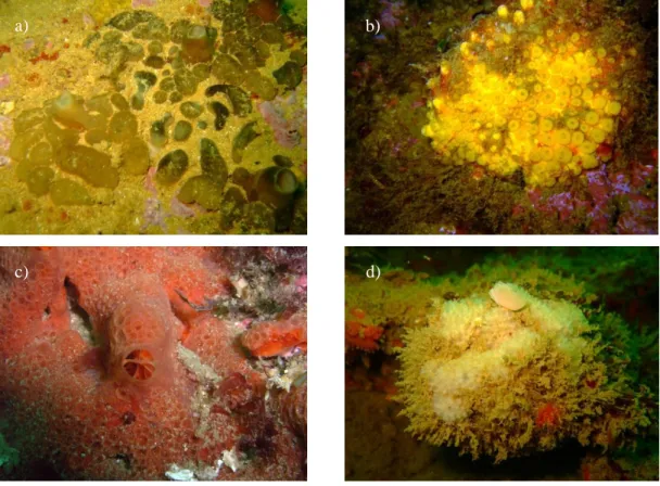
Figure 1.3 – Cliona viridis (a), Cliona celata (b), Phorbas fictitius (c) and Dysidea fragilis (d) pictured in situ, off the Algarve coast. Pictures by Francisco Pires, 2013.
Another member of this genus is Cliona celata, part of a cryptic species complex whose taxonomy is currently unsettled, reason why it is generally referred to as Cliona
celata complex 59. Since its first depiction in the 1900’s, this has been one of the most studied sponge species, with its most defining characteristic being the “bright sulfur yellow” tone, as seen in Figure 1.3b 60. Among the studied sponges, this species is,
a) b)
28 according to the World Register of Marine Species, the one with the most extensive distribution. It is present in the Atlantic and Pacific Oceans, as well as in the Mediterranean Sea and in the South African coast. Evidence suggesting HMA in C.
celata was found in 2015 when exemplars collected in Korea revealed relatively high
diversity, when compared to prokaryomes of known LMA sponges 61.
Figure 1.4 – World distribution of the studied sponges, based on the World Porifera
Database (http://www.marinespecies.org/porifera/). Blue, green, pink and brown areas depict P. fictitius, C. celata, C. viridis and D fragilis, respectively. Compound image of all distributions produced by Dr. Bart Vanhoorne (WoRMS) Data Management Team. Accessed on the 17th of April 2015.
Phorbas fictitius is externally characterized by its bright red to pinkish colouration,
predominant oscula across the ectosome, and can either develop massively or stay incrusting. This sponge species has been shown to be the most abundant along the coast of Algarve by Pires, 2012, being its distribution influenced the most by depth and spatial variability 62. It ranges from the mid-Atlantic Madeira archipelago and Mediterranean Sea to the North Atlantic, occurring also in the Norwegian coast and North Sea (Figure 1.4). The genus Phorbas has not yet been studied regarding its microbial abundance, although, two studies have so far characterized 2 and 7 exemplars from the same order (Poecilosclerida) as LMA 31,63. However, in 2012, Uriz et al. found sponges of the Hemimycale genus (Poecilosclerida) to be highly populated by “Calcibacteria”, which encompassed about 60% of the sponge’s microbiota total weight
64.
As is typical of members of the order Dyctioceratida, Dysidea fragilis possesses an intricate, concentrically defined spongin skeleton. Typically encrusting but often found in massive form, this sponge’s superficial morphology shows a complex rugged
29 network of interconnecting conules.The distribution of D. fragilis ranges from the Atlantic African and Portuguese coasts to the Mediterranean Sea and the White Sea, in the northern coast of Russia (see Figure 1.4). Although no molecular studies have specifically targeted this species, other exemplars of the same genus were studied. The Red Sea sponge Dysidea avara has been considered to be a LMA sponge as determined
2. Methodology
2.1. EMP dataset generation and processing 2.1.1. Sample collection
Twenty-six sponge specimens were collected off the coast of Algarve, southern Portugal by means of SCUBA diving, in October 2012. All were photographed in situ, before being collected. Each specimen was sampled in a hermetic plastic bag with ambient seawater, placed into cooling boxes and transported to the laboratory within 2 hours, where they were immediately processed. In the laboratory, all samples were photographed before being thoroughly rinsed in sterile calcium/magnesium free artificial seawater (ASW). They were further examined for the presence of obvious epibionts, which if present were removed. Sponge specimens were then cut into 0.25g pieces of sponge endosome using sterilized scalpel and tweezers. Backup pieces of sponge were kept at 96% ethanol for further taxonomical identification.
The specimens were then labelled with an “ALG” prefix indicating sampling location (Algarve), followed by “12”, indicating the sampling year (2012), and an individual code number. All samples were frozen at -80ºC and sent on dry ice to Wüerzburg University for DNA extraction. Metadata were recorded while sampling took place, according to EMP standard procedures (www.earthmicrobiome.org).
2.1.2. DNA extraction, sequencing and primary processing
In order to achieve standardization regarding this step, the Sponge Microbiology Consortium decided that all samples should be processed in three laboratory hubs, located in Europe, Australia and United States of America (USA), with the same methodology. This way, all extractions were carried out under the same protocol, therefore minimizing the user biases associated to handling by several users, for example.
Nucleic acid extraction was performed with the PowerLyzer PowerSoil DNA Isolation Kit” (MoBio Laboratories, Inc.), allowing for high throughput extraction of metagenomic material from elevated numbers of environmental samples at a same time (www.earthmicrobiome.org).
Ribosomal ribonucleic acid (rRNA) forms complex RNA-protein domains, forming ribosomes, which play vital roles in microbial translation. The 16S rRNA (consisting of
about 1500 nucleotides) is part of the small subunit (SSU) of the prokaryotic 70S ribosome and is known to be highly conserved, for which it has in the last decades been widely used as a barcode for bacteria and archaea. The 16S rRNA gene is divided into 9 hypervariable regions, which are utilized either individually or in pairs, e.g. V7-V8, according to primer design. In this case, the V4 region of the 16S rRNA gene was utilized after intense scrutiny regarding its phylogenetic resolution by Caporaso and colleagues 65. For such, paired-end 16S rRNA gene communitarian DNA sequencing was performed on an Illumina HiSeq (Illumina, Inc.) platform at the EMP headquarters in the USA, by means of a specifically designed primer set 66 utilizing standard Illumina barcodes 65. These primers (515F/806R 65) were built in order to amplify both bacterial and archaeal ribosomal 16S-V4 rRNA gene fragments from metagenomic DNA samples.
Illumina-generated libraries were then processed by means of mothur v1.31.2 54. Sequence reads were subjected to quality control, whereby those shorter than 100bp or containing homopolymers larger than 8bp were removed. Further, sequence limits of the SILVA reference alignment were trimmed based on the V4 region, in order to create a custom database generated only for this region of the 16S rRNA gene, creating a temporary general taxonomy file which allowed for pre-exclusion of non-assignable sequences. This trimmed SILVA alignment (version 115, June 2013) served as a template to align all sequences. Using uchime 67, chimeric sequences were removed
(chimera-checking). Any sequences aligning outside of the 16S-V4 region were then excluded. Pairwise distances between sequences were calculated using a 95% cut-off, in order to then cluster sequences into OTUs, with similarities higher or equal to 97% based on the ‘furthest neighbour’ method 68. This method allowed for OTUs to be
created with less computation requirements, given the pre-calculation of pairwise distances, further making sure that all sequences within a same OTU presented 97% or more similarity from each other.
An abundance matrix was then generated, containing the number of sequences (putative individuals) within an OTU (putative species) present in a given sample. Custom EMP scripts were utilized in order to eliminate OTUs containing only one sequence and samples with less than 500 generated sequence reads (poor sequencing output). This outputted a “.shared” file, containing all Earth Microbiome Project samples, which was distributed among all partners and further processed in-house as
one of the base-files for the present study. This file is a regular abundance matrix, which in its structure includes columns depicting OTU cut-off level, sample identification, the total number of OTUs for each sample, followed by the abundance in sequences for each OTU.
Using SILVA as the chosen ‘gold’ standard for alignment of all sequences, a taxonomical assignment database was therefore created, with regard to other online 16S taxonomy-dedicated databases available at the time. The representative sequences of each OTU were then randomly picked and, after the V4 region-specific trimming of each database, the taxonomical classifications of SILVA, Greengenes and RDP databases were imputed into a “.database” file 69–71. This gave the end-user the ability to
choose accordingly to their preferred online 16S taxonomy database, as all three differ in several aspects.
2.1.3. Local dataset processing for Algarve samples
To filter the general EMP “.shared” file, a “.accnos” file was generated by imputing the identification codes of samples collected in Algarve and separating them by a paragraph to create a list of samples for mothur to filter. Using the “get.groups” command of mothur it was then possible to generate a new filtered “.shared” file, containing only the samples corresponding to this study (“ALG”) and the associated OTUs and sequences. Further, OTUs not part of this study were automatically removed by mothur. The file was further processed in order to filter out OTUs to which only one sequence was assigned to (singletons) by means of a workaround (see Annex IV), given that mothur is not equipped for singleton-directed filtering. Therefore, singletons were removed from the filtered “.shared” file (“filter.shared”) and the recovered OTUs were listed (“list.otulabels”), so that, coming back to the original file they could be removed, using “remove.otulabels” (see Annex IV). This new “.shared” file was saved for posterior analysis.
The total number of sequences within each sample was then calculated by the “count.groups” command. Analysing the latter output, it was possible to know the lowest number of sequences across all samples (11.203 sequences), by which all samples were normalized, using the “sub.sample” command and its ‘size’ option. This command created the first major data input file (designated P1), which would later be subjected to ecological and statistical analysis, in which all “ALG” samples were
discriminated with no pooling of replicates. This was achieved by means of the “merge.groups” command, which used a “.design” file, which listed of the samples in this study associating each with their common designation, in this case the corresponding sponge taxonomical identification (Cliona viridis, Cliona celata,
Phorbas fictitius, Dysidea fragilis). The samples that originated from the same sponge
species were then pooled together, allowing for further species-specific analysis in a dedicated cumulative (pooled) dataset (this file was designated P2).
The “.database” file was edited using a custom script for the Ubuntu command line interface (CLI), in order to isolate the Greengenes taxonomy (see Annex III). The “.database” file was necessary for several steps within the mothur pipeline. This allowed the conversion of the “.shared” and “.database” files to “.biom” format, recognized by QIIME. This allowed the usage of specific QIIME commands.
2.2. Ecological analysis
Outputting ecological information related to the present dataset consisted of three general pathways: R-based outputs and direct outputs from mothur and QIIME software packages.
A custom R script (see Annex 1) was written with the objective to import the abundance matrix corresponding to the normalized unmerged and merged datasets (respectively P1 and P2), and a non-normalized dataset, as well as the filtered EMP metadata. All abundance matrices were previously formatted by means of the Ubuntu CLI, in order to safely be subjected to the subsequent processing steps (removal of ‘numOtus’ and ‘label’ columns). By using the “vegan” R package, the abundance matrix was in first place transformed following the Hellinger method (“decostand”, in vegan), and then matched with the metadata file 72. The Hellinger transformation involves
taking the square root of relative abundance values. Other methods are known to be highly biased by large amounts of zeros in a given matrix 73. This allowed for
correspondence between the two files; therefore between sponge specimens and sponge identification. Further, an averaged normalized abundance matrix was generated, by means of a custom R script and the Ubuntu CLI (see Annex II).
Species richness, diversity (Shannon-Weaver index) and evenness (Pielou’s index) were calculated using to vegan-based functions. While the first is a direct measurement of the number of species (OTUs) present in each sample, the second accounts for