Universidade do Algarve
Faculdade de Ciências e Técnologia
Mestrado Realizado no Instituto de Medicina
Molecular
Control of somite number/size in
zebrafish
upon reduction of progenitor cells
Lara Margarida Marques Saraiva de Carvalho
Universidade do Algarve
Faculdade de Ciências e técnologia
Mestrado Realizado no Instituto de Medicina
Molecular
Control of somite number/size in
zebrafish
upon reduction of progenitor cells
Lara Margarida Marques Saraiva de Carvalho Tese de mestrado orientada por:
Professora Doutora Rita Fior
Professora Doutora Isabel Palmeirm
Agradecimentos
Agradeço á minha orientadora Rita Fior por ser uma força da natureza que esteve sempre disponível e presente ao longo de todo o trabalho, que sempre puxou para dar o meu melhor e que muito contribui para o resultado final do mesmo.
Á Leonor Saúde por me ter dado esta oportunidade e ter apoiado esta ideia desde o inicio sempre com muito entusiasmo para que eu conseguisse alcançar este objectivo.
Á Professora Doutora Isabel Palmeirim por ter aceite em ser a minha orientadora interna.
Á Sara Fernandes pelo enorme interesse demonstrado pelo trabalho, pelas conversas cientificas que tanto me ajudaram e pela eterna disponibilidade em ajudar.
Á Professora Doutora Alexandra Chicharro por me ter acompanhado em todos os procedimentos necessários para a concretização deste trabalho
Ao António temudo pelos conhecimentos em microscopia que transmitiu com imensa paciência.
Á Aida pelas muitas horas que tomou conta sózinha da nossa fish facility e sem a qual era completamente impossível eu ter conseguido finalizar este projeto e manter o bem estar dos nossos peixinhos.
A todos os amigos e colaboradores que trabalham comigo na Fish Facility e no laboratório pela simpatia, amizade e compreensão que demonstraram neste ano de trabalho que deu muito prazer mas também muito cansaço.
Quero agradecer e dizer que estou de volta a todos os amigos que me deram força e acreditaram desde o inicio em mim e esperaram ansiosamente que acabasse em especial á Mariana, Raquel e Silvia.
Á minha familia por acreditarem em mim e me darem valor em especial á Bela que sempre esteve disponível para me ajudar neste trabalho e aos meus pais que sempre estiveram presentes e me ajudaram em todas as situações da minha vida.
Ao Nuno por sempre me incentivar a ser melhor e me ajudar sempre a realizar os sonhos
Abstract
Somites are transient embryonic structures, formed in a sequential and rhythmic manner from the presomitic mesoderm (PSM) in a process termed somitogenesis. The somites are precursors of the repetitive structures of vertebrates: the vertebral column with their ribs and associated skeletal muscles.
Each new pair of somites is generated from the PSM in a sequentially and rhythmic manner: a new pair of somites is formed every 30 minutes in the zebrafish, 2 hours in the mouse and 4-5 hours in humans. The total number of somites is also species specific and varies from ~30 in humans and zebrafish, to ~60 in mouse. Amazingly, embryos where large portions of PSM progenitor cells, at the blastula stage or later stages of development, were physically removed develop into smaller embryos, yet they form the same number of somites (Cooke, 1975). These experiments illustrate the regulative capacity and robustness of somite formation and lead to the clock and wavefront model of somitogenesis (Cooke and Zeeeman, 1975). A plethora of molecular and genetic evidences have emerged to support the clock and wavefront model. However, how the embryos regulate the total somite number has not been so much explored.
Therefore, we set out to re-visit the work of Cooke (1975) using new genetic tools available in zebrafish, where it is possible to reduce the size of the mesoderm progenitor population genetically at different time-points of development and access its impact on somite number and size.
We made use of heat-shock transgenic lines hsp70:msgn1, hsp70:dkk1:GFP and hsp70:fgfdn:GFP – that allowed us to modulate the mesoderm progenitor population upon a heat-shocked during gastrulation, trunk or tail development by interfering with the levels of Mesogenin1, Wnt and Fgf signalling, respectively. We conclude that Wnt signalling plays a role not only in the regulation of the total somite number but also in regulating somite size.
RESUMO
Em vertebrados a estrutura metamérica mais óbvia, e que caracteriza esta classe, é a coluna vertebral, constituída pelas vértebras, costelas e músculos esqueléticos associados. Estas estruturas derivam dos sómitos, que são pares bilaterais de segmentos de mesoderme formados a partir de células da mesoderme pré-somítica (MPC). As células progenitoras da mesoderme (CPM) produzem continuamente as células da MPC que posteriormente se diferenciam em sómitos.
Os sómitos são formados de uma forma sequencial e rítmica de anterior (cabeça) para posterior (cauda), através de um processo designado por somitogénese. O número total de sómitos varia entre espécies de vertebrados, no entanto este número é constante e especifico de cada espécie por exemplo: os humanos formam ~30 sómitos e o ratinho ~60.
O peixe zebra forma 30-32 sómitos, um par de 30 em 30 minutos a 28ºC até completar o número final. Mesmo com uma temperatura de desenvolvimento de 18ºC o tamanho e o número final de sómitos mantém-se (Mara and Holley, 2007). Experiências em embriões de Xenopus manipulados, onde foram fisicamente retiradas porções da blástula, desenvolveram-se em embriões dois terços mais pequenos que o normal, no entanto com o mesmo número total de sómitos que embriões não manipulados. Este estudo verificou que nestes embriões mais pequenos cada sómito continha menos células que embriões não manipulados (Cooke,1975), demonstrando que o mecanismo que regula o número total de sómitos não é um processo físico e não necessita de um número específico de células.
Esta capacidade extraordinária de regulação da segmentação levou à formulação de um modelo conhecido como relógio e frente de onda. O relógio faz com que as MPC entrem em ciclos de activação e repressão de vários genes da via Notch ou seja causa oscilações na transcrição de genes nas células da MPC. As células só conseguem formar um sómito durante um período especifico dentro de cada ciclo do relógio. A frente de onda representa a progressão de anterior para posterior do desenvolvimento do embrião.
Com o crescimento axial, novas células são adicionadas a zona posterior. Assim neste modelo o tamanho e velocidade de formação de cada sómito é determinado pela rapidez da onda e a frequência do relógio.
Durante estes últimos anos surgiram na literatura muitas evidências para o modelo do relógio e frente de onda (reviewed Holley, 2007) No entanto a maioria dos estudos realizados sobre a frente de onda centraram-se no controlo do tamanho dos sómitos e não no número total de sómitos formados.
Surge então a pergunta, como é que este mecanismo sabe quando é que deve parar para obter o número correcto de sómitos, ou por outras palavras como são distribuídas as células progenitoras de forma a atingir o numero total final exacto?
O estado indiferenciado das CPM é regulado por mecanismos moleculares. Em peixe zebra o “loop” auto-regulador Wnt/Ntl e as moléculas que interferem com este “loop” são essenciais na manutenção do estado indiferenciado das células da MPC (Martin and Kimelman, 2008).
Estudos preliminares no nosso laboratório sugerem que quando a redução da população de células progenitoras ocorre cedo no desenvolvimento o embrião alcança o número específico de sómitos ajustando o seu tamanho, no entanto quando esta redução ocorre mais tarde no desenvolvimento esta regulação já não é conseguida.
Para compreender um pouco melhor esta questão revisitámos o trabalho de Cooke, recorrendo a ferramentas genéticas disponíveis em peixe zebra que permitem modular a população de células progenitoras em várias fases do desenvolvimento do peixe zebra e estudar o seu impacto na regulação do número e tamanho dos sómitos.
Para alcançar este objectivo recorremos a linhas transgénicas de peixe zebra, que activam a expressão de genes específicos através de um choque térmico em qualquer fase do desenvolvimento escolhida. Peixes heterozigóticos foram cruzados com linhas selvagens gerando uma progenia onde 50% dos peixes são transgénicos e 50% selvagens.
Foram escolhidos diferentes estádios de desenvolvimento para activação dos genes: gastrulação, desenvolvimento do tronco e desenvolvimento da cauda.
Uma das linhas escolhidas foi a hsp70:msgn1, que após a sua activação faz com que haja uma sobre-expressão de Mesogenin1. Msgn1 regula negativamente o loop Wnt/Ntl/Fgf. A linha hsp70:dkk:GFP foi escolhida por Dkk1 ser um reconhecido inibidor da via Wnt. FGF tem um papel importante em definir a posição da frente de determinação no modelo do “relógio e frente de onda” e na padronização da mesoderme do tronco e cauda. Para inibir Fgf utilizámos uma linha dominante-negativa do receptor 1 do Fgf.
Após o choque térmico nos embriões das 3 linhas escolhidas nos três estadios do desenvolvimento o número e tamanho dos sómitos foram analisados 48 horas-pós-fertilização.
A sobre-expresssão de Mesogenin1 durante a gastrulação, originou embriões com sómitos mais pequenos e com um numero total muito semelhante aos embriões selvagens controlo. No entanto, quando a sobre- expresssão de Mesogenin1 ocorre durante a segmentação, os embriões apenas conseguem fazer dois terços do numero total de sómitos em relação aos selvagens controlo.
A inibição de Wnt em todos os estádios de desenvolvimento analisados neste estudo deu origem a embriões que conseguiram um número total de sómitos muito semelhante aos embriões selvagens.
No entanto, verificámos que a capacidade regulativa do embrião em alcançar o número total de sómitos perde-se quando estes ficam sujeitos a uma pequena inibição de Fgf em qualquer altura do desenvolvimento embrionário.
Assim, concluímos não só que a via Wnt regula a formação do número total de sómitos em qualquer estádio do desenvolvimento, mas também tem um papel regulador no seu tamanho, levando-nos a propor que a via Wnt em peixe zebra também participa na frente de onda. Por outro lado, verificámos que a sinalização Fgf apenas tem um papel regulador ao nível do tamanho dos sómitos.
Por fim, este trabalho leva-nos a propor um modelo em duas fazes de frente de onda: uma fase a nível do botão caudal - que controla a velocidade da saída das células do botão caudal para a MPS posterior regulada pela sinalização Wnt, e uma segunda fase regulada pela sinalização FGF que controla a velocidade de diferenciação da MPS anterior em sómitos.
Abbreviations
bHLH basic helix loop helix
Dkk1 dickkopf homolog 1
Fgf fibroblast growth factor
her 1 and her7 hairy/ enhancer of split – related
hs heat-shock
Hsp70 heat-shock protein 70
MPC mesodermal progenitor cells
Msgn1 mesogenin-1
ntl no tail
PFA paraformaldehyde
PSM presomitic mesoderm
SEM standard error mean
Table of contents
Agradecimentos i
Abstract ii
Resumo iii
Abbreviations vii
Table of contents viii
1. Introduction 1
1.1 Somitogenesis 1
1.2 Clock and Wavefront Model 3
1.2.1 How the clock works 3
1.2.2 Determination front 5
1.3 Regulation of PSM progenitor cells 7
1.3.1 Mesoderm induction 7
1.3.2 Mesoderm Specification / patterning 7
1.3.3 Maintenance of mesodermal progenitor cells 8 1.4 PSM maturation: time-line markers 10 1.4.1 PSM Maturation: from tailbud to PSM 11
2. Objectives 12
3. Materials and Methods 13
3.1 Zebrafish lines 14
3.2 Embryos heat-shock protocol 14
3.3 In situ hybridization 14
3.3.1 Antisense RNA probes 15
3.4 Somite length measures 15
4. Results 16
4.1.1 Impact of mesogenin-1 overexpression on total somite number at different
developmental time points 18
4.1.2 Impact of mesogenin-1 overexpression on somite size
at different developmental time points. 21
4.1.3 Expression of ntl and tbx24 in embryos
over expressing mesogenin-1 23
4.2. Impact of Wnt signalling inhibition 25
4.2.1 Impact of Wnt signalling inhibition on total somite
number at different developmental time-points 25 4.2.2 Impact of Wnt signalling inhibition on somite size
at different developmental time points 28
4.2.3 Expression of ntl and tbx24 under wnt signalling
down-regulation 30
4.3. Impact of fgf signalling inhibition 32
4.3.1 Impact of FGF signalling inhibition on total somite number
at different developmental time-points 32
4.3.2 The impact of fgf signalling inhibition on somite size
at different developmental time points. 34
4.3.3 Expression of ntl and tbx24 under fgf signalling
down-regulation 35
4.4. Comparison between the effect of Wnt and FGF signalling
inhibition and msgn-1 overexpression 38
5. Discussion 39
5.1 Msgn1 overexpression has a different impact on the regulation
of total somite number in different developmental stages. 41 5.2 Embryos reach the total somite number throughout development when the mesoderm progenitor cell population is reduced by Wnt inhibition 42
5.3 FGF inhibition disrupts the mechanisms that regulate the total number of somites formed 44 6. Conclusion 46 7. Future Work 48 Appendix 49 References 53
1. INTRODUCTION
Somites are transient mesodermal structures, formed in a sequential and rhythmic manner from the presomitic mesoderm (PSM) in a process termed somitogenesis. The somites are precursors of the characteristic repetitive structures of vertebrates, namely the vertebral column with its’ vertebrae, associated skeletal muscles and the dermis of the back.
The number of somites formed during somitogenesis varies widely between vertebrates but is species-specific (Richardson et al, 1998). For instance, humans form 33 somites, chick 55, mouse 65, zebrafish 30-32 somites, while snakes have several hundred’s (Gomez et al, 2008). The periodicity of somite formation is also species-specific ranging, for instance, from 30 minutes in zebrafish to 90 minutes in chicken and 120 minutes in mouse (Dequéant and Pourquié, 2008).
1.1 Somitogenesis
Somitogenesis is the process by which somites are formed sequentially, in an anterior to posterior direction and in a periodic manner from the PSM. The first pair of somites is located at the anterior tip of the trunk paraxial mesoderm and the last produced is located more posteriorly (Figure 1). Cells from the anterior PSM undergo a mesenchymal-to-epithelial transition to originate a somite at the same time that mesoderm progenitor cells (MPC) continuously enter the posterior end known as the tail bud to feed the PSM. The MPCs are a population of multipotent precursors, which is maintained to contribute to further caudal development (Gont et al.,1993).
Figure 1- Cartoon of a 13-somite zebrafish embryo in a lateral (left) and dorsal (right) view. Somites are represented in blue and tailbud in red
Somitogenesis is a very robust process. For instance, zebrafish embryos raised at 18ºC will form the same total number of somites, and these will have the same size as embryos raised at 28ºC. However, in the embryos raised at lower temperatures the somites are formed more slowly, in other words the rhythm of somite formation slows down as does the entire process of embryonic development, but the wild-type total number of somites is always reached.
Experiments done in Xenopus laevis where two thirds of the blastula or cells from tailbud were removed surgically develop into embryos two-thirds smaller than normal, yet these embryos formed the same number of somites and at the same rate as un-manipulated sibling embryos (Cooke, 1975). Similar observations have been made in the chick embryo where the removal or addition of presumptive somitic mesoderm only alters the size but not the total number of somites formed from the segmental plate (Menkes & Sandor,1977).
Furthermore, knypek;trilobite double zebrafish mutants, which are much shorter than a wild-type zebrafish embryo due to a severe convergence extension defect, form very small somites, but achieve the total somite number (Henry et al. 2000). Thus, the embryo “knows” the species-specific number of somites that it needs to generate and divides the available progenitor cell population accordingly; i.e. the goal is not to have a specific number of cells per somite but a species-specific number of somites.
1.2 Clock and Wavefront Model
The regulative capacity of vertebrate segmentation led to the proposal that somitogenesis is controlled by a “clock and wavefront”. In this model cells are only able to form a segment during a brief period within each cycle of the somite clock. The wavefront represents the progression of tissue maturation and cell differentiation that sweeps head-to-tail along the primary axis of the embryo (Cooke and Zeeman 1975; Cooke 1998). In this model, a somite forms when the wavefront encounters a group of cells in the correct, permissive phase of the clock. Thus, somite length and rate of formation are dependent on the frequency of the clock/oscillator and the velocity of the wavefront. The regulative capacity of this mechanism allows the embryo to parse cells into segments at a rate that would retain enough cells to populate the most posterior somites. During the past 10 years, molecular evidence for both a clock and a wavefront has emerged (Pourquié 2003; Rida et al. 2004).
1.2.1 How the clock works
The first evidence for a molecular oscillator – “clock” came from the observation of the periodic expression of the chick gene hairy1 in PSM cells. Hairy1 is a basic helix-loop-helix transcription factor belonging to the Hairy/enhancer of split family. This gene has a striped expression pattern that moves through the unsegment mesoderm in a posterior to anterior direction.
This pattern repeats itself with the same periodicity of somite formation (Palmeirim et al., 1997).
Since then, many genes with an analogous dynamic pattern of expression – termed cyclic genes - have been found in zebrafish, Xenopus and mouse. The majority of these genes belong to the Notch and Wnt signalling pathways (Giudicelli and Lewis, 2004). In zebrafish all the cyclic genes known to date belong to the Notch pathway and include not only a number of Hairy/Enhancer of Split-related genes such as her1 and her7 but also the Notch ligands deltaC and deltaD (Dequéant and Pourquié, 2008).
How these oscillations occur has been a subject much discussed. Expression of deltaC in Notch mutants reveals that PSM cells still express
deltaC in a cyclic way but the expression levels varied considerably between
neighbouring cells. This led to the idea that the main function of Notch signalling is to keep the oscillations of individual neighbouring cells in synchrony (Jiang et al., 2000). Morpholino experiments targeting her1 and her7 lead to a break- down of their cycling expression in the PSM and revealed that they negatively regulate their own and each other’s expression (Holley et al, 2000; Oates and Ho, 2002). Based on this knowledge, Lewis (2003) proposed a mathematical model were her1 and her7 oscillate as a result of a delayed negative feedback loop, in which the Her1/7 proteins act on the her1/7 promoters to inhibit their own transcription. In addition this model postulates that the oscillating levels of these proteins also drive oscillating expression of the Notch ligand DeltaC, and thereby activate Notch cyclically in the neighbouring cells. More recently several corroborating studies have emerged in support of the idea that Notch signalling serves to maintain synchrony in the PSM but is not necessary for oscillations in individual cells (Mara et al., 2007; Ozbudack and Lewis, 2008).
In zebrafish there is no direct evidence that Wnt plays a role in the clock, however in mouse Auleha et al. (2003) have shown that the Wnt/βcatenin cascade plays a major role in the clock. Their data reveal that the expression pattern of cyclic genes belonging to the Notch pathway is disrupted in the absence of Wnt signalling. (Auleha et al.,2003).
When the stripes of gene expression reach the anterior PSM their oscillations cease and the cells acquire their segmental identity. The border of unsegment mesoderm and the competence to become segmented mesoderm is known as determination front or wavefront. (Dubrulle et al., 2001; Baker et al., 2006).
1.2.2 Determination front
The determination front is a virtual spatial barrier beyond which cells are committed to differentiate and become a somite. This determination state is the result of two opposing signals: a differentiation signal that comes from the anterior PSM and an undiferentiation signal that comes from the posterior PSM/ tail bud.
It has been extensively shown that there is a graded expression of Fibroblast Growth Factor (FGF) signalling within the PSM: high concentration in the posterior/tailbud fading away towards the anterior PSM (Dubrulle et al., 2001, reviwed in Mara and Holley, 2007). FGF is involved in the maintenance of the mesenchymal/undifferentiated state (Corral et al., 2003) as opposed to the counteracting gradient of retinoic acid (RA), which is produced in the anterior PSM and fades away towards the posterior PSM. RA is involved in driving the differentiation and epithelialization of somites (Corral and Storey, 2004, Figure 2).
Evidence for the contribution of FGF signalling to the wavefront mechanism comes from experiments conducted in chicken and zebrafish. When beads soaked in FGF8 were placed in the anterior PSM (therefore displacing the FGF gradient towards a more anterior position) smaller somites were formed. Conversely, chemical inhibition of FGF signalling in the anterior PSM, (therefore displacing the gradient towards a more posterior position) caused an increase in somite size. These experiments showed that stimulating or blocking
FGF signalling alters the position of the determination front and consequently, the position of the somitic boundaries (Dubrulle et al., 2001; Swada et al., 2001).
Mouse experiments have shown the existence of another gradient parallel to the FGF: the Wnt signalling gradient in the PSM, highest in the posterior and lower in the anterior (Aulehla et al., 2003). Reducing the activity of β-catenin led to a posterior shifting of somites (leading to larger somites) and somite-specific gene expression, whereas elevated β-catenin activity had the opposite effect suggesting that the level of Wnt/β-catenin activity controls the anterior-posterior position of segment boundary formation in the PSM (Dunty et al., 2008), thereby controlling the position of the wavefront (Aulehla et al., 2008).
However, currently there is no evidence of the contribution of Wnt signalling to the zebrafish wavefront mechanism (Holley, 2007).
Figure 2 – Schematic representation of the clock and wavefront model. The wave of cyclic gene expression controlled by the segmentation clock oscillator is shown in green on the left side of the embryos. When competent cells pass through the determination front they receive a signal from the clock. In this model, the size of the segment (the future somite) is defined by the distance travelled by the wavefront during one oscillation of the segmentation clock (Adapted from Dequéant and Pourquié, 2008).
1.3 Regulation of PSM progenitor cells
We can view the history of a typical paraxial mesoderm cell as a time line, in which a mesoderm cell changes its position and goes through a multistep process of induction, specification and differentiation.
1.3.1 Mesoderm induction
This process requires the collaboration of several signalling pathways such as Nodal, FGF, Wnt and bone morphogenetic protein (BMP) to achieve induction, patterning, maintenance and differentiation of the mesoderm. Broadly speaking, Nodal signalling is essential to induce and pattern the mesoderm, BMP to pattern and FGF and Wnt signalling no only to pattern but also maintain mesodermal fates (reviewed in Holley, 2006a). Besides loss-of-function studies that indicate this hierarchy of events, gain-of-function experiments have shown that injection of bmp4, nodal, and wnt8 mRNA into blastomeres, which would normally give rise to ectoderm, induces instead ectopic tails (Agathon et al., 2003). Indicating that in mouse wnt induces tail formation.
1.3.2 Mesoderm Specification / patterning
In zebrafish, PSM progenitors cells come from the ventral and lateral margin of the blastula, which as been specified as mesoderm mainly by Nodal signalling. Although the somites of the trunk and tail seem identical, several genetic evidences point out that different genes/signalling pathways may have more prominent roles in the patterning and regulation of trunk vs tail formation. Mutants in the T-box family of transcription factors that lay downstream of these signalling pathways illustrate the different genetic requirements between regions of the zebrafish body: for instance, spadetail/tbx16 mutants (spt), lack trunk somites but form tail, whereas no tail (ntl) mutants have trunk somites but lack tail (Kimmel et al., 1989, Schulte Merker et al, 1994). In fact, it has been shown very elegantly, by genetic studies and transplantation experiments that the progenitors of the anterior trunk (somites 1 to 9), posterior trunk (somites 10 to 15), and tail (somites 16 to 30) are specified before gastrulation by the combination of Nodal, Fgf and BMP signalling (Szeto and Kimelman, 2006). Nodal is essential for specification of anterior trunk, FGF signalling specifies
posterior trunk and BMP specifies the tail. Interestingly, Szeto and Kimelman (2006) further show that these progenitors that are already before epiboly get mixed in the tailbud progenitor region and emerge at different times from the tail bud into the PSM-as if they had a “timer” that would control their exit-time from the tail bud.
1.3.3 Maintenance of mesodermal progenitor cells
The maintenance of mesodermal progenitor cells (MPC) is essential for the normal and complete development of the vertebrate body, since a premature depletion of these progenitors may lead to truncated embryos. Therefore the number of progenitors and the rate at which their progeny differentiates and moves from the tailbud into the PSM must be tightly controlled, so that the embryo retains enough progenitor cells to form the most posterior somites.
Martin and Kimelman (2008) have recently shown that the maintenance of this population of cells is dependent on an auto-regulatory loop, established between canonical Wnt signalling and the T-box transcription factor no tail (ntl) and. This study showed that any inhibition of this loop gives rise to embryos with a truncated body axis (Martin and Kimelman, 2008).
no tail (ntl) is one of the zebrafish brachyury homologues, part of the T-
box family of genes expressed in the progenitor cells population throughout somitogenesis (Griffin and Kimelman, 2002). Zebrafish ntl mutants display truncated bodies- only form 18 somites and lack notochord (Schulte-Merker et al.,1994).
Wnt signalling is crucial to maintain the mesoderm progenitor population ensuring the correct formation of the posterior trunk and tail somites. In zebrafish, embryos lacking both Wnt3a and Wnt8 only form 10–12 somites (Thorpe et al., 2005) and mouse mutants for Wnt3a only form the anterior-most 7-9 somites and completely lack a tailbud (Takada et al., 1994).
FGF signalling is also thought to contribute to the maintenance of the progenitor population since fgf8;fgf24 double morphants only form the most anterior 2–3 somites (Draper et al., 2003) and Griffin et al (1989) showed that FGF is necessary for the maintenance of spt expression, a factor essential for the trunk mesoderm. In addition, it has been shown that Fgf also engages a positive feedback loop with Ntl, however this loop seems to be more involved in notochord formation than of PSM (Griffin et al 1995, Martin and Kimelman, 2008).
In summary Wnt/ Ntl/ Fgf are essential to the maintenance of the mesodermal progenitor cells, Ntl and Wnt contribute to the formation of posterior trunk and tail somites, while FGF seems to have a more prominent role in the correct formation of trunk somites (Figure 3).
1.4 PSM maturation: time-line markers
After the induction and specification/patterning events that occur before gastrulation, after the epiboly and gastrulation movements occur mesoderm progenitors intermingle in the tail bud region. Their time-line of maturation and differentiation continues and during normal development, mesoderm progenitors located in tail bud region, express the progenitor markers like ntl,
fgf8, wnt3a and wnt8 (Griffin and Kimelman, 2002). The progeny of these cells
that are destined to become PSM move ventrally within the tailbud, where they start expressing msgn1, spt and tbx6l in addition to ntl, in their way to the posterior PSM (Kanki and Ho, 1997;Griffin and Kimelman, 2002). When cells reach the posterior PSM, they downregulate ntl expression but maintain expression of msgn1, spt and tbx6l (Griffin and Kimelman, 2002; Amacher et al., 2002). Later, as cells get displaced from the posterior to the intermediate PSM, they start to express tbx24 and will continue to express this gene until the somite border is completed (Nikaido et al., 2002)(Figure 4)
1.4.1 PSM Maturation: from tailbud to PSM
Griffin and Kimelman (2002) suggested that for PSM progenitors to progress from the tailbud into the PSM, they must downregulate progenitor markers like ntl and wnt8 and that spt contributes to this regulation.
Recent work from our lab (Fior et al 2012) support and confirm this model. Fior et al (2012) have shown that the progenitor proteins Ntl, Wnt and Fgf promote expression of msgn1, an essential regulator of paraxial mesoderm maturation. Mesogenin1 and Spt double mutants lack all somites (trunk and tail) and present an enlarged tail bud full of PSM progenitor cells, unable to progress along the differentiation cascade (Fior et al, 2012). In this study, by using an inducible msgn1 transgenic line, it was shown that Msgn1 promotes PSM differentiation by negatively regulating the wnt/ntl/fgf loop, allowing progenitors to advance to the next step of the cascade: expression of tbx24 (an intermediate- anterior PSM marker). These results were also confirmed by a short pulse inhibition of Wnt signalling that results in similar posterior expansion of tbx24 ie a premature differentiation of PSM (Fior et al, 2012).
2. OBJECTIVES
Somites are transient embryonic structures fundamental to the layout of the vertebrate body plan (Andrade et al., 2007). The correct formation of the somites is of extreme importance, as they will give rise to segmented structures such as vertebrae, intervertebral disks, ribs, skeletal muscles and dermis of the back.
Several theoretical models tried to explain the rhythm and precision in size/number of somite formation, and strong evidence has emerged to support the “clock and wavefront” model (Cooke & Zeeman, 1976). This model postulates the existence of two phenomena accounting for periodic somite formation: on one hand, there is an intrinsic clock in the PSM cells based on an oscillator that determines the periodicity of somite formation; on the other hand and concomitantly, a molecular wavefront of differentiation that defines somite size and number. The regulative capacity of this mechanism allows the embryo to parse cells into segments at a rate that retain enough cells to contribute to the most posterior somites.
During the past 30 years, many molecular evidences for both a clock and and a wavefront have emerged (reviewed in Dubrulle, 2001; Aulehla, 2003; Aulehla et al., 2008). However, studies on the wavefront have been focused mainly in the control of somite size not so much on the control of total somite number. Preliminary results from our lab using a heat-shock transgenic line that modulates the number of progenitor PSM cells suggested that when the reduction of the progenitor population occurs during early development (gastrulation stages), the embryo is still able to reach the total-species-specific- somite number (by reducing their size). However, when the reduction of the number of progenitors occurs later during segmentation this regulation is no longer achieved.
Therefore we set out to revisit the early work of Cooke using new genetic tools available in zebrafish, where it is possible to reduce the size of the mesoderm progenitor population genetically at different time-points of zebrafish development and access its impact on somite number and size.
3. MATERIALS AND METHODS 3.1 Zebrafish lines
In this work we used three different heat-shock transgenic lines. These lines express the genes msgn1, dkk1 and a dominant-negative form of the fgfdr1 under the heat-shock hsp70 promoter. Transgenic lines Hsp70:HA-msgn1 (Fior et al., 2012), hsp70:dkk1:GFP (Stoick-Cooper et al., 2007) , hsp70:fgfdnr1:GFP (Lee et al., 2005) were raised and maintained at 28˚C.
Msgn1 codes for a bHLH transcription factor that regulates paraxial mesoderm differentiation and cell movement (Fior et al, 2012)
Dkk1: Dickkopf-1 related protein that negatively modulates the Wnt pathway by having a strict inhibitory effect on the Wnt receptor Frizzled (Glinka et al., 1998). Fgfdnr1: fgf dominat negative receptor 1 the construct is predicted to block all the signalling downstream of Fgfr1 (Lee et al., 2005).
3.2 Embryos heat-shock protocol
Embryos from the above transgenic lines were obtained by mating heterozygote fish with wild-type lines, obtaining in this way a progeny composed of 50% transgenic and 50% wild-type siblings, which were raised at 25˚C.
All transgenic lines and wt siblings were heat-shocked at 39˚C but with different durations: hsp70:msgn1 line during 40 minutes, hsp70:dkk1:GFP and the hsp70:fgfdnr1:GFP for 5 minutes.
Embryos recover at 25˚C for the indicated time and were sorted by either by their phenotype in case of Hsp70:msgn1 or by GFP for hsp70:dkk1:GFP, hsp70:fgfdnr1:GFP lines.
3.3 In situ hybridization
The in situ hybridization technique allows specific nuclei acid sequences to be detected in embryonic tissues.
Embryos were processed for whole-mount in situ hybridization either at 48 hpf or 4 hours after heat-shock by fixing in 4% PFA overnight and transferred to 100% methanol.
Whole-mount in situ hybridization was performed as previously described (Thisse and Thisse, 2008) with some modifications, using digoxigenin (DIG) labelled antisense RNA probes for cb1045, ntl and tbx24.
3.3.1 Antisense RNA probes
Antisense RNA probes were created upon in vitro transcription from a linearized DNA template using appropriate T7, T3 or SP6 RNA polymerases. During transcription, a DNA sequence is read by RNA polymerase, which produces a complementary, antiparallel RNA strand.
The transcription is carried out at 37ºC for 2 hours in sterilized tubes in a water bath. Transcription mix: linearized DNA, RNA polymerase, transcription buffer, digoxigenin, RNase inhibitor and water. In order to stop the reaction the tube was put on ice, to digest the DNA after the two hours DNase was added, The followed RNA precipitation was carried out at -70ºC for 30 minutes by adding EDTA, LiCl and 100% ethanol, Tubes were then centrifuged at 4ºC for 30 minutes. The pellet was washed with 70% ethanol, air dried and dissolved in a suitable volume of ultrapure water. The probe was store at -20ºC until it was used in in situ hybridization.
3.4 Somite length measures
Embryos were photograph using a DMRT2 Leica stereomicroscope with 10x and 20x amplification and analysed in Imaje J. (W. S. Rasband,” http://imagej.nih.gov/ij/.”). Length of somite was achieve by drawing a line through groups of 3 somites and measured according to the picture in Fig. 5.
4. RESULTS
In order to investigate the regulative capacity of somite number/size formation upon reduction of the mesodermal progenitor population at different developmental stages, we made use of three different transgenic heat-shock transgenic lines, which regulate the mesoderm progenitor niche in different ways.
By using heat-shock lines we can activate the expression of specific genes in a time-controlled manner i.e. at any time point of embryonic development we can turn-on transcription by transferring the embryos to a permissive temperature. Note that in all experimental situations, the heterozygous transgenic line was out-crossed with wild type fish generating batches with 50% transgenics and 50% wild types, which were all subjected to the same experimental conditions.
As noted in the introduction the mesoderm progenitor population is specified by a combination of signalling pathways involving FGF and Wnt (Aulehla et al., 2003; Aulehla et al., 2008; Wahl et al., 2007; Delfini et al., 2005).
It has been shown that Wnt signalling is essential not only for early steps of patterning and specification but also crucial to maintain the mesoderm progenitor pool throughout development (Martin and Kimelman, 2008, ref).
FGF signalling on the other hand besides its essential role in patterning and specification of the early embryo plays a crucial role in the positioning of the wavefront of determination (Dubrulle et al., 2001, Martin and Kimelman, 2008).
Therefore in order to reduce the progenitor pool at different time points of embryonic development and subsequently assess the effect of mesoderm progenitor reduction on total somite number and size we resorted to a hsp70:dkk1:GFP transgenic line to inhibit Wnt signalling and a hsp70:fgfdnr1:GFP to inhibit Fgf signalling. However, since Wnt and FGF signalling are essential for early patterning and specification we first set out to define the shortest pulse that would activate GFP, in order to avoid extreme
effects on early specification / patterning events. After testing several time intervals for the heat-shock we observed that 5 minutes was enough to activate GFP and produce a discernible phenotype.
Msgn1 has been shown to regulate PSM differentiation, cell movement and the regulation of the mesoderm progenitor population (Fior et al, 2012). Msgn1 is activated by the mesoderm progenitor maintenance genes fgf, ntl and
wnt (Fior et al, 2012). However, Msgn1 negatively regulates these genes while
at the same time promoting PSM differentiation (tbx24 expression). This negative feedback suggests that the loop between Msgn1 and wnt/ntl/fgf regulates the homeostasis of the taibud progenitor population. Therefore we used the hsp70:msgn1 transgenic line to promote PSM differentiation and reduce the progenitor pool (by inhibiting wnt, ntl and fgf) at different time points of embryonic development and subsequently assess the impact of mesoderm progenitor depletion on the total somite number and size.
Szeto and Kimelman (2006) have shown that the zebrafish early body development is divided into three areas with different genetic requirements: anterior trunk, posterior trunk and tail. It has been shown that different signalling pathways may play more prominent roles in a body-region-specific manner (Szeto and Kimelman, 2006; Agathon et al., 2003). Based on that knowledge we chose to analyse the impact of mesodermal progenitor cell depletion on the somitogenesis process when the heat-shock lines were activated during gastrulation, trunk and the tail stages.
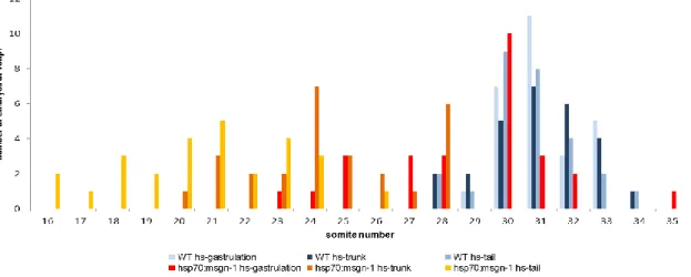
In order to determine if the total number of somites formed is affected by the activation of the referred genes, embryos were heat-shocked at three different time points of development and analyzed at the 48 hpf stage by in situ hybridization with a cb1045 probe. This probe is a very good marker for somite boundaries allowing us to count the total somite number and measure somite length.
To measure somite size we choose to measure the length of two groups of three somites along the anterior-posterior axis. The first group corresponding to the 3 somites formed before the heat-shock and the second group, to 5
somites formed after the heat-shock. We reasoned that 5 somites after heat- shock would give a sufficient delay to see the effect, assuming that cells already in anterior PSM would be committed and would not be so much affected by the misregulation of the signalling pathways. However, as we will see this may not be necessarily true.
A different strategy was adopted for the measurements made on embryos heat-shocked at gastrulation. In these embryos two groups of 3 somites were measured, with the first group corresponding to trunk somites (somites) and the second to approximately the last (somites) tail somites formed.
To assess the impact that each transgenic line has on the mesodermal progenitor cell population, upon heat-shock, we analysed the expression of two of T-box genes: the gene ntl that is expressed in the tailbud in mesoderm progenitors and the intermediate/anterior PSM marker tbx24. These mesodermal markers provide an indication of the changes produced in the size of the mesodermal progenitor population and in the differentiation of the paraxial mesoderm.
4.1. Impact of mesogenin1 overexpression
4.1.1 Impact of mesogenin1 overexpression on the total somite number at different developmental time points
hsp70:msgn1 transgenics together with their wt siblings were subjected to heat- shock either during gastrulation or segmentation (trunk or tail).
When embryos were heat-shocked during gastrulation, we observed a striking reduction in the overall body length of the transgenics. However when the total number of somites was counted we observed only a small reduction in the somite number of hsp70:msgn1 embryos (AVG=28.8 somites; SEM=0.5) in relation to their wild-type siblings (AVG=31.1 somites; SEM=0.2) (Figure 6A). Nonetheless, this reduction of approximately 2 somites in 31 is statistically significant by Student´s T-test ( p<0.001) (appendix1 A).
Figure 6 - The impact of mesogenin1 overexpression in the somites number.
Heat-shocked embryos from hsp70:msgn1 line were raised until 48 hours-post- fertilization.
(A-C) The number of somites from 27 embryos from three different batches were counted after stained with a cb1045 riboprobe. Average of the somites number ± standard error mean from hsp70:msgn1 and their wild-type siblings.
(A´-C´) Wild-type larvae 48 hours-post-fertilization after heat-shock at gastrulation, trunk and tail respectively. (A´´-C´´) Larvae from hsp70:msgn1 line in the same conditions of the wild-type. All the 27 larvae where embryos overexpressed
In contrast, when the pulse of msgn1 was given later in development (at trunk or tail developmental stages) the total number of somites formed in the transgenics is, on average, much lower than that formed in their wild type siblings. When embryos were heat-shocked during trunk formation we observed an average reduction of 10 somites in hsp70:msgn1 embryos (AVG=20,7 somites; SEM=0.4) in relation to their wild-type siblings (AVG=30.7; SEM=0.2) (p=9.548E-24) (Figure 6B). However, when embryos were heat-shocked during tail formation we observed a less striking reduction in somite number (an average reduction of approximately 6 somites) in hsp70:msgn1 embryos (AVG=24.5 somites; SEM=0.4) in relation to their wild-type siblings (AVG=31 somites; SEM=0.2) (p=5.196E-16) (Figure 6C).
Comparing the number of somites between the hsp70:msgn1 larvae that were subjected to heat-shock at gastrulation, trunk or tail (Figure 7) we observe that when the thermic shift is given at gastrulation 59.3% of the transgenic larvae form 30 somites or more and 40.7% form less than 30 somites. When the heat-shock was given at the other two stages (trunk and tail) none of the transgenic larvae reach 30 somites. In conclusion, only when Msgn1 is over- expressed at gastrulation stage can the embryos achieve a total number somites similar to the wild-type embryos (Figure 6A).
Figure 7 – Average of somite number at 48 hours-post-fertilization from
hsp70:msgn1line.
Somites from 27 larvae were counted from each time point after an heat-shock at gastrulation, trunk and tail stages.
4.1.2 Impact of mesogenin1 overexpression on somite size at different developmental time points.
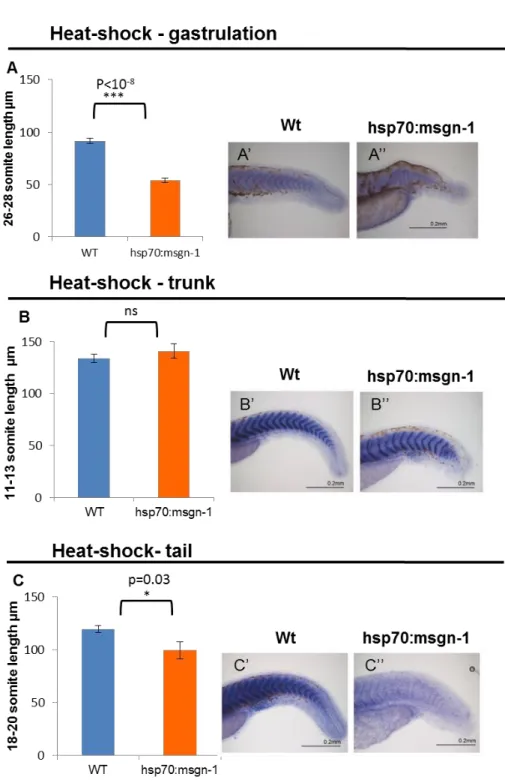
Next, in order to correlate the changes in total somite number with somite size we measured the somite lengths in the 3 experimental conditions as explained above. When hsp70:msgn1 embryos were heat-shocked during gastrulation the overall body length was clearly reduced but the total somite number was barely unchanged, therefore we predicted that somite size should be severely reduced. As expected, we observed that somites at the middle of the anterior-posterior axis (somites 16-18) have a 27% reduction in size (wt: AVG=114µm, SEM=1.19; msgn1: AVG=83µm, SEM=3.3 p=8.8x10⁻⁹) (appendix 2 A) and the last tail somites formed have a 41% size reduction compared to those of their wild-type siblings (wt: AVG=91.5µm, SEM=2.5; msgn1: AVG=53µm, SEM=2.3; p=2.2x10⁻⁹) (Figure 8A)
In contrast, when hsp70:msgn1 embryos were heat-shocked at trunk developmental stages we found a slight increase (2%) in somite length (somites 4-6 and 11-13) (appendix 2 B). However, when compared to the somite lengths of the wild-type embryos this difference was not considered significant according to Student´s T-test (after-heat-shock wt: AVG=130µm; SEM=4;
msgn1: AVG=140.818µm; SEM=6.875; p=0.4) (appendix 2 B; Figure 8B).
Analysing tail heat-shock results (Figure 8C) we can observe that somite length (somite 18-20) after heat-shock for the msgn1 line is smaller 10% than the pre-heat-shock. This difference is statistically significant, as determined by Student´s T-test (after-heat-shock wt: AVG=119.5µm; SEM=3.487; msgn1: AVG=99.800µm; SEM=8.137; p=0.03) (appendix 2 C).
In conclusion, embryos that are subjected to a thermic shift at gastrulation and tail level suffer a reduction of somite length.
Figure 8- The impact of overexpression from mesogenin1 in somite length.
Two groups of somites were measured from hsp70:msgn1 line one pre heat-shock and other after heat-shock. The graphs represent the average of length ± standard error mean of 3 somites post heat-shock
(A) Length of somites 26-28 when embryos were heat-shocked at gastrulation (n=12). (B) Length of somites 11-13 when heat-shock was given at trunk developmental stages (n=11). (C) Length of somites 18-20 when embryos were heat-shocked at tail
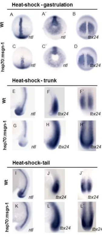
4.1.3 Expression of ntl and tbx24 in embryos over expressing
mesogenin1
Previous studies have shown that Mesogenin1 promotes the differentiation of mesodermal cells and inhibits the wnt/ntl/fgf progenitor loop (Fior et al., 2012). Our data confirm these results: we observed a clear decrease in the expression of ntl in all stages of heat-shock (Figure 9. C;G;K) and the expression of tbx24 was expanded in the PSM and extended into the region of the formed somites (Figure 9. H;L). However, during gastrulation the effect on tbx24 expansion was not observed, possibly indicating that msgn1 during anterior trunk development does not have a prominent role promoting PSM differentiation. These results confirm that we were able to reduce the pool of MPCs with this transgenic line.
Figure 9 – Expression of ntl
and tbx24 in embryos over expressing mesogenin-1. (A-A´;B;C-C´;D) Heat- shocked embryos at gastrulation stage;
(E;F-F´;G;H-H´) heat-shock was given at trunk stage; (I;J-J´;K;L-L`) heat-shock was given at tail stage.
(A-A´;B;E;F-F´;I;J-J´) wild- type embryos ; (C-C´;D;G;H-H´;K;L-L´) embryos from hsp70:msg-1 line. (A-A´;C-C´;E;G;I;K) ntl espression; (B;D;F-F´;H-H´;J-J´) tbx24 expression
(A-D) Posterior view embryo with dorsal to the top;
(A´-B´) animal pole to the top; (E-L) lateral view;
(F´;H´;J´;L´) posterior view with the dorsal to the top.
4.2. Impact of Wnt signalling inhibition
4.2.1 Impact of Wnt signalling inhibition on the total somite number at different developmental time-points
hsp70:dkk1:GFP transgenic line together with their wt siblings were subjected to heat-shock either during gastrulation or segmentation (trunk or tail developmental stages).
hsp70:dkk1:GFP transgenic embryos when heat-shocked during gastrulation, although their overall body length is shorter, we only observed a small reduction in the total somite number of hsp70:dkk1:GFP embryos (AVG=27 somites; SEM=0.78) in relation to their wild-type siblings (AVG=30.5 somites; SEM= 0.27) (Figure 10A). This reduction of approximately 3 somites in 31 is statistically significant, as determined by Student´s T-test (p<0.001) (appendix 1 B).
Similarly, but in contrast to the hsp70:msgn1, when heat-shock is given later during segmentation (trunk and tail) hsp70:dkk1:GFP transgenic embryos also present a small reduction in the average total number of somites formed compared to their wild-type siblings. hsp70:dkk1:GFP embryos heat-shocked during trunk formation have an average reduction of 3 somites (AVG=28.2 somites; SEM=0.44) in relation to their wild-type siblings (AVG=31 somites; SEM=0.27) (Figure 10. B). When the pulse of dkk1 is given at tail we observe a reduction of 2 somites in the average total somite number (Figure 10. C) (AVG=28.2 somites; SEM=0.44) in relation to the wild-type siblings. This small reduction in the total number of somites from heat-shocked embryos at trunk and tail stage is statistically significant by Student´s T-test (p<0.0001, appendix 1 B).
Comparing the number of somites between the dkk1 larvae that were subjected to heat-shock during gastrulation, trunk and tail (Figure 11), we observe that the total number of somites formed in these larvae is similar in the three experimental conditions and is slightly reduced in relation to their wild-type siblings.
Figure 10 - The impact of Wnt inhibition in total somites number.
Heat-shocked embryos from hsp70:dkk1:GFP line were raised until 48 hours-post- fertilization. (A-C) Average of the total somites number ± standard error mean from hsp70:dkk1:GFP and their wild-type siblings from 27 embryos from three different batches after stained with a cb1045 riboprobe.
(A´-C´) Wild-type larvae 48 hours-post-fertilization after heat-shock at gastrulation, trunk and tail respectively. (A´´- C´´) larvae from hsp70:dkk1:GFP line in the same conditions of the wild-type.
Although the slight reduction in 2-3 somites is statistically different, the majority of the embryos (55% in gastrulation heat-shock, 48% in trunk and tail heat-shock embryos) display a total number of somites that would normally be considered in the wild type category (between 29 and 30).
Figure 11 – Average of somite number at 48 hours-post-fertilization from
hsp70:dkk1:GFP line.
Somites from 27 larvae were counted from each time point of heat-shock at gastrulation, trunk and tail developmental stages.
4.2.2 Impact of Wnt signalling inhibition on somite size at different developmental time points
When hsp70:dkk1:GFP embryos were heat-shocked during gastrulation the overall body length was reduced but the total somite number was barely unchanged therefore we predicted that the somite size should be severely reduced as it happened with the hsp70:msgn1 line. In fact we observed a striking 55% reduction in size in relation to their wt siblings either at the level of trunk or tail (somites 16-18: wt-AVG=130,7 µm; SEM=2,504; hsp70:dkk1:GFP- AVG=58,3; SEM=3,239; p<10-12; somites 25-27: wt-AVG=109,8µm; SEM=2,284; hsp70:DKK1:GFP-AVG=48,7µm; SEM=1,777; p<10-13 ,Figure 12 A, appendix 3 A).
Somites from hsp70:dkk1:GFP embryos heat-shocked at trunk are 2,7% smaller than their wild-type siblings (wt-AVG=143,875µm; SEM=4,576; hsp70:DKK1:GFP-AVG=123,750µm; SEM=4,178; p≤0.005) (Figure 12. B).
In contrast, heat-shocked embryos at tail stage present somites 12.3% bigger than their wild-type siblings (after-heat-shock wt: AVG=110,7µm; SEM=1,764; dkk: AVG=120,4µm; SEM=3,745; p=0.03, Figure 12. C).
In summary, Wnt inhibition during gastrulation and trunk causes a reduction in somite size but during tail leads to an increase in the size of somites.
Figure 12 – The impact of dkk1 overexpression in the somite length.
Two groups of somites were measured from hsp70:dkk1:GFP line one pre heat-shock and other after heat-shock. The graphs represent the average of length ± standard error mean of 3 somites post heat-shock
(A) Length of somites 25-27 when embryos were heat-shocked at gastrulation (n=10). (B) Length of somites 11-13 when heat-shock was given at trunk developmental stages (n=8). (C) Length of somites 18-20 when embryos were heat-shocked at tail developmental stages (n=10).
4.2.3 Expression of ntl and tbx24 upon Wnt signalling down-regulation In order to bypass the severe effects of Wnt signalling inhibition, we reduced the time of heat-shock from 1 hour (Martin and Kimelman, 2008) to 5 minutes. Therefore, in order to check whether the short pulse still had a similar effect on the reduction of MPCs and PSM differentiation, we performed in situ hybridization for ntl and tbx24. In fact we did observe a severe downregulation of ntl expression in the tail bud (Figure 13. C´, G´, K´). The expression of tbx24 was more intense when heat-shock was performed during gastrulation and clearly expanded to the posterior PSM when embryos were heat-shocked at trunk and tail developmental stages (Figure 13. H, L).
Figure 13 - Expression of ntl
and tbx24h in embryos over expressing DKK
(A-A´;B;C-C´;D) Heat-shocked embryos at gastrulation stage; (E-E´;F-F´;G-G´;H-H´) heat- shock was given at trunk stage;
(I-I´;J;K-K´;L) heat-shock was given at tail stage.
(A-A´;B;E-E´;F-F´;I-I´;J-J´) wild-type embryos (C-C´;D;G-G´;H-H´;K-K´;L) embryos from hsp70:DKK1:GFP line. (A-A´;C-C´;E-E´;G-G´;I-I´;K-K´) ntl espression. (B;D;F-F´;H-H´;J;L) tbx24 expression
(A-D) Posterior view embryo with dorsal to the top;
(A´;C´) animal pole to the top; (E-L) lateral view;
(E´- I´)(K´) posterior view with the dorsal to the top.
4.3. Impact of FGF signalling inhibition
Previous studies have shown that interfering with FGF signalling leads to severe dorsalized phenotypes and loss of the posterior structures (Griffin et al., 1995; Draper et al., 2003; Furthauer et al., 2004)). Therefore, like with Wnt signalling, in order to bypass the crucial roles of FGF signalling in specification and patterning, we reduced the time of heat-shock from 1 hour (Nechiporuck et al., 2007; Martin and Kimelman, 2008) to 5 minutes, since this was the minimal time that elicited the appearance of GFP
4.3.1 Impact of FGF signalling inhibition on total somite number at different developmental time-points
hsp70:fgfdnr1:GFP transgenics together with their wt siblings were subjected to heat-shock either during gastrulation or segmentation (trunk or tail developmental stages). Considering the variability of the phenotype observed 48 hours-post-heat-shocks at gastrulation stage (figure 14. C-C´´) we decided to increase numbers and analyse embryos from four batches instead of the three batches analysed for the other lines. We observed that hsp70:fgfdnr1:GFP transgenics embryos when heat-shocked at gastrulation have a severe reduction of the total body length and a corresponding reduction in the total somite number (AVG=20 somites; SEM=1.08) in relation to their wild-type siblings (AVG=30.9 somites; SEM=0.27, Figure 14. A). This reduction of approximately one third of the somites (10 in 30.9) is highly statistically significant as determined by Student´s T-test (p<0.0001, appendix 1 C).
When the pulse of FGF inhibition is delivered later in development (at trunk or tail developmental stages) the average reduction in the total number of somites formed compared to the wild-type is very similar: 8 somites instead of 10 (hsp70:fgfdnr1:GFP-AVG-trunk=22.6 somites, SEM=0.75 vs wt-AVG- trunk=30.9 somites; SEM=0.27, hsp70:fgfdnr1:GFP-AVG-tail=23.6 somites; SEM=0.52 (hsp70:fgfdnr1:GFP-AVG-tail=23.6 somites; SEM=0.52 vs wt-AVG- tail=31 somites; SEM=0.22, Figure 14. B, C).
Figure 14 - The impact of FGF inhibition in somite number. Heat-shocked embryos
from hsp70:fgfdnr1:GFP line were raised until 48 hours-post.fertilization.
(A) Average of somites numbers from 36 embryos from four different batches were counted after stained with a cb1045 riboprobe when heat-shock was given at gastrulation stage. (D) (E) Average of the somites number ± standard error mean from hsp70:fgfdnr1:GFP and their wild-type siblings from 27 heat-shocked embryos at trunk and tail stages respectively. (B;D´;E´) Wild-type larvae 48 hours-post-fertilization after heat-shock at gastrulation, trunk and tail respectively. (C-C´-C´´; D´´;E´´´) Larvae from hsp70:fgfdnr1:GFP line in the same conditions of the wild-type.
These reductions in the total number of somites in embryos heat- shocked at the trunk and tail stages are highly statistically significant as determined by Student´s T-test (p<0.0001, appendix 1 C).
In summary, the total number of somites formed upon inhibition of FGF in all time-points of development chosen for this work is severely reduced in relation to wild-type embryos (Figure 15).
Figure 15 - Somite number at 48 hours-post-fertilization from hsp70:fgfdnr1:GFP line.
Somites from 36 larvae heat-shocked embryos at gastrulation time-point and 27 larvae were counted from heat-shocked embryos at tail and trunk time points.
4.3.2. The impact of Fgf signalling inhibition on somite size at different developmental time points.
When hsp70:fgfdnr1 embryos were heat-shocked during gastrulation we observed that somites 16 to 18 have a 13% reduction in size (wt-AVG=128µm, SEMst error=4.5; hsp70:fgfdnr1-AVG=111µm, SEM=1.1 p=0.0018, appendix 2) and the last somites formed have a 20% reduction in size compared to wild-type siblings (wt-AVG=118µm, SEM=3.4; hsp70:fgfdnr1-AVG=93 µm, SEM=2.3 p<10-4, Figure 16. A).
In contrast, when hsp70:fgfdnr1 embryos were heat-shocked at trunk stage we found a slight increase in somite size (1.5%) in relation to the wild- type (post-heat-shock wt-AVG=124,333µm, SEM=3.884; hsp70:fgfdnr1- AVG=122,667µm; SEM=16.501; p=0.903, Figure 16. B, appendix 4 B). The embryos heat-shocked at tail stage exhibit a reduction of 4.4% in somite size, relative to their wild-type siblings (post-heat-shock wt-AVG=136,556µm, SEM=5,180; hsp70:fgfdnr1-AVG=133µm; SEM=3; p=0.674, Figure 16. C).
Both measurements made at trunk and tail developmental stages were not statistically significant according to Student´s T-test, although we could clearly detect the differences. This discrepancy between observed and
statistically difference could be due maybe to the reduced numbers of embryos analyzed (n=9) but also to the region of the embryo that we have analysed. These results should be further validated in the future with more measurements in more embryos but also in different regions of the embryo.
4.3.3. Expression of ntl and tbx24 under Fgf signalling down-regulation The inhibition of Fgf signalling cause in all time points of inhibition a small decrease in the expression of ntl (Figure 17. A, C, E, G, I).
With exception of the heat-shock delivered during gastrulation, inhibition of FGF signalling during trunk and tail stages we observed a severe reduction in
tbx24 expression- possibly a reduction in the anterior expression domain
Figure 16 - The impact of Fgf inhibition in somite length.
Two groups of somites were measured from hsp70:fgfdnr1:GFP line one pre heat- shock and other after heat-shock. The graphs represent the average of length ± standard error mean of 3 somites post heat-shock
(A) Length of somites 18-20 when embryos were heat-shocked at gastrulation (n=9). (B) Length of somites 11-13 when heat-shock was given at trunk developmental stages (n=9). (C) Length of somites 18-20 when embryos were heat-shocked at tail developmental stages (n=9).
Figure 17 – Expression of ntl
and tbx24h in embryos that fgf is down-regulated.
(A-A´;B;C-C´;D) Heat-shocked embryos at gastrulation stage; (E-E´;F;G-G´;H) heat-shock was given at trunk stage; (I;J-J´;K;L- L´) heat-shock was given at tail stage. (A-A´;B;E-E´;F;H;I;J-J´) wild- type embryos (C-C´;D;G-G´;K;L-L´) embryos from hsp70:fgfdnR1:GFP line. (A-A´;C-C´;E-E´;G-G´;I;K) ntl espression. (B;D;F;H)(J-J´)(L-L´) tbx24 expression
(A-D) Posterior view embryo with dorsal to the top;
(A´;C´) animal pole to the top; (I-K;E-H) lateral view;
(E´;G´;L´;J´) posterior view with the dorsal to the top.
4.4 Comparison between the effect of Msgn1 overexpression and Wnt and FGF signalling inhibition
At all time-points of heat-shock the inhibition of FGF signalling had a greater impact on the total number of somites formed in comparison to the other lines. In all situations the total somite number was severely reduced and embryos were not able to adapt and spare progenitor cells to achieve the total somite number.
In contrast, embryos that were subjected to a short inhibition of Wnt signalling in all time points were capable of forming approximately the same number of somites as wild-type embryos (Figure 13A), suggesting that upon a Wnt challenge embryos are able to adapt and regulate the number of cells to achieve the total species-specific number.
Msgn1 overexpression resulted in somewhat intermediate result: when heat-shock is delivered during gastrulation embryos are able to regulate their total somite number by regulating somite size. However, when the same pulse of Msgn1 overexpression is delivered during segmentation, the embryos are no longer able to adapt and form less somites (Figure 13B).
Figure 18 – Comparison between the three lines analysed in this study. A. Normalized
somites number considering wild-types average of somite number=1. B. Normalized post heat-shock somite length, considering wild-type average of somite length=1.
5. DISCUSSION
The clock and wavefront model considers that somite size should be
proportional to the number of cells entering the PSM in each oscillation cycle of the segmentation clock, while the total number of somites should be equal to the total time for which production of PSM cells continues, divided by the length of that cycle.
In other words, somite size can change when the speed of wavefront or the period of the clock is altered. The model would predict that inhibition of the signalling pathways involved in the wavefront (posteriorization of the anterior limit of the wavefront) will lead to the formation of larger somites (Sawada et et al., 2001) and an anterior expansion (ectopic activation) leads to the formation of smaller somites (Aulehla et al., 2003). This has been clearly shown using beads soaked in Fgf or in Fgf inhibitor in chick and zebrafish (Dubrulle et al., 2001). On the other hand, slowing the speed of the clock should lead to an increase in segment size, and in fact this has been shown recently to occur in the zebrafish her6 mutant (Schroter and Oates, 2010) while an acceleration of the clock would lead to a reduction in somite size (Figure 19).
Figure 19 – Summary of predictions of the impact of altering independently the position of the wavefront or the speed of the clock can have on somite size.
“The total number of somites should be equal to the total time for which production of PSM cells continues, divided by the length of that cycle.” If we disentangle this sentence in a mathematical formula:
total somite number = total time of PSM production / length of the cycle
This means it is possible to change the total somite number either by altering the total time of PSM production or by altering the clock rate: a slower clock leads to a reduction in total somite number and an accelerated clock leads to an increase in somite number (considering that the total time of PSM production is the same). Indeed, Schroter and Oates (2010) observed that a slower clock leads to a reduction in total somite number
How can the total time of PSM production be altered? One possibility is to regulate the rate of PSM differentiation from the progenitor pool. A slower rate of PSM differentiation should lead to an increase in somite number (in fact recent work from our lab Fior et al, (2012) provided such an example) while acceleration of PSM differentiation should lead to a reduction of somite number (Figure20).
Figure 20 – Summary of predictions of the impact of altering independently the rate of differentiation or the speed of the clock can have on total somite number