Fabrícia Magalhães Araújo
I, Mavilde da Luz Gonçalves Pedreira
I, Ariane Ferreira Machado Avelar
I,
Márcia Lurdes de Cácia Pradella-Hallinan
II, Miriam Harumi Tsunemi
III, Eliana Moreira Pinheiro
II Universidade Federal de São Paulo, Escola Paulista de Enfermagem. São Paulo, Brazil. II Universidade Federal de São Paulo, Escola Paulista de Medicina. São Paulo, Brazil.
III Universidade Estadual Paulista Júlio de Mesquita Filho, Institute of Biosciences. Botucatu, SãoPaulo, Brazil.
How to cite this article:
Araújo FM, Pedreira MLG, Avelar AFM, Pradella-Hallinan MLC, Tsunemi MH, Pinheiro EM. Sleep and salivary cortisol in preterm neonates: a clinical, randomized, controlled, crossover study. Rev Bras Enferm [Internet]. 2018;71(Suppl 3):1358-65.
[Thematic Issue: Health of woman and child] DOI: http://dx.doi.org/10.1590/0034-7167-2017-0546
Submission: 08-03-2017 Approval: 10-07-2017
ABSTRACT
Objective: Analyze the infl uence of ear protectors on the baseline levels of salivary cortisol and response and total sleep time of preterm neonates during two periods of environmental management of a neonatal intermediate care unit. Method: A clinical, randomized, controlled and crossover study conducted with 12 preterm neonates. The use of ear protectors was randomized in two periods. Sleep evaluation was performed using one Alice 5 Polysomnography System and unstructured observation. Results:
No signifi cant difference was observed between the baseline levels of salivary cortisol and response in preterm neonates from the control and experimental groups, and no statistical signifi cance was observed between the total sleep time of both groups. No relationship was observed between the baseline levels of cortisol and response and total sleep time. Conclusion: Ear protectors in preterm neonates did not infl uence the salivary cortisol level and total sleep time in the studied periods.
Descriptors: Infant, Premature; Hydrocortisone; Sleep; Nursing Care; Stress, Physiological.
RESUMO
Objetivo: Verifi car a infl uência do uso de protetores auriculares nos níveis de cortisol salivar basal e resposta e no tempo total de sono de prematuros durante dois períodos de manejo do ambiente de uma unidade de cuidado intermediário neonatal.
Método: Ensaio clínico, randomizado, controlado e cruzado conduzido em 12 prematuros. O uso de protetores auriculares foi randomizado em dois períodos. A avaliação do sono foi realizada por meio do polissonígrafo Alice 5 e da observação não estruturada. Resultados: Não houve diferença signifi cante entre os níveis de cortisol salivar basal e resposta nos prematuros dos grupos controle e experimental, não havendo também signifi cância estatística entre o tempo total de sono dos dois grupos. Não foi observada relação entre os níveis de cortisol basal e resposta e o tempo total de sono. Conclusão: Os protetores auriculares nos prematuros não infl uenciaram o nível de cortisol salivar e o tempo total de sono nos períodos estudados.
Descritores: Recém-Nascido Prematuro; Hidrocortisona; Sono; Cuidados de Enfermagem; Estresse Fisiológico.
RESUMEN
Objetivo: Certifi car la infl uencia del uso de protectores auriculares en los niveles de cortisol salival basal y la respuesta y en el tiempo total de sueño de prematuros durante dos períodos de manejo del ambiente de una unidad de cuidado intermedio neonatal. Método: Ensayo clínico, aleatorio, controlado y cruzado conducido en 12 prematuros. El uso de protectores auriculares ha sido aleatorio en dos períodos. La evaluación del sueño ha sido realizada por medio del aparato de polisomnografía Alice 5 y de la observación no estructurada. Resultados: No ha habido diferencia signifi cante entre los niveles de cortisol salival basal y la respuesta en los prematuros de los grupos control y experimental, no habiendo también signifi cancia estadística entre el tiempo total de sueño de los dos grupos. No ha sido observada la relación entre los niveles de cortisol basal y la respuesta y el
Sleep and salivary cortisol in preterm neonates: a clinical,
randomized, controlled, crossover study
INTRODUCTION
Sleep is an essential physiological need for proper devel-opment of newborns, especially preterm neonates, since it contributes to body homeostasis and well-being(1), and it is es-sential for neurological maturation, preservation of brain plas-ticity, long-term memory consolidation, and the establishment of emotional health and immune function, and it contributes to cellular restoration in preterm neonates (PTN)(2-4).
Preterm neonates present polyphasic sleep, alternating three stages: active, quiet, and indeterminate(5). During these stages, physiological changes occur in the body, such as a de-crease in the level of cortisol, a corticosteroid hormone pro-duced by the hypothalamic-pituitary-adrenal axis(6-7).
Cortisol is an important biological marker of stress response in neonates(8). The cortex of adrenal glands is responsible for
the secretion of this hormone present in blood plasma and bound to carrier proteins, but cortisol can also be detected in its free form, in urine and salivary secretion(9-10). The analysis
of free salivary cortisol level in PTNs has been widely used in clinical studies because it is a noninvasive practice(7).
This glucocorticoid, just like sleep, is controlled by the cir-cadian cycle, but as it is absent in neonates, the release of salivary cortisol oscillates during the 24 hours(7,11).
Increased cortisol level causes important physiological and behavioral changes, such as increased metabolic rate, with con-sequent loss of body mass, glucose tolerance, increased frequen-cy of apnea and higher intracranial pressure, and it may cause intraventricular hemorrhage. In addition, it causes reduced per-ception of painful stimuli and increased irritability and crying(8).
During hospitalization, PTNs are exposed to excessive stimuli – constant presence of lighting, high levels of sound pressure in the environment and inside the incubators, constant handling and ambient temperature variation – which can generate stress and trigger frequent interruptions of sleep cycles(2,5,12).
Although the literature clearly shows the various stimuli from both neonatal unit and care environments may cause stress and sleep deprivation in neonates, a lack of knowledge of clinical studies correlating the level of salivary cortisol with sleep vari-ables of preterm neonates during hospitalization.
Based on the relationship between stress and sleep and its im-portance for infant development, studies are required to enable the implementation of more comprehensive protocols to guide the clinical practice, aiming to protect and promote sleep and the consequent development of hospitalized neonates.
OBJECTIVE
Analyze the influence of ear protectors (EPs) on the salivary cortisol level of PTNs, on the total sleep time and respective sleep stages during two periods of environmental management.
METHOD
Ethical aspects
This study was approved by the Research Ethics Committee of Universidade Federal de São Paulo and registered in Clini-cal Trials. Data collection started after an informed consent form was signed by the legal guardians of the neonates.
Design, study site and period
This is a randomized controlled crossover study conducted in a neonatal intermediate care unit (NICU) of a university hospital in São Paulo. This unit has some strategies of devel-opmental care, such as minimum neonate handling and en-vironmental management in two specific periods of the day to ensure the sleep and rest of the PTNs. This strategy, called “sleep time,” is performed in the morning (10 am to 11 am) and in the afternoon (4 pm to 5 pm). In these periods, the mul-tidisciplinary team of the NICU avoids handling the neonates, as far as possible, reduces lighting in the environment, and minimizes the noise level inside the rooms. Data collection was conducted between July 2013 and February 2015.
Population and sample
The study sample consisted of PTNs of gestational age of less than 37 weeks, at least 72 hours of life, weight be-tween 1,200 and 2,000 g, who were in incubators, presenting clinically stable conditions, with test of otoacoustic emission evoked by the presence of transient stimulus. Neonates in pho-totherapy, invasive and non-invasive mechanical ventilation, presenting congenital malformations, periventricular hemor-rhage grades II, III and IV, who took a depressor of the cen-tral nervous system, analgesic or corticoid medication were excluded from the study, as well as those whose mothers had a history of using illicit drugs during pregnancy. The CON-SORT diagram was used to include preterm neonates in the study. The inclusion and exclusion criteria were defined with the purpose to eliminate possible influences on sleep and the salivary cortisol level.
The sample was calculated by assuming the use of EPs during environmental management periods reduces the level of sali-vary cortisol and favors extended total sleep time of PTNs. Then, the calculation was based on nine samples of saliva initially ob-tained in the environmental management periods in the morn-ing shift from PTNs who used or did not use EPs. It should be noted that, in some samples collected from this initial sample, the amount of saliva required for cortisol level analysis was not obtained; then, the minimum value of 0.5 ng/ml detectable in the ELISA test was accepted for sample calculation.
The mean values of cortisol level before (2.29±1.44 ng/ ml) and after (1.17±0.93 ng/ml) of the intervention provided a difference of 1.12 ng/ml. Considering this difference, the
Eliana Moreira Pinheiro E-mail: [email protected]
CORRESPONDING AUTHOR
tiempo total de sueño. Conclusión: Los protectores auriculares en los prematuros no han influenciado el nivel de cortisol salival y el tiempo total de sueño en los períodos estudiados.
significance level of 5%, and 80% statistical power, it would take 12 PTNs to detect this difference with a statistically signif-icant result. The paired t-test of STATISTICA® software,
Okla-homa, USA, was used to calculate the sample.
Saliva samples were collected from 32 PTNs. However, due to insufficient amount of saliva collected for the cortisol level analysis, only 12 preterm neonates were included in the study, observing the sample calculation guidelines.
Study protocol
The experimental variable is the use of EP in PTNs during one of the NICU environment management periods, constitut-ing the experimental group. The EPs used in the study were MiniMuffs® Neonatal Noise Attenuators, manufactured by
Na-tus® Pediatrics, California, USA, which reduce the sound
pres-sure levels from 7 to 12 decibels(13-15).
EPs are oval-shaped anatomical devices that allow proper po-sitioning on the ears. They have soft foam and hydrogel adhesive on the outer edges for skin attachment. Considering the possibil-ity of detachment of these devices in the PTNs, a substance with polyethylene and cellulose was used, which helps protect the skin and improves the adhesion of the protectors. The EPs were placed on the PTNs by one of the researchers, on average 12 minutes before starting the NICU environmental management, and removed immediately after the end of these periods.
The control group consisted of PTNs that did not use EPs during one of the periods of NICU environment management.
The salivary cortisol levels of PTNs with and without the use of PAs before the start (baseline cortisol) and at the end (cortisol response) of environmental management periods and total sleep time and respective sleep stages were the outcome variables.
Saliva was collected from the PTNs with the help of oph-thalmic sponges (Merocel®, USA) of high capacity of
absorp-tion of body fluids, attached to a flexible rod(16). Two initial
samples were obtained 20 minutes before the environmental management period (baseline cortisol) and two samples were collected immediately after the end of the same period (cortisol response), before the removal of EPs, totalizing four samples from each neonate. Prior to collection, all neonates underwent oral hygiene with gauze and sterile water to remove milk resi-dues that could influence the analysis(17). The procedure of
sa-liva collection from the oral cavity was performed by one of the researchers and lasted, on average, eight minutes.
All saliva samples were placed in Eppendorf tubes and stored under refrigeration, with the sponges upwards, and sent to the laboratory for analysis. The samples were centrifuged at 3,000 rpm for 5 minutes in a refrigerated device and stored in a freezer at −18°C to −20°C until the analysis(16,18).
The laboratory analysis used the ELISA method, which de-termines the direct immunoenzymatic concentration of corti-sol. In this method, the minimum sensitivity for the detection of salivary cortisol level is 0.5 ng/ml(19).
The evaluation of total sleep time and respective sleep stag-es (active, quiet or indeterminate) used one Alice 5 Polysom-nography System (Philips Respironics®, Pennsylvania, USA),
an instrument that performs sleep records of PTNs(20). The
installation of this system on the PTNs was performed by a
polysomnography expert. This procedure started, on average, two hours before the morning environmental management pe-riod. Sleep data records started, on average, one hour before the beginning of the environmental management, and after that, they were continuously recorded until the researcher fin-ished the last collection of saliva after the afternoon environ-mental management.
The polysomnography analysis observed the guidelines from the American Academy of Sleep Medicine (AASM), ver-sion 2.0, and it was performed by a pediatric neurologist spe-cialized in sleep medicine(21).
Each PTN was submitted to two periods of environmental management, using the EPs (experimental group) in one period, and not using the EPs in the other period (control group). Ran-domization was obtained using the Latin square design method, which enabled the distribution of the use or non-use of EPs in the two periods of NICU environmental management.
Result analysis and statistics
Data collected were stored in Microsoft Excel® software
and analyzed in SPSS Statistics® 20.0, Chicago, USA, using
de-scriptive statistical analysis, parametric tests (paired t-test), and linear regression when the required prerequisites were met for these tests (normality and homogeneity). Then, the effect of total sleep time and baseline levels of salivary cortisol and response on independent variables was estimated. Non-para-metric test (Spearman’s correlation) was performed to evaluate the strength of the relationship between the variables. The R Program, New Zealand – USA, was also used in the construc-tion of mixed linear models to evaluate whether the random effect of using ear protectors was the same on each neonate. All analyses used significance level of 5% (p ≤ 0.05).
RESULTS
Table 1 shows the demographic and clinical characteristics of the PTNs that met the eligibility criteria defined for this study.
The neonates presented moderate prematurity, most of them classified as small for the gestational age, and very low birth weight. Cesarean delivery and female preterm neonates were predominant. Of note, more than half mothers used pre-natal corticosteroid, and the same proportion of PTNs used caffeine during hospitalization.
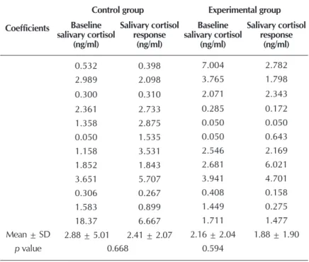
The baseline levels of salivary cortisol and response of the PTNs from the control and experimental groups during the environmental management periods of the neonatal unit did not present a statistically significant difference (Table 2). How-ever, the mean values of these levels show a more significant reduction of the values in the control group when compared to the experimental group.
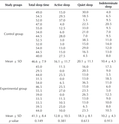
The total sleep time of PTNs was higher in the control group, mean of 5.3 minutes more when compared to the neonates from the exper-imental group. Regarding the sleep stages, the predominance of quiet sleep was observed in both groups. The use of EPs in preterm neonates did not show statistical significance between the two groups analyzed in this study (Table 3).
Spearman’s linear correlation between the total sleep time and sleep stages showed a strong relationship only with active sleep in the experimental group (ρ = 0.617; p = 0.033), indicating that longer total sleep time in the PTNs is related to more frequent active sleep. In the control group, a moderate relationship was observed between the total sleep time and quiet sleep (ρ = 0.560; p = 0.058).
Table 4 presents the relationship between baseline salivary cortisol and response and total sleep time of neonates during the environmental management periods in the control and experi-mental groups. This relationship is not strong and negative in both groups. Even without sta-tistical significance, this negative correlation in-dicates that the high level of cortisol implies a shorter sleep time of the PTNs in both groups.
The behavior of the variables: gestational age corrected by the last menstrual period (LMP), cur-rent weight, non-use of caffeine by the PTNs and prenatal corticosteroid by the mothers of the neo-nates in relation to the total sleep time and base-line salivary cortisol and response was estimated using multiple linear regression and did not show statistical significance. However, the control group showed a 7-minute increase in total sleep time of the PTNs whose mothers did not use prenatal corticosteroids and 2.1 minutes in the absence of caffeine. In the experimental group, the increase in total sleep time was 0.8 minutes and 2.3 min-utes for the respective criteria. This model explains 13.53% of the variation in total sleep time.
The linear regression analysis of baseline levels of salivary cortisol and response with the same variables shows a trend towards re-duced levels in PTNs who were not exposed to these substances, except for salivary cortisol response in the experimental group not ex-posed to caffeine, with tendency to increased level (2.2 ng/ml). This analysis explains 15.4% of baseline levels of salivary cortisol and re-sponse of the neonates.
The evaluation of the mean frequency of central apneas during the environmental man-agement periods using the paired t-test showed a higher occurrence in the control group (M = 37.4 ± 31.6) when compared to the ex-perimental group (M = 22.9 ± 26.0), p = 0.028.
Table 1 – Characteristics of preterm neonates evaluated in this study, São Paulo, Brazil
Variables Mean ± SD n (%)
Age
Gestational age acc. to LMP (weeks) 32.5 ± 2.1 12 (100.0) Gestational age acc. to LMP – corrected (weeks) 33.0 ± 1.4 12 (100.0) Chronological age (days) 18.3 ± 6.7 12 (100.0) Weight
At birth (grams) 1499.2 ± 330.4 12 (100.0) Current (grams) 1635.4 ± 218.7 12 (100.0) Birth
Cesarean – 11 (91.7)
Vaginal – 1 (8.3)
Sex
Female – 8 (66.7)
Male – 4 (33.3)
Size according to the gestational age
SGA – 8 (66.7)
AGA – 4 (33.3)
Diagnosis
Respiratory – 6 (50.0)
Hematologic – 4 (33.3)
Digestive – 2 (16.7)
Use of pre-natal corticosteroid
Yes – 7 (58.3)
No – 5 (41.7)
Use of caffeine
Yes – 7 (58.3)
No – 5 (41.7)
Note: SD – standard deviation; LMP – last menstrual period; SGA – small for gestational age; AGA – adequate for gestational age.
Table 2 – Baseline levels of salivary cortisol and response of preterm neo-nates from the control and experimental groups during the environ-mental management periods of a neonatal unit, São Paulo, Brazil
Coefficients
Control group Experimental group
Baseline salivary cortisol
(ng/ml)
Salivary cortisol response
(ng/ml)
Baseline salivary cortisol
(ng/ml)
Salivary cortisol response
(ng/ml)
0.532 0.398 7.004 2.782 2.989 2.098 3.765 1.798 0.300 0.310 2.071 2.343 2.361 2.733 0.285 0.172 1.358 2.875 0.050 0.050 0.050 1.535 0.050 0.643 1.158 3.531 2.546 2.169 1.852 1.843 2.681 6.021 3.651 5.707 3.941 4.701 0.306 0.267 0.408 0.158 1.583 0.899 1.449 0.275 18.37 6.667 1.711 1.477 Mean ± SD 2.88 ± 5.01 2.41 ± 2.07 2.16 ± 2.04 1.88 ± 1.90
p value 0.668 0.594
For a better evaluation of the frequency of central apnea and other parameters, the mixed linear models were adjusted, consid-ering only the moment (use of ear protec-tors) as a random effect.
The effect of EPs on central apnea, cor-rected gestational age considering the LMP and use of caffeine in relation to baseline levels of salivary cortisol and response based on the factors of moment of differ-entiation between these levels and total sleep time of PTNs (Table 5) found statis-tical significance only between total sleep time and the occurrence of central apnea (p < 0.035).
This model shows that the occurrence of central apnea increased the total sleep time by 0.11 minutes when the EPs were used. However, no relationship was found between baseline levels of salivary cortisol and response and the variables previously mentioned at the moments when the PTNs used the EPs. These variables also did not present any type of relation with the active, quiet and indeterminate sleep stages evalu-ated in the mixed linear model (Table 5).
DISCUSSION
The baseline levels of salivary cortisol and response and total sleep time and the respective sleep stages did not differ significantly in relation to the use of ear protectors in the environmental manage-ment periods of the investigated NICU.
The comparison of the results of this study is limited due to the lack of other studies using a similar methodological design, and it is the first study to associ-ate the use of EPs with the variables of salivary cortisol and sleep of PTNs, cor-roborated by the sleep analysis using a polysomnography system, allowing an accurate evaluation during the study pe-riod, thus contributing to result reliability. Among the studies on the use of EPs, only two of them included sleep in the evaluated variables, showing it favors the sleep of PTNs. However, in these studies, data were collected with the application of scales that classified/quantified sleep from direct observation of the behavioral states of the neonates, thus establishing scores that range from alertness to differ-ent classifications of sleep(13,18).
No significant correlation was observed between the baseline levels of salivary
Table 3 – Total sleep time and respective sleep stages, in minutes, of the preterm neonates from the control and experimental groups during the envi-ronmental management periods of a neonatal unit, São Paulo, Brazil
Study groups Total sleep time Active sleep Quiet sleep Indeterminate sleep
Control group
49.0 15.0 30.0 4.0
54.5 29.5 18.5 6.5
52.0 37.0 5.5 9.5
47.0 4.0 22.5 20.5
55.0 12.5 33.0 9.5
34.0 6.0 21.0 7.0
44.5 28.0 7.0 9.5
52.5 3.0 38.5 11.0
32.0 3.0 15.0 14.0
54.0 13.0 29.0 12.0
44.5 15.0 16.5 13.0
40.0 27.5 4.5 8.0
Mean ± SD 46.6 ± 7.9 16.1 ± 11.7 20.1 ± 11.1 10.4 ± 4.3
Experimental group
45.0 11.5 16.0 17.5
29.5 0.0 20.5 9.0
44.0 25.5 13.0 5.5
31.5 0.0 13.0 18.5
54.0 6.5 36.5 11.0
46.5 25.5 15.0 6.0
55.5 27.0 23.5 5.0
39.0 0.0 26.5 12.5
33.5 11.5 13.0 9.0
33.5 10.5 13.0 10.0
39.5 25.0 6.5 8.0
44.0 10.0 23.5 10.5
Mean ± SD 41.3 ± 8.4 12.8 ± 10.5 18.3 ± 8.1 10.2 ± 4.3
p value 0.149 0.381 0.613 0.913
Note: SD – standard deviation.
Table 5 – Relation between baseline salivary cortisol and response and total sleep time with central apnea frequency, gestational age corrected according to the last menstrual period and caffeine use by preterm neonates when using ear protectors, São Paulo, Brazil
Variables Estimate Standard error p value
Baseline salivary cortisol and response
Intercept 0.82 5.65 –
Central apnea – 0.00 0.01 0.573 Age corrected acc. to LMP 0.03 0.16 0.742 Use of caffeine – 0.10 0.71 0.811 Ear protector – 0.17 0.51 0.742 Table 4 – Spearman’s linear correlation coefficient between total sleep time and
baseline levels of salivary cortisol and response of preterm neonates from the control and experimental groups in the environmental management periods of a neonatal unit, São Paulo, Brazil
Variables
Control group (n = 12)
Experimental group (n = 12) Correlation
coefficient p value
Correlation
coefficient p value
TST Baseline salivary cortisol Salivary cortisol response
– 0.154 – 0.371 0.632 0.235 – 0.214 – 0.035 0.503 0.914
Note: TST – total sleep time.
cortisol and response and total sleep time in the environmen-tal management periods in which the PTNs used ear protectors. However, even without statistical significance, the negative cor-relation found in the present study indicates that total sleep time increases with reduced cortisol level, suggesting that further studies are required to evaluate this correlation.
A randomized crossover study conducted at the neonatal ICU that develops the Newborn Individualized Developmental Care and Assessment Program (NIDCAP) used the EDIN (échelle
dou-leur inconfort nouveau-né) scale to assess stress response, sleep
quality, pain, heart rate, oxygen saturation, and the impact of environmental measures through the application of some behav-ioral interventions such as positioning, touching, non-nutritive sucking during the weighing procedure in term neonates and PTNs. This study did not show a significant difference in base-line levels of salivary cortisol and response, but in the physio-logical parameters and in behavioral responses of neonates from both the control and experimental groups(22).
The strong positive correlation between baseline levels of salivary cortisol and response of the PTNs found in this study shows that these levels practically did not vary between the control and experimental groups. Also, it was not possible to identify the presence of cortisol circadian rhythm in the envi-ronmental management periods of the NICU, which may be a consequence of the immaturity of the neonates(23).
The duration of the environmental management periods set by the service where this study was conducted may not have been sufficient to promote the adaptation of the neonates to the EPs and the consequent reduction of the level of salivary cortisol response in the neonates. This factor may explain the absence of a statistically significant difference between the baseline levels of salivary cortisol and response in the control and experimental groups.
Studies that used EPs to evaluate behavioral aspects, such as sleep, motor response, pain and physiological parameters like body temperature, oxygen saturation, blood pressure, and heart and respiratory rates in PTNs, performed the interven-tion for periods of two hours or more(13-15,24-25).
The baseline levels of salivary cortisol and response of preterm neonates in this study did not differ significantly when compared to other studies. The literature shows absent
parameters of these levels in neonates of different gestational ages(17,26).
When evaluating the mean values of total sleep time and the different sleep stages, the control group showed a longer time of quiet sleep. This finding does not agree with a study that evaluated the use of EPs in PTNs and found a longer time of quiet sleep when these devices were used(13). In addition, in
the experimental group, the use of EPs favored active sleep. This unprecedented result is very important, since this sleep stage is fundamental for the PTNs, especially for their neurosensory de-velopment, indicating the need for further studies(1-2).
A higher frequency of apnea and a longer sleep time were observed when the PTNs used the EPs. No significant relation-ship was observed between the occurrence of central apnea and the different sleep stages. A study that used a polysomnography system and evaluated the effect of thermoregulation on the oc-currence of apnea episodes in PTNs during the quiet and active sleep stages showed a higher frequency of central apnea during active sleep, which was associated with loss of body heat(27).
Study limitations
One study limitation referred to the challenge to obtain a sufficient amount of saliva for analysis due to the peculiar hy-posalivation of PTNs. Further studies should be conducted for a longer period, since the time of adaptation of neonates to the EPs may not have been sufficient to show reduced salivary cortisol level.
Contributions to the nursing area
The proposed intervention allowed a longer time of active sleep to the preterm neonates.
CONCLUSION
The use of EPs on the PTNs did not influence the baseline levels of salivary cortisol and response and total sleep time during the environment management periods of an NICU. However, the use of EPs favored longer active sleep time, es-sential for the neurosensory development. The longer total sleep time when the PTNs used the ear protectors provided more episodes of central apnea.
FUNDING
This study was conducted with the financial support from Fundação de Amparo à Pesquisa do Estado de São Paulo (Fapesp), protocol nº 2012/50365-2.
Variables Estimate Standard error p value
Total sleep time Intercept 39.42 36.23 – Central apnea 0.11 0.05 0.035* Age corrected acc. to LMP 0.16 1.06 0.872 Use of caffeine – 5.63 4.59 0.229 Ear protector – 4.42 3.27 0.186
Note: LMP – last menstrual period; *p < 0.05. Table 5 (concluded)
REFERENCES
1. Arditi-Babchuk H, Feldman R, Eidelman AI. Rapid eye movement (REM) in premature neonates and developmental outcome at 6 months. Infant Behav Dev [Internet]. 2009[cited 2016 Jun 15];32(1):27-32. Available from: http://www.sciencedirect.com/science/ article/ pii/S0163638308000775?via%3Dihub
rhythm of premature infants. Chronobiol Int [Internet]. 2014[cited 2016 Jun 16];31(9):1062-74. Available from: http://www. tandfonline. com/doi/full/10.3109/07420528.2014.948183?scroll=top&needAccess=true
3. Calciolari G, Montirosso R. The sleep protection in the preterm infants. J Matern Fetal Neonatal Med [Internet]. 2011[cited 2016 Jun 26];24(Suppl-1):12-4. Available from: http://www.tandfonline.com/doi/abs/10.3109/14767058.2011. 607563?journalCode=ijmf20
4. Miller DJ, Duka T, Stimpson CD, Schapiro SJ, Baze WB, McArthur MJ, et al. Prolonged myelination in human neocortical evolution. Proc Natl Acad Sci USA[Internet]. 2012[cited 2016 Jul 20];109(41):16480-5. Available from: http://www.pnas.org/content/109/41/16480.full
5. Bonan KC, Pimentel Filho JC, Tristão RM, Jesus JA, Campos Jr D. Sleep deprivation, pain and prematurity: a review study. Arq Neuropsiquiatr [Internet]. 2015[cited 2016 Jul 12];73(2):147-54. Available form: http://www.scielo.br/pdf/anp/v73n2/0004-282X-anp-73-02-147.pdf
6. Mörelius E, He HG, Shorey S. Salivary cortisol reactivity in preterm infants in neonatal intensive care: an integrative review. Int J Environ Res Public Health [Internet]. 2016[cited 2016 Jul 23];13(3):337-51. Available from: https://www.ncbi.nlm.nih.gov/pmc/ articles/ PMC4809000/pdf/ijerph-13-00337.pdf
7. Silva ML, Mallozi MC, Ferrari GF. Salivary cortisol to assess the hypothalamic-pituitary-adrenal axis in healthy children under 3 years old. J Pediatr[Internet]. 2007[cited 2016 Jun 30];83(2):121-6. Available from: http://www.jped.com.br/conteudo/07-83-02121/port.pdf
8. Tollenaar MS, Beijers R, Jansen J, Riksen-Walraven JM, Weerth C. Solitary sleeping in young infants is associated with heightened cortisol reactivity to a bathing session but not to a vaccination. Psychoneuroendocrinol[Internet]. 2012[cited 2016 Jun 30];37(2):167-77. Available from: http://www.sciencedirect.com/science/article/pii/S0306453011001065
9. Grunau RE, Holsti L, Haley DW, Oberlander T, Weinberg J, Solimano A, et al. Neonatal procedural pain exposure predicts lower cortisol and behavioral reactivity in preterm infants in the NICU. Pain [Internet]. 2005[cited 2016 Jun 26];113(3):293-300. Available from: https://www.ncbi.nlm.nih.gov/pmc/articles/PMC1447527/
10. Herrington CJ, Olomu IN, Geller SM. Salivary cortisol as indicators of pain in preterm infants: a pilot study. Clin Nurs Res[Internet]. 2004[cited 2016 Jun 25];13(1):53-68. Available from: https://www.ncbi.nlm.nih.gov/pubmed/14768767
11. Custodio RJ, Martinelli Jr CE, Milani SL, Simões AL, Castro M, Moreira AC. The emergence of the cortisol circadian rhythm in monozygotic and dizygotic twin infants: the twin-pair synchrony. Clin Endocrinol[Internet]. 2007[cited 2016 Jun 30];66(2):192-7. Available from: https://www.ncbi.nlm.nih.gov/pmc/articles/PMC1859886/
12. Smith SL, Haley S, Slater H, Moyer-Mileur LJ. Heart rate variability during caregiving and sleep after massage therapy in preterm infants. Early Hum Dev [Internet]. 2013[cited 2016 Jul 25];89(8):525-9. Available from: https://www.ncbi.nlm.nih.gov/pmc/ articles/PMC3647010/
13. Duran R, Ciftdemir NA, Ozbek UV, Berberoğlu U, Durankuş F, Süt N, et al. The effects of noise reduction by earmuffs on the physiologic and behavioral responses in very low birth weight preterm infants. Int J Pediatr Otorhinolaryngol [Internet]. 2012[cited 2016 Jun 26];76(10):1490-3. Available from: http://www.sciencedirect.com/science/article/pii/ S0165587612003874
14. Abdeyazdan Z, Ghassemi S, Marofi M. The effects of earmuff on physiologic and motor responses in premature infants admitted in neonatal intensive care unit. Iran J Nurs Midwifery Res [Internet]. 2014[cited 2016 Jun 25];19(2):107-12. Available from: https:// www.ncbi.nlm.nih.gov/pmc/articles/PMC4020017/
15. Abujarir R, Salama H, Greer W, Thani MA, Vlisda F. The impact of earmuffs on vital signs in a neonatal intensive care unit. Neonatol Today [Internet]. 2012[cited 2016 Jun 25];7(2):1-8. Available from: http://www.neonatologytoday.net/newsletters/nt-feb12.pdf
16. Weerth C, Jansen J, Vos MH, Maitimu I, Lentjes EG. A new device for collecting saliva for cortisol determination. Psychon euroendocrinol[Internet]. 2007[cited 2016 Jun 24];32(8-10):1144-8. Available from: http://www.psyneuen-journal.com/article/ S0306-4530(07)00200-4/pdf
17. Cândia MF, Osaku EF, Leite MA, Toccolini B, Costa NL, Teixeira SN, et al. Influence of prone positioning on premature newborn infant stress assessed by means of salivary cortisol measurement: pilot study. Rev Bras Ter Intensiva[Internet]. 2014[cited 2016 Jul 10];26(2):169-75. Available from: http://www.scielo.br/pdf/rbti/v26n2/0103-507X-rbti-26-02-0169.pdf
18. Neu M, Goldstein M, Gao D, Laudenslager ML. Salivary cortisol in preterm infants: validation of a simple method for collecting saliva for cortisol determination. Early Hum Dev [Internet]. 2007[cited 2016 Jun 25];83(1):47-54. Available from: http://www. sciencedirect.com/science/article/pii/S0378378206001319
19. DiaMetra. Cortisol Saliva for routine analysis, direct immunoenzymatic determination of cortisol in saliva [Internet]. 2017[cited 2016 Jul 10]. Available from: http://www.diametra.com/home/register.html
20. American Academy of Sleep Medicine. The AASM Manual for the Scoring of Sleep and Associated Events Rules, Terminology and Techinical Specifications. Version 2.0. Illions (US): AASM; 2012. 58 p.
21. American Academy of Sleep Medicine. International Classification of Sleep Disorders, revised. Diagnostic and coding manual. 2th ed. Westchester: AASM; 2005. 208 p.
22. Catelin C, Tordjman S, Morin V, Oger E, Sizun J. Clinical, physiologic, and biologic impact of environmental and behavioral interventions in neonates during a routine nursing procedure. J Pain [Internet]. 2005[cited 2016 Jun 26];6(12):791-7. Available from: http://50.28.28.219/wp-content/uploads/2013/12/Catelin-2005-Clinical-psychological-and-biologic.pdf
preterm infants. Clin Endocrinol[Internet]. 2000[cited 2016 Jul 6];52(4):423-6. Available from: http://onlinelibrary.wiley.com/ doi/10.1111/j.1365-2265.2000.tb00561.x/pdf
24. Aita M, Johnston C, Goulet C, Oberlander TF, Snider L. Intervention minimizing preterm infants’ exposure to NICU light and noise. Clin Nurs Res [Internet]. 2013[cited 2016 Jun 27];22(3):337-58. Available from: http://journals.sagepub.com/doi/ pdf/10.1177/1054773812469223
25. Abdeyazdan Z, Ghasemi S, Marofi M,Berjis N. Motor responses and weight gaining in neonates through use of two methods of earmuff and receiving silence in NICU. Sci World J[Internet]. 2014[cited 2016 Jun 15];2014:1-5. Available from: http://dx.doi. org/10.1155/2014/864780
26. Cabral DM, Antonini SR, Custódio RJ, Martinelli Jr CE, Silva CA. Measurement of salivary cortisol as a marker of stress in newborns in a neonatal intensive care unit. Horm Res Paediatr [Internet]. 2013[cited 2016 Jun 30];79(6):373-8. Available from: https://www.researchgate.net/ publication/241696910_Measurement_of_Salivary_Cortisol_as_a_Marker_of_Stress_in_Newborns_in_a_Neonatal_Intensive_Care_Unit