The role of fetal-maternal microchimerism as a
natural-born healer in integrity improvement of maternal
damaged kidney
_______________________________________________
Abdol-Mohammad Kajbafzadeh
1, Shabnam Sabetkish
1, Nastaran Sabetkish
11 Pediatric Urology and Regenerative Medicine Research Center, Section of Tissue Engineering and Stem
Cells Therapy, Children’s Hospital Medical Center, Tehran University of Medical Sciences, Tehran, Iran
ABSTRACT
ARTICLE
INFO
______________________________________________________________ ______________________
Purpose: To identify the fetal stem cell (FSC) response to maternal renal injury with emphasis on renal integrity improvement and Y chromosome detection in damaged maternal kidney.
Materials and Methods: Eight non-green fluorescent protein (GFP) transgenic Sprague-Dawley rats were mated with GFP-positive transgenic male rats. Renal damage was induced on the right kidney at gestational day 11. The same procedure was performed in eight non-pregnant rats as control group. Three months after delivery, right ne-phrectomy was performed in order to evaluate the injured kidney. The fresh perfused kidneys were stained with anti-GFP antibody. Polymerase chain reaction (PCR) assay was also performed for the Y chromosome detection. Cell culture was performed to detect the GFP-positive cells. Technetium-99m-DMSA renal scan and single-photon emission computed tomography (SPECT) were performed after renal damage induction and 3 months later to evaluate the improvement of renal integrity.
Results: The presence of FSCs was confirmed by immune histochemical staining as well as immunofluorescent imaging of the damaged part. Gradient PCR of female rat purified DNA demonstrated the presence of Y-chromosome in the damaged maternal kidney. Moreover, the culture of kidney cells showed GPF- positive cells by immuno-fluorescence microscopy. The acute renal scar was repaired and the integrity of dam-aged kidney reached to near normal levels in experimental group as shown in DMSA scan. However, no significant improvement was observed in control group.
Conclusion: FSC seems to be the main mechanism in repairing of the maternal renal injury during pregnancy as indicated by Y chromosome and GFP-positive cells in the sub-cultured medium.
INTRODUCTION
Fetal maternal cell trafficking (FMCT) can be defined as the presence of cells originating from genetically distinct individual without evidence of immunological response. FMCT is considered to be the trafficking of semi-allogenic
fetal cells into the maternal circulation that may culminate in a mixtue of both maternal and fetal cells in maternal tissue during and after pregnancy. Several studies have demonstrated the persistence of FMCT in the CD34+ population for more than 30 years after delivery (1). Male cell markers have been applied in most studies
Keywords:
Fetal Stem Cells;
Y Chromosome; Technetium Tc 99m Dimercaptosuccinic Acid; Green Fluorescent Proteins
Int Braz J Urol. 2018; 44: 608-16
_____________________
Submitted for publication: May 26, 2017
_____________________
Accepted after revision: September 04, 2017
_____________________
because of its simplicity in identification of FMCT. In addition, FMCT is derived from both male and female fetus (2). Immune tolerance of the mother to the fetus and vice versa also appears to develop by this phenomenon (3). Migration, engraftment and differentiation of fetal stem cells (FSCs) to several maternal tissues during the pregnancy may happen, especially in damaged host organ (4-6). FSCs may have a crucial role in healing maternal damaged organs during the pregnancy by passing through the placenta and entering the maternal blood circulation. FSCs have also the ability to migrate to sites of the affected maternal organs, differentiate, and proliferate locally.
However, methods to determine the role of fetal progenitor cells in treating damaged organs during the pregnancy are still laborious. The accre-tion of FSCs in the local damaged organs may be due to the consequence of the disease; or caused by the response to tissue injury. Furthermore, it has been considered that FSCs have the ability to gain tissue specific markers as they migrate to the envi-ronment of damaged maternal organ (74). To date, the role of FSCs in functional improvement of the damaged organ has not been well evaluated.
In the current animal model, we used trans-genic male rats expressing green fluorescent pro-tein (GFP) in order to achieve the best sensitivity for better detection of FSCs in the maternal damaged kidney. The aim of the current study was to investi-gate the multilineage capacity of FSCs in repairing of maternal injured kidney as well as improving its functional capacity in a rat model of FMCT by detecting Y-chromosome and GFP-positive cells in the damaged part of maternal kidney.
MATERIALS AND METHODS
Animal model of FMCT and the subsequent induction of renal damage
The local ethics committee approved the experimental protocol. The principles of laboratory animal care (NIH publication no. 85-23, revised 1985) were respected for animal treatment.
Eight non-GFP female Sprague-Dawley rats weighting 220-310g were mated with GFP-positive male rats. All rats were maintained in standard sin-gle cages on a 12h darkness/12h light cycle with
the best access to standard feed and water ad li-bitum in our laboratory. The rats were examined at 8h interval for detection of vaginal plague. The identification of vaginal plague was consi-dered as gestational day 0 (GD0). Afterwards, fe-male rats were kept in separate cages with con-summative diet.
Renal mass ablation by specially designed diathermy or electrocautery probe has been em-ployed by several studies (8-11). At GD 11, the rats were anesthetized by administering intraperitoneal Ketamine (40mg/kg) and Xylazine (4mg/kg) and the right lateral flank was shaved and swabbed with a solution of 0.5% chlorhexidine in 70% alco-hol (Hibitane®). For right kidney exposure, a late-ral incision was made before separating the kidney from the adrenal gland and perirenal fat. Then, the renal damage (focal burning) was performed on the lateral upper pole of the right kidney. The degree of burning could be precisely controlled by the period of contact with the kidney. In summary, the right upper renal pole was cauterized for a period of 4 seconds by the application of conventional cautery to avoid the incidence of necrosis as an irreversi-ble renal injury. The maintenance of anesthesia was obtained by the injection of 0.75mg/kg Ketamine with 20 minutes intervals. The rats were then kept in specific cages under intensive care for termina-ting the gestational period. The same procedure was also performed in eight non-pregnant rats as con-trol group to compare the role of FSCs in repair of damaged maternal kidney.
Immunohistochemistry and immunofluorescence of fixed frozen samples
next step, slides were blocked in 10% normal serum with 1% bovine serum albumin (BSA) in TBS for 2h, after being washed in Tris buffe-red saline (TBS) plus 0.025% Triton X-100. Ap-propriate dilution of antibodies was applied for overnight incubation of the slides.
The fresh frozen samples were also analyzed by immunofluorescent microscopy to verify the cell trafficking of GFP-positive cells in the kidney of non-GFP rat. Other organs, in-cluding liver and lung were also analyzed by immunofluorescent microscopy in order to de-tect homing of GFP-positive cells in undama-ged tissues.
PCR
For PCR of the Y chromosome which was performed on genomic DNA, upper pole renal DNA was extracted using the QIAamp® DNA kit (Qiagen AG, Basel, Switzerland) after nephrec-tomy of the damaged kidneys. The ubiquitous β-globin gene was amplified by PCR for the pur-pose of controlling the quality of extracted DNA and the lack of PCR inhibitors (12). Y chromo-some DNA amplification was performed by the application of 1 microgram of extracted DNA by means of a single PCR assay with the primer set CNX43-F (5’-TTC CTT TGA CTT CAG CCT CC-3’), CNX43-R (5’-GTG TTA CAG CGA AAG GCA G-3’), KH-1F (5’-GAG AGA GGC ACA AGT TGG C-3’), and KH-1R (5’-GCC TCC TGG AAA AAG GGC C-3’). The PCR steps included denaturation at 94ºC for 4 minutes, 38 cycles of amplifica-tion, and elongation at 72ºC for 10 minutes. Af-ter electrophoresis with a 1.5% agarose gel at different temperatures, ethidium bromide stai-ning was utilized to observe the results under UV trans-illumination.
Cell Culture
After dissection of the damaged kidney in sterile condition, the upper pole of the kidney was cut into 5- to 10mm-thick coronal slices. The fragments were washed in chilled basal medium. The fragments from the damaged section were minced into 1- to 2mm pieces with crossed bla-des. Tissue fragments (an approximate amount of 5mL) were transferred to a tube containing
20mL of warm collagenase-trypsin solution. The tissue was incubated in an orbital shaker with gentle agitation within an incubator at 37ºC for 1h. Subsequently, 20mL of basal medium contai-ning 0.05mg/mL DNase was added and the su-pernatant was gathered at 20-min intervals with gentle trituration. The collected supernatant was diluted with an equivalent volume of complete culture medium, dispensed into aliquots in 50mL tubes, and centrifuged at 100g for 15 min. Then, each pellet was resuspend in 45mL of complete medium and seeded 15 into one 75cm2 flask. The sub-cultured cells were analyzed by immuno-fluorescence microscopy in order to detect the GFP-positive FSCs.
Technetium-99m DMSA renal scan and
sin-gle-photon emission computed tomography (SPECT)
After induction of renal damage and three months after delivery in experimental group and three months after renal damage in control group, Technetium-99m-DMSA solution was injected into the tail vein of rats to deter-mine the differential renal function of the da-maged kidney in four rats of each control and experimental groups. The mean renal function was obtained in both groups. As DMSA solution needs approximately 4-6 hours to travel around the blood stream to reach the kidneys, anesthesia was maintained during this period. The progress of the DMSA solution through the kidneys was traced with a single-head rotating camera. For SPECT, an Elscint® SP-1 computer was applied to produce images representing slices through the kidneys in different planes in four rats of each control and experimental groups. The ima-ges obtained by SPECT are functional in nature rather than being purely anatomical.
RESULTS
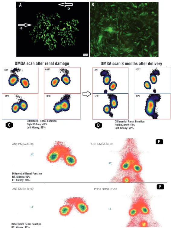
the maternal peripheral circulation and they also contributed to the repairing process of maternal kidney by migrating to the damaged parts for further differentiation. As depicted in Figure-1 (A) GFP positive cells formed both tubular and interstitial lesions. However, no detectable focal aggregation of GFP-positive cells was found in immunofluorescent microscopy of lung and liver as samples of undamaged organs.
The presence of Y-chromosome in the da-maged maternal kidney was obviously proved by the gradient PCR of female rat purified DNA. Re-garding the fact that the exact temperature for PCR of this specific primer was unknown, gra-dient PCR method was applied for a purified DNA sample at different temperatures. Results of the gradient PCR showed that the most appropriate temperature for this specimen was 54ºC. Gradient PCR at 49ºC was applied in order to reassure the specific and non-specific binding of the primer.
The sub-cultured cells from the damaged part of the non-GFP kidney were viewed with immunofluorescent microscopy. The GFP-posi-tive renal cells were observed in company with undamaged maternal renal cells (Figure-1B).
Renal imaging was performed by the ap-plication of Technetium-99m-DMSA solution the high percentage of which in the renal cor-tex results in the high gamma flow. Each of the static scintiphotos was obtained by the applica-tion of a high-resoluapplica-tion collimator 1 hour after administration of Technetium-99m-DMSA solu-tion. The result of DMSA scan after induction of renal damage in experimental group
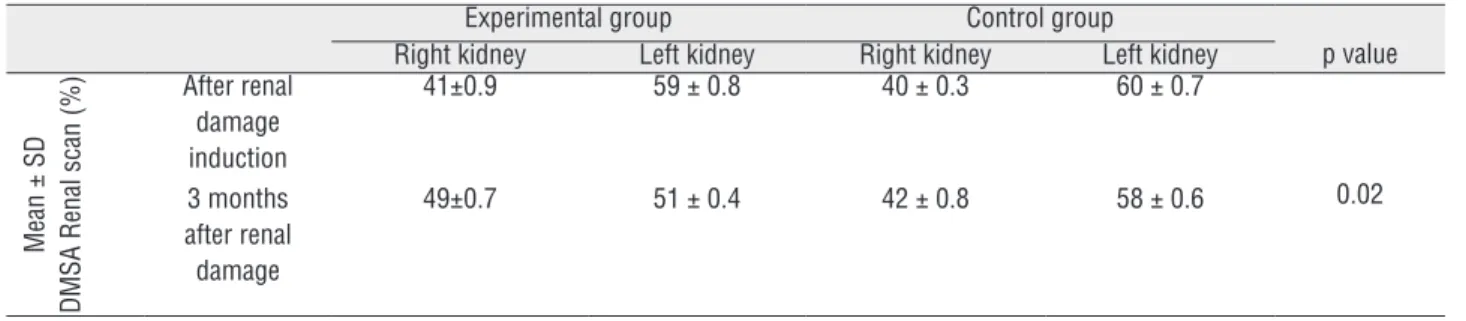
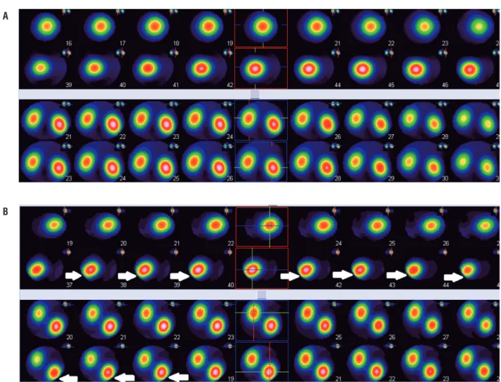
revea-led decreased renal function in the right kidney in which the damage was induced (41%±0.9% versus 59%±0.8%). However, similar uptake in both damaged (right) and normal kidneys (left) with uniform distribution pattern of renal ac-tivity was obtained 3 months after delivery (49%±0.7% versus 51%±0.4%). The results of DMSA scan in control group revealed no signi-ficant improvement in function of right kidney (mean±SD) after 3 months of renal damage in-duction (40%±0.3% versus 60%±0.7% after re-nal damage and 42%±0.8% versus 58%±0.6% ,3 months postoperatively). DMSA scan of preg-nant and non-pregpreg-nant rats confirmed the role of FSCs in repairing the damaged part of the kidney (Table-1). The split renal function des-cribed in the DMSA scan was from average data of the groups (Figures 1-C-F). The results of DMSA scan were compatible with the outcomes obtained from SPECT. SPECT of control group showed renal damage in several continuous sections while satisfactory results without any detectable renal impairment were obtained in none of the sections in experimental group 3 months after operation (Figure-2).
DISCUSSION
The current experimental study provided an overview about the role of FSCs in healing ma-ternal renal damage during pregnancy where the GFP is set as the marker of fetal-origin cells in the maternal damaged part of kidney. The focus of the current study was on determining the role of FCSs in maternal renal function improvement.
Table 1 - Comparing the renal function after renal damage induction and 3 months postoperatively in pregnant (experimental group) and non-pregnant (control group) rats.
Experimental group Control group
p value Right kidney Left kidney Right kidney Left kidney
Mean ± SD
DMSA Renal scan (%)
After renal damage induction
41±0.9 59 ± 0.8 40 ± 0.3 60 ± 0.7
0.02 3 months
after renal damage
Figure 1 - (A) Immunofluorescence of damaged part of maternal non-GFP kidney. Arrow a shows the damaged part of maternal kidney which is covered by GFP-positive FSCs. Arrow b shows the undamaged non-GFP maternal kidney; (B) Fetal GFP-positive cells in company with maternal non-GFP renal cells after culture of the damaged part from the upper pole. Technetium-99m-DMSA scan of pregnant rats (experimental group); (C) differential renal function after induction of renal damage confirmed decreased renal function on right kidney; (D) Renal function was similar in both kidneys three months postoperatively. Technetium-99m-DMSA scan of non-pregnant rats (control group); (E) differential renal function after induction of renal damage confirmed decreased renal function on right kidney; (F) Renal function did not improve significantly three months after renal damage induction
A
C D
E
F B
DMSA scan after renal damage
ANT ANT
LPO LPO
POST POST
RPO RPO
Differential Renal Function Right Kidney: 41% Left Kidney: 59%
RT.
LT.
RT.
LT. Differential Renal Function
RT. Kidney: 40% LT. Kidney: 60%
Differential Renal Function RT. Kidney: 42% LT. Kidney: 58%
POST DMSA-Tc-99 POST DMSA-Tc-99 ANT DMSA-Tc-99
ANT DMSA-Tc-99
Differential Renal Function Right Kidney: 41% Left Kidney: 59%
A
B
Figure 2 - (A) SPECT in the experimental group three months after delivery showed satisfactory results without any significant impairment as compared with control group; (B) SPECT in the control group showed that the renal injury persisted after three months of damage induction. The arrows demonstrate the damage in several sections while such impairment was not detected in experimental group.
It has been shown that the frequency of FMCT can be affected by histocompatibility be-tween the mother and fetus (13). Approximately, 4 weeks post-gestation, FSCs can be detected in maternal circulation (14). In one study, the per-sistence of fetal leukocytes containing a Y body has been documented in the peripheral blood of women that were pregnant for the first time for more than 1 year after delivery (15). In another study in 1974, the persistence of fetal leukocytes in the maternal circulation was shown after ges-tation (16). In the study of Ciaranfi et al., notice-able male lymphocytes were detected in maternal
(3). Accordingly, the frequency of these progenitor cells reaches to its maximum level at 18-19th day of gestation (3).
FSCs have the capability to increase lo-cally in the injured organ during and after preg-nancy. In several maternal damaged tissues such as kidney, specific FMCT have been detected in both mice and humans. In the study of Khosro-tehrani et al. the liver of a pregnant murine mo-del was injured by carbon tetrachloride and it was demonstrated that the number of FSCs increased in the affected organ (20). In one study on GFP mice, Streptozotocin (STZ) was injected in order to induce maternal beta cell injury and investigate the persistence of FSCs in the pancreas. The re-sults showed fetal DNA and EGFP+ cells in mater-nal bone marrow, kidney, pancreas and liver (21). Recently, it was concluded that cigarette smoke exposure in the pregnant mouse model, leads to FSC retention in the maternal injured lung (22). In another study, functional blood vessels has been formed in maternal inflammatory skin by fetal en-dothelial progenitor cells during pregnancy (23). In the study of Wang et al., it has been shown that fetal GFP-positivecells migrate to damaged liver and kidney that were exposed to alcohol and gen-tamicin, respectively (24).
It has been demonstrated that the morpholo-gical appearances of the FSCs within different mater-nal tissues was similar to differentiated matermater-nal cells in mice during pregnancy (18).
Their results were in accordance with the outcomes of the current study in which we revea-led that the morphological appearance of FSCs in the damaged kidney after three months of delivery was indistinguishable from the maternal counterparts. In spite of the fact that no specific tissue marker was applied in the present study which is considered as one of the its limitations, the improvement of renal function, and the successful cell culture with the re-nal cell protocol can approve the differentiation of these FSCs to renal cells.
In the study of Perin et al. stem cells from human amniotic fluids were injected into embryonic kidneys of murine. The results demonstrated the po-tential of these stem cells to differentiate into renal vessels culminating in early development of the kid-ney (25). In spite of the fact that it has been mentioned
that FSCs have an ineffective role on maternal health , the results of the current study expressed opposi-te results as confirmed by DMSA scan and SPECT. It has been shown that no significant improvement was detected in renal function of control group in which unpregnant rats were evaluated. The role of FSCs in functional improvement was not evaluated in previous articles which is one of the benefits of the current study. SPECT study may be also helpful when the size of the renal damage in small which cannot be detected with planer study. In the present study, this method was applied for the first time to estimate the renal function of rats in the field of FMCT.
In the present study, we sought to determi-ne the prolonged persistence of FSCs in damage part of maternal kidney even after the pregnancy. Therefore, we decided to apply GFP-positive trans-genic male rats to identify the FSCs more easily by their green color. Using this method, we achieved high sensitivity detection of FSCs in the section of the injured kidney. The ability of FSCs in mi-grating to damaged maternal tubular epithelial cells was demonstrated by immune histochemical analysis with anti-GFP antibody. The domicilia-tion of GFP-positive FSCs was confirmed by im-munofluorescent microscopy according to their special immunofluorescent staining.
By PCR amplification of Y-chromosome sequences, we confirmed the persistence of FSCs in the blood circulation of the pregnant rat and the migration of Y-chromosome DNA of the male fetus to the damaged region of the kidney for fur-ther regeneration. In the present study, we confirm that FMCT may contribute to organ regeneration and repair. FSCs were only detected in injured re-gion of the kidney and no FSC was distinguished in non-injured organs. These outcomes are in ac-cordance with previous studies in which FSCs mi-grated to the maternal organs after induction of injury (20, 24). However, functional evaluation of the damaged organ was also studied to determine the role of FSCs in improvement of the impaired organ which was not previously evaluated in pre-vious studies.
we decided to perform the renal damage at GD 11 which is in the middle of gestational period. In the current study, we showed that FSCs impro-ved renal function and increased the proliferative response in the damaged kidney compared to the normal one.
This study has also other limitations. Al-though the trend of improvement in functional parameters can ensure a persistent outcome in tissue repair, renal specific markers were not ap-plied. However, renal function improvement, and detection of Y chromosome and GFP-positive cells in the sub-cultured medium indicated the ability of FSCs in migration from the peripheral circula-tion to injured maternal kidney and differentiate into renal cells. Considering the fact that serum creatinine and glomerular filtration rate are easier and cheaper modalities in bilateral renal injuries, their application was not feasible in this model in which unilateral renal damage was created. So, we focused on imaging techniques rather than these laboratory tests.
While medical research teams are now trying to estimate the advantages and disadvan-tages of FMCT in human societies, this study de-monstrated a perfect result of this phenomenon in functional improvement of the maternal damaged kidney in rat model. Therefore, we concluded that fetal GFP-positivecells have the potential to per-sist after delivery and domiciliate in injured kid-neys with dynamic respond. It can be also realized that FSCs in maternal tissues have the ability to act as a reservoir of stem cells and pregnancy is a protection against susceptibility to several dise-ases. However, further studies are required to es-timate the role of FSCs in repairing the maternal damaged organs by introduction of FSCs to ma-ternal organ after harvesting and expanding the cells in vitro. Additionally, further investigations are required to estimate the efficiency of FMCT in human pregnancies.
CONCLUSIONS
In this study, we investigated the role of FSC migration and homing in their final destina-tion in different target organs and tissue regene-ration. We sought to characterize FSCs with
mul-tilineage potential that migrate to the maternal organs. The results of the current study revealed that FSCs play a crucial role in repairing maternal damaged kidney and improve its impaired func-tion without prior in vitro manipulafunc-tion. However, more studies are required to conclusively demons-trate the role of FMCT in human maternal dama-ged organs.
ABBREVIATIONS
FSCs = Fetal Stem Cells
GFP = Green Fluorescent Protein
DMSA = Technetium 99m-Dimercaptosuccinic
Acid
SPECT = Single-photon emission computed
tomo-graphy
FMCT = Fetal maternal cell trafficking
FUNDING
This study was funded by Tehran Universi-ty of Medical Sciences (grant number=32246)
CONFLICT OF INTEREST
None declared.
REFERENCES
1. Baptiste N, Friedlander P, Chen X, Prives C. The proline-rich domain of p53 is required for cooperation with anti-neoplastic agents to promote apoptosis of tumor cells. Oncogene. 2002;21:9-21.
2. Dawe GS, Tan XW, Xiao ZC. Cell migration from baby to mother. Cell Adh Migr. 2007;1:19-27.
3. Pritchard S, Hoffman AM, Johnson KL, Bianchi DW. Pregnancy-associated progenitor cells: an under-recognized potential source of stem cells in maternal lung. Placenta. 2011;32(Suppl 4):S298-303.
4. Seppanen E, Fisk NM, Khosrotehrani K. Pregnancy-acquired fetal progenitor cells. J Reprod Immunol. 2013;97:27-35. 5. Bhattacharya N, Stubblefield P. Fetomaternal Cell Trafficking:
A Window into the Long-Term Health Effects of Treating Disease with Fetal Cell/Tissue Transplants? Human Fetal Tissue Transplantation: Springer; 2013; pp. 15-23.
7. Khosrotehrani K, Johnson KL, Cha DH, Salomon RN, Bianchi DW. Transfer of fetal cells with multilineage potential to maternal tissue. JAMA. 2004;292:75-80.
8. Boudet J, Man NK, Pils P, Sausse A, Funck-Brentano JL. Experimental chronic renal failure in the rat by electrocoagulation of the renal cortex. Kidney Int. 1978;14:82-6.
9. Gibb IA, Hamilton DN. An experimental model of chronic renal failure in mice. Clin Immunol Immunopathol. 1985;35:276-84.
10. Gagnon RF, Ansari M. Development and progression of uremic changes in the mouse with surgically induced renal failure. Nephron. 1990;54:70-6.
11. Chow K-M, Liu Z-C, Chang TM-S. Animal remnant kidney model of chronic renal failure revisited. Hong Kong Journal of Nephrology. 2003;5:57-64.
12. Saiki RK, Gelfand DH, Stoffel S, Scharf SJ, Higuchi R, Horn GT, et al. Primer-directed enzymatic amplification of DNA with a thermostable DNA polymerase. Science. 1988;239:487-91. 13. Nelson JL. HLA relationships of pregnancy, microchimerism
and autoimune disease. J Reprod Immunol. 2001;52(1-2):77-84.
14. Thomas MR, Williamson R, Craft I, Yazdani N, Rodeck CH. Y chromosome sequence DNA amplified from peripheral blood of women in early pregnancy. Lancet. 1994;343:413-4. 15. Schröder J, Tiilikainen A, De la Chapelle A. Fetal leukocytes in
the maternal circulation after delivery. I. Cytological aspects. Transplantation. 1974;17:346-54.
16. Tiilikainen A, Schröder J, De la Chapelle A. Fetal leukocytes in the maternal circulation after delivery. II. Masking of HL-A antigens. Transplantation. 1974;17:355-60.
17. Ciaranfi A, Curchod A, Odartchenko N. [Post-partum survival of fetal lymphocytes in the maternal blood]. Schweiz Med Wochenschr. 1977;107:134-8.
18. Khosrotehrani K, Johnson KL, Guégan S, Stroh H, Bianchi DW. Natural history of fetal cell microchimerism during and following murine pregnancy. J Reprod Immunol. 2005;66:1-12.
19. Zhang G, Zhao Y, Li XM, Kong J. Fetal cell microchimerism in the maternal mouse spinal cord. Neurosci Bull. 2014;30:81-9.
20. Khosrotehrani K, Reyes RR, Johnson KL, Freeman RB, Salomon RN, Peter I, et al. Fetal cells participate over time in the response to specific types of murine maternal hepatic injury. Hum Reprod. 2007;22:654-61.
21. Sunami R, Komuro M, Yuminamochi T, Hoshi K, Hirata S. Fetal cell microchimerism develops through the migration of fetus-derived cells to the maternal organs early after implantation. J Reprod Immunol. 2010;84:117-23.
22. Vogelgesang A, Scapin C, Barone C, Tam E, Blumental Perry A, Dammann CE. Cigarette smoke exposure during pregnancy alters fetomaternal cell trafficking leading to retention of microchimeric cells in the maternal lung. PLoS One. 2014;9:e88285.
23. Nguyen Huu S, Oster M, Uzan S, Chareyre F, Aractingi S, Khosrotehrani K. Maternal neoangiogenesis during pregnancy partly derives from fetal endotelial progenitor cells. Proc Natl Acad Sci U S A. 2007;104:1871-6.
24. Wang Y, Iwatani H, Ito T, Horimoto N, Yamato M, Matsui I, et al. Fetal cells in mother rats contribute to the remodeling of liver and kidney after injury. Biochem Biophys Res Commun. 2004;325:961-7.
25. Perin L, Giuliani S, Jin D, Sedrakyan S, Carraro G, Habibian R, et al. Renal differentiation of amniotic fluid stem cells. Cell Prolif. 2007;40:936-48.
_______________________ Correspondence address: