Daniela Maria Ramos Pereira
Universidade do Minho
Escola de Ciências
Modulation of the immune response to
mycobacteria: implications on protection
and pathology
Tese de Mestrado
Escola de Ciências – Genética Molecular
Trabalho efectuado sob a orientação de:
Doutor Jorge Manuel Rolo Pedrosa
Professor Associado da Escola de Ciências da
Saúde da Universidade do Minho, Braga, Portugal
e co-orientação de:
Doutor António Gil Pereira de Castro
Professor Associado da Escola de Ciências da
Saúde da Universidade do Minho, Braga, Portugal
Daniela Maria Ramos Pereira
Universidade do Minho
Escola de Ciências
Modulation of the immune response to
mycobacteria: implications on protection
and pathology
Modulação da resposta imunológica na
infecção por micobactérias: implicações na
protecção e patologia
ii
DECLARAÇÃO
Nome: Daniela Maria Ramos Pereira
Endereço electrónico: [email protected] Telefone: +351 967429042
Número do Bilhete de Identidade: 12093144
Título da dissertação:
Modulation of the immune response to mycobacteria: implications on protection and pathology.
Modulação da resposta imunológica na infecção por micobactérias: implicações na protecção e patologia.
Orientador:
Doutor Jorge Manuel Rolo Pedrosa Co-orientador:
Doutor António Gil Pereira de Castro
Ano de conclusão: 2009
Designação do Ramo de Conhecimento do Mestrado: Ciências – Genética Molecular
É AUTORIZADA A REPRODUÇÃO INTEGRAL DESTA TESE/TRABALHO
APENAS PARA EFEITOS DE INVESTIGAÇÃO, MEDIANTE DECLARAÇÃO
ESCRITA DO INTERESSADO, QUE A TAL SE COMPROMETE
Universidade do Minho, 30/10/2009
The work presented in this thesis was done in the Laboratory of Immunology of Infection in the Life and Health Sciences Research Institute (ICVS), Minho University. The financial support was given by PTDC/SAU-MII/70895/2006 and by Health and Human Development Services of the Calouste Gulbenkian Foundation.
A
GRADECIMENTOS
O trabalho que fui desenvolvendo durante estes anos contou com a colaboração, a vários níveis, de um conjunto de pessoas a quem gostaria de agradecer.
Obrigada ao fantástico I3.02, do qual tenho orgulho em pertencer.
Ao Professor Doutor Jorge Pedrosa, meu orientador deste mestrado, a quem admiro o rigor, clareza e ética em comunicar e discutir ciência. Obrigada pela oportunidade que me concedeu em fazer investigação e por valorizar a minha formação científica.
Ao Professor Doutor Gil Castro, o meu mestre desta jornada, pela liberdade que me facultou no desenvolvimento do projecto, pelo conhecimento que me transmitiu, por ter acreditado em mim e pela forma informal mas rigorosa com que sempre me acompanhou.
À Professora Doutora Paula Sampaio pela supervisão e acompanhamento.
À Doutora Margarida Saraiva, pelo sentido crítico, disponibilidade em discutir o projecto, e pelas peripécias engraçadas do Gui.
É muito fácil falar dos motivos que tenho para agradecer à Doutora Andrea, pois foi a minha “guru” do laboratório:). A ti recorri mil e uma vezes para aprender as técnicas, planear experiências, discutir resultados. Obrigada pela confiança que depositaste em mim, mesmo quando as coisas corriam mal, dando-me força para fazer cada vez melhor! A tua experiência e amizade foram pilares nesta jornada.
À Jenny, por muitos momentos partilhados, desde calos em Lisboa, ao trabalho de Jenniela. Obrigada pela tua amizade, e por me compreenderes tão bem (deve ser do ascendente em escorpião;). À Carole, cá estamos:)! A entre-ajuda que mantivemos fez com que fosse mais fácil superar os obstáculos. É a força do crepúsculo incandescente! Obrigada por teres sido tão boa companheira. Maria, obrigada pela tua ajuda nas duras maratonas de P3 e pelos fins de tarde a jogar “mau futebol”! Agradeço à Alex por ser sempre prestável, por não ter parado de falar naquele dia de P3 (é que sabes mesmo integrar pessoas!) e pelas cantorias super afinadas
“que aaaaaaaltuuura a árvore tem…”. À Teresinha, ao Bernardo, e ao Diogo que, além de tudo, partilham comigo, respectivamente, o Benfica, o Afonso Henriques e agora o Mestrado. Obrigada
também à Susana, Cláudia, Palmira, Cláudio e Margarida por estarem sempre disponíveis a ajudar e por serem porreiros, pá!
Ao Nuno, pelas longas conversas, “peladinhas”, pela amizade. À Rita, Guida, Raquel, Rexona e Sofia, por acompanharem mais uma etapa da minha vida. Ao Tiago, por se ter lembrado de mim;). À Cristiana e ao Mooji, porque eles é que sabem.
Aos meus pais e à Mariana, pelo apoio incondicional. Obrigada pela paciência e sabedoria de estarem presentes sempre que precisei e ausentes quando necessário.
Abstract
Effective control of Mycobacterium tuberculosis infection requires the induction of inflammatory T helper (Th) 1 responses and avoidance of tissue damage. However, little is known about the mechanisms that rule the fine balance between protection and pathology in this infection. The anti-inflammatory cytokine IL-10 is important for the regulation of the immune response to several pathogens. However, despite the fact that elevated levels of IL-10 have been detected in tuberculosis patients, the role of IL-10 during M. tuberculosis infection is not clearly understood.
In this study, we evaluated the impact of a transient over-expression of IL-10, during either the innate or the adaptive phases of the immune response, in the outcome of infection. For this, we used a novel animal model, the PMT-10 mice, which over-express IL-10 under the control of a zinc-inducible promoter. These transgenic animals were infected with M. tuberculosis, either via the intranasal or intravenous route and the expression of IL-10 was induced at different stages of the infection.
Over-expression of IL-10 early after infection did not influence the capacity of PMT-10 mice to control bacterial growth, regardless the route of inoculation. In contrast, over-expression of IL-10 during the chronic phase of infection caused significant effects in the progression of M. tuberculosis infection. Interestingly, whereas in intravenous infection, late IL-10 induction resulted in a transient increase in susceptibility of PMT-10 mice to M. tuberculosis, in intranasal infection, induction of IL-10 led to an increased resistance of PMT-10 mice that was maintained until the end of the experiment. However, no major differences were observed between induced and non-induced PMT-10 mice concerning the dynamic of cells or cytokine expression during infection that could explain the different outcomes observed.
Although the cellular and molecular mechanisms underlying the differences observed remain unknown, altogether our data suggest that the impact of IL-10 expression during an infection with M. tuberculosis is variable. Indeed, we show for the first time that the timing of IL-10 expression as well as the route of inoculation can determine the outcome of M. tuberculosis
infection. Further understanding of our observations might be useful in the context of immunomodulatory strategies based on IL-10 expression or suppression.
Resumo
O controlo efectivo da infecção por Mycobacterium tuberculosis requer o desenvolvimento de respostas inflamatórias do tipo T de ajuda (Th) 1. No entanto, pouco ainda se sabe acerca dos mecanismos que regulam o equilíbrio entre patologia e protecção no contexto desta infecção. A IL-10 é uma citoquina com propriedades anti-inflamatórias e, portanto, importante na regulação da resposta imunológica a patogénios. Contudo, e apesar da IL-10 se encontrar em elevadas quantidades em pacientes com tuberculose, o papel da IL-10 em infecções por M. tuberculosis não está completamente esclarecido.
Neste estudo avaliámos o impacto da sobre-expressão transiente de IL-10, nas fases inata ou adquirida da resposta imunológica, na evolução da infecção. Para isso, utilizamos um novo modelo animal, os ratinhos PMT-10, que sobre-expressam IL-10 sobre o controlo de um promotor induzido pelo zinco. Estes ratinhos transgénicos foram infectados com M. tuberculosis
pela via intranasal ou intravenosa e a expressão de IL-10 foi induzida em diferentes fases da infecção.
A sobre-expressão de IL-10 cedo após infecção não influenciou a capacidade dos ratinhos PMT-10 em controlar o crescimento bacteriano, independentemente da via de inoculação. Pelo contrário, a sobre-expressão de IL-10 durante a fase crónica da infecção por M. tuberculosis
provocou efeitos significativos na progressão da infecção. Curiosamente, após a indução de IL-10, enquanto na infecção intravenosa, os PMT-10 apresentaram um aumento de susceptibilidade transiente, na infecção intranasal, os PMT-10 exibiram uma protecção superior aos ratinhos não induzidos. Contudo, não se observaram diferenças relevantes entre PMT-10 induzidos e não induzidos, em relação à dinâmica de células e citoquinas analisadas e mais frequentemente expressas durante a infecção por M. tuberculosis.
Apesar dos mecanismos celulares e moleculares responsáveis pelas diferenças obtidas permanecerem por explicar, os nossos resultados sugerem que o impacto da expressão de IL-10 numa infecção por M. tuberculosis é variável. Assim, demonstramos neste estudo que o momento da expressão de IL-10, bem como a via de inoculação pode determinar o resultado da infecção por M. tuberculosis. A compreensão mais dissecada destes resultados poderá constituir uma mais-valia no desenho de novas estratégias de imuno-modulação baseadas na expressão ou supressão da IL-10.
T
ABLE OF CONTENTS1. Introduction 1
1.1. Current status of Tuberculosis – Overview of the disease worldwide 1
1.2. Immune response to M. tuberculosis – Effector mechanisms 3
1.2.1. Innate immune response 3
1.2.2. Adaptive immune response 7
1.3. Modulation of the immune responses: Role of IL-10 9
1.3.1. IL-10 in infectious diseases and in therapy strategies 9
1.3.2. IL-10 sources and targets 10
1.3.3. IL-10 in mycobacterial infections 11
1.4. Aims 14
2. Material and methods 15
3. Results 19
3.1. Effect of IL-10 over-expression early after infection with M. tuberculosis: impact on the innate and adaptive immune responses 19
3.2. Effect of IL-10 over-expression late after infection with M. tuberculosis: impact on the ongoing immune responses 30
4. Discussion 43
5. Conclusion 52
A
BBREVIATIONSBCG Bacille Calmette-Guérin TLR Toll-like receptor
CFU Colony forming units TNF Tumor necrosis factor
CR Complement receptor WHO World health organization DCS Dendritic cells
HIV Human immunodeficiency virus
IFN Interferon
iNOS Inducible nitric oxide synthase
kDa Kilodalton
LAM Lipoarabinomannan
LN Lymph node
MHC Major histocompatibility complex
MT Metallothioneic
MyD88 Myeloid differentiation protein 88
NK Natural killer
NO Nitric oxide
PCR Polymerase chain reaction
PI3P Phosphatidylinositol 3-phosphate
RNI Reactive nitrogen intermediates
TACO Tryptophan-aspartate containing coat protein
TGF Transforming growth factor
1. I
NTRODUCTION
1.1. Current status of Tuberculosis – Overview of the
disease worldwide
Despite decades of research on chemotherapy for the disease and development of preventive vaccines, tuberculosis remains a leading global health threat.
Tuberculosis kills more people per year than any other single infectious disease. Its causative agent, Mycobacterium tuberculosis, is a slow growing bacillus transmitted primarily by the respiratory route, and although it can cause disease in most organs, pulmonary tuberculosis is the most common (1).
Primary infection leads to active disease in only a minority (about 10%) of infected individuals (2) probably due to the lack of initiation of an appropriate immune response. In the remaining 90 % of cases the immune system controls the infection, but the pathogen is not eradicated. This clinical latency, although not contagious, can persist throughout the person’s lifetime. However, reactivation of the latent infection can occur due to perturbations in the host immune response, such as co-infection with the human immunodeficiency virus (HIV), malnutrition, or immunossupressive medication (3).
According to the World Health Organization (WHO) 2009 report (4), the incidence of tuberculosis is increasing worldwide and reached 9,27 million new cases in 2007, even though the number of incident cases per capita is modestly declining. The more affected countries, regarding total number of tuberculosis cases, are India (2.0 million), China (1.3 million), Indonesia (0,53 million), Nigeria (0.46 million) and South Africa (0.46 million), explaining why the Asiatic and African continents have the higher fraction of global cases, with 55% and 31%, respectively. Small proportions of cases were reported in Eastern Mediterranean Region (6%), Europe (5%) and America (3%).
Regarding the European continent, most countries experience a steady decrease in the tuberculosis incidence rate over the last few decades, reaching an overall rate of 17 cases per 100,000 population, although substantial increases were observed in Malta, Iceland, Ireland and Greece. In Portugal, the incidence of tuberculosis has been decreasing 7,3% per year since
2003, with its lowest rate of 29,5 cases (per 100 000 population) in 2007. Nevertheless, Portugal is currently the fifth country in Europe with higher case rates of tuberculosis, following Estonia, Bulgaria, Lithuania and Romania (5).
One of the factors closely associated with the increasing incidence of tuberculosis is the HIV epidemic. In fact, 1,37 million of tuberculosis incident cases (14%) are HIV positive, with the African Region accounting for the higher fraction of global cases (79%). More dramatic is the mortality data, revealing that deaths from tuberculosis among HIV patients account for 27% of the estimated 1,7 million tuberculosis deaths that occurred in 2007 (4).
In addition, the emergence of extensively drug-resistant tuberculosis seriously increases the concerns with regard to transmission and propagation of the disease. Among the 9,27 million episodes of tuberculosis, an estimated 0,5 million cases of multidrug resistant tuberculosis were reported, being India, China, Russian and South Africa the countries with largest number of cases (4). Moreover, in Europe, drug-resistant tuberculosis constitutes the strongest determinant of death among tuberculosis patients (6).
The currently vaccine used against tuberculosis is bacilli Calmette-Guerin (BCG), a live attenuated strain of M. bovis. This vaccine, developed around the turn of the last century and first administered in 1921, is given to a large proportion of newborn infants throughout the world, however, the protection that BCG confers against tuberculosis is both incomplete and variable (7).
Thus, it is expected that an increased understanding of disease pathogenesis will contribute to the design of more effective vaccines and new therapeutic strategies (like immunotherapy), which will certainly improve the outcome of individual patients and limit the spread of M. tuberculosis around the world.
1.2. Immune response to
M. tuberculosis
– Effector
mechanisms
1.2.1. Innate immune response
The differences, among individuals, in the outcome of infection with M. tuberculosis may, in part, be explained by the variable efficiency of various innate host defense mechanisms. Immune recognition, phagocytosis, cytokine production and effector mechanisms all contribute to innate immunity.
Alveolar macrophages, residing in the distal airways, avidly engulf inhaled bacteria and are the first cells to be infected by M. tuberculosis (8). A variety of phagocytic receptors are involved in the binding and internalization of the bacteria, demonstrating the complex structure of its cell wall structure.
M. tuberculosis can invade host macrophages after opsonization with complement factor C3, which is followed by binding and uptake through complement receptor 1 (CR1), CR3, and CR4 (9). Blocking CR drastically reduces binding and invasion of M. tuberculosis but does not abolish it, suggesting that other receptors participate in their uptake. Consistent with this, M. tuberculosis has also been demonstrated to bind to the mannose receptor, which recognizes terminal mannose residues on mycobacteria (10). This occurs more frequently with virulent strains of M. tuberculosis, suggesting that this route of entrance is advantageous to the bacillus. In fact, phagocytosis through the mannose receptor does not induce anion superoxide production by macrophages (11), and instead triggers anti-inflammatory signals (12). M. tuberculosis can also be internalized through the type A scavenger receptor and Fcγ receptors that result in respiratory burst, and in an inflammatory type of response by macrophages (13).
The receptors involved in the phagocytic entry may lead to differences in signal transduction, immune activation and, thus, have a major impact on the survival chances of M. tuberculosis once inside the macrophage. However, most of these interactions have been demonstrated in vitro, and their relative importance in vivo remains to be revealed.
Once internalized, M. tuberculosis resides in the phagosome that fuses with early endosomes, arresting iron and other nutrients important for mycobacteria survival (14). Thereafter, M. tuberculosis products either located in the cell wall, like lipoarabinomannan (LAM), or released to the cytosol, as the phosphatase SapM and the kinase PKnG, are able to impair the transfer of the phagosome to the lysosome (15-17), where bacterial degradation occurs. The inhibition of phagosome-lysosome fusion by M. tuberculosis is due its capacity to interfere with host glycolipids and proteins that regulate this process, such as PI3P (phosphatidylinositol 3-phosphate) (18), TACO (Tryptophan-aspartate containing coat protein) (19) and LRG-47 (47-kilodalton guanosine triphosphatase) (20). As a result, M. tuberculosis is able to survive within a phagosome only mildly acidified (21).
The vesicle trafficking events also deliver M. tuberculosis to endosomes-related compartments for antigen degradation and subsequent binding to major histocompatibility complex (MHC) class II molecules. Peptide-loaded MHC-II molecules are shuttled to plasma membrane and presented to T cells, initiating a specific immune response (22). However, in vitro studies show that M. tuberculosis is also able to subvert this pathway. Indeed, prolonged infection of macrophages with M. tuberculosis inhibits the expression of MHC-II and class II transactivator (CIITA), (23, 24) by blocking chromatin remodeling at the CIITA promoter (25), thus resulting in decreased MHC-II antigen presentation.
Besides expressing phagocytic receptors, macrophages and dendritic cells (DCs) also express several pattern recognition receptors, such as Toll-like receptors (TLRs) that recognize specific molecular patterns expressed by pathogens. All the TLRs have, at least, one signaling pathway dependent on intracellular adaptor molecule myeloid differentiation factor 88 (MyD88) (26). Thus, MyD88 deficient mice (MyD88-/-) are highly susceptible to aerogenic M. tuberculosis
infection, implying that TLR signaling plays an important role in mycobacteria resistance (27). A major consequence of M. tuberculosis interaction with TLRs is the secretion of cytokines and chemokines that activate the macrophage and regulate the development of an antigen-specific adaptive immune response.
The 19 kilodalton (kDa) lipoprotein, a secreted antigen of M. tuberculosis, was the first ligand shown to interact specifically with TLR2 to induce tumor necrosis factor (TNF) and nitric oxide (NO) production from both murine and human macrophages (28). Other in vitro studies show that the immunostimulatory responses to M. tuberculosis LAM, and phosphatidylinositol
mannoside (PIM) are also mediated by TLR2 (29, 30). But interestingly, despite a large collection of TLR2 agonists present on the tuberculosis bacillus, in vivo studies in the mouse model indicate that TLR2 is not essential for host resistance against low dose M. tuberculosis infection (31-33). Besides TLR2, other TLRs may be involved in immune recognition of M. tuberculosis. An undefined heat-labile cell-associated mycobacterial factor was found to be a ligand to TLR4 (30), however, TLR4-/- mice do not show a compromised resistance to tuberculosis following an aerosol
challenge (32, 34). Also, TLR9 was shown to bind GpG dinucleotides in bacterial DNA, inducing a rapid antimycobacterial response in macrophages, in a phospholipase D-dependent manner (35). Therefore, the understanding of the in vivo role played by of the various TLRs in the host defense against M. tuberculosis infection awaits further experimentation.
As it was mentioned above, recognition of M. tuberculosis by phagocytic cells leads to cell activation and induction of cytokine production, which itself induces further macrophage activation and cytokine production, in a complex dynamic process in order to kill the mycobacterial infection.
One of the most important pro-inflammatory cytokines produced by macrophages in response to M. tuberculosis infection is TNF. The role of this cytokine is of great clinical relevance as TNF-blocking drugs, used as anti-inflammatory therapy to rheumatoid arthritis, are associated with reactivation of latent tuberculosis in humans (36, 37). Also, in mouse models, disruption of the TNF gene or neutralization of TNF lead to disorganized granulomas, a characteristic and crucial feature of tuberculosis, compromising the containment of M. tuberculosis (38). Importantly, TNF, along with the cytokine interferon gamma (IFNγ) produced mainly by T- lymphocytes induce NO production in macrophages by activating the inducible nitric oxide synthase (iNOS) enzyme (39). NO reacts with oxygen radicals forming reactive nitrogen intermediates (RNI) that can kill or inhibit M. tuberculosis growth. However, in some cases, and despite the high toxicity of these products, M. tuberculosis is able to persist within this environment. This resistance is based on multiple strategies developed by the bacilli, such as the mycobacterial proteasome and the production of KatG, a catalase-peroxidase that can inactivate RNI within phagosomes (40, 41).
Finally, TNF can also induce apoptotic cell death in macrophages during M. tuberculosis
replication, TNF-mediated apoptosis represents an innate defense mechanism that slows the increase of bacterial load following infection. In fact, several studies associate the virulence of M. tuberculosis strains with apoptosis (43-45), indicating that virulent strains actively suppress apoptosis by interfering with TNF signaling and by up-regulating the expression of anti-apoptotic molecules, such as Mcl-1 (46).
It is therefore accepted that M. tuberculosis persistence in the host is, in part, due to its cell wall resistance and to the capacity of bacilli to modify effector functions of macrophages and DCs. However, some inhibitory mechanisms can be intentionally initiated by the host to counteract detrimental side-effects of a pro-inflammatory response. Whether triggered by the host or by M. tuberculosis, the macrophages and DCs are able to produce anti-inflammatory cytokines, such as IL-10. The specific role of IL-10 during M. tuberculosis infection will be discussed in section 1.3. However, tumor growth factor (TGF-β) is also an anti-inflammatory cytokine that, like IL-10, is produced during the chronic phase of M. tuberculosis infection (47, 48), suggesting an important immunomodulatory role for this cytokine.
Beside macrophages and DCs, neutrophils and natural killer (NK) cells also participate in the innate immune response.
NK cells are recruited early during M. tuberculosis infection and are a primary source of IFNγ, either in response to IL-12 or IL-18 production by macrophages and DCs, reinforcing the bactericidal capacity of these cells (49). Unlike T-cells, NK cells do not recognize mycobacterial antigens presented by MHC class I or class II molecules. Whether these cells are essential to innate resistance to M. tuberculosis is not completely proved (50, 51).
Circulating neutrophils are also recruited to the lungs early after infection. Their role in host defense against M. tuberculosis is supported by studies showing that depletion of neutrophils early after intravenously challenge with M. tuberculosis compromises the immune response against mycobacterial infection (52-55). Moreover, an in vitro study suggested that the phagocytosis of apoptotic neutrophils by macrophages decreases the viability of intracellular M. tuberculosis, due to the uptake of neutrophil antimicrobial peptides (56). Yet, the mechanisms by which neutrophils can mediate antimicrobial activity against M. tuberculosis are not completely understood.
In the majority of individuals exposed to M. tuberculosis, the innate response alone cannot protect from infection, and effector T cells of the adaptive immune response are necessary to restrict bacterial growth and to mediate protection. Therefore, following M. tuberculosis
phagocytosis and concomitant TLR activation, the next step in the development of host immunity is the transport of pathogen from the lung to the draining lymph node (LN), where the process of adaptive immune response is initiated.
1.2.2. Adaptive immune response
After aerosol exposure, DCs infected by M. tuberculosis are capable of migrating to the LN where naive T cells can be activated (8). This activation requires expression of antigen in the context of MHC, costimulatory molecules, and the necessary cytokines that promote T cell differentiation (57).
The antigen specific T helper (Th) 1 cells are induced mainly in the presence of IL-12 (58, 59) and are actively involved in the control of tuberculosis. This CD4 T cell subset is able to produce IFNγ to full activate the antimicrobial mechanisms of macrophages. Murine studies have shown that IFNγ -/- mice rapidly succumb to M. tuberculosis infection (60, 61) and antibody
depletion of CD4 T cells, similarly, decreases resistance to M. tuberculosis infection (62). In humans, the loss of CD4 T cells in HIV patients greatly increases susceptibility to both acute and reactivation tuberculosis (63). In addition, individuals carrying defective genes for IFNγ receptor or IL-12 receptor (64) are more susceptible to intracellular pathogens, including low virulent mycobacteria such as BCG.
Various studies investigated the role of CD8 T cells, during a M. tuberculosis infection. These cells are limited to either cytotoxicity activity or secretion of IFNγ (65), which happens when DCs take up apoptotic vesicles containing M. tuberculosis antigens and cross-present them on MHC Class I (66, 67). However, the impact of CD8 T cells in the protection against M. tuberculosis is not completely understood, as the absence of CD8 T cells in a low dose aerosol model of infection had little impact in disease progression, until after 200 days post infection (68, 69).
Investigators have recently identified Th17 cells as another subset of CD4 T cells that is detected in mouse models and in humans exposed to M. tuberculosis (70). The differentiation of this T cell lineage is induced in the murine model by the action of TGF-β and IL-6 (71), but also
requires IL-23 (72) to become a persistent population. The first studies on the role of Th17 cells during mycobacterial infection indicate that these cells mediate neutrophil recruitment during inflammatory responses (73, 74), and can be negatively regulated by IFNγ (75). However, a protective role for Th17 in tuberculosis has not been yet demonstrated. It has been shown that Th17 cells are induced after vaccination and promote an accelerated IFNγ response during subsequent M. tuberculosis infection (76). Studies in our laboratory, on the other hand, showed that M. tuberculosis infected mice, repeatedly challenged with mycobacterial antigen, have an enhanced pulmonary pathology with an increased lesion size that is dependent on both IL-23 and IL-17 (A. Cruz et al, unpublished).
The migration of macrophages and of differentiated T cells to the site of infection culminates in the formation of a granuloma. This structure encompasses the bacilli, residing within the macrophages, and functions as an immune microenvironment to facilitate interactions between T cells, macrophages and the produced cytokines (77). In addition, the granuloma serves to wall off the bacteria from the rest of the lung, limiting its spread (78). However, in some cases, M. tuberculosis may induce chronic immunopathology leading to lesions that undergo caseation necrosis, that provide excellent conditions for bacterial extracellular growth, and give rise to cavities that allow the bacillus to spread through the airways to other parts of the lung and outside the environment (3).
1.3. Modulation of the immune response: Role of IL-10
1.3.1. IL-10 in infectious diseases and in therapy strategies
During an infectious disease, the host immune system needs to respond with sufficient intensity and duration to control and eliminate the infection. However, strong antimicrobial effector mechanisms can often cause significant collateral damage to the host, which sometimes is more harmful than the infection itself. Thus, the presence of anti-inflammatory cytokines is important for the safe resolution of infection, as it is confirmed by the critical role of IL-10 in a variety of infectious diseases (79).
Thus, it has been shown that reduction of IL-10, using monoclonal antibodies or IL-10
-/-mice, during Plasmodium spp. (80-82) and Toxoplasma gondi infections (83, 84), resulted in the onset of severe or even fatal immunopathology, caused by excessive IFNγ mediated responses. In addition, injection of mice with recombinant IL-10 or over-expression of this cytokine in transgenic mice enhanced their survival during toxic-shock-like syndromes, by reducing the levels of TNF, IFNγ and MIP-2 (85, 86).
However, the presence of anti-inflammatory cytokines can also delay or impair protective immune responses. This is supported by studies showing that reduction of IL-10 decreased the survival of intracellular pathogens, such as Listeria monocytogenes (87), Candida albicans (88), and Leishmania major (89). In addition, excessive or untimely IL-10 production inhibit the pro-inflammatory responses to T. cruzi (90), Plasmodium yoelli (91), Leishmania spp (92, 93) and lymphocytic choriomeningitis virus (94) to the extent that these pathogens escape immune control, which can lead to either fulminate or chronic non-healing disease.
Therefore, the resolution of infection requires a coordinated response in which pro-inflammatory mechanisms clear the pathogen and are then down-modulated by IL-10, before immunopathology occurs. IL-10 has itself to be strictly controlled to avoid an inefficient response or the development of chronicity.
The profound immunosuppressive effects of IL-10 have prompted a variety of clinical studies to employ recombinant IL-10 to treat patients with immune mediated inflammatory diseases. The clinical effects of recombinant IL-10 have, however, been quite heterogeneous. For instance, whereas almost no effect was seen in rheumatoid arthritis, significant response was
observed in psoriasis (95). Patients with psoriasis that received recombinant IL-10 had significant clinical benefits, decreasing the size of psoriatic areas as well as its severity index (96). However, in some other studies with larger numbers of patients and more severe forms of psoriasis, the systemic administration of IL-10 resulted in only temporary clinical improvement but not remission (97). More encouraging results were observed when IL-10 was delivered locally to the area of inflammation rather than systemically (98). In experimental autoimmune encephalomyelitis, an animal model for human multiple sclerosis, Cua et al showed that, for optimum therapeutic activity, IL-10 had to be delivered directly to the central nervous system by an adenovirus vector, whereas systemic administration was ineffective (99). This study suggested that the localization and timing of IL-10 production or administration may determine its effectiveness. The local delivery of IL-10 to sites of inflammation has been a major challenge, and new experimental approaches are being undertaken in this area, such as bacteria delivery systems (100) or nanoparticles-in-microsphere oral systems (101).
1.3.2. IL-10 sources and targets
IL-10 is produced by different types of immune cells, from both myeloid and lymphoid lineages (79). Monocytes, macrophages and DCs are important producers of IL-10, generally when induced by TLR2 but also TLR4 ligands (102). B cells are also a potentially important source of IL-10 (103), as are some granulocytes, including eosinophils and mast cells (104). In addition, many subsets of T cells can a produce IL-10, such as Th2 cells, inducible regulatory T cells, which are generated in the periphery, and natural regulatory T cells, which are generated in the thymus (105). Interestingly, Th1 and, more recently, Th17 cells were also found to coproduce IL-10, (106-108) together with IFNγ or IL-17, when induced by particularly strong antigen dose and inflammatory responses, allowing the immune responses to be inherently self-regulating (109).
Most hematopoietic cells express the IL-10 receptor and, therefore, IL-10 is able to down-regulate many steps in the pathway of both the innate and adaptive immunity.
IL-10 produced early in the immune response can act in antigen presenting cells in an autocrine way, down-regulating the expression of MHC class II and the costimulatory molecule B7-1/B7-2 (110, 111). In addition, IL-10 can inhibit the production of pro-inflammatory cytokines, including IL-1α and β, IL-6, IL-12, IL-18, and TNF, as well as the expression of
chemokines implicated in the recruitment of monocytes, DCs, neutrophils, and T cells (MCP1, MCP5, RANTES, IL-8, IP-10, and MIP-2) (79). As a result, the effects of IL-10 in macrophages and DCs indirectly compromise the activation of effector T cells and subsequent initiation of adaptive immunity.
In addition, IL-10 can act directly on T cells to limit their proliferation and production of cytokines, such as IL-2, IFN-γ, IL-4 and IL-5 (112, 113) or may promote the differentiation of naïve T cells into IL-10-producing regulatory T cells (105) that, in turn, may directly influence macrophages, DCs and effector T cells (114).
In contrast, IL-10 also have stimulatory effects on mast cells (115), and in B cells by up-regulating the expression of MHC class II molecules and enhancing IgA responses (114), and induces the recruitment, proliferation and cytotoxic activity of NK cells and CD8 T cells (116).
1.3.3. IL-10 in mycobacterial infections
There are evidences demonstrating that Il-10 is produced in patients with tuberculosis. Indeed, T cells expressing both IFNγ and IL-10 have been isolated from the bronchoalveolar lavage fluid of tuberculosis patients (117), and expression of IL-10 mRNA has been demonstrated in circulating mononuclear cells of the pleural fluid (118). Nevertheless, the role of this cytokine during M. tuberculosis infection is not clearly understood. That IL-10 may be important in tuberculosis is suggested by reports associating the risk of developing tuberculosis with the presence of human polymorphisms. For instance, SLC11 A1, a tuberculosis susceptibility locus, has been associated with increased innate IL-10 response that may lead to a tendency towards the development of primary progressive tuberculosis (119). In addition, studies in Turkish and Cambodian populations found a decrease in the frequency of the allele IL10 – 1082/, related to progression in lung tuberculosis (120, 121). However, in other populations (122-124) no differences in the IL-10 genotype frequencies were observed among tuberculosis patients.
In vitro and ex vivo studies indicate that IL-10 inhibit or modulate the immune response against M. tuberculosis. Several studies demonstrate that IL-10 antagonizes the activation of macrophages infected with M. tuberculosis by down-regulating the production of TNF and IL-12 (125-127), as well as the expression of costimulatory molecules (128). Furthermore, IL-10 inhibits the activation of CD4 T cells and the production of IFNγ in response to M. tuberculosis
These works suggest that IL-10 may suppress the development of the immune response against M. tuberculosis. However, Jung et al showed that IL-10-/- mice display an identical
capacity to control M. tuberculosis infection as wild-type mice, despite the stronger Th1-mediated immunity generated early in the infection (131). In the other hand, it was recently reported that IL-10-/- mice infected with M. tuberculosis display exacerbated immunopathology, thereupon
succumb at very late stage of infection (132). In contrast, IL-10-/- mice are more resistance to
BCG infection than wild-type mice (131, 133), probably due to enhanced levels of TNF and iNOS in the granuloma. In addition, IL-10 neutralizing antibodies in M. avium infection resulted in enhanced bacterial clearance (134, 135).
Transgenic mice that over-express IL-10 show increased susceptibility to M. tuberculosis
and other mycobacterial species. However, these works are not consensual in revealing what immune mechanisms are impaired by IL-10, and whether they correlate with the increased susceptibility to mycobacterial infection.
Turner et al. showed that mice over-expressing IL-10 in activated T cells during M. tuberculosis infection, exhibited an impaired Th1 development, as characterized by decreased numbers of activated T cells in the blood and lung tissue as compared to wild-type mice (136). However, Murray et al, in another T cell-specific IL-10 transgenic model, showed that after BCG infection IL-10 had little effect on T cell function, but rather acted primarily in the costimulatory functions of macrophages (137).
In models in which IL-10 is specifically over-expressed by macrophages, the Th1 response induced upon mycobacterial infection seemed to be unaffected. Feng et al (138) and Lang et al
(139), showed that IL-10 over-expression deactivates macrophage by inhibiting TNF and IL-12 in
M. avium and BCG infection, respectively. However, Schreiber et al (140) suggested that after M. tuberculosis infection, over-expression of IL-10 does not impair effector cytokines, but instead induces an alternative activation pathway in lung macrophage that inhibits RNI production.
Altogether, the results of these studies are difficult to compare as, besides using different mycobacterial species, other experimental settings diverge, such as the cellular source of IL-10, the route of infection and the time points assessed.
Therefore, further studies are needed to dissect the role of IL-10 during mycobacterial infections. We propose, that before dissecting the precise mechanisms inhibited by IL-10 which impact the outcome of M. tuberculosis infection, it is important to firstly identify when they occur during the infectious process.
In the previous studies, although the increased bacterial loads detected in IL-10 transgenic mice occurred during the chronic phase of infection, we question whether: (i) it was a consequence of an impairment in the early innate mechanisms that affected long-term cellular responses and, therefore, influenced the disease progression, or (ii) IL-10 was able to hinder the immune system when acquired immunity was established, causing increased bacterial replication. The fact that the transgenic mice models used so far over-express IL-10 constitutively during the infection, hampers any attempt to address these 2 questions. Assessing separately the consequences of IL-10 during these two singular periods of the host immune response against M. tuberculosis is, undoubtedly, a more reliable approach that would further allow us to unravel the cause-effect mechanism underlying IL-10 regulation.
For this purpose we used a novel model of IL-10-expressing transgenic mice, the PMT-10 mice, which over-express IL-10 under the control of a zinc-inducible methionine promoter. In this context, we evaluated the impact of a transient over-expression of IL-10, during either the innate or the adaptive phases of the host immune response, in the outcome of M. tuberculosis infection.
1.4. Aims
The aim of this thesis is to understand the role of IL-10 in M. tuberculosis infection. We propose to:
- Determine the impact of high expression of IL-10 in the outcome of intranasal or intravenous M. tuberculosis infection;
- Distinguish the progression of the disease when IL-10 is over-expressed during the early or late stages of M. tuberculosis infection;
2. M
ATERIAL AND
M
ETHODS
Animals. PMT10 animals on a C57BL/6 background were produced by Drs. P Vieira and AG
Castro. A mouse IL-10 cDNA sequence was cloned in the p169ZT vector (141), which carries the sheep metallothioneinc (MT) Ia promoter (142), a β-globin splice site and the SV40 polyadenylation signal. The resulting vector (pMT-IL10) was injected in C57BL/6 eggs and transgenic founders were identified by PCR using MT specific primers.
IL-10 over-expression was induced by giving a 2% sucrose solution with 50 mM of zinc sulfate to animals ad libitum. As the IL-10 promoter is associated with a metalloprotein, the presence of zinc in this solution induces its activation. Serum levels of IL-10 could be measured 3 days after induction. A group of transgenic littermates were supplied with regular water, as a control. All animals were bred under specific pathogen-free conditions in our animal facilities.
Bacteria and infection. The H37Rv strain of M. tuberculosis was grown in Proskauer Beck
medium containing 0.05% Tween 80 to mid-log phase and prepared for infection of mice with 102
colony-forming units (CFU) via the respiratory route or 105 CFU via the intravenous route. Before
intranasal infection, mice were anesthetized with Ketamine (Imalgene 1000)/Medetomidine (Domitor).
Bacterial load determination. Infected mice were killed by CO2 asphyxiation and the organs
were aseptically excised. Each of the organs was individually homogenized in phosphate-buffered saline (PBS), and serial dilutions of the organ homogenate were plated on nutrient 7H11 medium (Middlebrook). CFUs were counted after 3 weeks of incubation at 37°C.
Cell preparation and culture. Lung cell suspensions were prepared by removing the lung
aseptically and sectioning it in ice-cold DMEM (Mediatech-Cellgro) using sterile razor blades. Dissected lung tissue was then incubated in DMEM containing collagenase IX (0.7 mg/ml; Sigma- Aldrich) at 37°C for 30 min. Digested lung, lymph node and spleen tissues were gently dispersed by passage through a 40- μm pore size nylon tissue strainer (Falcon; BD Biosciences);
the resultant single-cell suspension was treated with ACK solution to remove any residual red blood cells, washed twice, and counted. These cells were used for flow cytometric analysis.
Flow cytometry analysis. Single cell suspensions from the lungs or draining LN were prepared
as described above and cells were stained with labeled antibodies specific for CD4 (RM4-5), CD11c (N418), I-A/I-E (M5/114.15.2) purchased from Biolegend and CD11b (M1/70), GR-1 (RB6-8C5), CD44 (IM7), CD62L (MEL-14) purchased from BD Biosciences. Cells were analyzed using a BD Biosciences FACSCalibur and data analyzed using FlowJo7 software.
Real-time PCR. Total RNA from cell suspensions was extracted with TRIzol® Reagent
(Invitrogen, San Diego, CA) according to the manufacturer’s instructions. Reverse transcription was done with whole RNA in a final volume of 20 μl using SuperScript II (Invitrogen) and Oligo(dT) (Roche) according to the manufacturer’s instructions. The cDNA was then subjected to real-time PCR for quantification of ubiquitin, TNF and IFNγ in 10 μl with SYBR Green Supermix (Bio-Rad) in CFX 96 Real-Time (Bio-Rad) system. Primers had the following sequences: ubiquitin sense: 5’- TGG CTA TTA ATT ATT CGG TCT GCA -3’, antisense: 5’- GCA AGT GGC TAG AGT GCA GAG TAA -3’; TNF sense: 5’- GCC ACC ACG CTC TTC TGT CT -3’, antisense: 5’- TGA GGG TCT GGG CCA TAG AAC -3’; IFNγ sense: 5’- TGGCAAAAGGATGGTGACATG-3’, antisense: 5’- GACTCCTTTTCCGCTTCCTGA - 3’. All reactions were performed using the following cycling parameters: 1 cycle of 95ºC for 15 min, followed by 3 cycles of 95ºC for 15 min, 58ºC for 20 min and 72ºC for 15 min, and 2 amplification cycles of 65ºC for 1.3 min and 95ºC for 15min; and 1 cooling cycle of 35ºC for 1.3 min.
Cytokine ELISA. The concentration of IL-10 in sera was determined at appropriate dilutions by
the mouse IL-10 Ready-SET-Go ELISA kit (eBiosciences)
Histological studies. All excised tissues were fixed in 10% phosphate-buffered formalin.
Sections of organs (4 μm) were processed for light microscopic studies after hematoxylin-eosin or Ziehl-Neelsen staining and analyzed using morphometric tool of BX61 Olympus microscope. This tool determines the area defined by the squared pixel value for each granuloma. For
immunohistochemistry, paraffin was removed from the formalin-fixed lung sections, which were then washed with xylene, alcohol and PBS. Antigens were ‘unmasked’ with Citrate Buffer Heat induced Epitope Retrieval (Lab Vision). Sections were probed with goat anti-mouse specific for inducible nitric oxide synthase (M-19.G; Santa Cruz Biotechnology), which was detected with Alexa Fluor 568–conjugated polyclonal goat anti-rabbit (Invitrogen). DAPI (4¢,6-diamido-2-phenylindole hydrochloride) was used to counterstain tissues and to detect nuclei. Pictures were obtained with a BX61 Olympus microscope.
Statistical analysis. The results are given as means ± SE. Statistical significance was
3.
R
ESULTS
3.1. Effect of IL-10 over-expression early after infection with
M.
tuberculosis
: impact on the innate and acquired immune responses.
Early immunity against M. tuberculosis infection encompasses several critical events, as activation of macrophages and DCs in the lung, dissemination of M. tuberculosis to the draining LN and local priming of T cells (8). Several studies show that IL-10 is able to down-regulate the activation of macrophages and DCs and, therefore, the generation of Th1-mediated responses (79). In that sense, we questioned whether over-expression of IL-10 during the early phase of the immunological response against M. tuberculosis affects the outcome of intranasal or intravenous experimental infections.
In the intranasal infection model, a group of PMT-10 mice, was induced to over-express IL-10 between days 4 and 13 post infection, a period in which bacilli disseminate from the lung to the LN, where priming of T cells starts to occur. As for the intravenous model, i-PMT-10 mice were induced to over-express IL-10 between 3 days before M. tuberculosis challenge and 6 days post infection. We decided, in this latter model, to induce IL-10 before infection, as upon intravenous infections, dissemination of bacteria to the spleen and T cell activation occur almost immediately, thus resulting in T cell activation earlier than observed for intranasal infections. The IL-10 promoter was induced by administering, in the drinking water, a solution of zinc sulfate to PMT-10 mice. In both models, two groups were analysed: infected induced PMT-10 (i-PMT-10) and, as a control, infected non-induced PMT-10 littermates.
i-PMT-10 mice transiently over-express IL-10 early after intranasal or intravenous M. tuberculosis infection.
In order to confirm that i-PMT-10 mice, in both models of infection, transiently over-expressed IL-10 early after M. tuberculosis exposure, we measured by immunoassay the amounts of IL-10 in the serum of infected animals.
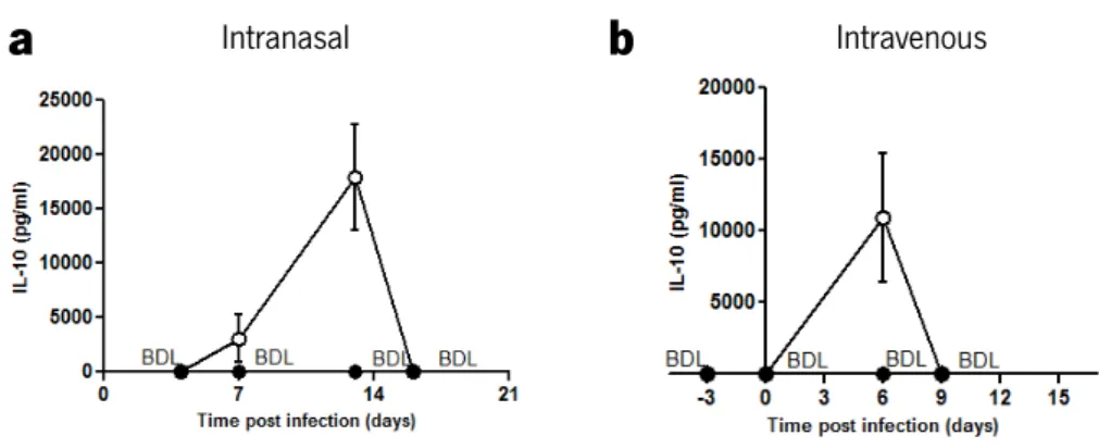
As shown in figure 1, in both models, the levels of circulating IL-10 in control littermates were below the detection limit (BDL) of the assay, at all the time points analyzed. After intranasal
infection (Fig. 1a), IL-10 levels could be detected at day 7 (3 days after zinc-mediated induction) in i-PMT-10 mice, being higher at 13 days post infection (the day of the end of the zinc treatment). At day 16 post infection, as in control PMT-10 mice, circulating IL-10 could no longer be detected in i-PMT-10 mice. Also, in the intravenous infection (Fig. 1b), i-PMT-10 mice showed transient increased IL-10 production, until 6 days after infection.
a
b
FIGURE 1. i-PMT-10 mice transiently over-produce IL-10 during intranasal or intravenous M. tuberculosis
infection. i-PMT-10 mice ({) were induced to over-express IL-10 (a) between 4 and 13 days after intranasal M. tuberculosis infection, or (b) 3 days before intravenous challenge until 6 days of infection. At indicated time points, IL-10 concentration was determined in the serum of i-PMT-10 and control littermates (z) by ELISA. Data represent the mean ± SEM from four mice per group from one experiment (a) or one representative of two independent experiments (b).
Over-expression of IL-10 during the early immune response against M. tuberculosis does not impact the outcome of either intranasal or intravenous infection.
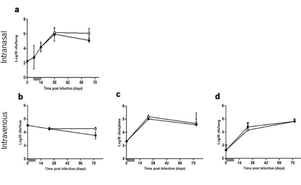
At various time points after M. tuberculosis infection, organs were collected and the bacterial loads were compared between PMT-10 and i-PMT-10. As shown in figure 2a, after intranasal infection, the bacterial load in lungs of non-induced littermates progressively increased till reaching approximately 6 logs on day 28, after which the bacterial growth was controlled at a stationary level, as previously described (143). Lungs from i-PMT-10 mice showed similar level of bacterial burdens than control mice, over the course of infection (Fig. 2a). Similarly, both control
Intravenous Intranasal
In tran as al In tra ven ou s
and i-PMT-10 mice infected via intravenous route were equally capable of controlling the bacterial burden in the liver, spleen and lung (Fig. 2b-d).
Our data suggest that an early over-expression of IL-10 does not impair the host resistance to M. tuberculosis infection.
FIGURE 2. Early IL-10 over-expression does not affect the resistance of i-PMT-10 mice to either intranasal or intravenous M. tuberculosis infection. i-PMT-10 mice ({) and control littermates (z) were infected intranasally (a) or intravenously (b-d) with 2,3 log10 CFU or 105 CFU of M. tuberculosis, respectively. i-PMT-10 mice over-expressed IL-10, as shown in figure 1, during the time-frame indicated (grey bar). Lungs (a and d), livers (b) and spleens (c) were removed at the indicated time points, and the number of viable bacteria were determined by plating serial dilutions of organ homogenates on Middlebrook 7H11 medium. Data represent the mean ± SEM from six (a) or five (b-d) mice per group from one experiment (a) or one representative of two independent experiments (b-d).
IL-10 over-expression affects the granuloma size developed after intravenous but not intranasal M. tuberculosis infection.
A protective immune response against pathogens needs to limit tissue damage while clearing the pathogen (144). IL-10 has been clearly associated to the regulation of inflammation and pathology prevention (79). Due to anti-inflammatory properties of IL-10, we were prompted to compare the extension of the inflammatory process in PMT-10 and i-PMT-10 infected with M. tuberculosis. For that, infected organs were collected, and morphometric analysis of the lesions was performed.
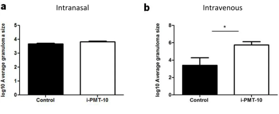
During the course of intranasal or intravenous M. tuberculosis infection, both i-PMT-10 mice and control littermates developed granulomatous lesions. These lesions, typical of mycobacterial infections, are defined as a focal accumulation of mononuclear cells, forming an organized structure with centrally located macrophages surrounded by a lymphocyte cuff (78). Thus, granulomas are thought to afford the T cell-macrophage contact and cooperation necessary for an effective antimycobacterial defense. On the other hand, granulomas displace and destroy adjacent lung tissue and may necrotize at the center leading to cavity formation, which is the most relevant sequelae of the chronic inflammatory response to M. tuberculosis (77). Importantly, lungs of i-PMT-10 mice, infected by the intranasal route, presented a granuloma size similar to that of control littermates, as shown in figure 3a. On the other hand, in livers of intravenously infected mice, it was evident that i-PMT-10 mice developed larger granulomas than non-induced PMT-10 mice (Fig. 3b), although histopathological analysis of the livers revealed that the 2 groups of mice developed similar number of granulomas per field after intravenous infection (data not shown).
These data show that an early over-expression of IL-10, while not impacting the bacterial growth in both infection models, induces, in the intravenous infection a larger inflammatory area at later time points.
FIGURE 3. Morphometric analysis of the granulomas developed by intranasally or intravenously infected mice. i-PMT-10 mice (white bars) and control littermates (black bars) were infected intranasally (a) or intravenously (b) with 2,3 log10 CFU or 105 CFU of M. tuberculosis, respectively. At 64 days (a) or 72 days (b) post infection morphometric analysis of granuloma was performed in lungs (a) and livers (b) sections of infected mice. Data represent the mean ± SEM from six (a) or five (b) mice per group from one experiment (a) or one representative of two independent experiments (b). * p < 0.05
Early IL-10 over-expression induces a delay in the expansion of several populations of leukocytes in the lymphoid organs of intranasal or intravenously infected mice.
We next investigated whether an intact cellular response was present in i-PMT-10 mice, despite the high levels of IL-10 observed in these animals, during the initiation of the response. To test this, we next evaluated, in the course of intranasal or intravenous infections, the kinetics of the cellular populations, previously described to be inhibited by IL-10.
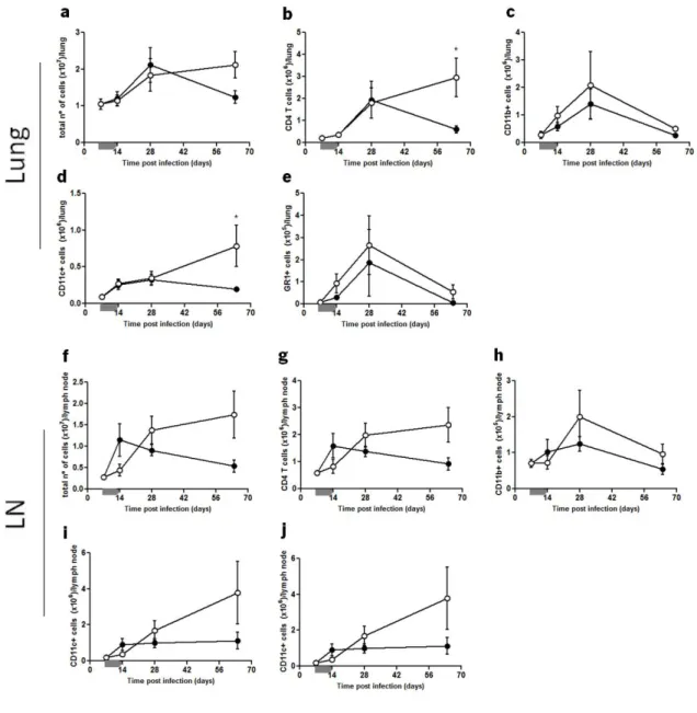
In the intranasal model, shown in figure 4, the total number of cells in the lungs from both induced and control PMT-10 mice followed similar kinetics over time (Fig. 4a). However, by analyzing, by flow cytometry, separately the major leukocyte subsets (Fig 4b-e), it became evident that the number of CD4 T cells and CD11c cells similar in both groups until day 28 post infection, were significantly increased at day 64 in i-PMT-10 mice (Fig. 4b,d). As for the LN, the total number of cells in control mice increased until day 14 post infection, before undergoing a slow decline until day 64 (Fig. 4f). However, in i-PMT-10 mice, the total number of cells increased at a slower rate and peaked at day 28, sustaining the same amounts throughout the infection.
This delay in i-PMT-10 mice was evident in all cell types analyzed (Fig. 4g-j) and the relative proportion of each cell type was not affected over time (data not shown).
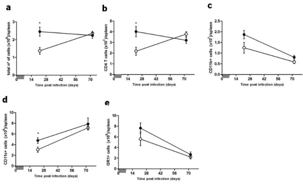
FIGURE 4. Kinetics of cellular expansion in the lungs and LN of intranasally infected mice. i-PMT-10 mice ({) and control littermates (z) were infected intranasally with 2,3 log10 CFU of M. tuberculosis. i-PMT-10 mice over-expressed IL-10, as shown in figure 1a, during the time-frame indicated (grey bar). At different time points, single cell suspensions from lungs (a-e) and LN (f-j) were prepared, stained with antibodies specific for CD4, CD11b, CD11c, GR-1 and analyzed by flow cytometry. The total number of cells of lungs (a) and LN (f) was determined using the Newbauer chamber. Data represent the mean ± SEM from six mice per group from one experiment. * p< 0.05.
The intravenous model resembled the major features of the intranasal model. As seen in figure 5a, after M. tuberculosis intravenous infection, while the total number of cells in the spleen of control mice roughly stabilized after day 21, the number of splenocytes in i-PMT-10 mice was significantly lower at day 21, after which it progressively increased until day 72, reaching the same amounts as in control mice. This difference was observed for the major cell population analyzed, being more pronounced in CD4 T cells and CD11c cells (Fig. 5b,d), in accordance with the observed in the intranasal model of infection. In addition, it seems that after intravenous infection, the increased granuloma size observed in the livers of i-PMT-10 mice at day 72 is not related to an increased cellular response at this time point, suggesting that perhaps IL-10 altered, instead, the organization/conformation of the granuloma.
FIGURE 5. Early over-expression of IL-10 induces a delay in the expansion of cells in the spleen of intravenously infected mice. i-PMT-10 mice ({) and control littermates (z) were infected intravenously with 105 CFU of M. tuberculosis. i-PMT-10 mice expressed IL-10, as shown in figure 1b, during the time-frame indicated (grey bar). At different time points, single cell suspensions from spleens were prepared and stained with antibodies specific for CD4, CD11b, CD11c and GR-1 and analyzed by flow cytometry. The total number of cells of speens (a) was determined using the Newbauer chamber. Data represent the mean ± SEM from five mice per group from one representative of two independent experiments. * p < 0.05.
Altogether, our data points out to a potential contribution of IL-10 over-expression to an initial delay on the cellular dynamics observed on the LN of intranasally infected i-PMT-10 mice and on the spleens of intravenously infected i-PMT-10 mice. Of note, this delay is timely related to the peak of IL-10 production observed in i-PMT-10 mice, under our experimental conditions. Interestingly, however, is the later increase of certain cellular populations observed in the intranasal model of infection in the lungs of i-PMT-10 mice. It is tempting to suggest that this increase might be an attempt of the immune response to overcome any down-modulating effects of IL-10.
After intranasal M. tuberculosis infection, early over-expression of IL-10 induces an increased expansion of activated CD4 T cells in both lungs and LN.
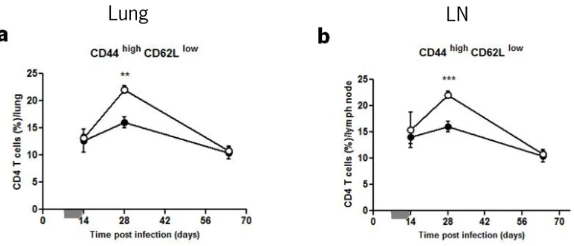
It has been described that activation of naïve T lymphocytes in the draining LN, which occurs at about 14 days upon intranasal or aerosol M. tuberculosis infection (145), and their subsequent migration to the site of infection, are essential for the induction/potentiating of host defensive mechanisms against M. tuberculosis (8). Since the delay observed in intranasally infected i-PMT-10 mice occurred at around day 14 and affected CD4 T cells, we hypothesized that the early over-expression of IL-10, between day 4 to 13 post intranasal infection, could be impairing the activation of CD4 T cells in the LN and lungs of i-PMT-10 mice. To test this hypothesis, we measured, by flow cytometry, the expression of CD44 and CD62L during the course of infection. We found that, in the CD4 gate, the CD44high CD62Llow population, indicative of
T cell activation, was significantly increased, in both LN and lungs of i-PMT-10 mice at 28 days post infection, as compared to littermates control (Fig. 6). Our results thus suggest that the early expression of IL-10 during M. tuberculosis infection, not only impair CD4 T cell activation, when IL-10 expression was at its peak, but instead seemed to potentiate it afterwards. Again, it is possible that this increased emergence of activated CD4 T cells occurred to counteract the delayed cellular expansion detected in the LN of intranasally infected i-PMT-10 mice.
Lung
LN
FIGURE 6. Early over-expression of IL-10 induces an increased expansion of activated CD4 T cells at 28 days post intranasal infection. i-PMT-10 mice ({) and control littermates (z) were infected intranasally with 2,3 log10 CFU of M. tuberculosis. i-PMT-10 mice were over-expressed IL-10, as shown in figure 1, during the time-frame indicated (grey bar). At different time points, single cell suspensions from lungs (a) and LN (b) were prepared and stained with surface and activation markers for flow cytometry analysis of activated CD44high CD62Llow CD4 T cells. Data represent the mean ± SEM from six mice per group from one experiment. ** p< 0.01, and ***p <0.001
Early IL-10 does not impair the expression of IFNγ and TNF during the course
of infection in either the intranasal or intravenous models.
Several studies demonstrate the requirement of IFNγ and TNF for macrophage activation and initial control of M. tuberculosis infection (61, 146). Since i-PMT-10 mice were able to control the infection by M. tuberculosis, despite the fact that IL-10 has been extensively described to suppress the production of the former cytokines, we asked whether IFNγ and/or TNF were normally expressed in infected i-PMT-10 mice.
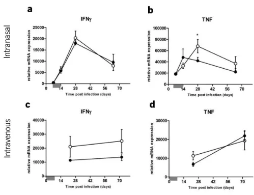
Semi-quantitative real-time PCR analysis showed that, after intranasal M. tuberculosis
infection, IFNγ expression in the lungs of control mice, started to progressively increase after day 7, reaching a peak at day 28, consistent with the influx of Th1 cells to the lung (8), before undergoing a slow decline until the end of the experiment (Fig. 7a). Infected i-PMT-10 mice showed identical amounts of IFNγ transcripts, in the lungs, over time. As shown in figure 7b, the expression of TNF in control littermates increased after intranasal M. tuberculosis infection peaking at day 14, before decreasing to near basal levels. However, in i-PMT-10 mice, TNF expression progressed more slowly until day 14, but continued to increase until day 28, reaching
In tran as al In tra ven ou s
significantly higher levels than control littermates. Interestingly, this high induction of TNF in the lungs of i-PMT-10 mice occurred at the time of increased activation of CD4 T cells, suggesting that 2 weeks after the IL-10 over-expression period, i-PMT-10 mice reacted with an increased inflammatory response.
After M. tuberculosis infection via the intravenous route, expression of IFNγ was highly induced in the spleens from both induced and control PMT-10 mice at day 21, maintaining the same level throughout the infection (Fig. 7c). The kinetics of TNF expression was also comparable in the spleens of both groups of mice (Fig. 7d), showing the inability of IL-10 to impair the induction of pro-inflammatory cytokines after M. tuberculosis infection in our experimental settings.
FIGURE 7. Early over-expression of IL-10 does not impair the expression of IFNγ and TNF after intranasal or intravenous infection. i-PMT-10 mice ({) and control littermates (z) were infected intranasally (a,b) or intravenously (c,d) with 2,3 log10 CFU or 105 CFU of M. tuberculosis, respectively. i-PMT-10 mice over-expressed IL-10, as shown in figure 1, during the time-frame indicated (grey bar). Gene expression of IFNγ (a,c) and TNF (b,d) was determined in lungs (a,b) or spleen (c,d) homogenates by real time PCR. Data represent the mean ± SEM from six (a,b) or five (c,d) mice per group from one experiment (a,b) or one representative of two independent experiments (c,d). * p < 0.05.
Taken together, the data presented in part 3.1 suggest that over-expression of IL-10, early after intranasal or intravenous M. tuberculosis infection, induces a delayed expansion of lymphoid populations that is not sufficient to affect the outcome of infection, probably due to the fact that IFNγ and TNF were not impaired in the time points analyzed. Regarding the intranasal model, whether the late, but increased, inflammatory response detected in the lungs of i-PMT-10 mice was critical or redundant for the resolution of infection is still not answered.
3.2. Effect of IL-10 over-expression late after infection by M.
tuberculosis: impact on the ongoing immune response.
The adaptive immune response generated in the individuals who are exposed to M. tuberculosis, although protective, does not induce sterilizing immunity. These individuals, therefore, remain latently infected, and are vulnerable to disease reactivation when their immune surveillance weakens, or when their immune response is compromised (3). Elevated levels of IL-10 were detected in individuals with active tuberculosis (117, 118). However, it is unclear whether IL-10 plays a role in promoting the reactivation of tuberculosis in chronic latent infected individuals. Since IL-10 has been shown to inhibit both T cell proliferation and IFNγ production we decided to investigate whether high amounts of IL-10 in a phase of infection where latency is established could compromise the immune response and increase host susceptibility.
To address this question, PMT-10 mice, infected via the intranasal or intravenous route by
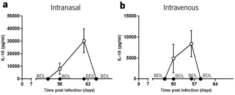
M. tuberculosis, were induced to over-express IL-10 during the late phase of infection, more precisely between days 53 to 62 in intranasal model, or between days 47 to 56, in intravenous model. At various time points thereafter, IL-10 was measured in the blood by immunoassay (Fig. 8). As expected, in both infection models, whereas IL-10 was not detected in the serum of control animals, in i-PMT-10 mice, circulating IL-10 was detected 3 days after zinc administration, specifically at day 56 or 50 after intranasal (Fig. 8a) or intravenous (Fig. 8b) infection, respectively, and was highly produced until the end of the induction period.
a
b
FIGURE 8. i-PMT-10 mice transiently over-produce IL-10 during the late phase of intranasal or intravenous M. tuberculosis infection. i-PMT-10 ({) mice were induced to over-express IL-10 between 53 and 62 days (a) or between 47 and 56 days (b) after intranasal or intravenous M. tuberculosis infection, respectively. At various time points IL-10 concentration was determined in the serum of i-PMT-10 and control littermates (z) by ELISA. Data represent the mean ± SEM from four mice per group from one experiment (a) or one representative of two independent experiments (b).
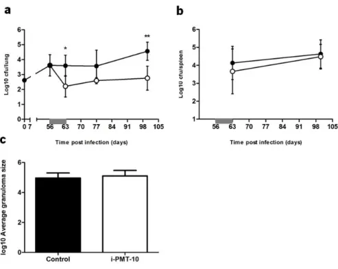
IL-10 over-expression during the late phase of M. tuberculosis infection resulted in different outcomes of infection, depending on the route of infection.
At various time points after M. tuberculosis infection, organs were collected and the bacterial loads were compared between PMT-10 and i-PMT-10 mice. After intranasal infection, non-induced PMT-10 mice were able to control the bacterial burdens in the lung during the course of infection, as expected (Fig. 9a). Surprisingly, i-PMT-10 mice displayed lower bacterial loads in the lungs as compared to control mice, after day 63 of intranasal infection and this difference was sustained until the end of the experiment, at 100 days post infection. As for progression of intranasal infection in the spleen, no differences were observed between the 2 groups of mice (Fig. 9b). Histopathological analysis of the lungs, performed at the end point of the experiment (100 days), showed similar areas of inflammatory process in both i-PMT-10 and control mice (Fig. 9c). These data are in line with the ones obtained upon early induction of IL-10 in the intranasal model of infection, although no increase in protection was observed before.
Intravenous Intranasal
FIGURE 9. Late over-expression of IL-10 increases the resistance of i-PMT-10 mice to intranasal M. tuberculosis infection. i-PMT-10 ({) and control littermates (z) were infected intranasally with 2,6 log10 CFU of M. tuberculosis H37Rv. i-PMT-10 mice over-expressed IL-10, as shown in figure 8a, during the time-frame indicated (grey bar). Lungs (a) and spleens (b) were removed at the indicated time points, and number of viable bacteria was determined by plating serial dilutions of organ homogenates on Middlebrook 7H11 medium. (c) At 100 days post infection, morphometric analysis of the granulomas was performed in lungs sections of infected mice. Data represent the mean ± SEM from six mice per group. * p < 0.05, **p < 0.01.
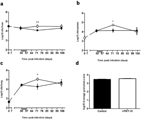
In contrast, i-PMT-10 mice challenged with M. tuberculosis by the intravenous route developed increased bacterial loads at day 72, as compared to control mice, in the liver, spleen and lung (Fig. 10). Interestingly, at day 105 post infection, i-PMT-10 mice were able to decrease the bacterial replication until the levels observed in control mice, therefore suggesting that, in this model, the over-expression of IL-10 induced a transient increase in susceptibility, which was recovered as soon as the effect of IL-10 waned off.
Despite the increased bacterial load in the livers of i-PMT-10 mice, the number (data not shown) and size (Fig. 10d) of granuloma per field at 72 days was similar to that of control mice.
FIGURE 10. i-PMT-10 mice are transiently more susceptible to intravenous M. tuberculosis than control littermates. i-PMT-10 ({) and control littermates (z) were infected intravenously with 105 CFU of M.
tuberculosis. i-PMT-10 over-expressed IL-10, as shown in figure 8b, at the time-frame indicated (grey bar). Livers (a), spleens (b) and lungs (c) were removed at the indicated time points, and number of viable bacteria was determined by plating serial dilutions of organ homogenates on Middlebrook 7H11 medium. (d) After 72 days post infection morphometric analysis of average granuloma size was performed in livers sections of infected mice. Data represent the mean ± SEM from five mice per group from one representative of two independent experiments. * p< 0.05 and ** p< 0.01.
Considering that the over-expression of IL-10 during the late phase of M. tuberculosis
infection resulted in different outcomes of infection, depending on the route of infection, we will from now on analyze each experiment separately, in an effort to further dissect the observed differences.