BJRS
RADIATION SCIENCES
06-03 (2018) 01-13Evaluation of some dosimetric properties of a dedicated
plane parallel ionization chamber for radiotherapy
electron beams
J. O. Silva
a; F. B. C. Nonato
b; F. G. A. Sampaio
c; L. V. E. Caldas
daUniversidade Federal de Goiás/Instituto de Física, Campus Samambaia, Goiânia, GO, Brazil b Centro Universitário Anhanguera de Santo André, Santo André, SP, Brazil
c Instituto de Radioterapia e Megavoltagem, Ribeirão Preto, SP, Brazil
d Instituto de Pesquisas Energéticas e Nucleares (IPEN-CNEN/SP), São Paulo, SP, Brazil
ABSTRACT
Ionization chambers are the reference detectors for electron beam dosimetry. In this paper a dedicated radio-therapy plane parallel ionization chamber manufactured with low cost materials is presented for dosimetry in electron beams. The ionization chamber tested has a sensitive volume of 0.4 cm³. Both the collecting electrode and the guard ring were painted with a homogeneous mixture of nail polish and graphite. The dedicated ioniza-tion chamber presented a linear response with electron absorbed dose within the range 0.5 to 8.0 Gy, an increase of its response with the field size increasing, an angular dependence within ±5°, as recommended by interna-tional standards, and a polarity effect of 0.78% according to the field size. Considering the results obtained, it is possible to conclude that the plane parallel ionization chamber tested in this work presents potential use for elec -tron beam dosimetry in clinical routine.
Keywords: ionization chambers, electron beams, radiation dosimetry, radiotherapy.
1.
INTRODUCTION
The most widespread use of ionizing radiation by humans is found in medical field applications. Among them, one of the major uses of radiation in medicine is in radiotherapy, that is applied to eliminate or inhibit the cancerous tumors by using high energy X-rays or electron beams (telether-apy). It is important to guarantee the maximum radiation dose to the disease and to avoid unneces-sary irradiation of healthy tissues. This is accomplished with a well established dose planning fol-lowed by the radiation protection of patients and a high accuracy quality control program [1].
The dosimetry of electron beams in radiotherapy can be performed either by plane parallel or by cylindrical ionization chambers for any electron energy range [2,3], but plane parallel ion chambers are mandatory for electron beam energies lower than 10 MeV [3-5]. Ionization chambers are the reference detectors for electron beam dosimetry, and it is essential to apply correction factors to their response as for humidity, temperature and pressure, ion recombination and polarity changes
[4-8].
At the Calibration Laboratory of IPEN the research group has developed several ionization chambers for radiation dosimetry in medical irradiation equipment (therapy and diagnostic energy range) and laboratory applications (as secondary standards) made with cheap accessible materials with high metrological performance [9-13].
The dedicated plane parallel ionization chamber utilized in this work has been characterized previously in standard electron and photon beams, as reported by Nonato et al. [10], and it uses a homogeneous mixture of nail polish and powdered graphite applied to a PMMA base as collecting electrode. It presents a polarity effect of 4% for LINAC electron beams with nominal energy of 6 MeV and around 3% for 12 MeV electron beams, both for the depth of 0.7Rp (practical range in
centimeters). These results on the polarity effect are less than the value of 5% for the polarity effect of the Advanced Markus ionization chamber when irradiated with electron beams with nominal en-ergy of 4 MeV[14]. However, the polarity effect is not a concern if the ion chamber is always uti-lized at the same conditions of polarity and tension [15]. Besides the polarity effect, other tests are necessary to evaluate its use in clinical electron beam dosimetry [16].
The ionization chamber investigated was the first one developed by the IPEN research group us-ing the described mixture for the collectus-ing electrode. The most common material utilized for the collecting electrodes of the IPEN ionization chambers was a uniformly spread graphite spray in var-ious base types [11-13,17]. The mixture of colorless nail varnish and powered graphite was an alter-native way for collecting electrode material because of its easy acquisition and application on a PMMA base with an excellent electrical conductivity [18].
The objective of this work was to verify the performance of the dedicated plane parallel ioniza-tion chamber in clinical dosimetry tests with high energy electron beams as linearity of the response with absorbed dose in a PMMA phantom, angular dependence, field size dependence and the polar-ity effect as a function of the field size in a PMMA water equivalent phantom.
2.
MATERIALS AND METHODS
The ionization chamber tested in this work is a vented chamber with a sensitive volume of 0.4 cm³ and a distance of 0.2 cm between the electrodes. A scheme of this ionization chamber with its dimensions is presented in Figure 1. The chamber body, the collecting electrode and the guard ring are made of PMMA with a density of 1.17 g/cm³. The collecting electrode and the guard ring were painted with a homogeneous mixture of colorless nail polish and powdered graphite. More details about how the painting of the mixture was applied can be found in [18].
To establish the electric field, an aluminized polyester film with 1.87 mg/cm² of superficial den-sity was utilized as the entrance window. The primary characterization tests were made at the Cali-bration Laboratory (LCI) at IPEN/CNEN-SP [18].
A Siemens Primus linear accelerator at the Radiotherapy and Megavoltage Institute - IRMEV, Ribeirão Preto, Brazil, with electron beam energy of 7 MeV, was utilized as the irradiator system. A dose rate of 3 Gy/min, specified for dose to PMMA, and a source to surface distance of 100 cm were selected for all irradiations. The linear accelerator electron beams dosimetry was accom-plished according to the IAEA protocol [5]. Measurements of linearity of the response with ab-sorbed dose, angular dependence, field size dependence and the polarity effect as function of the field size were performed to investigate the ionization chamber response. The reference material for the absorbed dose was the PMMA.
A phantom made of various PMMA slab thicknesses with 30 × 30 cm2 was utilized to localize
number 4372, of the IRMEV, was utilized to polarize and to collect the readings of the ionization chamber. The measurements were corrected for the environmental standard conditions of tempera-ture and pressure, as described in IAEA TRS 398 for this radiation detector type [5]. In Figure 2 is presented a general experimental setup scheme for the measurements performed in this work.
Figure 2: Diagram of the general experimental setup for the measurements obtained with
the ionization chamber in high energy electron beams.
The un-certainties utilized in this work were from both types, A and B, as defined in the GUM [19]. For type A uncer-tainties the mean values and standard deviations from the measurements performed with the ioniza-tion chamber were considered. The resoluioniza-tions of the CNMC electrometer and auxiliary equipment (thermometer and barometer) were considered for type B uncertainties.
3.
RESULTS AND DISCUSSION
The ionization chamber was tested according to international recommendations [4,5,16]. The ionization chamber was pre-irradiated during 15 minutes before the irradiations to achieve response stability conditions [10]. The ionization chamber tested in this work presents a reproducible response in time with accuracy within ±0.5% limit variation [18] as recommended in the IEC
60731:2011 standard for reference-class dosimeters used in radiotherapy [16]. Other tests of response variation of the dedicated ionization chamber with time were presented previously [10].
The dedicated plane-parallel ionization chamber may be submitted to the maximum polarities of ±400 V, but the work tension of +300 V was chosen because this value was applied during the ionization chamber calibration at IPEN [16].
3.1. Linearity curve
This ionization chamber presented a linear response in an absorbed dose range of 0 to 0.6 Gy when submitted to a 90Sr+90Y standard source in a source-chamber distance of 11 cm [10]. In order
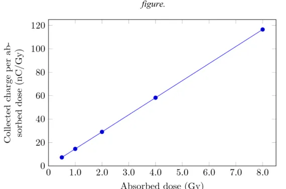
to verify the dedicated chamber response in a wider range, the linearity curve (response × absorbed dose) was obtained taking 3 measurements for each absorbed dose ranging from 0.5 Gy to 8.0 Gy for 7 MeV electron beams. The measurements for this test were performed with a 10 × 10 cm2 field
size, and at the depth of maximum dose. The ionization chamber was located at a reference source-chamber distance of 100 cm, and was polarized with +300 V.
The linearity curve is presented in Figure 3 where each point represents the mean value of 3 measurements, each set of three measurements with a repeatability around 0.5%. The correlation coefficient for the curve was calculated using the method of linear curve fitting based on least-squares fit, and it was exactly 1.000.
Figure 3: Linearity curve of the ionization chamber: response as function of the absorbed
dose, using a 7 MeV electron beam. The maximum uncertainty was 0.05%, not visible in the figure.
3.2. Angular dependence
The dependence of the ionization chamber response with the central axis angular orientation was verified in the angular dependence test. The ionization chamber entrance window was located in the PMMA phantom at the depth of maximum dose, at the reference distance of 100 cm. The ir-radiations were performed in a 7 MeV electron beam, for an absorbed dose of 1 Gy and a 10 × 10 cm2 field, which was the smallest field size available for the electron beams utilized in the
present work.
The radiation incident angle was changed from 0° to ±5°, in steps of 1°, in relation to the posi-tion of perpendicular incidence of the radiaposi-tion beam axis. The clockwise orientaposi-tion was taken as the positive one, and for each angle 3 measurements were carried out. In Figure 4 the ionization chamber performance in this test is presented, and each point represents the normalized response to the 0° mean value. All the uncertainties presented are the overall uncertainties with k = 2. Figure 4 shows that the results are within the value of ±1.0% for the tilt of ±5°, as recommended by IEC [16].
Figure 4: Angular dependence test results for the ionization chamber when exposed to a
3.3. Field size dependence
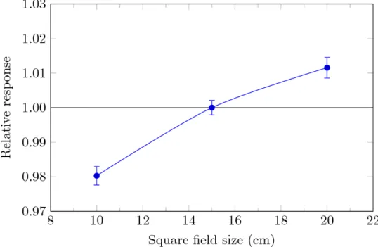
The variation of the ionization chamber response with the amount of scattered radiation was verified in the field size dependence test for 7 MeV electron beam. The detector was polarized with +300 V and it was positioned at the depth of maximum dose in the PMMA phantom, at the center of the radiation field, 100 cm from the electron source. This test was performed for an absorbed dose value of 1 Gy delivered at the depth of maximum dose, where the chamber reference point was located. In this study, field sizes with areas of 10 × 10, 15 × 15 and 20 × 20 cm2, defined by electron
applicators, were applied, and the mean value of 3 measurements for each field size was normalized in relation to the 15 × 15 cm2 field mean result. The results for this test are presented in Figure 5, in
which an increase in the ionization chamber response according to the increase of field dimensions can be observed. This occurs due to the higher amount of lateral scattered radiation in a larger PMMA phantom surface. It can be seen that the limit of response variation for this test is within ±2.0 %, as required by the IEC 60731 standard [16].
Figure 5: Field size dependence of the response of the ionization chamber utilized in this
work for 7 MeV electron beam. The fields are defined by electron applicators and the results were normalized to the mean result obtained for 15 × 15 cm² field.
3.4. The polarity effect as function of the field size
For this analysis, the same setup applied to the field size dependence test was utilized. The ion-ization chamber was polarized with ±300 V, to obtain the polarity ratio (Q-/Q+), and one more field
size (25 × 25 cm2) was added to this study. Figure 6 shows the polarity effect as function of the field
size for the ionization chamber tested in the present study. The ionization chamber presents a field size dependence of 0.78% with the change of polarizing voltage over the entire measured range of field sizes.
It can be seen that when the field dimensions are increased the polarity effect increases too. The results obtained agree with those published by Ramsey et al. [20], except for the 25 × 25 cm2 field
that has a lower value in comparison with that related to the 20 × 20 cm2 field. A similar behavior
was obtained for the commercial plate-parallel PS033 chamber. It presented a field size dependence of the polarity effect around 0.5% over a field size range from 4 × 4 cm2 to 25 × 25 cm2, when
irra-diated with a 6 MeV electron beam [21].
4.
CONCLUSION
A dedicated plane parallel ionization chamber was characterized in clinical electron beams of 7 MeV in terms of linearity of the response with absorbed dose, angular dependence, field size de-pendence and the polarity effect with the field size. The dedicated ionization chamber presented a linear response with the absorbed dose in a large dose range. The ionization chamber angular de-pendence was within international recommendations of ±1% for ±5°. It is sensitive to the scattered radiation increase due to changes in the field size, in accordance with an expected output factor curve. Polarity effects related to the change in radiation field size are present, but its behavior is similar to that of commercial ionization chambers. From the results obtained, it is possible to con-clude that the ionization chamber tested in this work presents potential use for electron beam dosimetry in clinical routine.
ACKNOWLEDGMENT
The authors are thankful to the Brazilian agencies CNPq (Project 301335/2016-8), FAPESP (Project 2008/57863-2) and MCTIC (Project INCT for Radiation Metrology in Medicine, 573659/2008-7), for partial financial support.
REFERENCES
[1] d’ERRICO, F. Dosimetric issues in radiation protection of radiotherapy patients. Radiat Prot
Dosim, v. 118, p. 205-212, 2006.
[2] MUIR, B. R.; McEWEN, M. R. Technical note: On the use of cylindrical ionization chambers for electron beam reference dosimetry. Med Phys, v. 44, n. 12, p. 6641-6646, 2017.
[3] ALMOND, P. R.; BIGGS, P. J.; COURSEY, B. M.; HANSON, W. F.; HUQ, M. S.; NATH, R. AAPM's TG-51 protocol for clinical reference dosimetry of high-energy photon and electron beams. Med Phys, v. 26, n. 9, p. 1847-1870, 1999.
[4] IAEA – International Atomic Energy Agency. The use of plane-parallel ionization chambers
in high-energy electron and photon beams: An international Code of Practice for dosimetry. IAEA TRS 381, Vienna: IAEA, 1995.
[5] IAEA – International Atomic Energy Agency. Absorbed dose determination in external
beam radiotherapy: An international code of practice for dosimetry based on standards of absorbed dose to water. IAEA TRS 398, Vienna: IAEA, 2000.
[6] PEARCE, J.; THOMAS, R.; DuSAUTOY, A. The characterization of the Advanced Markus ionization chamber for use in reference electron dosimetry in the UK. Phys Med Biol, v. 51, n 3, p. 473-483, 2006.
[7] GERBI, B. J.; KHAN, F. M. The polarity effect for commercially available plane-parallel ionization chambers. Med Phys, v. 14, n. 2, p. 210-215, 1987.
[8] HAVERCROFT, J. M.; KLEVENHAGEN, S. C. Ion recombination corrections for plane-parallel and thimble chambers in electron and photon radiation. Phys Med Biol, v. 38, n. 1, p. 25-38, 1993.
[9] NONATO, F. B. C.; SAKURABA, R. K.; da CRUZ, J. C.; CHIARA, A. C. M.; VERNUCIO, S. L.; MENEGUSSI, G; CALDAS, L. V. E. Development and characterization tests of a homemade ionization chamber with silver collecting electrode for use in electron beams.
Biomed Phys Eng Express, v. 2, n. 1, p. 015011, 2016.
[10] NONATO, F. B. C.; SAKURABA, R. K.; da CRUZ, J. C.; CALDAS, L. V. E. Characterization tests of a new parallel plate ionization chamber for use in electron beams.
Radiat Phys Chem, v. 104, p. 244-247, 2014.
[11] SILVA, J. O.; NONATO, F. B. C.; CALDAS, L. V. E. Characterization tests of a homemade ionization chamber in mammography standard radiation beams. Radiat Phys Chem, v. 95, p. 151-153, 2014.
[12] PERINI, A. P.; NEVES, L. P.; FERNADEZ-VAREA, J. M.; CASSOLA, V. F.; KRAMER, R.; KHOURY, H. J.; CALDAS, L. V. E. A new parallel-plate graphite ionization chamber as a
60Co gamma radiation reference instrument. Radiat Phys Chem, v. 95, p. 106-108, 2014.
[13] NEVES, L. P.; PERINI, A. P.; CALDAS, L. V. E. Development and characterization of a new cylindrical ionization chamber for dosimetry of 60Co beams. IEEE Trans Nucl Sci, v. 60,
p. 712-715, 2013.
[14] PEARCE, J.; THOMAS, R.; DuSAUTOY, A. The caracterization of the Advanced Markus ionization chamber for use in reference electron dosimetry in the UK. Phys Med Biol, v. 51, p. 473-483, 2006.
[15] IAEA – International Atomic Energy Agency. Calibration of reference dosimeters for
external beam radiotherapy. IAEA TRS 469, Vienna: IAEA, 2009.
[16] IEC – International Electrotechnical Commission. Medical electrical equipment
-Dosimeters with ionization chambers as used in radiotherapy. IEC 60731, Brussels: IEC,
2011.
[17] YOSHIZUMI, M.; CALDAS, L. V. E. A new ring-shaped graphite monitor ionization chamber. Nucl Instrum Meth Phys Res A, v. 219, n. 1, p. 207-210, 2010.
[18] NONATO, F. B. C. Design, construction and characterization of reference systems for electron beams of clinical accelerators. PhD thesis. Universidade de São Paulo, 2014. (In Portuguese)
[19] JCGM - Joint Committee for Guides in Metrology. Evaluation of measurement data —
Guide to the expression of uncertainty in measurement. JCGM 100:2008, Paris: JCGM,
2008.
[20] RAMSEY, C. R.; SPENCER, K. M.; OLIVER, A. L. Ionization chamber, electrometer, linear accelerator, field size, and energy dependence of the polarity effect in electron dosimetry.
[21] WILLIAMS, J. A.; AGARWAL, S. K. Energy-dependent polarity correction factors for four commercial ionization chambers used in electron dosimetry. Med Phys, v. 24, n. 5, p. 785-790, 1997.