Pneumocephalus is a rare consequence of epidural anesthesia, which may occur following inadvertent or unidentified dural puncture when the loss of resistance to air technique is applied to identify the epidural space. Headache is the most common symptom presented in this condition, usually with sudden onset. This case report describes an unusual presentation of diffuse pneumocephalus after an unidentified dural puncture. The patient (male, 67 years old) was submitted to epidural catheter placement for the treatment of acute exacerbation of ischemic chronic pain using loss of resistance to air technique. No cerebrospinal fluid or blood flashback was observed after needle withdrawal. Shortly after the intervention, the patient presented symptoms of lethargy, apathy, and hypophonia, which are not commonly associated with pneumocephalus. No motor or sensory deficits were detected. Cranial computed tomography showed air in the frontal horn of the left ventricle, subarachnoid space at interhemispheric fissure and basal cisterns, confirming the diagnosis of diffuse pneumocephalus. The patient remained under vigilance with oxygen therapy and the epidural catheter left in place. After 24 hours, cranial computed tomography showed air in the temporal and frontal horns of the left ventricle, with no air in the subarachnoid space. The patient presented no neurological signs or symptoms at this time. Although headache is the most common symptom presented in reported cases of pneumocephalus, this case shows the need for the clinician to be aware of other signs and symptoms that may be indicative of this condition, in order to properly diagnose and treat these patients.
Key words: Pneumocephalus, continuous epidural analgesia, ischemic chronic pain, loss-of-resistance to air technique, dural puncture, headache, unusual presentation
Pain Physician 2017; 20:E329-E334
Case Report
Pneumocephalus Following Unidentified Dural
Puncture: A Case Report with an Unusual
Neurological Presentation
From: 1 Anesthesiology, Intensive Care
and Emergency Department, Centro Hospitalar do Porto, Porto, Portugal;
2Neurosciences Department, Neurology,
Centro Hospitalar do Porto, Porto, Portugal Address Correspondence: Helena Dutra Figueira, MD Centro Hospitalar do Porto Anesthesiology, Intensive Care and
Emergency Department Largo Prof. Abel Salazar, 4099-001, Porto, Portugal E-mail: helenadfigueira@gmail.com Manuscript received: 04-28-2015 Revised manuscript received:
01-12-2016 Accepted for publication: 02-03-2016 Free full manuscript: www.painphysicianjournal.com
Helena Dutra Figueira MD1, Joana Guimarães MD1, Ana Luísa Sousa MD2, and Ana Margarida Regalado MD1
P
neumocephalus is a rare consequence of epidural anesthesia, resulting from injection of air into the subarachnoid or subdural space and cranial migration (1,2). It occurs when the loss- of-resistance to air (LORA) technique is used to identify the epidural space. There have been some reports in the literature of subarachnoid pneumocephalus after an epidural catheter placement, following inadvertent (2-4) or unidentified dural puncture (5-8).Continuous epidural analgesia with a tunneled catheter may be an option for pain management in some situations of chronic pain. We report a case of a patient with an acute exacerbation of ischemic chronic
pain that developed diffuse pneumocephalus after an unidentified dural puncture during epidural catheter placement using the LORA technique, with unusual neurological symptoms. Written informed consent was obtained from the patient for publication of this case report.
C
aseR
epoRtA 67 year-old man presented for analgesia with continuous epidural block due to acute exacerbation of chronic left lower extremity ischemic pain refrac-tory to noninvasive pain therapy. Past medical his-tory included severe peripheral artery disease
(Leriche-relief of symptoms after 4 hours. The epidural catheter was left in place maintaining effective continuous epi-dural analgesia the following days.
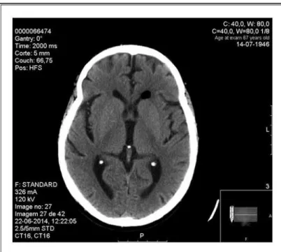
Cranial computed tomography performed 24 hours after the episode revealed air in the temporal and fron-tal horns of the left ventricle and no air in the subarach-noid space (Fig. 2). Clinical evaluation performed at this time showed no neurological signs or symptoms.
D
isCussionPneumocephalus is defined as the presence of gas within any of the intracranial compartments, either intra- or extra-axial, including the intraventricular, in-traparenchymal, subarachnoid, subdural, and epidural spaces (11).
The development of this condition requires a con-nection between the central nervous system and the external environment, and air must traverse through this passage. The postulated mechanisms for air entrap-ment include a ‘‘ball-valve’’ mechanism where air is forced through a dural rent but does not escape and an “inverted bottle” mechanism where drainage of spinal fluid leads to a negative intracranial pressure gradient being replaced by the influx of air (11). Pneumocepha-lus is most frequently associated with disruption of the skull after trauma (12) or surgical procedures, among which craniotomies are the leading causes (11). There are other less common causes, such as neoplasms caus-ing tumor erosion through the skull or skull base, in-fection with gas producing organisms, diagnostic and therapeutic procedures such as the lumbar puncture, or even can occur spontaneously through an osseous defect in the pneumatized temporal bone (13). It is also a rare but potential complication of epidural anesthesia that can occur after accidental dural puncture.
The most common way of identifying the epidural space for neuroaxial blocks is the loss-of-resistance technique using either an air or saline-filled syringe (14). Although the best method remains controversial, there is a tendency towards the increased use of saline (15,16). Potential complications with the use of the LORA technique include dural puncture with or without postdural puncture headache (PDPH), pneumocepha-lus, spinal cord and nerve-root compression, subcutane-ous emphysema, paresthesia, and incomplete analgesia (17). On the other hand, some advocate that the LORA technique improves the ability to identify cerebrospi-nal liquid in cases of accidental dural puncture (6). A meta-analysis of 4,422 patients failed to show overall risk differences for adverse outcomes (risk of difficult Fontaine classification stage IV), major right lower limb
amputation, CREST (calcinosis, Raynaud phenomenon, esophageal dysmotility, sclerodactily. and telangiecta-sia) syndrome, cardiovascular risk factors (hyperten-sion, type 2 diabetes, dyslipidemia), and ischemic heart disease.
A lumbar epidural with a tunneled catheter was placed under strict aseptic conditions, with the patient in the sitting position and properly monitored accord-ing to the American Society of Anesthesiologists stan-dards (9). A median approach using a G18 Tuohy needle to identify the epidural space with the LORA technique was successful on the second attempt. The catheter was inserted at the L3-L4 interspace and was advanced 4 cm into the epidural space. The volume of air used to iden-tify the epidural space was not stated in the records. Immediately after catheter insertion the patient expe-rienced a feeling of light-headedness, accompanied by pallor, diaphoresis, and bradycardia. He was positioned in lateral decubitus while the needle was still in place, recovering completely after repositioning. After the needle was withdrawn, no cerebrospinal fluid or blood flashback were observed during catheter aspiration. Af-ter the test dose (3 mL of lidocaine 2% with 1:200,000 epinephrine), 5 mL of lidocaine 1% were administered, resulting in analgesic block without evidence of sub-arachnoid block. A drug infusion balloon with an ad-justable infusion of ropivacaine 0.2% was connected to the epidural catheter.
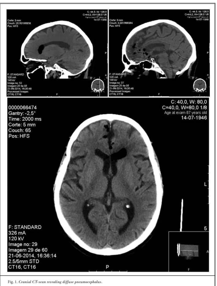
About 30 minutes after epidural insertion, the patient became apathetic and with slowed responses, without ever experiencing loss of consciousness. On observation, the patient was spontaneously awake but with diminished attention and lack of verbal or motor initiative. Although the patient showed slowing and prolonged speech latency, there was no language dis-turbance. The patient also presented a marked facial inexpression and hypophonia. Cranial nerve exam was otherwise normal and there were no motor or sensory deficits. Cranial computed tomography (CT) scan was performed immediately, revealing multiple small air bubbles with infracentimetric diameter in the subarach-noid space, at the interhemispheric fissure and basal cisterns, and a bigger air collection in the frontal horn of the left lateral ventricle, with an estimated volume of 1.52 cm3, according to the ABC/2 score (10). There were no signs of ischemic or hemorrhagic lesions (Fig. 1). Diagnosis of diffuse pneumocephalus was made. The patient remained under vigilance on bed rest with a high concentration oxygen face mask and showed
catheter insertion, incidence of intravascular catheter insertion, incidence of paresthesia, risk of accidental dural puncture, PDPH, and incidence of partial block) between the different mediums (liquid versus air) for the obstetric population (14). Instead, it revealed that the loss-of-resistance technique with liquid reduces the incidence of PDPH in chronic pain patients (14,18).
Pneumocephalus may arise when the LORA technique is used to identify the epidural space. The air used during LORA may gain access to the subarachnoid or subdural space by direct injection of air through a dural and arachnoid defect following an accidental dural tap, although pneumocephalus can also occur without evi-dence of dural puncture (1,18). In our case, there was no obvious dural puncture and no evidence of cerebrospinal fluid outflow via the needle or during catheter aspiration. It may have occurred during the insertion of the needle at the first or second attempt of epidural space identification, therefore being responsible for the presyncope symptoms described, or at the time the patient was positioned in lateral decubitus, lacerating the dura after catheter insertion and before needle withdraw. The catheter was prop-erly positioned in the epidural space since the infusion of local anesthetic promoted adequate analgesia without signs of spinal blockade.
The incidence of pneumocephalus is unknown and may be underestimated, since patients may be asymptomatic (1). There are no typical signs or symptoms, but headache is the most fre-quent presentation, usually with sudden onset (2) of a severe frontal headache (19). Gradual improvement of this symptom over
Fig. 2. Cranial CT-scan performed 24 hours after the epidural catheter
placement.
a few days is expected (20). Headache due to pneumocephalus is distinct from the classical PDPH, whose typical description is moderate to severe postural pain, which begins 24 – 48 hours after dural puncture (1). The first is believed to be caused by cranial migration of air causing meningeal irritation (1), while the PDPH is related to loss of cerebrospinal fluid after dural puncture leading to intra-cranial hypotension (21). In addition, the trapped expansion of intracranial air due to a ball valve effect resulting in mass effect can precipitate signs of a space-occupying lesion (focal neurologic deficits including cranial nerve palsies, hemiparesis, or hemiplegia) or increased intracranial pressure (vomit-ing, seizures, lethargy) and cardiovascular instability may arise (7). Symptoms duration and intensity depend on the distribution and amount of intracranial air (7). Also, a minority of patients describe “bruit hydro-aerique,” which is a characteristic sign of pneumocephalus (22), being described as a splashing noise heard by the patient on head movement. Our patient developed lethargy, apathy, and hypophonia, which are not com-monly associated with pneumocephalus, according to our best knowledge. We also speculate that the presyncope state could be an early sign of this complication as long as it may also be associated with vegetative symptoms and changes in blood pressure (19).
A cranial computed tomography scan was immediately performed for differential diagnosis between iatrogenic and other causes of brain injury. The presence of air density areas within the cranial cavity on computed tomography scan confirmed the diagnosis. Time course to symptoms resolu-tion was similar to other cases described in the literature, usually occurring over the course of several hours, while imaging find-ings may persist for over 7 days (5). Pneu-mocephalus usually resolves spontaneously after 3 – 5 days (2). Treatment is symptomatic with bed rest, hydration, analgesics, and caf-feine may be appropriate. Oxygen therapy facilitates denitrogenation and reabsorption of the air collected (6). Blood patches are
ineffective in pneumocephalus-induced headache and surgery is recommended only in tension pneumocepha-lus (7).
The majority of reported cases describing pneumo-cephalus after epidural technique occurs in obstetric patients (1-4,7). Furthermore, the most predominant symptom associated is headache (1-8). This unusual case emphasizes the need for the clinician to be aware of other symptoms that may lead to the diagnosis and appropriate treatment of these patients.
a
CknowleDgmentsAuthor Contributions: Drs. Helena Dutra Figueira, Joana Guimarães, Ana Luísa Sousa, and Ana Margarida Regalado had full access to all the data in the case report and take responsibility for the integrity of the data.
Drs. Helena Dutra Figueira, Joana Guimarães, Ana
Luísa Sousa, and Ana Margarida Regalado managed the literature searches and summaries of previous re-lated work and wrote the first draft of the manuscript. Drs. Helena Dutra Figueira, Joana Guimarães, Ana Luísa Sousa, and Ana Margarida Regalado provided revision for intellectual content and final approval of the manuscript.
The authors wish to thank editorial board of Pain Physician for review and criticism in improving the manuscript.
Disclaimer: There was no external funding in the preparation of this manuscript.
Conflict of interest: Each author certifies that he or she, or a member of his or her immediate family, has no commercial association (i.e., consultancies, stock own-ership, equity interest, patent/licensing arrangements, etc.) that might pose a conflict of interest in connection with the submitted manuscript.
R
efeRenCes1. Nafiu OO, Urquhart JC. Pneumo-cephalus with headache complicating labour epidural analgesia: Should we still be using air? Int J Obstet Anesth 2006; 15:237-239.
2. Velickovic IA, Rostislav P. Pneumo-cephalus complicated by postdural puncture headache after unintention-al durunintention-al puncture. Anesth Anunintention-alg 2007; 104:747-748.
3. Braga AFA, Potério GMB, Cremonesi E, David LH, Schimidtt R. Pneumo-cephalus after epidural anesthesia. Case report. Rev Bras Anestesiol 2001; 51:325-330.
4. Barbosa FT, Cunha RM, Rocha APC, Silva Junior HJL. Intraventricular pneu-mocephalus after accidental perfora-tion of the dura mater. Case report.
Rev Bras Anestesiol 2006; 56:511-517.
5. Syed SF, Garcon E. A Case of diffuse subarachnoid pneumocephalus after epidural injection. OMICS J Radiology 2013; 2:120.
6. McMurtrie R Jr., Jan R. Subarachnoid pneumocephalus: A rare complication of epidural catheter placement. J Clin
Anesth 2002; 14:539-542.
7. Gómez-Ríos MA, Fernández-Goti MC. Pneumocephalus after inadvertent du-ral puncture during epidudu-ral anesthe-sia. Images in anesthesiology.
Anesthe-siology 2013; 118:444.
8. Guarino AH, Wright NM. Pneumoceph-alus after lumbar epidural steroid injec-tion. Pain Physician 2005; 8:239-241. 9. ASA standards for basic anesthetic
mon-itoring: www.asahq.org/~/media/sites/ asahq/files/public/resources/standards- guidelines/standards-for-basic-anes-thetic-monitoring.pdf
10. Kothari RU, Brott T, Broderick JP, Barsan WG, Sauerbeck LR, Zuccarello M, Khoury J. The ABCs of measuring intracere-bral hemorrhage volumes. Stroke 1996; 27:1304-1305.
11. Schirmer CM, Heilman CB, Bhardwaj A. Pneumocephalus: Case illustrations and review. Neurocrit Care 2010; 13:152-158. 12. Keskil S, Baykaner K, Ceviker N, Isik S,
Cengel M, Orbay T. Clinical significance of acute traumatic intracranial pneumo-cephalus. Neurosurg Rev 1998; 21:10-13. 13. Abbati SG, Torino RR. Spontaneous
in-traparenchymal otogenic pneumoceph-alus: A case report and review of litera-ture. Surg Neurol Int 2012; 3:32.
14. Schier R, Guerra D, Aguilar J, Pratt GF, Hernandez M, Boddu K, Riedel B. Epi-dural space identification: A meta-anal-ysis of complications after air versus liq-uid as the medium for loss of resistance.
Anesth Analg 2009; 109:2012-2021.
15. Cowan CM, Moore EW. A survey of
epi-dural technique and accidental epi-dural puncture rates among obstetric an-aesthetists. Int J Obstet Anesth 2001; 10:11-16.
16. Wantman A, Hancox N, Howell PR. Techniques for identifying the epidural space: A survey of practice amongst anaesthetists in the UK. Anaesthesia 2006; 61:370-375.
17. Saberski LR, Kondamuri S, Osinu-bi OY. Identification of the epidural space: Is loss of resistance to air a safe technique? A review of the complica-tions related to the use of air. Reg
Anes-th 1997; 22:3-15.
18. Aida S, Taga K, Yamakura T, Endoh H, Shimoji K. Headache after attempted epidural block: The role of intrathecal air. Anesthesiology 1998; 88:76-81. 19. Laviola S ,Kirvelä M, Spoto MR, Tschuor
S, Alon E. Pneumocephalus with in-tense headache and unilateral pupil-lary dilatation after accidental dural puncture during epidural anesthe-sia for cesarean section. Anesth Analg 1999; 88:582-583.
20. Katz JA, Lukin R, Bridenbaugh PO, Gunzenhauser L. Subdural intracranial air: An unusual cause of headache after epidural steroid injection.
Anesthesiol-ogy 1991; 74:615-618.
Post-dural puncture headache: Pathogen-esis, prevention and Treatment. Br J
An-aesth 2003; 91:718-729.
22. Ladehoff M, Zachow D, Koch C, Nowak G, Echelmeyer A, Arnold H, Giese A. Cerebellar haemorrhage and tension
pneumocephalus after resection of a Pancoast tumour. Acta Neurochir (Wien) 2005; 147:561-564.