Escola de Engenharia
Rita Maria Barbosa Peixoto
Nanocomposite polymer beads for cell detection
and
in vitro
culturing
Dissertação de Mestrado
Mestrado Integrado em Engenharia Biológica
Trabalho efectuado sob a orientação de
Professor Doutor Miguel Gama,
Doutor Carlos Rodriguez-Abreu e
Doutora Ana Vila
Autor: Rita Maria Barbosa Peixoto
Título
Nanocomposite polymer beads for cell detection and in vitro culturing
Orientadores:
Professor Doutor Miguel Gama, Doutor Carlos Rodriguez-Abreu Doutora Ana Vila
Ano de conclusão: 2015
Mestrado Integrado em Engenharia Biológica
É AUTORIZADA A REPRODUÇÃO INTEGRAL DESTA TESE/TRABALHO APENAS PARA EFEITOS DE INVESTIGAÇÃO, MEDIANTE DECLARAÇÃO ESCRITA DO INTERESSADO, QUE A TAL SE COMPROMETE
Universidade do Minho, ___ /___ /___
I
Acknowledgments
This dissertation has been a great journey. It allowed me to develop my scientific knowledge and to grow personally. This was only possible due to the people who accompanied me throughout this project. Thus, I cannot finish this dissertation without expressing my sincere gratitude to all who supported, encouraged and accompanied me in this project.
I am especially grateful to Dr. Ana Vila and Dr. Carlos Rodriguez, my supervisors at International Iberian Nanotechnology Laboratory (INL), for the opportunity to work on this project and for sharing their knowledge.
I am also indebted to Prof. Dr. Miguel Gama for his guidance and for always being available to support and guide me.
I also want to thank to my team at INL, Yury Kolen’ko, Laura Salonen, Noelia Guldris, Juan Gallo, Jose Blanco and Rodrigo Maganha, who have been incredibly supportive and welcoming.
Over the course of my dissertation work, I received incredible support and encouragement from a numerous people at INL. To all of them, I am extremely thankful. Of particular note, I would like to acknowledge Elisabete Fernandes for her incredible generosity and friendship; and Alexandre Chícharo for his help in the lab and for teaching me many practical skills. I also must thank João Jacinto for listening me sympathetically and for our shared camaraderie as we together navigated through the masters’ project and the unique experience of working at INL.
Finally, I am eternally thankful to the most important people in my life – my family – for the unwavering support and motivation. I cannot express how deeply I am grateful for their love and their continual encouragement.
III
Abstract
Circulating tumor cells have an active role in the formation of metastasis. However, their scarce number in blood of cancer patients raises numerous technological challenges. Technologies for detection of these cells and the conceptions of metastatic progression are the cancer investigation allies for a new dimension of clinical research that demands modern and clinically viable trial designs.
This dissertation aims for the development of functionalized polymer beads with encapsulated magnetic nanoparticles for detection and quantification of circulating tumor cells and their in vitro culturing after isolation from blood samples. The project was divided into two main tasks: depletion of CD45 white cells for culture in vitro, and development of nanocomposites for cell detection and quantification.
For depletion of CD45 white cells, two kinds of beads were studied, the commercially available Dynabeads® and beads prepared at International Iberian Nanotechnology Laboratory (Braga, Portugal). For Dynabeads®, the number of depleted cells increased with the total amount of beads, whereas for INL beads this does not apply.
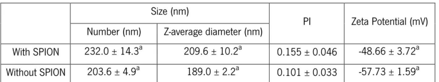
For preparation of the polymer beads, a nanoprecipitation method was used, with Poly(L-Lactide-co-glycolide) and superparamagnetic iron oxide nanoparticles as the main materials. The polymer was chosen due to its biodegradability. Since the magnetic beads will not be removed prior to in vitro culturing of the cells, biodegradability is an important aspect to avoid negative effects on cell growth. Initially, a preliminary study was performed to determine the optimal formulation for beads production. The best conditions included as non-solvent a mixture of ethanol and acetone in a volume ratio of 20:80, a solvent and non-solvent ratio of 0.02, and an amount of magnetic nanoparticles ten times smaller than the amount of polymer. The beads were characterized by Dynamic Light Scattering, in terms of particle size, polydispersity index and zeta potential; magnetic content through Thermogravimetric Analysis; and morphological features by Scanning Electron Microscopy. They presented average size values of 232 ± 14.30 nm. Their polydispersity index and zeta potential was 0.155 ± 0.046 and -48.66 ± 3.72 mV, respectively. The beads successfully encapsulated the iron oxide nanoparticles, with values of magnetic content of 51.5 ± 23 wt%. Lastly, the efficiency of nanocomposite beads was assessed by an immunomagnetic cell separation test. Beads formulated by nanoprecipitation were able to isolate 47 ± 16 % of cells.
IV
This dissertation focuses on polymeric beads for detection and quantification of cells. It has been demonstrated that biodegradable Poly(L-Lactide-co-glycolide) beads containing superparamagnetic iron oxide nanoparticles are an appropriate cell separation vehicle for cell culturing.
Keywords: Circulating Tumor Cells; nanocomposites polymer beads; immunomagnetic separation
V
Resumo
Células tumorais circulantes têm um papel activo na formação de metástases. No entanto, o seu número escasso no sangue de pacientes com cancro apresenta numerosos desafios tecnológicos. Tecnologias de detecção destas células e concepções de progressão metastática são aliados na investigação do cancro para uma nova dimensão na investigação clínica que procura ensaios experimentais modernos e clinicamente viáveis.
Esta dissertação tem como objectivo o desenvolvimento de beads poliméricas funcionalizadas com nanopartículas magnéticas encapsuladas para detecção e quantificação das células tumorais circulantes e a sua cultura in vitro após o seu isolamento em amostras de sangue. Este projecto encontra-se dividido em dois tópicos: depleção de células brancas CD45 para cultura in vitro, e desenvolvimento de nanocompósitos para detecção e quantificação celular.
Na depleção de células brancas CD45, duas beads foram analisadas, a disponível comercialmente Dynabeads® e beads preparadas no International Iberian Nanotechnology Laboratory (Braga, Portugal). Nas Dynabeads®, o número de células isoladas aumentou com a quantidade de beads, o mesmo não se verificou com as segundas.
Para a preparação das beads poliméricas, o método nanoprecipitação foi utilizado, sendo Poli(láctico-co-glicólico) e nanopartículas superparamagnéticas de óxido de ferro os materiais principais. Este polímero foi escolhido devido à sua biodegradabilidade. Uma vez que as beads magnéticas não serão removidas na cultura celular in vitro, biodegradabilidade é um aspecto importante de modo a evitar efeitos nefastos no crescimento celular. Inicialmente, um estudo preliminar foi realizado para determinar a formulação óptima para a sua produção. As melhores condições incluem como não solvente uma mistura de etanol e acetona num rácio volumétrico de 20:80, um rácio de solvente e não solvente de 0.02, e uma quantidade de nanopartículas poliméricas dez vezes mais pequena que a quantidade de polímero. As beads foram caracterizadas, por Dispersão Dinâmica de Luz, em termos de tamanho da partícula, índice de polidispersibilidade e potencial zeta; conteúdo magnético através Análise Termogravimétrica; e observação morfológica por Microscopia Electrónica de Varrimento. Estas apresentaram valores de tamanho médio de 232 ± 14.30 nm. O seu índice de polidispersibilidade e potencial zeta foi de 0.155 ± 0.046 e -48.66 ± 3.72 mV, respectivamente. As beads encapsularam com sucesso as partículas de óxido de ferro, obtendo um conteúdo magnético de 51.5 ± 23 wt%. Finalmente,
VI
a eficiência dos nanocompósitos foi avaliada com separação imunomagnética de células. Beads formuladas por nanoprecipitação conseguiram isolar 47 ± 16 % das células.
Esta dissertação foca-se em beads poliméricas para detecção e quantificação celular. Foi demonstrado que beads biodegradáveis de Poli(láctico-co-glicólico) contendo nanopartículas superparamagnéticas de óxido de ferro são veículos apropriados para separação celular.
Palavras-chave: Células tumorais circulantes; beads poliméricas de nanocompósitos; Separação imunomagnética
VII
Table of Contents
Acknowledgments I
Abstract III
Resumo V
Table of Contents VII
List of Figures XI
List of Tables XIII
General Nomenclature XV
Chapter I. Introduction 1
Chapter II. Background 7
2.1. Biodegradable polymeric biomaterials 9
2.1.1. Poly (L-Lactide-co-glycolide) (PLGA) 10
2.2. Iron oxide nanoparticles 11
2.3. Nanocomposite polymer beads for biomedical application: preparation, characterization and
functionalization 12
2.3.1. Preparation method 12
Nanoprecipitation 13
2.3.2. Characterization methods 14
Dynamic Light Scattering 14
Thermogravimetric Analysis (TGA) 15
Scanning Electron Microscopy (SEM) 15
2.3.3. Surface Functionalization 16
2.4. Circulating Tumor Cells (CTC) 17
2.4.1. Biological Properties of CTCs 18
Viability and Proliferation Capacity 18
Stem cell-like properties 19
VIII
Chapter III. Depletion of CD45 White Cells 23
3.1. Materials and Methods 25
3.1.1. Buffy coat extraction from blood samples 25
Materials 25
Methodology 25
3.1.2. Enrichment of CD45 white cells 25
Materials 25
Methodology 26
3.2. Results and Discussion 26
3.2.1. Dynabeads® 27
3.2.2. INL Beads 27
Chapter IV. Magnetic Nanocomposites 31
4.1. Materials and Methods 33
4.1.1. Preparation magnetic nanocomposites 33
Materials 33
Methodology 33
4.1.2. Characterization 34
Nanoparticle Size, Polydispersity Index and Zeta Potential 34
Magnetic content 34 Morphology 34 4.1.3. Data Analysis 35 4.1.4. Beads Functionalization 35 Materials 35 Methodology 35
4.1.5. Magnetic isolation of EpCAM cells 36
Materials 36
Methodology 36
4.2. Results and Discussion 36
4.2.1. Optimization of the formulation 37
4.2.2. Characterization 38
IX
Iron oxide content 39
Morphological observation 40
4.2.3. Magnetic separation of EpCAM cells 41
Chapter V. General Conclusions 43
5.1. Conclusions 45
5.2. Study limitations and future investigations 45
References 47
Annexes 57
Annex A. Percentage of white cells elimination 59
XI
List of Figures
Chapter II
Figure 2.1 – Chemical structure of PLGA and its monomers. Image adapted from Gentile et al. (2014)[28].
Figure 2.2 – Preparation of nanocomposites polymer bead by nanoprecipitation. Image adapted from Nicolas et al. (2013)[39].
Figure 2.3 – Reactions involving EDC and activation as an NHS ester. Image adapted from Thermo Scientific[52].
Figure 2.4 – Blood sample centrifuged showing different cell fractions.
Chapter III
Figure 3.1 – Percentage of depleted cells in relation to total volume (µL), i.e, the volume of the dispersion, of Dynabeads® per million of white cells.
Figure 3.2 – Percentage of depleted cells in relation to total volume (µL), i.e, the volume of the dispersion, of INL beads for one million of white cells.
Chapter IV
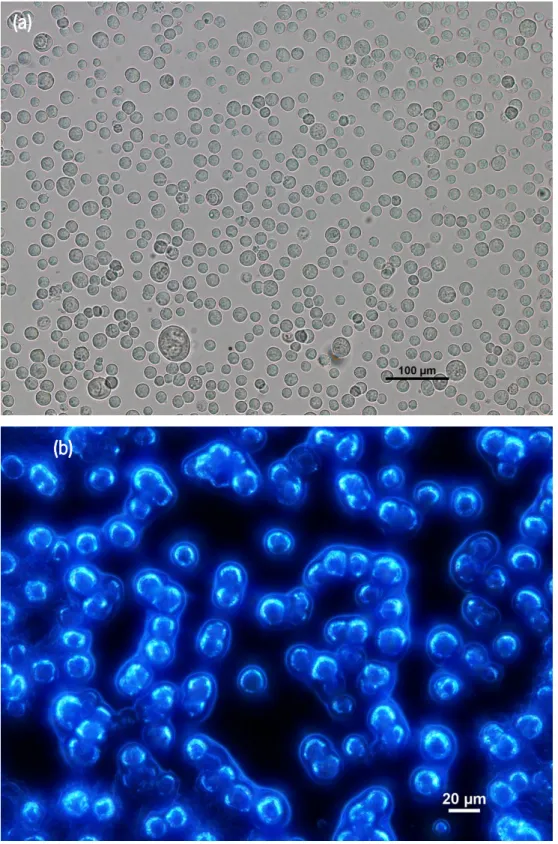
Figure 4.1 – Scanning electron microscopy (SEM) images of magnetic PLGA beads Figure 4.2 – Photomicrographs of SW480 cells with attached beads taken using (a) fluorescence microscope and (b) inverted fluorescence microscope.
XIII
List of Tables
Chapter IV
Table 4.1 – Optimal parameters for the preparation of magnetic polymer beads by nanoprecipitation.
Table 4.2 – Particle size, PI and zeta potential for PLGA beads with and without SPION by DLS.
Annexes
Table A.1 – Percentage of white cells elimination cells in relation to total volume (µL), i.e, the volume of the dispersion, by INL beads and Dynabeads®
Table B.1 – Different formulations tested for the development of magnetic nanocomposites Table B.2 – Values of particle size and PI of different formulations for development of magnetic nanocomposites.
XV
General Nomenclature
Abbreviations
ALDH1 Aldehyde dehydrogenase bcl-2 Cell lymphoma protein 2
BSA Bovine Serum Albumin
Ck Cytokeratin
CTCs Circulating tumor cells
CTM Circulating tumor microemboli D Translational diffusion coefficient DEP Dielectrophoretic
DEP-FFF Dielectrophoretic field-flow fractionation d(H) Hydrodynamic diameter
DLS Dynamic Light Scattering DPSS Diode-pumped solid-state
EDC 1ethyl-3-[3-dimethylaminopropyl] carbodiimide e.g. For example (exempli gratia)
EMT Epithelial-mesenchymal transition EpCAM Epithelial cell adhesion molecule EPISPOT Epithelial immunospot
FDA United States Food and Drug Administration
g grams
GA Glycolic acid
i.e. This is (id est)
IgG Immunoglobulin G
INL International Iberian Nanotechnology Laboratory ISET Isolation by size of epithelial tumor cells
k Boltzmann’s constant
kV Kilovolts
LA Lactic acid
MBCs Mononuclear blood cells
XVI min Minutes mL Millilitres mV Millivolts µL Microliter µm Micrometre
MOFF Multi-orifice flow fractionation
mol moles
nm Nanometre
NS Non solvent
PBS Phosphate buffered saline PCS Photon Correlation Spectroscopy PI Polydispersity index
PLA Poly(Lactic acid)
PLGA Poly(L-Lactide-co-Glycolide) RBCs Red blood cells
rcf Relative centrifugal force rpm Rotations per minute
S Solvent
SCLC Small-cell lung cancer
SEM Scanning Electron Microscopy
SPION Superparamagnetic iron oxide nanoparticles Sulfo-NHS N-hydroxysulfoccinimide
T Temperature
TM Trademark
TGA Thermogravimetric Analysis WBCs White blood cells
XVII Symbols α Alfa º Angle ºC degree Celsius CO2 Carbon Dioxide
Fe2+ Iron (II) ion Fe3+ Iron (III) ion
γ-Fe2O3 Maghemite Fe3O4 Magnetite ± Plus-minus % Percentage ® Registered trademark η Viscosity 𝜉 Zeta Potential
Peixoto, Rita (2015)
Nanocomposite polymer beads for cell detection and in vitro culture |1
Chapter I
Chapter I. Introduction
Peixoto, Rita (2015)
Nanocomposite polymer beads for cell detection and in vitro culture |3 Cancer is the leading cause of mortality and morbidity worldwide. In 2012, there were globally 32.6 million people with cancer, 14 million new cases and 8.2 million cancer related deaths. Cancer is the rapid formation of abnormal cells that grow uncontrollably beyond their usual boundaries. Some cancer cells are able to invade and propagate to distant organs[1]. This
process is denoted as metastasis. This is the most fearful aspect of cancer once it represents the cause of 90% of the deaths in cancer patients [3],[4].
Circulating tumor cells (CTCs) can detach from the primary tumor and are able to enter and circulate in the blood stream or the lymphatic system[4]. These cells have an active role in the
formation of metastasis. However, only a fraction is responsible for it, whereas the remaining CTCs can be originated from the damage caused by metastasis[5]. The first documented
microscopically observation of these cells was made in 1869 by Thomas Ashworth. When analysing a blood sample of a man with metastatic cancer, the Australian physician was faced with identical cells to the cancer itself, allowing him to claim that “One thing is certain, that if they [CTC] came from an existing cancer structure, they must have passed through the greater part of the circulatory system to have arrived at the internal saphena vein of the sound leg”[6].
Since then, CTCs have been increasingly a target of great interest in translational cancer research. With more than 400 clinical studies, CTCs are considered cancer biomarkers. The aims of the research performed on CTCs comprise prognostic information about the risk estimation of metastatic relapse or progression; the real time monitoring and stratification of treatments; identification of resistance mechanisms and therapeutic targets; understanding of the development of metastasis in cancer patients[7].
CTCs can be used as “liquid biopsy”, a promising alternative to invasive biopsies, as a source of tissue in non-malignant neoplasm. They can also contribute to the understanding of the tumor’s biology and tumor cell dissemination, to the formulation of new therapies and better prediction for clinical benefit[8][9].Their identification biomarkers are EpCAM and Cytokeratin 8, 18
and 19, however they do not allow the identification of biological behaviour (benign vs. malignant), only allowing the identification of tissue origin[10]. The expression of these biomarkers
could decrease or disappear during metastasis or dissemination, creating substantial heterogeneity[11].
The frequency of CTCs is usually 1 to 10 cells among 6×10⁶ leukocytes, 4×10⁹ erythrocytes and 2×10⁸ platelets per mL of blood[12],[13]. Their rarity presents tremendous
Chapter I. Introduction
4| Nanocomposites polymer beads for cell detection and in vitro culture
In the past two decades, significant technological efforts allowed the development of platforms to identify and count CTC reliably[14]. As these methods for CTC isolation develop,
connections between the cells and the disease have also been found. Results suggest that the treatment’s efficacy or cancer severity can be monitored with CTC blood analysis. In 2004, researchers established that the levels of CTC before and after the initial line of therapy for metastatic breast cancer are valuable predictors of overall survival[15]. These results were also
obtained in other types of cancer, including colorectal and prostate cancers[16]. Thereafter some
studies analysed the genetic mutations carried by the cells by comparison with the ones in the primary tumor or by correlation with the severity of the patient’s condition. In circulating lung-cancer cells, the emergence of mutations that cause drug resistance can increase the number of cells and contribute to a faster tumor progression[17]. In 2012, another research concluded that
the prostate cancer patient’s response to a drug can be predicted during treatment through identification of alterations in certain signalling pathways within CTC[18].
Additional studies focused on the physical and genetic characteristics of isolated CTCs to identify possible drug targets. CTCs can undergo measurable and controllable changes that make them more propitious to deposit into a new tissue, contributing to cancer dispersion. By blocking such modifications or signals, metastasis could be stopped. CTC makers capable of signal metastasis and offer novel drug targets were found in blood samples of breast cancer patients[19]
and mouse models of pancreatic cancer[20].
The investigation of the genomic and transcriptomic profiles of tumor cells represents an important stage to comprehend the biology of cancer. Notwithstanding, in a single cell analysis, cells even from a single blood sample presented different gene-expression patterns, portraying CTCs’ heterogeneous diversity[21].
The main objective of this project is to develop functionalized polymer beads with encapsulated magnetic nanoparticles for detection and quantification of circulating tumor cells and in vitro culturing of CTCs after their isolation from blood samples. The focus topics were:
I. Depletion of CD45 white cells in order to culture in vitro
II. Development of functionalized magnetic nanocomposites for detection and quantification of cells
Peixoto, Rita (2015)
Nanocomposite polymer beads for cell detection and in vitro culture |5 This dissertation is organized in five chapters. This one comprises the motivation, research aims and the outline of the dissertation. The second chapter, “Background”, reports a review of the theoretical foundations of this topic, providing to the reader the basic information to comprehend the main issues of this project. The project was divided into two parts “Depletion of CD45 White Cells” and “Magnetic Nanocomposites”. The first one corresponds to the third chapter and the second to the fourth chapter. In each chapter the materials and methodologies used are described, as well as the results obtained and their discussion. Finally, the fifth chapter, “General Conclusions”, presents the overall conclusions, limitations and recommendations for future work.
Chapter I. Introduction
Peixoto, Rita (2015)
Nanocomposite polymer beads for cell detection and in vitro culture |7
Chapter II
Chapter II. Background
Peixoto, Rita (2015)
Nanocomposite polymer beads for cell detection and in vitro culture |9 Nanomedicine is a process that uses molecular tools and molecular knowledge of the human body to diagnose, treat and prevent diseases and traumatic injuries, and preserve and improve human health[22]. Nanomedicine integrates physical, chemical and biological sciences at
a nanometre scale. One particularly promising area is that related to biomedical applications of magnetic nanomaterials. Magnetic nanomedicine is developing rapidly and there is already an extensive range of applications such as cell separation, cellular functions studies, biosensing, as well as a diversity of potential therapeutic and medical uses[23].
2.1. Biodegradable polymeric biomaterials
The emergence of novel biomedical technologies, such as regenerative medicine, tissue engineering, controlled drug delivery and bionanotechnology, required new biodegradable biomaterials[24].
Biomaterials are biocompatible materials intended to interact with biological systems to evaluate, treat, replace or enhance any function, tissue or organ[25]. Biocompatibility depends on
the biological environment and the tolerability of the specific interactions between the material and the tissue[26]. The molecular weight, solubility, hydrophilicity/hydrophobicity, and erosion
mechanism of the material can affect its biocompatibility[24].
Biodegradable materials, with natural or synthetic origins, are able to degrade in vivo by enzymatic and/or non-enzymatic mechanisms[27]. Their most essential properties are (i)
biocompatibility, i.e. the material should not induce an inflammatory or toxic response upon in contact with the tissue; (ii) its degradation time and the regeneration or healing time should match; (iii) the mechanical properties of the material should be appropriate for the application and the variation of these properties by degradation should be compatible with the regeneration or healing process; (iv) the by-products should be non-toxic and able to be metabolized and eliminated; and (v) the material’s permeability should be appropriate for the application[24].
Polymers have been widely used as biomaterials. Their versatility makes them more attractive than other materials like metals, alloys and ceramics[24]. Natural polymers, such as
polysaccharides and proteins, display numerous advantages, such as degradability and negligible toxicity. Nevertheless, several benefits are also reported for synthetic polymers, including
Chapter II. Background
10| Nanocomposites polymer beads for cell detection and in vitro culture
exceptional and reproducible physical and mechanical properties (such as tensile strength, degradation rate and elastic modulus) and highly controlled degradation properties[28].
There is a wide range of applications for biodegradable polymeric biomaterials including large implants, e.g. bone screws and plates; small implants, e.g. sutures and drug delivery vehicles; multifilament meshes or porous structures for tissue engineering; and plain membranes for tissue regeneration[29].
2.1.1. Poly (L-Lactide-co-glycolide) (PLGA)
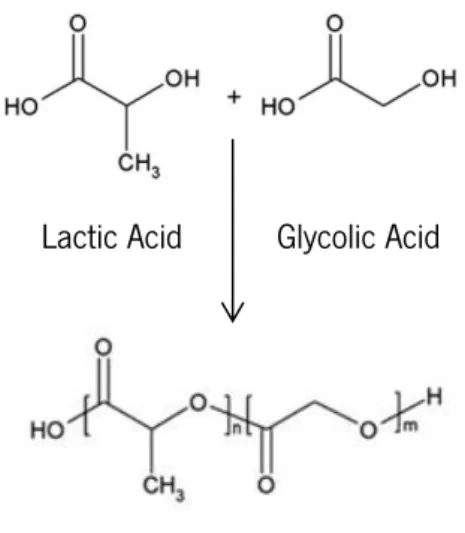
Poly (L-lactide-co-glycolide) (PLGA) is a copolymer of lactic acid (LA) and glycolic acid (GA) (Figure 2.1), prepared in different ratios. The different forms of PLGA are usually identified by the monomer’s ratio in the polymer, i.e., PLGA 70:30 indicates a copolymer with 70% of lactide and 30% of glycolide[30].
Figure 2.1 – Chemical structure of PLGA and its monomers. Image adapted from Gentile et al. (2014)[28].
PLGA can be synthesised using different processes that strongly influence the physical and chemical properties of the polymer. It can be dissolved by several solvents and processed into diverse shapes and sizes[30]. The physical properties of this biomaterial depend on multiple factors
including molecular weight, LA:GA ratio, exposure to water and storage temperature[31]. Its
molecular weight and polydispersity index can also affect the mechanical strength and influence polymer’s degree of crystallinity and melting point[27]. The degradation properties of this
copolymer can be controlled by the crystallinity, hydrophilic/hydrophobic balance and Lactic Acid Glycolic Acid
Peixoto, Rita (2015)
Nanocomposite polymer beads for cell detection and in vitro culture |11 composition (LA:GA ratio, molecular weight). In aqueous environment, PLGA degradation occurs by hydrolysis of its ester linkages through heterogeneous erosion. The resulting by-products are LA and GA[30].
PLGA is one of the most successful biodegradable polymers used in sutures, tissue engineering scaffolds, drug delivery devices, and micro- and nanoparticles. This is due to its appealing features: (i) biocompatibility and biodegradability; (ii) approved by FDA and European Medicine Agency for use in humans; (iii) enables formulation of a variety of structures and forms; (iv) controllable degradation rates[24]; (v) possibility to modify surface properties to provide better
interaction with biological materials[32]; and (vi) good cell adhesion and proliferation[24].
2.2. Iron oxide nanoparticles
Iron oxide is highly investigated due to its remarkable biocompatibility, chemical stability and superparamagnetic behaviour[33]. There is a great variety of iron oxides in nature. However,
magnetite (Fe3O4) and maghemite (γ-Fe2O3) are the most employed in biological and biomedical
applications[37]. Iron oxide nanoparticles (typically in the size range of 5–15 nm) can be
synthetically produced by several procedures[38]. The simplest, economical and
environmentally-friendly method is based on the co-precipitation approach, which implicates the concurrent precipitation of Fe2+ and Fe3+ ions in a basic aqueous media[39].
A superparamagnetic behaviour means that the particle has no residual magnetism after an external magnetic field is removed. This ability represents a significant advantage because prevents potential particle aggregation[34]. Superparamagnetic iron oxide nanoparticles (SPION)
have great potential for several biomedical applications, such as (i) cell therapy for cell labelling, targeting and separation; (ii) tissue repair; (iii) magnetic resonance imaging (MRI); (iv) hyperthermia; and (v) magnetic field-guided carriers for targeting drugs or radioactive therapies[35].
Chapter II. Background
12| Nanocomposites polymer beads for cell detection and in vitro culture
2.3. Nanocomposite polymer beads for biomedical application:
preparation, characterization and functionalization
Magnetic nanoparticles (MNPs) have gained significant attention among nanoscale materials for biomedical applications. They can act as successful magnetically recoverable catalysts, drug delivery and magnetic separation agents, magnetic resonance imaging devices and magnetic labels, among other[36]. The possibilities of design of magnetic nanocomposites are
vast in terms of decreasing functional space, adjusting magnetic characteristics, enhancing magnetic performance, multi-functionalization, and the development of materials with unique features. Magnetic polymeric nanocomposites intend to integrate various component materials with different features in a single material. These are usually composed of a core of an inorganic magnetic component with a dimension in the nanometre range (1 – 100 nm) surrounded by an organic polymer or vice versa. The organic-inorganic synergies bring new properties to the material that cannot be accomplished in single organic or inorganic materials[37].
The polymer can act as a template to regulate size, shape and organization; coating to protect from the surrounding medium; agent to provide mechanical properties and processability of the polymers to the magnetic material; functional element, adding its own optical, chemical or electrical features, augmenting others of the magnetic component, or developing new properties by interaction with the magnetic material. The magnetic properties can be affected by the size of the particle and by the polymer interphase. Besides, the structuring capacity of polymers can be utilized to control the magnetic interactions within the particles[37].
In biomedical applications, magnetic nanocomposites require to get close to or enter a biological entity. Therefore, the size of magnetic nanocomposites can be adapted, so they can be comparable to or be smaller than a cell (10 – 100 µm), a virus (20 – 450 nm), a protein (5 – 50 nm) or even a gene (2 nm of width and 10 – 100 nm of length). Furthermore, the interaction between the biological entities and magnetic nanocomposites can be aided by functionalization[23].
2.3.1. Preparation method
There are several methods developed and successfully used to prepare nanocomposite polymer beads. They can be prepared by direct polymerization of monomers, using techniques such as surfactant-free emulsion, micro emulsion, mini emulsion and interfacial polymerization,
Peixoto, Rita (2015)
Nanocomposite polymer beads for cell detection and in vitro culture |13 or from preformed polymers, using methods like solvent evaporation, nanoprecipitation, salting-out and dialysis. The preparation method has an important role in achieving the properties of interest[38]. The choice of the technique is made taking into consideration the properties of the
beads as well as characteristics of the methodology, such as, the simultaneous use of polymer and SPION.
Nanoprecipitation
The nanoprecipitation technique (also called solvent displacement method) for manufacturing nanostructures was developed by Fessi et al[39]. This is a straightforward, one step
method[40]. The nanoprecipitation system comprises two phases: an organic phase which consists
of synthetic, semi-synthetic or natural polymer, and polymer’s solvent or binary solvent blends; and an aqueous phase, the non-solvent or a mixture of non-solvents of the polymer[39].
In contrast to other preparation methods, nanoprecipitation does not require surfactants that might cause toxic effects or influence the surface characteristics of the nanostructures[41]. The
basic principle of this method is based on the rapid desolvation of the polymer when the organic phase is added to the non-solvent solution[40]. As soon as the polymer-containing solvent diffuses
into the dispersing medium, a decrease of interfacial tension between the phases occurs, increasing the surface area and precipitating the polymer (Figure 2.2)[39].
Figure 2.2 – Preparation of nanocomposites polymer bead by nanoprecipitation. Image adapted from Nicolas et al. (2013)[39].
The conditions of adding the organic phase to the aqueous phase determine the success of the method and affect the physicochemical properties of nanostructures. These conditions include the method of organic phase addition, aqueous phase agitation rate, organic phase to
Chapter II. Background
14| Nanocomposites polymer beads for cell detection and in vitro culture
aqueous phase ratio and the nature and concentration of their components[38]. The size and
shape of the particles can hardly be predicted. Nevertheless, by changing the conditions mentioned above it is comparatively easy to influence the particle formation[41].
2.3.2. Characterization methods
The nanocomposites polymer beads were characterized by Dynamic Light Scattering, in terms of particle size, polydispersity index and zeta potential; Thermogravimetric Analysis to determine their magnetic content; and Scanning Electron Microscopy for morphological observation.
Dynamic Light Scattering
Dynamic Light Scattering (DLS) or also known as Photon Correlation Spectroscopy (PCS) is a non-invasive and useful technique to measure the particle size, polydispersity index (PI), zeta potential and, in some cases, the shapes of particles in solution. Normally, this technique is used in the characterization of emulsions, polymers, micelles, proteins, nanoparticles or colloids[43],[44].
The particle size is the diameter of nanoparticles and the polydispersity index (PI) indicates the homogeneity of the sample, i.e. the distribution of nanoparticles size. The PI is a dimensionless variable that ranges between 0 and 1; values greater than 0.3 indicate that the sample is polydisperse, in other words, has a very broad size distribution[42].
The measurements of these parameters are achieved by analysis the scattered light generated when a laser is focused on the sample. Due to the different particles sizes, the scattered light will have different directions and different intensities[44],[45].
Particles in a fluid are subject to Brownian motion, which is the random movement of the particles resulting from the collisions with other particles and molecules in the fluid. Larger particles have slower a Brownian motion and smaller particles move more rapidly. DLS measures the fluctuations in scattered light intensity caused by Brownian motion and relates it to the size of the particles. The diameter of particles can be estimated using the Stokes Einstein equation (Equation 1.1)[44],[45].
d(H) = 3πηDkT Equation 1.1
Peixoto, Rita (2015)
Nanocomposite polymer beads for cell detection and in vitro culture |15 Hydrodynamic diameter, d(H);
Boltzmann’s constant (k); Absolute temperature (T); Viscosity (η);
Translational diffusion coefficient (D).
The Z-average diameter or average size estimated by DLS is calculated from the intensity distribution using the cumulants method, and can be converted to a number distribution using the dispersant and particle refractive index and some instrumental constants[43].
Other parameter that can be measured by DLS is the Zeta Potential (𝜉). It correlates with the electrostatic charge at the surface of the particle. This measurement can provide information related to the stability of dispersions, emulsions and suspensions[44],[46].
Thermogravimetric Analysis (TGA)
Thermogravimetric Analysis is a technique in which the sample’s weight is monitored as a function of temperature or time as the sample is subjected to a temperature program in a controlled atmosphere[46].
A TGA system consists of a precision balance and a furnace that is heated or cooled during the experiment. The sample environment is controlled by a sample purge gas. This system can quantify loss of water, loss of solvent, decarboxylation, oxidation, decomposition, among others[46].
The results from a thermogravimetric analysis can be presented by a thermogravimetric curve (weight versus temperature or time); or a differential thermogravimetric curve (rate of loss of weight versus temperature). In those curves, some features can be identified. These features include: a plateau, which indicates a constant weight; a curved section (the slope indicates the rate of weight loss); an inflection that can imply a formation of an intermediate compound result of disturbances in the heating rate. The results can be affected by the heating rate; sample weight; the geometry of the crucible; and the atmosphere[47].
Scanning Electron Microscopy (SEM)
Scanning electron microscopy (SEM) is a method for high-resolution imaging. It uses a focused beam constituted by high-energy electrons to provide images with information about the
Chapter II. Background
16| Nanocomposites polymer beads for cell detection and in vitro culture
samples’ external morphology, chemical composition, crystalline structure and orientation of elements of the sample. This type of electron microscope has a large depth of focus. It has also higher resolution (between 1 nm to 10 nm), so that closely spaced samples can be magnified at higher levels. Furthermore, SEM uses electromagnets instead of lenses which permits more control on the degree of magnification[49],[50].
An electron gun at the top of the microscope produces a beam of electrons. The beam goes vertically through the microscope, which is kept in vacuum. It passes through electromagnetic fields that focus the beam towards the sample. Electrons and X-rays are expelled from the sample when hit by the beam. Detectors convert these X-rays, backscattered electrons and secondary electrons into a signal. The signal is also converted into the final image[49],[50].
2.3.3. Surface Functionalization
Polymeric materials suitable for biomedical applications must be biocompatible so their insertion into the body does not cause any adverse reaction. Functional groups, attached on the surface of the polymer, are generally responsible for their biocompatibility. Therefore, the appropriate surface functionalization of the polymer can efficiently affect the recognition and cooperation with bio-assemblies and living cells[50]. In the functionalization process, several
biological entities can be used, e.g., proteins, antibodies, ligands and enzymes[23]. In the case of
PLGA beads, the carboxylates (–COOH) present in the surface of the polymer are activated by Sulfo-NHS (N-hydroxysulfosuccinimide) and EDC (carbodiimide), followed by the addition of protein A and antibody. In the presence of a carbodiimide such as EDC, carboxylates can react with Sulfo-NHS. This reaction results in a semi-stable NHS ester, which can form a stable amide bond when reacting with primary amines (–NH2) (Figure 2.3). The use of Sulfo-NHS is not mandatory for carbodiimide reactions; however it enhances greatly the coupling efficiency and makes it possible to execute a two-step reaction[51]. To the amide bond created in this reaction will
be attached a protein and the corresponding antibody in order to be possible the connection between the bead and the cell.
Peixoto, Rita (2015)
Nanocomposite polymer beads for cell detection and in vitro culture |17 Figure 2.3 – Reactions involving EDC and activation as an NHS ester. Image adapted from Thermo Scientific[52].
2.4. Circulating Tumor Cells (CTC)
During carcinogenesis, some cells within the primary tumor can obtain traits of invasiveness and mobility that allow them to enter into the blood stream or the lymphatic system[4].After intravasation, a circulating tumor cells (CTC) can die by apoptosis; become an
isolated cell; or aggregate with other CTCs creating circulating tumor microemboli (CTM). CTM cannot extravasate, however, due to its resistance to apoptosis and great proliferative capacity; it can rupture the capillary walls and originate a metastasis. On the contrary, the isolated cells can extravasate to distant organs, remaining as dormant solitary cells (disseminated tumor cells) or undergoing proliferation creating metastasis[52].
A CTC possesses certain characteristics that distinguish it from other cells. For a cell to be defined as CTC must contain an intracellular nucleus, a round to oval morphology and a size of, at least 4×4 µm². It also should stain for EpCAM and CK 8, 18 and 19, but do not stain for CD45[12].
CTCs are rare events with a frequency of 1 to 10 CTC per mL of blood in patients with metastatic tumor[16]. Their number can be an indicator of the tumor aggressiveness[5], and
therefore CTC analysis represents a great advantage for treatment. Furthermore, a peripheral blood analysis is easier than a bone marrow biopsy, which enables a real-time monitoring of the metastatic progression[3]. Sulfo-NHS Unstable reactive o-Acylisourea Ester Carboxylate molecule Semi-stable amine-reactive NHS ester Stable amide bond EDC
Chapter II. Background
18| Nanocomposites polymer beads for cell detection and in vitro culture
2.4.1. Biological Properties of CTCs
The heterogeneity of CTC morphology within and between patients[13],[54] can suggest
variances in their biological properties[8].
Viability and Proliferation Capacity
In the blood, CTCs can be differentiated between apoptotic or viable and cycling or non-cycling[4] These features influence the response to treatment and the success of the metastasis formation[5],[9].
The monoclonal antibody M30 can be used to identify apoptotic cells. During apoptosis, caspase-cleavage of Cytokeratin 18 occurs creating a neo-epitope which is recognized by the M30. If the staining reaction with this antibody is positive, the cells are at the beginning of the apoptotic cascade, whereas if it is negative the cells are viable or necrotic[54].
The first assay that aimed the detection of viable disseminating breast tumor cells was the EPISPOT (Epithelial Immunospot). In this test, the cells were detected according to their secretion of the tumor markers Cytokeratin 19 and MUC-1[55]. Another study concluded that the addition of
the marker M30 to classic cytokeratin staining allow the assessment of the ratio of viable to dead CTCs[56]. Apoptotic cells were found in 43% of patients with small-cell lung cancer (SCLC) and,
curiously, within CTM none of the cells exhibited an apoptotic nuclear morphology[57].
In contrast with M30, the B-cell lymphoma protein 2 (bcl-2) regulates apoptosis, suggesting a viable cell, wherein its expression predicts the response to a selected endocrine and chemotherapies[59],[60]. In 18 of 30 SCLC patients, bcl-2 was found in CTC and CTM[57] It was also
detected bcl-2 expression in an average of 62% of the CTCs in 36 of 83 metastatic breast cancer patients[60].
Concerning the proliferative capacity of a CTC, this is essential for their colonization in distant organs[8]. Ki67 is a universally expressed nuclear non-histone protein recognized as a
proliferation marker[61]. When the antigen is absent or rare in the CTC, the cell might display a
non-cycling phenotype that allows it to survival to chemotherapies[4]. It was reported that
approximately 25% of CTCs in breast cancer patients expressed Ki67[62], whilst in another study,
Peixoto, Rita (2015)
Nanocomposite polymer beads for cell detection and in vitro culture |19 Stem cell-like properties
Some studies acknowledged subpopulations of CTCs with stem cell-like phenotypes[8]. In a
study involving 30 patients with metastatic breast cancer, 80% of the patients revealed CK+/CD44+/CD24-/low phenotype among their CTCs[64]. It was also perceived the presence of a
less common population of aldehyde dehydrogenase 1 (ALDH1) high/CD24-/low. ALDH1 is a stem cell marker associated with deficient prognostic in breast cancer[65] and, usually, with EMT
(epithelial-mesenchymal transition) markers[66].
CTCs with the self-renewal potency and resistance to several chemotherapeutics characteristic of stem cells can represent a more aggressive group of CTCs[5],[9].
2.4.2. Enrichment of CTCs
The rare occurrence of CTCs demands a selective enrichment of these cells and/or a systematic removal of mononuclear blood cells (MBCs) and red blood cells (RBCs) in order to detect them in the blood of cancer patients. Numerous methods to overcome the restrictions linked to the low concentration of these cells have been developed. The different techniques are based on the physical (e.g., size, density, electric charge and deformability) or biological (e.g., surface protein expression and invasion capacity) properties that differentiate CTCs from the normal hematopoietic cells[67].
Enrichment of CTCs based on their physical properties does not require labelling. This group includes (i) density gradient centrifugation (Ficoll, OncoQuick™), an easy and cheap method, feasible with EpCAM positive and negative tumor cells, but with low specificity and loss of some CTCs[68]; (ii) filtration through filters like ISET® (Isolation by size of epithelial tumor
cells)[69] or three dimensional microfilter device[70]; this method is appropriate for EpCAM positive
and negative tumor cells, but a small amount of CTCs can be lost and large hematopoietic cells can be retained in the pores of the filter; (iii) a versatile label free biochip that uses differences in size (larger) and deformability (stiffer) of CTCs from blood cells[71]; (iv) a microfluidic device using
a separation technique that combines multi-orifice flow fractionation (MOFF) and dielectrophoretic (DEP), an electric method to manipulate cells in accordance with their size and membrane properties[72]; and (v) a dielectrophoretic field-flow fractionation (DEP-FFF) device, that
Chapter II. Background
20| Nanocomposites polymer beads for cell detection and in vitro culture
Separation based on CTCs biological properties can be performed by immunological procedures either by positive selection or negative selection. Immunomagnetic systems are based in the attachment of magnetic particles to cells via antibodies. When the mixed population is placed in a magnetic field, cells with attached beads will be attracted to the magnet and hence separated from the cells without beads[68]. Positive selection is usually executed with antibodies
against the protein EpCAM existent in the outer surface of CTCs. Among the numerous EpCAM-based CTC detection technologies, the CellSearch® system is the only one approved by FDA. Through this system, CTCs count have been associated with prediction of survival in cancer patients[15]. The AdnaTest®, another marker-based research tool, positively enriches CTCs from a
blood sample. With this methodology, the presence or disappearance of CTCs was proven to be a prognostic and predictive marker on metastatic breast cancer[74]. Microfluidic platforms like CTC-
or HB-Chip are also promising alternatives to selectively capture EpCAM-positive CTCs in patients with cancer[75]. CTC-iChip enhances the chance of systematic removal of MBCs and RBCs by
combining a chip-based platform with an affinity-based strategy. Other commercially available technology is the IsoFlux® system. It isolates EpCAM-positive cells from blood samples through its magnetic isolation zones[67]. To avoid sample volume restraints, CellCollector™ enriches CTCs
in vivo directly from the vein of the patient[76]. Negative selection, i.e. depletion of CD45-positive
cells is preferable when CTCs lack of adequate expression of EpCAM. This strategy can be combined with density gradient centrifugation or RBC lysis to displace undesirable RBCs and to improve the yield[77]. Nevertheless, these methods can contribute to loss of CTCs resulting in
false-negative results. The Parsortix system is a separation device that isolates viable CTCs according to their physical features. Alternative methodology Vita Assay™, a cell adhesion matrix method, isolates viable and invasive tumor cells[67].
In some cases, a pre-treatment of blood to obtain the buffy coat is necessary for the successful application of these methods. Buffy coat is the fraction of anticoagulated blood that contains most of the leukocytes, platelets and tumor cells. When a blood sample settles or is spun in a centrifuge, red blood cells, white blood cells and other blood components separate into layers according to their density (Figure 2.4). Due to their buoyancy, CTCs are in the same fraction as the white blood cells[78]. This process can, however, destroy some cells, decreasing cell
Peixoto, Rita (2015)
Nanocomposite polymer beads for cell detection and in vitro culture |21 Figure 2.4 – Blood sample centrifuged showing different cell fractions.
Plasma
(55% of total blood) Buffy Coat
Leukocytes & platelets (<1% of total blood) Erythrocytes (45% of total blood)
Chapter II. Background
Peixoto, Rita (2015)
Nanocomposite polymer beads for cell detection and in vitro culture |23
Chapter III
Chapter III. Depletion of CD45 white cells
Peixoto, Rita (2015)
Nanocomposite polymer beads for cell detection and in vitro culture |25 This project has two parts: depletion of CD45 white cells in order to culture in vitro and, development of functionalized magnetic nanocomposites for detection and quantification of cells. This chapter is focused on the first area and is divided in two sections, “Materials and Methods” and “Results and Discussion”.
3.1. Materials and Methods
Depletion of CD45 white cells includes the extraction of buffy coat from blood samples and its magnetic isolation.
3.1.1. Buffy coat extraction from blood samples
Materials
The histopaque, Phosphate Buffered Saline (PBS) and Albumin from bovine serum (BSA) were purchased from Sigma-Aldrich Chemical Co. Ltd (St. Louis, MO, USA). The blood samples were obtained from voluntary donors.
Methodology
A dilution media (PBS 2% BSA) was added to the blood sample previous to the addition to Histopaque in a ratio of 1:2. A buffy coat was obtained through centrifugation (Eppendorf, Germany) at 400 rcf for 30 minutes, with no brake. From the three layers, the buffy coat was removed and the cells counted by optical microscopy (Eclipse TS100 Inverted Routine Microscope, Nikon, Japan) using Neubauer Chamber.
3.1.2. Enrichment of CD45 white cells
Materials
Dynabeads® Magnetic beads with 4.5 µm diameter, 12% to 17% of magnetic content and coated with IgG2a antibody specific for a CD45 membrane antigen were purchased from Invitrogen by Life Technologies (California, USA).
Chapter III. Depletion of CD45 white cells
26| Nanocomposites polymer beads for cell detection and in vitro culture
Beads with 140 nm with 40% of magnetic content and functionalized with anti-CD45 prepared at International Iberian Nanotechnology Laboratory (Braga, Portugal) were used[80]. The
purified anti-human CD45 was purchased from BioLegend (San Diego, CA, USA). And finally, Phosphate Buffered Saline and Albumin from bovine serum were obtained from Sigma-Aldrich Chemical Co. Ltd (St. Louis, MO, USA).
Methodology
Two different magnetic beads were used to deplete the white cells extracted from blood samples. Commercial beads, Dynabeads®, and beads prepared at International Iberian Nanotechnology Laboratory (INL beads) were used. The methodology used was based on the protocol created by Life Technologies[81] and applied to both cases. However, for INL beads, a
functionalization step was required. This step included an incubation of INL beads with anti-CD45 for 1 hour at 4ºC with a sample mixture (Life Technologies, USA), followed by magnetic separation with a permanent magnet, removal of supernatant and pellet re-suspension in PBS 2% BSA. For the depletion process, beads were incubated with cells for 30 minutes at 4ºC with constant agitation (Life Technologies, USA). After incubation, magnetic separation was carried out with a permanent magnet. The supernatant was removed and the pellet re-suspended in PBS 2% BSA. The white cells with beads were quantified by optical microscopy (Eclipse TS100 Inverted Routine Microscope, Nikon, Japan) using Neubauer Chamber.
3.2. Results and Discussion
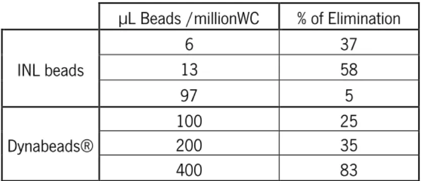
CD45 white cells were depleted using the immunomagnetic separation method. Two magnetic beads were tested, Dynabeads® and INL beads. Both beads are superparamagnetic and made of polystyrene. However, they have different sizes and magnetic content. Dynabeads® have a 4.5 µm diameter and 12% to 17% of magnetic content, whereas INL beads size is 140 nm and their magnetic content 40%. The number of beads per cell also differs, being 1 bead per cell in case of the Dynabeads® and 475 ± 25 beads per cell in case of INL beads. Furthermore, the antibody used in Dynabeads® is IgG2a while in INL beads is anti-CD45. These studies were performed in order to determine the conditions in which the higher percentage of white cells elimination, i.e., depleted cells, is obtained in both cases.
Peixoto, Rita (2015)
Nanocomposite polymer beads for cell detection and in vitro culture |27
3.2.1. Dynabeads®
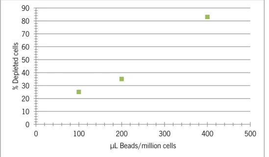
The commercial magnetic beads were tested in three different conditions. For each, 1×10⁷ white cells, 100, 200 and 400 µL of Dynabeads® were added. The results obtained are described in Figure 3.1 (Annex A).
Figure 3.1 - Percentage of depleted cells in relation to total volume (µL), i.e, the volume of the dispersion, of Dynabeads® per million of white cells.
A direct proportionality between the volume of beads and the amount of depleted cells was verified, achieving 83% of depleted cells with a volume of 400 µL per million of white cells.
3.2.2. INL Beads
The same protocol for Dynabeads® was used for INL beads. However, a functionalization process with anti-CD45 was required. As for Dynabeads®, the volume of beads was increased and the results are displayed in Figure 3.2 (Annex A).
0 10 20 30 40 50 60 70 80 90 0 100 200 300 400 500 % De pl et ed ce lls µL Beads/million cells
Chapter III. Depletion of CD45 white cells
28| Nanocomposites polymer beads for cell detection and in vitro culture
Figure 3.2 – Percentage of depleted cells in relation to total volume (µL), i.e, the volume of the dispersion, of INL beads for one million of white cells.
INL beads present an increase of elimination when the total volume of beads is increased from 6 µL to 12 µL. However, with a volume of 97 µL, a very small elimination is observed, being only 5% the cells eliminated. A possible explanation for this event can be aggregation of the beads. Different volumes of beads were tested, but the results were not coherent. With the increase of volume no changes in the percentage of elimination was verified. This could be an indicator that it is necessary more antibody on the surface in order to facilitate the interaction between beads and cell.
Taking into account the purpose of this part, the higher percentage of white cells elimination obtained by Dyanbeads® is 83% with 400 µL/million cells and and by INL beads is 58% with 13 µL/million cells.
Although the volumes used are lower than the ones used to test the Dynabeads®, it is possible to observe that INL beads obtained a higher percentage of depleted cells with less volume. INL beads eliminates 58% of cells with less than 100 million of cells while that amount of Dynabeads® only eliminates 25% of white cells. Besides, to eliminate 37% of cells, it is necessary 6 µL INL beads/million of cells, whereas with Dynabeads® 200 µL are needed. Nevertheless, the percentage of depleted cells drops drastically in the case of INL beads when the volume is increased to 100 µL. This is not verified on Dynabeads. As mentioned previously, the percentage of depleted cells increase as the volume of beads also is increased. It is also important to
0 10 20 30 40 50 60 70 0 20 40 60 80 100 120 % De pl et ed Ce lls µL Beads/million cells
Peixoto, Rita (2015)
Nanocomposite polymer beads for cell detection and in vitro culture |29 emphasize that these beads have different size and concentration, and thus it is not possible to draw conclusions.
Chapter III. Depletion of CD45 white cells
Peixoto, Rita (2015)
Nanocomposite polymer beads for cell detection and in vitro culture |31
Chapter IV
Chapter IV. Magnetic Nanocomposites
Peixoto, Rita (2015)
Nanocomposite polymer beads for cell detection and in vitro culture |33 The main objective of this project was the development of nanocomposites polymer beads with encapsulated magnetic nanoparticle for cell detection and their further characterization. Preparation of magnetic polymer beads was performed through nanoprecipitation method using the polymer PLGA and SPION.
4.1. Materials and Methods
The achievement of magnetic nanocomposites involves their preparation, characterization and data analysis, functionalization and, finally, their efficacy in magnetic separation of cells.
4.1.1. Preparation magnetic nanocomposites
Materials
The polymer Poly(l-lactide-co-glycolide) 70:30 was purchased from Polysciences, Inc. (Warrington, PA, USA). Iron oxide nanoparticles prepared in International Iberian Nanotechnology Laboratory (Braga, Portugal) were used. The solvent dichloromethane with MW 84.93 g/mol was obtained from Fisher Scientific UK (Loughborough, United Kingdom). Acetone with 58.08 g/mol was purchased from Sigma-Aldrich Chemical Co. Ltd (St. Louis, MO, USA) and ethanol with 46.07 g/mol from Scarlau (Barcelona, Spain).
Methodology
The preparation of polymer beads with encapsulated magnetic nanoparticles was based on the methodology described by Csaba and Alonso (2006). Nanoprecipitation followed by centrifugation was the production method used, with PLGA and iron oxide nanoparticles as the main materials. Through an optimization study, the optimal conditions (selected based in the particle size and PI) were determined. Briefly, 12.5 mg of PLGA and 1.25 mg of iron oxide nanoparticles were dissolved in dichloromethane (500 µL). Then, the solution was poured into a beaker containing a mixture of 80% ethanol and 20% acetone (18 mL and 4.5 mL, respectively) under constant and rapid agitation using an overhead stirrer (VWR International Ltd, UK). Then, 100 mL of mili-Q water were added. Afterward, the solvents were evaporated utilizing a rotary evaporator (IKA-Werke, Germany) with a vacuum pump (Vaccuumbrand, Germany). After size
Chapter IV. Magnetic Nanocomposites
34| Nanocomposites polymer beads for cell detection and in vitro culture
and PI measurement, the sample was centrifuged (Eppendorf, Germany). The optimal velocity and time was determined (selected based in the particle size and PI), being at 4500 rpm during 1 minute.
4.1.2. Characterization
Nanoparticle Size, Polydispersity Index and Zeta Potential
Size, polydispersity index (PI) and zeta potential of beads were measured using dynamic light scattering (DLS) (Horiba Scientific SZ-100, USA) equipped with a diode-pumped solid-state (DPSS) laser with a wavelength of 532 nm. For size and PI determination, quartz cells (Sigma-Aldrich Chemical Co. Ltd, USA) were used and for zeta potential disposable zeta potential cells were used (Sigma-Aldrich Chemical Co. Ltd, USA). All measurements were carried out by triplicate at 25ºC.
Magnetic content
The iron oxide content of polymeric beads was measured using thermogravimetric analysis
(TGA) (Mettler-Toledo TGA/DSC1 STARe system). The measurements were performed in an environment with a constant argon flow rate of 20 mL/min The program used had three steps: it starts with temperature increasing from 21ºC to 100ºC for 10 K/min; then the temperature stabilize at 100ºC for 10 minutes; and finally, the temperature increases again up to 900ºC for 20 K/min.
Morphology
The surface morphology of the magnetic polymer beads and their size was determined by scanning electron microscopy (SEM) using a Quanta ESEM (FEI, USA) operating at a voltage of 3 kV. For the analysis, a silicon wafer with a drop of sample was dried in vacuum. This wafer was then attached to the SEM specimen stub.
Peixoto, Rita (2015)
Nanocomposite polymer beads for cell detection and in vitro culture |35
4.1.3. Data Analysis
Data analyses were performed using Microsoft Windows Excel 2010. Three samples of magnetic beads were prepared. All data were reported as the mean standard deviation (SD) from at least three values. A T test (α=0.05) was employed to assess the statistical significance of the results. Experimental results were considered statistically significant at 95% confidence level (p<0.05). SEM images were treated with ImageJ (Version 1.49 (Free), Wayne Rasband National Institute of Health, USA).
4.1.4. Beads Functionalization
Materials
EDC (1ethyl-3-[3-dimethylaminopropyl] carbodiimide) and Sulfo-NHS (N-hydroxysulfoccinimide) with MW 191.7 g/mol and 217.14 g/mol, respectively, were purchased from Thermo Scientific (Paisley, UK). The surfactant Tween 20 was obtained from Sigma-Aldrich Chemical Co. Ltd (St. Louis, MO, USA). Protein A with 46.76 g/mol was purchased from Thermo Scientific (Paisley, UK) and the antibody Alexa 488 Fluor-Anti EpCAM from BioLegend (San Diego, CA, USA). Phosphate Buffered Saline (PBS) and Albumin from bovine serum (BSA) were obtained from Sigma-Aldrich Chemical Co. Ltd (St. Louis, MO, USA). Cell staining buffer was purchased from BioLegend (San Diego, CA, USA).
Methodology
The functionalization process of the selected polymer beads was based on the methodology used by Vila et al. (2014). Briefly, the carboxylic groups of PLGA present on the surface of the magnetic beads were activated using EDC and Sulfo-NHS (24 mg/mL and 64 mg/mL, respectively) during 15 minutes at room temperature and constant agitation with a sample mixture (Life Technologies, USA). This step enables the formation of amide bonds. To avoid bead aggregation, the sample was centrifuged with a Tween 20 bed, at 10500 rpm during 10 minutes, for a quick removal of the two reagents. Then, the polymer beads were incubated with protein A (1 mg/mL) during 3 hours at 4ºC with a sample mixture (Life Technologies, USA). Thereafter, the beads with protein A were centrifuged and re-suspended in PBS 2% BSA and Cell Staining Buffer (50:50).
Chapter IV. Magnetic Nanocomposites
36| Nanocomposites polymer beads for cell detection and in vitro culture
After coating the surface of the beads with protein A, the anti-EpCAM was attached by incubation during 1 hour at 4ºC and constant agitation (sample mixture, Life Technologies, USA). To remove free antibody (i.e. that not attached to beads), a magnetic separation was performed and the supernatant was re-suspended in PBS 2% BSA.
4.1.5. Magnetic isolation of EpCAM cells
Materials
SW480 colorectal cell line was purchased from ATCC (Virginia, USA). Phosphate Buffered Saline (PBS) and Albumin from bovine serum (BSA) were obtained from Sigma-Aldrich Chemical Co. Ltd (St. Louis, MO, USA).
Methodology
The next step to the beads functionalization was to test their interaction with cells. The SW480 cell line was maintained in Dulbecco's Modified Eagle's Medium (DMEM) (ATCC, USA) supplemented with 10% Fetal Bovine Serum (FBS) (ATCC, USA) and 1% Penicillin-Streptomycin Solution (ATCC, USA) at 37 °C in a humidified incubator under 5% CO2. After harvesting the cells, a centrifugation at 1200 rpm for 5 min (Eppendorf, Germany) was performed, being the supernatant discarded and the pellet re-suspended in PBS 2% BSA. Anti-EpCAM polymer beads (150 µL beads per million cells) were incubated with SW480 cells for 1 hour at 4ºC with constant rotation (Life Technologies, USA). After incubation, magnetic separation with a magnet was carried out and the supernatant removed. The pellet was re-suspended with PBS 2% BSA and the cells were counted by optical microscopy (Eclipse TS100 Inverted Routine Microscope, Nikon, Japan) using Neubauer Chamber and observed by fluorescence microscopy (Inverted Metallurgical Microscope ECLIPSE MA200 and Industrial Microscope ECLIPSE, Nikon, Japan).
4.2. Results and Discussion
Magnetic polymer beads were produced using the nanoprecipitation method followed by centrifugation, with PLGA, SPION as main materials. Based on the methodology described by Csaba et al (2006), due the great complexity of the method and the use of a different
Peixoto, Rita (2015)
Nanocomposite polymer beads for cell detection and in vitro culture |37 encapsulated component, it was necessary to test different formulations for their preparation. Thus, initially an optimization was performed and the optimal formulation determined.
4.2.1. Optimization of the formulation
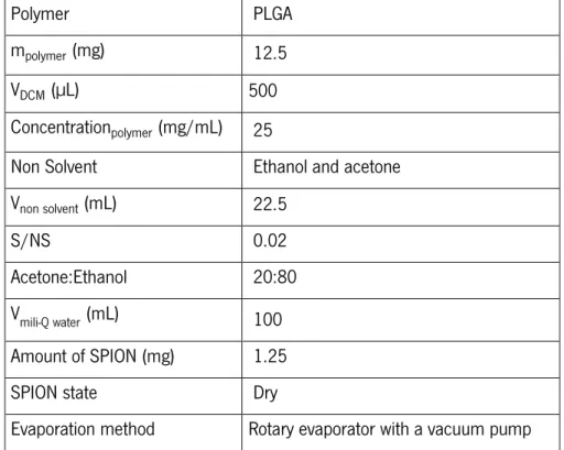
The particle size and polydispersity index (PI) are important parameters to evaluate the stability and homogeneity of beads in solution. Therefore, the particle size and PI of different formulations were analysed using DLS in order to optimize the formulation. The preparation conditions of PLGA beads have been optimized in terms of the polymer (PLA and PLGA), polymer concentration, iron oxide content and physical state (dried or liquid), magnetic susceptibility (i.e. how quickly the beads migrate to the magnet), ratio between solvent and non-solvent (S/NS), nonsolvent (ethanol or ethanol and acetone), acetone/ethanol ratio, mili-Q water volume, and evaporation method (Heating plate or rotary evaporator with a vacuum pump). In Annex B the different formulations tested are summarized. The optimal parameters are described in table 4.1.
Table 4.1 – Optimal parameters for the preparation of magnetic polymer beads by nanoprecipitation.
Polymer PLGA
mpolymer (mg) 12.5
VDCM (µL) 500
Concentrationpolymer (mg/mL) 25
Non Solvent Ethanol and acetone
Vnon solvent (mL) 22.5
S/NS 0.02
Acetone:Ethanol 20:80
Vmili-Q water (mL) 100
Amount of SPION (mg) 1.25
SPION state Dry