Acupuncture effects on delayed
onset muscle soreness
RICARDO MANUEL TAVARES CARDOSO
DISSERTAÇÃO DE MESTRADO APRESENTADA
AO INSTITUTO DE CIÊNCIAS BIOMÉDICAS ABEL SALAZAR
DA UNIVERSIDADE DO PORTO EM
MEDICINA TRADICIONAL CHINESA
RICARDO MANUEL TAVARES CARDOSO
ACUPUNCTURE EFFECTS ON DELAYED ONSET MUSCLE
SORENESS
Dissertação de Candidatura ao Grau de
Mestre em Medicina Tradicional Chinesa
submetida ao Instituto de Ciências
Biomédicas Abel Salazar da Universidade
do Porto.
Orientador – Doutor Henry Johannes Greten
Categoria – Professor Associado
Afiliação – Instituto de Ciências Biomédicas
Abel Salazar, Universidade do Porto.
Co-Orientador – Doutor José Lumini de Oliveira
Categoria – Professor Auxiliar
Acknowledgments
To my family for all their background support that has enabled me to develop my
own education.
To my girlfriend Ana Santos for all the support, care and understanding.
To Prof. Dr. Henry Johannes Greten, for his encouragement, support, supervision
and to introduce me to this wonderful world of Chinese Medicine and for all of his
inspiring lectures and teachings.
To Prof. Dr. José António Rodrigues Lumini de Oliveira, for all the support,
availability and supervision.
To Maria João, for all the support, teachings and always willing to help.
To Dr. Isabel Santos, for authorizing the study in the Hospital-School of Fernando
Pessoa University.
To all colleagues of Hospital-School of Fernando Pessoa University who helped
me in the realization of the study.
To all the participants of this study, because without them this wouldn`t be
possible.
To Petra for all her support.
To Bruno Ramos for all his support and availability.
To all the colleagues and friends of this master and specialization with whom I
learned and shared unforgettable moments.
Resumo
Introdução: Apesar de haver vários estudos sobre a sensação retardada de desconforto
muscular (SRDM), a explicação dos mecanismos referentes a esta condição clínica ainda se encontra em discussão, assim como as suas estratégias de tratamento e prevenção. Os estudos científicos mostram que não há consenso sobre os efeitos da acupunctura na SRDM.
Objectivo: Avaliar os efeitos da acupunctura na SRDM.
Métodos: 45 participantes (média ± s idade 25,38 ± 4,77 anos; peso 65,76 ± 9,88 kg;
altura 169 ± 0,09 cm e índice de massa corporal 22,94 ± 2,51 kg/m2) dos dois géneros (19 homens, 26 mulheres), depois de completarem um questionário de triagem e fornecerem o consentimento informado escrito, foram distribuídos de forma aleatória em três grupos (grupo de acupunctura verdadeira, grupo de acupunctura falsa e grupo controlo – AV, AF e GC). O desconforto muscular (DM), limiar de dor à pressão (LDP), amplitude articular (AA), salto vertical (SV) e avaliações do isocinético foram realizados antes e após (imediatamente e 24h) um protocolo de exercícios de dano muscular (EDM) em que os indivíduos realizaram cinco séries de 20 drop jumps num step a uma altura de 0,6m, com 10 segundos de intervalo entre saltos e 2 minutos de descanso entre séries. A análise estatística foi realizada através do Microsoft Excel 2010, para determinar a média e desvio padrão da amostra e dos parâmetros avaliados, e o IBM SPSS Statistics 22 foi usado para realizar o teste Kruskal-Wallis para a análise das diferenças entre grupos em diferentes momentos e bem como para o Teste de Friedman com o Teste Bonferroni post-hoc, para obter as diferenças entre grupos ao longo dos diferentes momentos.
Resultados: O protocolo de EDM foi bem sucedido ao induzir SRDM. Quando os grupos
de AV, AF e GC foram comparados, verificou-se que a AV teve melhores resultados em todos os parâmetros avaliados, especialmente no DM, LDP e SV. Contudo, o grupo de AF demonstrou melhores resultados que o GC, quando o DM e LDP foram comparados, mas não foram resultados com diferença estatisticamente significativa.
Conclusão: Os resultados demonstram que, a acupunctura aplicada depois de um
protocolo de EDM, teve efeitos de redução do DM e melhoria no LDP, AA, SV e medições no isocinético.
Palavras-Chave: Acupunctura, Medicina Tradicional Chinesa, sensação retardada de
Abstract
Introduction: Despite of the many studies on the subject of delayed onset muscle
soreness (DOMS), the explanation of the mechanisms underlying this clinical condition is still under discussion, as well as the strategies for its prevention and treatment. Scientific studies have shown no consensus of the effects of acupuncture on DOMS.
Objective: To evaluate acupuncture effects on DOMS.
Methods: 45 participants (mean ± s age 25,38 ± 4,77 years; weight 65,76 ± 9,88 kg;
height 169 ± 0,09 cm and body mass index 22,94 ± 2,51 Kg/m2) of both genders (19 males, 26 females), after completing a screening questionnaire and providing written informed consent, were randomly distributed into three groups (verum acupuncture group, sham acupuncture group and a control group – VA, SA and CG) . Muscle soreness (MS), pressure pain threshold (PPT), range of motion (ROM), vertical jump (VJ) and Isokinetic evaluations were performed before and after (immediately and 24h) a exercise induced muscle damage (EIMD) protocol which subjects performing a five sets of 20 drop jumps from a height of 0,6m step, with a 10 seconds interval between jumps and 2 minutes rest period between sets. Statistical analysis was performed using Microsoft Excel 2010 to determine the mean and standard deviations of the sample and outcome measures, and IBM SPSS Statistics 22 was used to perform a Kruskal-Wallis test with for analysis of differences between groups in the different moments and a Friedman test with a Bonferroni post-hoc test, to assess differences within the group along the different moments.
Results: The protocol of EIMD showed success in inducing DOMS. When VA, SA and
CG groups were compared, it was found that VA had better results in all outcome measures, especially in MS, PPT and VJ. However, SA group showed better results than CG, when MS and PPT were compared, but they were not statistically significant.
Conclusion: The results showed that acupuncture applied after an EIMD protocol, had an
effect on MS reduction and an improvement on PPT, ROM, VJ and Isokinetic measures.
Key words: Acupuncture, Traditional Chinese Medicine, delayed onset muscle soreness,
Contents
Chapter 1
Introduction………1
1. Exercise induced muscle damage……….2
2. Delayed Onset Muscle Soreness………..4
3. Factors that influence DOMS………...20
4. Models of DOMS and EIMD……….24
5. Prevention and treatment strategies for DOMS………...25
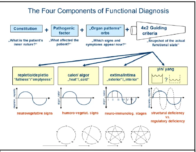
6. The Heidelberg Model of Traditional Chinese Medicine………..27
6.1. TCM diagnostics according to the Heidelberg model……….………..34
6.2. DOMS according Heidelberg model of TCM……….……….38
7. “Leopard Spot” technique……….39
8. Neurophysiological basis of acupuncture………...42
9. The impact of the Heidelberg Model of TCM on current acupuncture research………..43
Chapter 2 Methods……….………..45 Chapter 3 Results……….………64 Chapter 4 Discussion………..……….76 Chapter 5 Conclusions……….………85
References……….……….…A
Annexes………I
INDEX OF FIGURES
Figure 1 – Possible Mechanisms of Exercise Induced Muscle Damage……….3
Figure 2 - Schematic showing possible sequence of injury in DOMS (Connolly, Sayers and Mchugh, 2003)………..6
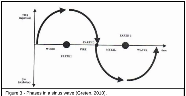
Figure 3 - Phases in a sinus wave (Greten,2010)……… ………..30
Figure 4 - Diagnostic pathways (Greten, 2010)………31
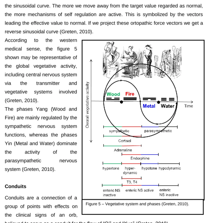
Figure 5 – Vegetative system and phases (Greten, 2010)………..32
Figure 6 – The stomachal conduit (Greten, 2010)………33
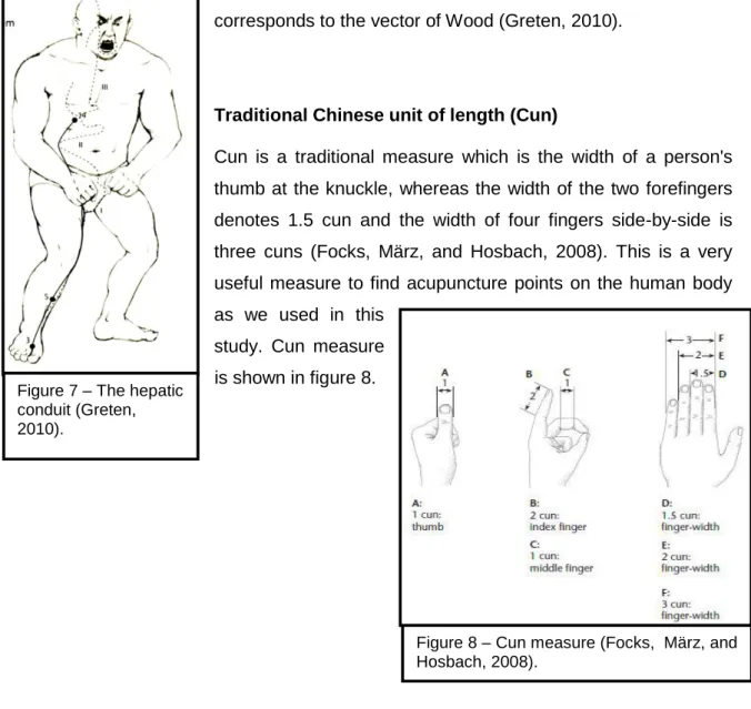
Figure 7 – The hepatic conduit (Greten, 2010)……….34
Figure 8 – Cun measure (Focks, März, and Hosbach, 2008)………34
Figure 9 – The four components of the functional diagnosis in TCM (Greten, 2010)……….37
Figure 10 – “Leopard Spot” technique in a participant……….40
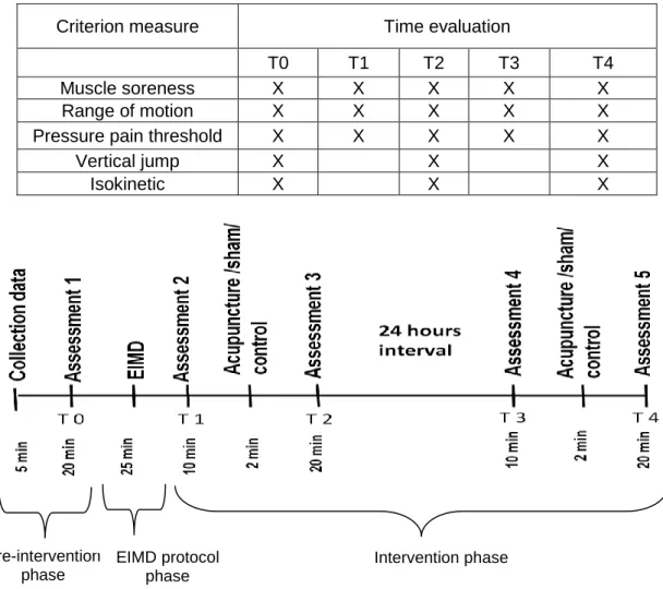
Figure 11 – Criterion Measure Testing according time evaluation and experiment flow-chart….….47 Figure 12 – S34 localization (Focks, März and Hosbach, 2008)………49
Figure 13 – S36 localization (Focks, März and Hosbach, 2008)………50
Figure 14 – H3 localization (Focks, März and Hosbach, 2008)………..50
Figure 15 – V57 localization (Focks, März and Hosbach, 2008)………51
Figure 16 - Sample diagram………...52
Figure 17 – Squat position for measure muscle soreness………..57
Figure 18 – PPT measure……….……58
Figure 19 – Participant performing the squat jump test……….……..59
Figure 20 – Participant at start position at left and performing isokinetic evaluation at right.………60
Figure 21 – Participant performing EIMD. Starts from up to down and left to right………….………61
Figure 22 - MS scores of the VA, SA and CG groups (mean ±SD). * vs. CG (p<0.05)…….………65
Figure 23 - PPT scores of VA, SA and CG groups (mean ±SD). * vs. CG (p<0.05). # vs. SA (p<0.05). ………66
Figure 24 - ROM scores of the VA, SA and CG groups (mean ±SD)………68
Figure 25 - VJ scores of VA, SA and CG groups (mean ±SD). * vs. CG (p<0.05)……….……70
Figure 26 - PT/BW scores of the VA, SA and CG groups (mean ±SD)……….…72
INDEX OF TABLES
Table 1 - Clinical signs and symptoms of DOMS (Sethi, 2012)………...……….5 Table 2 - Sample data characterization………..52 Table 3 - Differences within and between the groups (VA, SA and CG) in the different moments in MS (VAS)………64 Table 4 - Differences between the groups (VA, SA and CG) in the different moments in MS (VAS)………..………64 Table 5 - Differences within and between the groups (VA, SA and CG) in the different moments in PPT (kgf/cm2)………..………..65 Table 6 - Differences between the groups (VA, SA and CG) in the different moments in PPT (kgf/cm2)………..……….66 Table 7 - Differences within and between the groups (VA, SA and CG) in the different moments in ROM (º)……….……….67 Table 8 - Differences between the groups (VA, SA and CG) in the different moments in ROM (º)………..…..68 Table 9 - Differences within and between the groups (VA, SA and CG) in the different moments in VJ (cm)……….………..69 Table 10 - Differences between the groups (VA, SA and CG) in the different moments in VJ (cm)………..………..69 Table 11 - Differences within and between the groups (VA, SA and CG) in the different moments in VJ (cm)……….…………..70 Table 12 - Differences between the groups (VA, SA and CG) in the different moments in PT/BW
(%)……….………. 71
Table 13 - Differences within and between the groups (VA, SA and CG) in the different moments in AVG PT (N.m)………..……….72 Table 14 - Differences between the groups (VA, SA and CG) in the different moments in AVG PT (N.m). ……….…73
LIST OF ABBREVIATIONS
EIMD Exercise-induced muscle damage DOMS Delayed Onset Muscle Soreness RBE Repeated-bout effect
MVC Maximal Voluntary Contraction ROM Range of Motion
CK Creatine kinase
LDH Lactate dehydrogenase MRI Magnetic Resonance Imaging US Ultrasound
CNS Central nervous system LFF Low frequency fatigue HFF High frequency fatigue SR Sarcoplasmic reticulum EE Eccentric exercise ATP Adenosine triphosphate ROS Reactive Oxygen Species O2 Oxygen
EPOC The over consumption of oxygen post-exercise HMB Beta-hydoxy-beta-methylbutyrate
TCM Traditional Chinese Medicine VA Verum acupuncture group SA Sham acupuncture group CA Control acupuncture group VAS Visual analogue scale PT Peak torque
PT/BW Peak torque/body weight AVG PT Average peak torque BMI Body mass index MS Muscle soreness PPT Pressure pain threshold ROM Range of Motion
1
IntroductionDelayed onset muscle soreness (DOMS) is a common form of muscle soreness, experienced by individuals who perform unaccustomed exercise and consequently exercise induced muscle damage (EIMD), that typically involves an eccentric component that peaks between 24 and 48 hours post-exercise and that spontaneously disappears within 5 to 7 days (Aminian-Far et al., 2011; Torres et al., 2012).
However, it is not just novel strength training exercises or eccentric contractions that can cause DOMS. In the working environment, a common problem is muscle tenderness, soreness and pain, especially for workers frequently exposed to unilateral high repetitive movement‟s tasks (Andersen, Hansen, Mortensen, Zebis, 2011). Furthermore, as muscle pain has been associated with decreased performance in the form of decreased rapid force capacity and maximal muscle strength, continuous overload causing muscle soreness and pain may not only have adverse functional implications during working hours but also during activities of daily living during non-working hours (Andersen, Holtermann, Jørgensen, Sjøgaard, 2008; Andersen, Nielsen, Søgaard, Andersen, Skotte, Sjøgaard, 2008).
Acupuncture has shown to increase of muscular power (Huang, et al., 2007; Hubscher, et al., 2010; Ozerkan, et al., 2007; Yang, et al., 2006; Zhou, et al., 2012), to improve microcirculation (Kuo, Lin, Ho, 2004), to decrease inflammatory processes (Moon et al, 2007), to release endogenous endorphins (Hwang et al, 2002), to inhibit spinal and supraspinal nociceptive transmission (Ikeda, Asai, Murase, 2000; Rong et al, 2005), as well as to improve vertical jump (Sousa, 2012).
Therefore, from a theoretical standpoint, acupuncture might be an attractive, beneficial, low-cost, quick and low-risk treatment strategy for DOMS treatment, improving performance on athletes and productivity in workers.
The major aim of this thesis is to evaluate the effects of acupuncture on DOMS.
To fulfill this goal is presented a theoretical background data based on an extensive literature research with the intention to support the following experimental study.
The theoretical fundamentals are presented in the first part of the thesis (chapter one) which is comprised of a review on EIMD, DOMS, it´s physiology, consequences, factors that influence and models to induce DOMS. It´s prevention and treatment strategies are presented followed by an explanation of the Heidelberg Model of Traditional Chinese Medicine and neurophysiological basis of acupuncture.
The theoretical fundamentals are expected to provide the needed support for the following detailed description of the methods (chapter two). In the third chapter are exposed the
2
results and in the chapter four are presented the discussion, that is supported by literature findings, the limitations and suggestions. Finally, the thesis is enclosed with the conclusions (chapter five).
1. Exercise induced muscle damage
In the early 1900‟s Dr. Theodore Hough observed a long lasting decline in force production and increased soreness following an exercise of the finger flexors (Hough, 1900), but only in the early 1980‟s, research on this exercise-induced muscle damage (EIMD) really begun (Thiebaud, 2012).
Unaccustomed muscle work, especially that which is eccentric in nature, can cause post-exercise soreness, usually referred to as delay onset muscle soreness (DOMS) ( Lewis, Ruby, Bush-Joseph, 2012; Proske, Morgan, 2001; Yu, Malm, Thornell, 2002; Zainuddin, Newton, Sacco, Nosaka, 2005). Muscle soreness can negatively interfere with the activities of daily living as well as sports performance. Research commonly suggests that DOMS is the result of an inflammatory process caused by micro-tears in the muscle fibers during unaccustomed repetitive activity and/or eccentric contractions (Barbe, Barr, 2006; Barr, Barbe, Clark, 2004) but it has also been suggested that muscle soreness can occur without micro-trauma (Zainuddin, Newton, Sacco, Nosaka, 2005).
However, it is not just unaccustomed strength training exercises or eccentric contractions that can cause muscle soreness. In the working environment, a common problem is muscle tenderness, soreness and pain, especially for workers frequently exposed to unilateral high repetitive movements tasks. As an example, computer workers commonly experience soreness of several different neck/shoulder muscles (Andersen, Hansen, Mortensen, Zebis, 2011). Andersen and coworkers reported that 33% and 29% of blue and white-collar workers respectively, within the general working population, suffered from neck/shoulder pain (Andersen, Mortensen, Hansen, Burr, 2011). Furthermore, it seems that highly repetitive movement tasks can physically alter the muscle fiber itself. In a study of Andersen, Suetta, Andersen, Kjaer and Sjøgaard (2008), they identified grossly hypertrophied type 1 muscle fibers with poor capillarization – so called megafibers - in the trapezius muscle of women with trapezius myalgia who worked in monotonous jobs, indicating that high intensity or eccentric muscle contractions are not a prerequisite to cause muscle soreness. Furthermore, as muscle pain has been associated with decreased performance in the form of decreased rapid force capacity and maximal muscle strength, continuous overload causing muscle soreness and pain may not only
3
have adverse functional implications during working hours but also during activities of day-to-day living (Andersen, Holtermann, Jørgensen, Sjøgaard, 2008; Andersen, Nielsen, Søgaard, Andersen, Skotte, Sjøgaard, 2008).
Figure 1 outlines the possible mechanisms of muscle damage. Several events happen to induce muscle damage.
Specifically, the type of exercise producing the damage seems to be particularly important, as lengthening contractions tend to produce the greatest amount of muscle damage.
It was noted by Newham et al. (1983) that little damage was caused by shortening contractions
but significantly greater damage was found in muscles performing lengthening contractions. In addition, maximal isometric contractions performed at 90 degrees elbow flexion produce no muscle damage (Nosaka, Newton, Sacco 2002) but when they are performed at longer muscle lengths (20 degrees elbow flexion) muscle damage occurs (Philippou et al., 2004). Thus, it seems that the initial event producing muscle damage results from mechanical overload to the muscle fiber when it contracts at long muscle lengths or during lengthening, eccentric contractions. Mechanical factors that contribute to the amount of EIMD include the number of contractions, force, specific force, and contraction velocity. As the number of lengthening contractions increase, a greater amount of EIMD is found (Talbot, Morgan, 1998). Force and specific force are particularly important factors producing EIMD. For example, McCully and Faulkner (1986) found that decreases in maximum isometric force were highly correlated with histological muscle damage and that this injury was related to the amount of peak force produced during eccentric contractions. Another study found that the peak total force produced during the eccentric contractions, decreased muscle performance independent of lengthening velocity or muscle length change (Warren et al., 1993). Lieber and Friden (1993) suggest that high forces did not necessarily dictate the amount of muscle damage produced but rather it was the muscle fiber strain produced during the lengthening contractions. In addition, they observed that faster lengthening velocities produced more muscle damage than slower velocities. Another study reported that higher specific torque eccentric
Figure 1 – Possible Mechanisms of Exercise Induced Muscle Damage. E-C coupling= Excitation-contraction coupling. TRP= Transient receptor potential (Thiebaud, 2012).
4
contractions resulted in greater amounts of muscle damage compared to lower specific torque eccentric contractions when matching contraction velocity range of motion, active muscle, and contraction number (Black et al., 2008). All of these studies indicate that mechanical factors play a role in EIMD, and the mechanical damage produced by these factors translates into damage at the muscle fiber level.
Normally, EIMD results in symptoms such as DOMS, tenderness, edema, and muscle stiffness.
2. Delayed Onset Muscle Soreness
DOMS is classified as a type I muscle strain injury (Gulick, Kimura, 1996; Safran, Seaber, Garrett, 1989) which the main symptoms are tenderness and/or stiffness to palpation and/or movement (Gulick, Kimura, 1996). Although the pathology associated with DOMS is usually subclinical (Armstrong, Warren, 1993), the sensations experienced with this EIMD, can vary from slight muscle stiffness, which rapidly disappears during daily routine activities, to severe debilitating pain which restricts movement. Tenderness is usually concentrated in the distal portion of the muscle (Armstrong, 1984; Armstrong, Warren, 1993; Garrett, 1996; Garrett, 1990; MacIntyre, Reid, McKenzie, 1995; Noonan, Garrett, 1992) and becomes progressively diffuse in 24–48 hours post exercise (MacIntyre, Reid, McKenzie, 1995). This localization of pain can be attributed to a high concentration of muscle pain receptors in the connective tissue of the myotendinous region (Newham, Mills, Quigley, et al., 1982). The myotendinous junction is characterized by a membrane which is continuous, extensively folded and interconnected with the muscle cells (Noonan, Garrett, 1992). The oblique arrangement of the muscle fibers just prior to the myotendinous junction reduces their ability to withstand high tensile forces (Friden, Sfakianos, Hargens, 1986; Noonan, Garrett, 1992; Tidball, 1991). As a result, the contractile element of the muscle fibers in the myotendinous junction is vulnerable to microscopic damage. In table 1 are presented the clinical signs and symptoms of DOMS.
5
2.1. Physiology of DOMSDOMS is normally associated with unusual physical activities that develop large amounts of power (Cheung et al., 2003). The intensity level of discomfort rises within the first 24 hours, after the end of the activity, reaching the peak between 24 and 72 hours (Gleeson et al., 1998) and, depending on its severity level, it can last up to 10 days before it completely disappears (Cheung et al., 2003).
The origin of the muscular injury, associated with DOMS (Gleeson et al., 1998), has been the subject of a lot of research and theories. Clarkson and Sayers (1999), state that this damage is initiated by metabolic changes, followed by mechanical changes, causing inflammatory response and oxidative stress (Close et al., 2004).Other inflammatory mediators and the synthesis of acute phase proteins seem to have an important role on repairing this injury (Clarkson and Sayers, 1999). A schematic of the events associated with DOMS is presented in Figure 2.
Table 1 - Clinical signs and symptoms of DOMS (Sethi, 2012).
Muscle soreness and aching peaks between 24 and 48 hours post-exercise and that spontaneously disappears within 5 to 7 days.
Tenderness with palpation throughout the involved muscle belly or at the myotendinous junction. Increased soreness with passive lengthening or active contraction of the involved muscle. Local edema and warmth.
Muscle stiffness reflected by spontaneous muscle shortening before the onset of pain. Decreased range of motion during the time course of muscle soreness.
Deceased muscle strength prior to onset of muscle soreness that persists for up to 1 to 2 weeks after soreness has remitted.
6
2.1.1. Mechanical Changes2.1.1.1. Sarcomere Length
Studies show that are mainly the eccentric contractions the ones that cause this type of muscular damage (Hamill et al.,1991). In this type of contraction, the muscle stretches while exercising power, resulting on a tension and on a significantly bigger damage than in a concentric contraction (Clarkson and Sayers, 1999).
Morgan (1990), states that the muscular injury induced by the eccentric exercise (EE) is the result of a non-uniform length, by the sarcomeres when the activated muscle is stretched beyond its ideal length. So, if the sarcomeres are progressively stretched more than what they should be, they will become weaker decreasing their ability for force production (Morgan and Proske, 2004). Therefore, since the sarcomeres are not aligned throughout any myofibril, this non-uniform length causes a possible micro rupture of the myofibrils, exposing membranes and T tubules to big strains (Morgan and Proske, 2004). The type I fibers (slow fibers) have a most robust structure unlike those of type II (fast fibers), which have weaker and narrower Z lines, becoming themselves more vulnerable to mechanical ruptures induced by stretching, and might cause more muscular pain in muscle with predominantly this type of fibers (Cheung et al., 2003).
After exercise the muscle suffers an adaptation phase. The Z line works as the origin to the formation of new sarcomeres and the continuous stretching of the muscular fibers during successive contractions (eccentrics) contributes to protein synthesis and muscular growth (Fridén, 1984). This author made a study where five subjects did a EE program during two months and checked that these stresses, when repeated for a long period,
Figure 2 - Schematic showing possible sequence of injury in DOMS (Connolly, Sayers and Mchugh, 2003).
7
might induce structural changes on the muscular level, being able to create a reorganization of the muscular fibers which are the most affected, resulting in a better elasticity of those fibers, reducing the risk of mechanical injury throughout a better overlap between the actin and the myosin. As a result of this adaptation, when the muscular fibers are exposed to high stress, the sarcomeres are initially too much stretched out to develop this maximum stress, which causes more sarcomeres to be recruited, with a reduction on its length (Morgan, 1990).
2.1.1.2. Conjunctive tissue injury
Brown et al. (1997a), state that the muscular injury not only damages the muscular cells but there is also an increase of the collagen breakdown index on the days after EE, which indicates an injury on the connective tissue. According to Cheung et al. (2003), DOMS might be associated with the injury and the inflammation of the non-contracted connective tissue, which causes painful sensations when the muscle is palpated, stretched or activated.
Brown et al. (1997b) observed that, after having induced EE, the biggest perception of pain was on the distal portion of the muscle, extending itself up to the myotendinous junction and on the muscular belly the sensation was minimal. This junction has a membrane that is continuous, bended extensively and interdigitated with the muscular cells. The oblique disposition of the muscular fibers slightly before the myotendinous junction reduces its own capacity to resist to higher tensions, causing that the contracted elements of the muscular fibers of this junction are vulnerable to microscopic damages (Cheung et al., 2003).
2.1.1.3. Muscular spasm
Some studies verified an increase of the activity in muscular rest, which could indicate a tonic spasm of the motor units (Bobber et al.cit. in Cheung et al. 2003). Cheung et al. (2003) consider that it might lead to the compression of the blood vessels, ischemia and accumulation of painful substances, creating a vicious cycle, where a posterior stimulation of the nervous pain terminations might cause reflexive muscular spasms and prolonged conditions of ischemia.
8
2.1.2. Metabolic changes2.1.2.1. Lactic acid
Gleeson et al. (1998), two days after having induced the DOMS through an EE, put the subjects of the experimental group on a cycloergometer starting at 150W, with increases of 50W, each 2 minutes, until fatigue. They verified a significant increase of the lactate concentration on the blood, which they connected with the increase of the glycogenesis due to an increase of the type II fibers recruiting. According to Powers and Howley (2000), the lactic acid production has been considered as an indication of increase on the anaerobic metabolism of the muscle that is being contracted, due to low levels of oxygen (O2) (hypoxia) on the muscular cells. They also refer that, one of the hypothesis for the accumulation of lactic acid is due to the speed of the exercise, which causes an increase of adrenaline and noradrenaline levels, from 50-60% of the maximum VO2, stimulating the glycolytic task and, therefore, the lactic acid production. Another explanation is associated with type II fibers recruitment on an intense and fast exercise, which promotes the lactate dehydrogenase isoenzyme affinity to pyruvic acid and thus increasing the formation of lactic acid. The muscular pain resulting from EE might also induce an increase of the muscular membrane permeability, creating an increase of lactic flux (Gleeson et al., 1998). On a muscular injury, after EE, the capacity to generate power might be compromised and, to maintain the power level, there will be an increment of a metabolic effort to excessively request fibers that, in a reduced number, aren‟t damaged (Gleeson et al., 1998). However, this metabolic toxic final product doesn‟t appear to be related with DOMS, because these lactic acid levels return to the values of a pre-exercise phase within an hour post-exercise (Cheung et al., 2003). Thus, lactic acid contributes to the muscular fatigue felt right after the exercise (Powers and Howley, 2000) but doesn‟t contribute to the DOMS felt between 24-48 hours post- exercise (Croisier et al., 2003).
2.1.2.2. Enzymatic flux
2.1.2.2.1. Creatine Kinase (CK)
The CK is an intramuscular enzyme responsible for the appropriate maintenance levels of adenosine triphosphate (ATP) during a muscular contraction (Fridén and Lieber, 2001). We can find it on the skeletal and cardiac muscle and is a reliable indicator of the muscular membrane permeability or sarcoplasmmatic reticulum (SR) (Cheung et al., 2003).
Numerous studies associate CK values with the magnitude of the muscular injury, although this correlation isn‟t necessarily proved. Cheung et al. (2003), refer that the CK
9
values under normal conditions are around 100 IU/L, but only 12h after EE rupture of the Z lines and damages on the sarcolemma, and an increase of muscular cell membrane permeability is observed. This allows a diffusion of soluble muscular enzymes, like CK, to the interstitial fluid, increasing, thus, its values to 40000 IU/L. Despite that, discrepancy between the peak of the CK values (5 days post-exercise) and the muscular pain peak doesn´t explain the pain resulting from DOMS.
2.1.2.2.2. Calcium homeostasis
Calcium (Ca2+) is usually stored on the SR (Powers and Howley, 2000). Damages on this structure and on its permeability, previously referred, will also increase the amount of intracellular Ca2+ due to its storage at the mitochondrial level (Clarkson and Sayers, 1999).
Clarkson and Sayers (1999) assert that the forced increase on the fibers stretch will affect the Ca2+ channels on the membrane, increasing its flux to the muscle interior. This increment might take the mitochondria to a cell breathing inhibition, causing a break on the ATP regeneration, which is necessary to actively take the Ca2+ back to the SR when the muscle relaxes (Cheung et al., 2003). They also state that this Ca2+ accumulation might also activate proteases and phospholipases, causing a bigger injury to the sarcolemma due to the production of prostaglandins and other substances resulting in apoptosis (REF mitochondrial pathway of apoptoisis).
2.1.3. Inflammation
According to Hume et al. (2004), high stress forces caused by the eccentric contraction causes a rupture of the protein structure on the muscular fibers, especially of the weakened Z lines. Damages on type II fibers increases the permeability of little blood vessels, producing an inflammatory response which increases the interstitial fluid rich in proteins, that produces the characteristic edema after EIMD and the increased cell number on the injured tissue on the first 4 to 20 hours after EE (Ahmadi et al., 2008). First there is an invasion of the neutrophils followed by the monocytes, on the injury spot (Cheung et al., 2003). The monocytes converted into macrophages which also increase in quantity until a 48 hours peak, post-exercise, producing prostaglandins that, on the other hand, sensitize the sensitive nervous terminations, type III and IV, to mechanical, chemical or thermic stimulations (Fridén, 1984). The accumulation of histamine, potassium and cyanine, due to phagocytosis and cellular necrosis, in addition with the high pressure of the edematous tissue and high temperature, will activate the nociceptors from the muscle and from the myotendinous junction (Fridén, 1984). These events may lead to the pain sensation felt in DOMS, resulting in an increased pain with movement
10
related with the respective increase of the intramuscular pressure that creates a mechanical stimulus on the sensitive receptors of pain (group IV) already synthesized by prostaglandins. Another explanation for the pain in DOMS, might be the inflammation of the perimysium and epimysium (Malm, 2001). According to this author, muscular pain might occur because of the liberation of substances from the muscular cells or endothelial cells, mastocytes or macrophages. Most of these substances, like bradykinin, P substance, PGE2 (a type of prostaglandin), are known for causing pain, as nociceptors are found on the interstitial space on the skeletal muscle (Babenko et al., 1999). Despite this hypothesis, Croisier et al. (1996), didn´t find any significant differences of PGE2 increases after concentric and EE, suggesting that it might not be involved on the pain sensation and, therefore, on DOMS.
According to Clarkson and Sayers (1999), the macrophages, like neutrophils, can also contribute to the tissue injury through the production of free radicals and cytotoxic enzymes.
In the process of repairing the muscle injury, the macrophages have an important role, on the first 12 hours post- exercise, on the removal of cellular waste (Clarkson and Sayers, 1999). The acute phase proteins, on the other hand, usually maintain the cellular homeostasis, restoring injured proteins and/or routes proteins so that the lysosomes proceed to their digestion and posterior degradation (Kilgore et al., 1998).
2.1.4. Oxidative stress (OS) 2.1.4.1. Oxygen (O2)
According to Powers and Howley (2000), the O2 debt (the over consumption of oxygen post-exercise - EPOC) is the O2 consumption over the rest level on the post-exercise period. There are, however, several factors that contribute to the EPOC. First, a part of the consumed O2 on the beginning of the recovery period is used to resynthesize the creatine phosphate stored on the muscles and to replenish the O2 “stock” on muscles and blood. The other factors already contribute, however, to the “slow” portion of EPOC such as: the high body temperature, the O2 necessary to convert the lactic acid into glucose and the high blood levels of adrenalin and noradrenalin (Powers and Howley, 2000). As previously referred, type II fibers are the most affected ones after this kind of exercise (Fridén, 1984), which makes that, after this injury, there‟s a decreasing on the type II fibers recruitment and a bigger solicitation of type I fibers to an activity (Ahmadi et al., 2008). This authors refer that this slow fibers possess a big oxidative capacity and its recruitment to a certain activity might result on a bigger consume of O2 in relation with fast fibers. They also add that, after EE, the increase of the intramuscular pressure, of the
11
vasodilatation, and the content of the water, on the muscular level, might change the blood flux and the muscular oxygenation. They also refer that, the possible changes on the recruiting level of muscular fibers after muscular injury induction through EE, can also influence the tissue oxygenation.
Ahmadi et al. (2008) verified a decrease on the saturation of O2 rest on the tissue level 4 days after EE. On the other hand, Laaksonen et al. (2006) detected a local blood flux increase from 2 to 4 days after EE and a respective increase on the saturation of tissue rest oxygen. Kano et al. (2005) observed, in rats after EE, that the O2 supplement doesn‟t get at the same time when the injured muscle demands it. Ahmadi et al. (2008) suggest that there is an increase of the cardiorespiratory answer so that it compensates the delay of the O2 supplement. However, they verified that the blood flux and the O2 consumption on muscular level returns to their normal values on the first 24 hours without having a conclusion related with the change on the saturation of O2 rest on the tissue level, leaving open its relationship with the DOMS.
Moreover, according to Schneider and Oliveira (2004), periods of intense exercise might increase the oxidative stress due to temporary hypoxia and reoxygenation, which happens on the exercised muscle according to the cycles of contractions and relaxing of the muscles. During the contraction, the vascular compression creates ischemia and, therefore, hypoxia. In relaxation, the reperfusion occurs and, consequently, reoxygenation, normally associated with OS (REF).
2.1.4.2. Reactive Oxygen Species (ROS)
The ROS production, after exercise, was always assumed as harmful to the muscle. Hellsten et al. (1997) report that, after EE, on the next 4 days, the ROS production is the result of a second inflammatory process, with leukocyte invasion containing xanthine oxidase. This enzyme uses oxygen as a receptive electron and generates the oxygen free radicals that might contribute to the aggravation of the muscular injury.
On the other hand, Lowe et al. (cit. in Close et al. 2004) observed that the produced ROS during EE have an important role on the removal of damaged cells, a necessary action that occurs before the muscular regeneration. However, Close et al. (2004) verified on their study that the production increase of ROS occurs when the pain peak is decreasing and the muscular function is returning to its normal values, which suggests that the ROS are not responsible for the start of the injury but have an important role on its recovery.
12
2.2. Consequences of DOMSEccentric exercises might affect non trained people but also the performance of athletes that compete on higher levels, especially in offseason trainings, when there is a reduction of the maximum power, decrease of the movement amplitude, sensibility increase, muscular pain, edema, muscular hardness and an increase of muscular proteins on the blood (Sayers and Dannecker, 2004).
Eccentric muscle contractions may induce skeletal muscle damage (Friden et al., 1983; Crameri et al., 2007), which manifests itself as a range of signs or symptoms. These symptoms are known to develop in different levels across individuals, including DOMS, swelling, decreased range of motion, prolonged muscle dysfunction and leakage of muscle proteins into blood circulation (Clarkson et al., 1992; Proske and Allen, 2005; Chen et al., 2011).
In fact, some studies reported that the damage profile or the time course of changes in some indirect markers of EIMD vary between muscles after unaccustomed isokinetic eccentric exercise (Jamurtas et al., 2005; Chen et al., 2011). For example, Chen et al. (2011) recently compared the damage profile of four limb muscle groups within the same individuals. They demonstrated that elbow flexors and extensors are equally more susceptible to muscle damage than leg muscles, but the changes in indirect markers of EIMD were smaller for the knee extensors compared with the knee flexors. This suggests that the findings based on an arm model would not be directly extrapolated to the lower limbs.
2.2.1. Maximum Power
Following eccentric activity in persons not accustomed to such exercise a profound reduction in eccentric, concentric and isometric torques [Maximal Voluntary Contraction (MVC) Torque] can be evident immediately following exercise which does not fully recover for many days, or weeks in some cases (Chapman, Newton, Sacco, and Nosaka, 2005; Clarkson et al., 1992; Newham, Jones, and Clarkson, 1987). The largest decrease in MVC torque is usually apparent immediately following the exercise activity with a gradual recovery of force generating ability over subsequent days or weeks (Nosaka, Clarkson, McGuiggin, and Byrne, 1991). Clarkson and Hubal (2002) note that it has still not been positively established exactly how force is lost following eccentric exercise. It seems that the exact mechanism remains to be elucidated. It is, however, thought that the decline in MVC torque following eccentric exercise is initially caused by the high mechanical stress negatively affecting structures involved with excitation-contraction coupling (Ingalls, Warren, Williams, Ward, and Armstrong, 1998b).
13
There are also other theories, one of which suggests that sarcomeres are non-uniformly stretched during lengthening contractions resulting in damage from „sarcomere popping‟ (Morgan, 1990; Morgan and Allen, 1999). Another suggests that the force loss after eccentric exercise may be due to damage at the level of tendon attachments or within the series elastic elements of muscle (Clarkson and Hubal, 2002).
Clarkson and Sayers (1999) refer that, after EE, there are power losses of about 50 to 60%, taking about 10 days to recover the normal values. They add that it‟s unlikely for the DOMS to result on a decrease of power, because this power is verified right after the exercise before the DOMS is exacerbated, and persists after the DOMS decreases. Ingalls et al. (1998) suggest that this power loss is due to problems on the linkage excitation-contraction and decrease on the concentration of contractible proteins. The fact that the sarcomeres have suffered successive stretches might have caused a deformation hard to return to its normal size after contraction (Clarkson and Sayers, 1999).
Croisier et al. (1996) observed that after eccentric effort, the maximum power, measured after 24 to 48 hours was less affected under the effect of ibuprofen. They refer that, this way, the increase on the production of inflammatory mediators, derivatives from the metabolism of the arachidonic acid, through cyclooxygenase, might be associated to DOMS or muscular injury.
2.2.2. Muscle Fatigue
Mammalian skeletal muscles are capable of generating enormous forces when appropriately activated. However, repeated attempts to reproduce equivalent force or power output are invariable met with failure, as characterized by an acute and progressive impairment in performance, which may persist for several days or even weeks. This phenomenon is usually named by neuromuscular fatigue. The etiology of muscle fatigue has interested exercise scientists for more than a century, yet definitive agents remain to be identified. The causes of fatigue during exercise include factors that reside in the brain (central fatigue) and in the muscles themselves (peripheral fatigue) (Ascensão et al., 2003).
Additionally, fatigue manifestations have been associated with muscular power decline generated during and after submaximal and maximum exercises, with the incapacity of maintaining certain exercise intensity on time, with decrease on the rate contraction and with the time increase of muscular relaxation (Allen, Lännergren, Westerblad, 1993; Bangsbo, 1997; Davis, Bailey, 1997; McKenna, 1992; Newsholme, Blomstrand, Ekblom, 1992; Pagala, Ravindran, Amaladevi, Namba, Grob, 1994; Sahlin, 1992a; Sahlin, 1992b). This phenomenon is also related with certain changes of some electromyographic parameters (Guével, Hogrel, Marini, 2000; Masuda, Masuda, Sadoyama, Inaki, Katsuta,
14
1999; Weir, Mahoney, Haan, Davis, 1999), especially during isometric and dynamic muscular contractions, maximum and submaximal, as well as with the variation of intra and extracellular concentrations of some metabolites and ions (Allen, Lännergren, Westerblad, 1995; Bangsbo, 1997; McKenna, 1992). Fatigue has been, also, suggested as a protection mechanism against possible deleterious effects of the skeletal muscular fiber integrity (Williams, Klug, 1995). In fact, muscular fatigue may result from homeostasis changes on the skeletal muscle itself, in other words, the result of the contractile power decrease independently from the neural impulse speed of conduction, usually named as fatigue with predominantly peripheral origin. It can also be the result of neural input changes that reach the muscle, expressed by a progressive reduction of speed and conduction frequency of voluntary impulse to the motoneurons during exercise, usually named as fatigue with a prominently central origin (Davis, 1995; Davis, Bailey, 1997; Fitts, Metzger, 1988). Additionally, it should be noted that muscular fatigue depends of type, duration and exercise intensity, recruited muscular fibers typology, the subject training level and environmental conditions of exercise realization (Davis, Fitts, 2001; Enoka, Stuart, 1992; Fitts, Metzger, 1988; Roberts, Smith, 1989).Fatigue of central origin translates into a voluntary or involuntary failure on the impulse conduction which promotes a reduction on the number of active motor units and a decrease of the motoneurons firing frequency (Stackhouse, Dean, Lee, Binder-Macload, 2000; Sunnerhagen, Carlsson, Sandberg, Stålberg, Hedberg, Grimby, 2000). The possible role of the central nervous system (CNS) on the origin of fatigue is, usually, studied using techniques designated by interpolated contractions (Allen, Lännergren, Westerblad, 1995; Stackhouse, Dean, Lee, Binder-Macload, 2000). On these studies, the maximum power voluntarily generated by the subject is compared with the power supra maximally produced by exogenous electrical stimulation of the motor nerve or by the muscle itself (Allen, Lännergren, Westerblad, 1993; Davis, Fitts, 2001). Initially, the results from some of those studies seemed to demonstrate that, on trained and motivated subjects, the overprint of a supra maxim electric stimulus didn‟t translate, usually, on a power increase in isolated muscles during fatigue (Davis, Bailey, 1997). This premise was used many times to conclude that the decrease of nervous activity on impulse conduction and, therefore, of the nervous system, didn‟t represent a factor leading to the installation of muscle fatigue. However, more recent studies prior to the ones mentioned above seem to indicate the existence of a sensorial feedback that inhibits the motoneurons discharge tax during fatigue, justifying the importance of central mechanisms on the maintenance of a certain power level (Davis, Bailey, 1997; Davis, Fitts, 2001; Gandevia, 2001). This inhibition may result from a mechanism of reflex feedback from the mechanoreceptors, namely from muscle spindles and/or Golgi tendon organs, or nervous terminations type III and IV, which seem to be
15
sensitive to the accumulation of some metabolites on muscular level during exercise Davis, Bailey, 1997; Gandevia, 2001).
However, it does not seem to exclude the important contribution of deficit on impulse conduction from superior brain regions as the cause of fatigue. Recent techniques using transcranial magnetic stimulation have, also, provided evidences about the role of superior mechanisms of the SNC on fatigue, particularly on the decrease of cortical activity, on corticospinal conduction of the nervous impulse, as well as on the activation of cerebral areas leading to a bigger dopamine production (Davis, Bailey, 1997; Davis, Fitts, 2001; Gandevia, 2001; Taylor, Allen, Butler, Gandevia, 2000).
Regardless of any terminological conflict with respect to some peripheral fatigue types (Segersted, SjØgaard, 2000), particularly between low frequency fatigue (LFF) and high frequency fatigue (HFF), it‟s obvious a framework of particularities that differentiates them. Therefore, LFF is characterized: by a sharp decrease of relative power generated by fibers, when stimulated at low frequency (10-30Hz), compared with frequencies of high stimulation (100Hz); by a slow power recovery and persistency of fatigue signs (expressed on the decrease of around 15-20% of the maximum tension generated by the fiber from the first recovery hour) on the absence of significant electrical or metabolic disturbances (Binder-Macleod, Russ, 1999; Chin, Balnave, Allen, 1997; Favero, 1999; Segersted, SjØgaard, 2000). It should be noted, however, that this type of fatigue isn‟t caused only by the realization of exercises with stimulation low frequencies (Binder-Macleod, Russ, 1999). Effectively, LFF is, fundamentally, characterized by its duration (hours or days), and the designation “long lasting fatigue” is the suggested terminological alternative (Chin, Balnave, Allen, 1997). The recovery from LFF is, probably, related with the protein turnover tax necessary to regeneration and reparation of muscular protein structures injured during and after the exercise. Some authors suggest that the loss of cellular homeostasis to the ion Ca2+, particularly its cytoplasmic increase, seems to be one of the most probable causes of LFF (Binder-Macleod, Russ, 1999; Chin, Allen, 1996; Segersted, SjØgaard, 2000).
On the other hand, HFF is characterized by power decrease during periods of high frequency stimulation (50-100Hz), and that it‟s reversible when the stimulation frequency decreases; by power decrease, followed by decrease of the amplitude and duration of the action potential and by the decrease of power, accentuated by the increase of Intracellular Na+ and extracellular K+ concentrations, lying recovery dependent from the fast replacement of ionic homeostasis (Jones, 1996; Segersted, SjØgaard, 2000). In fact, the increase of K+ interstitial concentrations, as a result of its movement to outside the cell during the action potential, has been referred by numerous authors as an important factor in fatigue development during intense exercise of short duration (Bangsbo, 1997;
16
Bangsbo, Madsen, Kiens, Richter,1996; Juel, Bangsbo, Graham, Saltin, 1990; Juel, Pilegaard, Nielsen, Bangsbo, 2000; Segersted, SjØgaard, 2000). This increase may result from the incapacity of maintaining the ionic gradient around the sarcoplasmic membrane of skeletal muscular fibers, by joint or isolated failure of the Na+/K+ membrane bombs responsible for reuptake K+ from the extracellular space to the cell interior. Consequently, there is a progressive decrease of the action potential amplitude, the sarcolemma excitation and T tubules, as well as a reduction of Ca2+ liberation to the cytoplasm and the produced power (McKenna, 1992; Segersted, SjØgaard, 2000). One of the hypothetical mechanisms suggested by Bangsbo (Bangsbo, 1997) to explain the relationship between interstitial accumulation of K+ and the fatigue development is the stimulation of group III and IV sensitive nervous fibers by K+. Effectively, the stimulation of these fibers seems to promote an inhibition at the cortical level and on spinal
2.2.3. Edema
As previously referred, the edema might be a consequence of the inflammation caused by EE, where the productions from the muscular injury, such as protein fragments, are slowly removed from the extracellular matrix through the lymphatic system, and might attract water, which causes a localized edema (Clarkson and Sayers, 1999). They also refer that the edema starts inside the muscle and spreads itself to the subcutaneous space 5 days after the exercise. Nosaka and Clarkson (1996) observed that the accumulated fluid is moved outside the perimysium 10 days after EE. The edema might be caused by other factors besides inflammation, like the increase of the protein metabolism on muscular cells and the consequent increase of the osmotic pressure due to muscle reoxygenation as previously mentioned (Malm, 2001).
2.2.4. Muscular stiffness
Muscular stiffness increases immediately after the EE and maintains high levels up to 4 days after exercise (Clarkson and Sayers, 1999). Chleboun et al. (1995) refer that the muscular edema and stiffness might be associated. Fridén (1984) refers that this stiffness is due to the myosin head lack of power, on the formation of bridges, to generate contraction and, this happens in order to prevent a future injury.
2.2.5. Sensibility
The increase of sensibility is more focused on the distal muscle part (Cheung et al., 2003). These authors add that the localization of pain in this myotendinous region might exist due to the high concentration of muscular pain receptors, on the conjunctive tissue.
17
2.2.6. Range of MotionRange of motion (ROM) of the elbow joint, determined by the difference between the flexed and stretched elbow joint angle, has been shown to decrease immediately following novel eccentric exercise of the elbow flexor muscles, reaching the smallest angle around three days post exercise and slowly recovering over the following days (Nosaka et al., 1991). Relaxed elbow joint angle, which is determined by the angle at the elbow while the arm is hanging freely by the side of the body, is similarly found to be most a pronounced 3 days post exercise, slowly recovering to baseline,10 days following exercise (Clarkson et al., 1992). The aetiology of the decreased ROM following eccentric exercise remains to be fully elucidated, however, previous research suggests that shortened non-contractile components, change in calcium homeostasis due to muscle damage, decreased strength, and/or swelling may be implicated (Chleboun, Howell, Conatser, and Giesey, 1998; Jones, Newham, and Clarkson, 1987). If swelling is involved it is not thought to play a significant role in the decreased ROM evident immediately following the lengthening contractions (Chleboun et al., 1998).
2.2.7. Limb Circumference
Following novel eccentric activity circumference of the exercised limb increases, usually peaking between three to five days post exercise (Clarkson et al., 1992; Howell, Gary, and Robert, 1993). The exact mechanism causing the increased circumference is not clear but has been suggested to be due to either swelling within the affected muscle fibers (Crenshaw, Thornell, and Friden, 1994), swelling of the connective tissue (Clarkson et al., 1992), or increased synthesis of connective tissue rather than fluid accumulation (Smith, 1991).
2.2.8. Intracellular Protein Release
Intracellular proteins such as CK, lactate dehydrogenase (LDH), myoglobin, and myosin heavy chain fragments are detectable in the blood of individuals who have performed novel eccentric exercise (Hirose et al., 2004; Nosaka et al., 1992; Sorichter, Puschendorf, and Mair, 1999). The most commonly used is CK (Ebbeling and Clarkson, 1989), which peaks about three to seven days post exercise and slowly returns to baseline levels thereafter (Newham, Jones, and Edwards, 1986; Nosaka et al., 1992). Each of the three listed proteins show delayed (24 to 48 hour) increases in the blood (Nosaka et al., 1992), suggesting that exit time from the muscle and / or the time taken to drain into the central circulation from the lymphatic system is protracted. The activity of CK in the blood
18
following unaccustomed eccentric activity is variable among subjects (Clarkson and Ebbeling, 1988) and although increased levels of this enzyme can be used as a marker of muscle damage, it is not recommended that it be used as a quantitative measure of the degree of muscle injury incurred (Clarkson et al., 1986).
2.2.9. Neural adaptations
Researchers note that DOMS and the repeated bout effect (RBE) may have a more systemic outcome, known as cross-transfer or cross education, where the strength gains and reduced soreness seen in conjunction with the RBE, carry over to seemingly uninvolved areas of the body. In a review of 13 studies, Munn et al. (2004) reported that an average increase in strength of 35% in the trained limb was accompanied by a significant 7.8% increase in the untrained limb. The results of this meta-analysis were further supported by Starbuck and Eston (2012). Subjects performed bicep exercises of one arm to induce DOMS and the RBE. Researchers discovered the strength increases associated with the RBE did carryover to the contra lateral arm (Starbuck and Eston, 2012). This suggests that DOMS may be centrally mediated and implies some degree of neural adaptation, as there was no direct training stimulus to the untrained muscles. Reductions in the indicators of EIMD, known as the RBE, have been well documented when eccentric exercises are repeated on ipsilateral muscles (Clarkson et al., 1992; Nosaka and Newton, 2002), while the existence of such adaptation on the contralateral limb remained poorly investigated.
Moreover, Willems and Ponte (2012) recently showed different impairments between dominant and non-dominant quadriceps femoris in maximal voluntary isometric force during early recovery after unilateral isometric contractions. The authors suggested that reduced central drive (i.e. central fatigue) that contributed to the reduction in maximal isometric force could be related to leg dominance. It may thus be speculated that dominant and non-dominant knee extensors are not equally resilient to EIMD following eccentric exercise. In addition to observe no significant differences between dominant and non-dominant arms for any EIMD markers, Newton et al. (2012) reported that some criterion measures (maximal voluntary isometric torque, upper arm circumference, plasma CK activity) showed significant smaller changes following the second bout. This latter data confirmed two previous studies (Howatson and Van Someren, 2007; Starbuck and Eston, 2012) demonstrating an initial bout of maximal eccentric exercise in one arm (i.e. elbow flexors of the right arm) provides protection from the symptoms of EIMD during a second eccentric bout in the contralateral arm (i.e. the left arm muscle). Only one investigation has attempted to examine the existence of cross transfer of the RBE in the lower limbs.
19
Connolly et al. (2002) observed a significant attenuation of pain after the second bout on the contralateral quadriceps when compared with the initial bout performed 2 weeks earlier. However, isometric muscle strength was not different between bouts. The authors suggested that this significant pain reduction was the result of habituation to pain from the initial bout and concluded that a contralateral effect was not evident. In their investigation, subjects performed a stepping protocol that could lead to insufficient damage unable to elicit a marked adaptation. Further investigations are thus required to address the issue of a potential contralateral RBE following maximal exercise on knee extensors.
2.2.10. Psychosocial mediators
Recently, pain research has recognized the interplay of psychosocial factors and physiological factors in the pain experience (Gatchel et al., 2007). It follows that these psychosocial factors may contribute to the perceived severity of DOMS symptoms. Biopsychosocial proponents suggest that the pain experience does not necessarily result from tissue damage; rather each individual‟s pain is dependent upon their genetics, history, current mental status, patient expectations, and socio-cultural influences (Gatchel et al., 2007; George et al., 2007). In one study, researchers took 19 males and 23 females with no history of shoulder pain, and naïve to exercise. Subjects were asked to complete surveys which measured fear of pain, pain catastrophizing and anxiety. Pain catastrophizing has been defined as an unrealistic interpretation of bodily sensations, which leads to the preoccupation that one has a serious problem, and is doomed for the worst outcome (Gatchel et al., 2007). The volunteers underwent shoulder external rotation exercises to induce DOMS and were evaluated 24h post exercise. Those that demonstrated a high fear of pain had more pronounced DOMS symptoms (George et al., 2007), that was supported as these authors investigated the role of fear and a specific gene associated with chronic pain. Subjects completed self-report pain questionnaires and were screened for having the COMT genotype (an enzyme linked to pain modulation). DOMS was induced by having participants perform shoulder exercises and were assessed 24, 48, and 72h post exercise. Those demonstrating high pain catastrophizing beliefs and having the gene associated with low COMT enzyme activity (higher pain sensitivity), were more likely to have elevated pain intensity (George et al., 2008). Trost and colleagues demonstrated a connection between fear avoidance beliefs and DOMS symptoms. Trost, (2011) induced DOMS on the trunk extensors of 30 participants. The researchers found that fearful participants had lower strength production and were hypervigilant to pain sensations. It should be noted that the induction of DOMS in these studies served as an experimental model of pain. Inducing DOMS provides more control
20
over the mechanism of injury in comparison to clinical pain. Intent notwithstanding, these studies indicate that psychological factors, including catastrophizing and fear, can influence the perceived severity of DOMS.
3. Factors that influence DOMS
Several factors such as age (Manfredi et al., 1991), gender (Rinard et al., 2000), training status (Dolezal, Potteiger, Jacobsen, and Benedict, 2000), prior exposure to eccentric exercise (Clarkson and Tremblay, 1988), intra-subject design (Clarkson, Byrnes, Gillisson, and Harper, 1987), race and genetics (Clarkson et al., 2005) have been proposed to influence the magnitude of changes in markers of exercise-induced muscle damage and DOMS following eccentric exercise.
3.1. Exercise type and intensity
The type of exercise is a major determinant of the magnitude of changes in markers of muscle damage (criterion measures). Research has shown conclusively that exercise incorporating eccentric contractions (actions) leads to greater changes than those of an isometric or concentric nature (Clarkson et al., 1986; Friden, Sjostrom, and Ekblom, 1983; Lavender and Nosaka, 2006a). It is also known that the type of eccentric exercise can affect the magnitude of change in these measures. Submaximal eccentric exercise has been reported to cause a similar magnitude of initial damage to that of a maximal bout, however, subsequent damage was smaller (Nosaka and Newton, 2002b). Clarkson and Tremblay (1988) also revealed that eccentric exercise that was lower in volume produced only a modest amount of damage when compared to a higher volume bout.
The velocity and range of motion of the eccentric exercise have also been shown to affect the magnitude of subsequent muscle damage. Chapman, Newton, Sacco and Nosaka (2005) reported that in untrained subjects, when time under tension is constant, fast velocity eccentric exercise produces a larger magnitude of muscle damage than slow velocity exercise. Some, but not all, research has shown that a greater magnitude of damage is caused by eccentric exercise which is performed at long compared to short muscle lengths (McHugh and Pasiakos, 2004; Nosaka and Sakamoto, 2001). Nosaka and Sakamoto (2001) noted that the greater changes following eccentric exercise at the longer ranges of motion appeared to be due to a larger magnitude of damage to the brachialis and biceps brachialis. In contrast, however, eccentric exercise of the human rectus femoris at a short muscle length induced greater muscle damage and produced a decline in peak torque greater than the corresponding long length (Paschalis et al., 2005).
21
3.2. Muscle GroupIt appears that responses to eccentric exercise are different between leg and arm muscles as the magnitude of muscle damage seems greater for arm muscles compared with leg muscles. However, little research has been conducted directly comparing the magnitude of muscle damage between different muscle groups employing the same relative intensity of eccentric exercise. A study by Jamurtas et al. (2005) had subjects perform submaximal eccentric exercise of the knee extensors and elbow flexors while relative intensity was controlled. The results suggested that the magnitude of muscle damage was greater and the recovery of muscle function was slower in the elbow flexor muscles. Whether such variability exists between other muscle groups remains to be elucidated.
3.3. Training
The majority of research focusing on EIMD has employed untrained subjects. Findings from these studies have provided important information in furthering our understanding of the effect of eccentric exercise on muscle function and DOMS, however, they do little to inform us how trained muscle responds to such exercise. In a review Falvo and Bloomer (2006) noted that there is little research that has investigated the response of “trained” individuals to EIMD. This is unfortunate as there is a wealth of research describing the neuromuscular and endocrine adaptations gained from exercise training.
When resistance exercise is employed as the training modality, muscles have been shown to improve their ability to produce force in all contraction modes and improvements in strength have been shown as early as during the first training session (Hakkinen, 1989). Early increases in strength are believed to be primarily neural in nature (Gabriel, Kamen, and Frost, 2006; Jones, Rutherford, and Parker, 1989) and may involve increases in maximal firing frequency, down regulation of inhibitory pathways (Aagaard, 2003) and increased motor unit synchronization (Gabriel et al., 2006). With chronic resistance training, peripheral adaptations such as muscular hypertrophy begin to contribute appreciably to the gains in strength (Deschenes and Kraemer, 2002). Increased absolute amounts of connective tissue have been reported in resistance-trained individuals (MacDougall, Sale, Alway, and Sutton, 1984) leading Stone (1992b) to speculate, that strength training may cause adaptations to these structures allowing them to better resist injury. Depending upon the view one takes of such neuromuscular adaptations it could be argued that resistance-trained individuals are more, equally, or less susceptible to EIMD. The increased strength may allow them to produce and absorb more force and hence increase their chance of incurring damage. Alternatively, the improved peripheral adaptations may provide more resilient muscle and tendon structures and render them