BRAZILIAN ARCHIVES OF BIOLOGY AND TECHNOLOGY
A N I N T E R N A T I O N A L J O U R N A L
Paper-Based Microfluidics Immunoassay for Detection of
Canine Distemper Virus
Thiago Mazzu-Nascimento
1,2, Fabiana Cristina Donofrio
3, Beatriz Cutilak Bianchi
4,
Regiane de Fátima Travensolo
4, João Batista Souza Junior
1, Daniel Angeli Moraes
1,
Giorgio Gianini Morbioli
1,2,5, Thiago Pinotti Segato
4, Laudemir Carlos Varanda
1, Emanuel
Carrilho
1,2*.
1 Instituto de Química de São Carlos, Universidade de São Paulo. 13566-590 São Carlos, SP, Brazil. 2Instituto Nacional de Ciência e Tecnologia de Bioanalítica. Campinas, SP, Brazil. 3Universidade Federal de Mato Grosso, Instituto de Ciências da Saúde. 78557267, Sinop, MT , Brazil. 4Partecurae Pesquisa e Desenvolvimento. 13560-460 São Carlos, SP, Brazil. 5School of Chemistry and Biochemistry, Georgia Institute of Technology, Atlanta, Georgia 30332, USA.
ABSTRACT
Paper-based devices present low-cost and are versatile, making them very attractive for clinical analysis. To manufacture those devices wax patterns are printed on paper surface and upon heating the wax permeates through the entire thickness of the paper, creating hydrophobic barriers that delimit test areas. Antibodies produced in rabbits against canine distemper virus (CDV) were physically adsorbed on the surface of gold nanoparticles (AuNPs) and incubated with CDV viral antigens, forming the immunocomplex. Anti-CDV antibodies were immobilized into the microchannels by physical adsorption, forming the test region. The test solution containing conjugated AuNPs was applied at the bottom of the microchannel and it was eluted with a phosphate buffer solution 0.01 M pH 7.4. When the solution containing the AuNPs reached the test zone the recognition of antigens contained on the immunocomplex occurred with the consequent development of a red line, which represents a positive outcome for the test. This method demonstrated the success of physical immobilization of antibodies on AuNPs and
the physical immobilization of antibodies on cellulose’s surface. This colorimetric assay brings simplicity and
versatility to clinical analyses, presenting potential for CDV diagnosis.
Key words: paper-based devices; wax printing; point-of-care testing; gold nanoparticles; canine distemper virus.
*
Author for correspondence: [email protected] http://dx.doi.org//1678-4324-2016160317
ISSN 1678-4324 Online Edition
H
Nascimento, TM et al. 2
INTRODUCTION
Paper-based microdevices provide low-cost, fast and portable analysis, being suitable for application in clinical analysis in needy regions 1,2. Results from paper-based tests can be obtained on-site, on qualitative and semi-quantitative basis, or digitalized and send to an analysis center, for quantitative analysis by digital means3. The use of wax printing is an inexpensive and practical method for manufacturing microfluidic paper devices and requires only a wax printer and a hot plate or a lab oven. The printer deposits the wax onto the surface of the paper, and the hot plate melts the wax, which permeates through the entire thickness of the paper. This process creates hydrophobic barriers and produce channels suitable for individualized chemical reactions 4.
Gold nanoparticles (AuNPs) have been extensively used in bioassays due to their optical properties and compatibility with biomolecules 5. Colloidal solutions of AuNPs can exhibit different colorations, depending on their size, shape, and solvation, which is attributed to the surface plasmon resonance phenomena 5. Several methods have been developed to optimize the production of AuNPs with controlled size and shape 6 and surface ligands, to distinct applications 7. AuNPs exhibit excellent affinity with various biomolecules, mostly through the interaction between those biomolecules with thiol and amino groups and the surface of AuNPs 7, being one of the preferential attachment point 8,9. Different organic molecules and biomolecules can be used to increase specificity of functionalized AuNPs bioassays with the target molecules, showing the versatility of the method 10.
The Turkevich method is the most common chemical route to produce AuNPs and uses tetrachloroauric acid (HAuCl4) and sodium citrate to reduce Au (III) cations to
metallic Au in aqueous solution. Other synthetic methods use different reducing agents, distinct reducing condition (photochemical reduction) or different reactional conditions, such as pH and temperature 11. The size of the nanoparticles can be tuned controlling the nucleation and growth of the nucleus formed at the beginning of the gold salt reduction by changing reaction temperature, reactional time and precursor concentration 11.
The use of AuNPs in immunoassays is based mainly on the combination of nanoparticles with biomolecules of interest, such as DNA, antibodies, amino acids, small peptides, among others 7,12,13, particularly antibodies, which enable the detection of specific antigens widely used in clinical diagnosis 11,14. One strategy for the bioconjugation of gold nanoparticles is physical adsorption through electrostatic interactions, which allow interactions between surfaces with opposing charges. Under physiological conditions antibodies have an overall positive charge 15, interacting electrostatically with the nanoparticles when in contact with them 7. This method is common in laboratories as it is simple and efficient. Besides the fact that this is the easiest approach to immobilize antibodies onto the AuNPs surface, it results in a random distribution of antibodies, what can diminishes the binding affinity between the antibody and antigen, resulting in loss of sensitivity in bioassays
7
. This inconvenience is minimized in qualitative and semi-quantitative readouts, such as in this low-cost bioassay, when in comparison with quantitative measurements.
Canine distemper virus (CDV) is an infectious disease caused by a virus, of the
Morbillivirus genus, Paramyxoviridae family, that infects domestic dogs and
carnivores 16. The main symptoms are vomiting, diarrhea and respiratory affections, besides affecting the nervous system, leading the seizures, paraplegia, coma, and death 16. Due to the fact that this disease presents a high mortality rate, its rapid diagnosis becomes extremely relevant. Among the most common diagnostic methods, the polymerase chain reaction (PCR) 17 and the immunoassays 18 are
prominent, but immunochromatography assays arise as a new alternative for rapid diagnosis of CDV 19. The functionalities of nanoparticles and the low cost of paper-based microdevices were combined to prospect a rapid and inexpensive test to CDV.
MATERIALS AND METHODS
Paper microdevice production
The platform was designed in the CorelDRAW software with a layout similar to that used in conventional lateral flow tests. The sheets of Whatman No 1 chromatographic paper were then cut to size A4 and inserted into the Xerox Phaser 8560 printer. Wax patterns were printed on the surface of the paper, creating the channels. Then, the printed microdevices were taken to a thermal press and heated for 2 min at a temperature of 150 °C to melt the wax on the surface and allow it to permeate to into the paper. The steps of manufacturing the paper microdevices are shown in Figure 1.
Figure 1. Steps involved in the production of the paper microdevices. (a) Designing layout. (b) Wax printing on the surface of the paper. (c) Heating wax printed paper with a heat press. (d) Several microdevices printed on a single sheet. (e) Example of a finished lateral flow microdevice.
Using the CorelDRAW software, microchannels of diameters ranging from 1 to 6 mm were designed in order to evaluate the most effective microchannel that lead to the smaller migration time.
Gold nanoparticles (AuNPs) synthesis
In order to synthesize particles with larger diameters and spherical shape two different methods were used: the Turkevich and the sodium borohydride methods.
Turkevich method
In a 250 mL round-bottom flask, 25.4 µ mol of HAuCl4.3H2O were added to 95 mL
Nascimento, TM et al. 4
Martin method
An aqueous solution consisting of HAuCl4.3H2O (50 mmol L−1) and HCl (50 mmol
L−1) was prepared. Another aqueous solution contained NaOH (50 mmol L−1) and NaBH4 (50 mmol L−1). For the synthesis of the AuNPs, 100 µL of the HAuCl4/HCl
solution were added to a test tube containing 9.6 mL of water at 25 °C, under agitation using a vortex-type mixer. 300 μL of the NaBH4/NaOH solution was
quickly injected into the HAuCl4/HCl solution. The solution changed from light
yellow to orange and then to red, indicating the formation of AuNPs, then the stirring was stopped after 1 min and the solution was stored in dark for further surface functionalization 21.
AuNPs Characterization
Colloidal AuNPs solutions were characterized by UV-Vis spectroscopy using a JASCO V-630 series instrument. The presence of the surface plasmon resonance band (SPR) near 520 nm indicates nanoparticles formation. The concentration of particles was calculated by using the extinction coefficient from the literature 8. The morphology of the nanoparticles was analyzed at the Structural Characterization using a PHILIPS CM120 transmission electron microscope (TEM) operating at 120 kV and a JEOL JEM-2100 transmission electron microscope operating at 200 kV. The samples were prepared to analysis as follows: a small portion of the sample was dispersed in isopropanol, and 10 µL of this dispersion was then dropped on a TEM copper grid containing a Formvar® film coated with a carbon sputtering. The solvent was allowed to evaporate slowly at room temperature, and the sample was kept in a vacuum for at least 24 h. The diameter of approximately 150 nanoparticles was performed using ImageJ software to estimate the average particle diameter.
Production of rabbit polyclonal antibodies against CDV and its partial purification
The production of rabbit polyclonal antibodies against CDV (anti-CDV) was approved by Ethics Committee on Animal Use of Universidade Federal de São
Carlos under No. 086/2012. Anti-CDV polyclonal was generated by immunizing
rabbits with intradermal injections of 1.0 mL of viral suspension (respectively, 1.0 mg mL−1) mixed with 1.0 mL Freund’s complete adjuvant, in order of determine the concentration of viral suspension. Subsequent injections of viral suspension mixed with incomplete adjuvant were given weekly for a period of 4 weeks and then monthly for a period of 3 months. The rabbits were bled 7 days after the last dose. The immunoglobulin fractions of both sera were separated by precipitation with ammonium sulfate (1.56 mol L−1; pH 6.5). Desalinization of the protein solution was performed using a dialysis sack (Sigma-Aldrich) with Molecular Weight Cut-Off (MWCO) of 12 kDa against PBS buffer solution (0.01 mol L-1, pH 7.4) 22. Immunoglobulin fractions were stored at –70 °C until use. This desalinization procedure by selective and passive diffusion through a semi-permeable membrane is common for protein purification, buffer exchange in protein solutions and also for sample preparation for ion exchange chromatography. Desalinization was used here to avoid gold nanoparticles aggregation during conjugation with anti-CDV.
AuNP conjugation with anti-CDV antibodies
Anti-CDV antibodieswere physically adsorbed on the surface of gold nanoparticles (AuNPs). 1.5 mL of gold nanoparticles solution were adjusted to pH 9.0 with 10 mmol L−1 borate buffer pH 9.2. Then, under constant stirring, 100 μL of the antibody solution (100 μg mL−1) were added and this solution was incubated under agitation for 20 min at 650 rpm. Then, 100 μL of an aqueous bovine serum albumin (BSA)
solution (1 μg mL−1) was added and the stirring was continued for another 20 min at 650 rpm. Finally, the solution was centrifuged at 14,000 rpm during 20 min. The supernatant was removed and the conjugated AuNPs precipitate was resuspended in 300 μL of a 2 mmol L−1 borate buffer (pH 7.4) 23.
Immunochromatographic trials
The immobilization of anti-CDV antibodies in cellulose occurred by manual deposition onto the microchannel forming a line. This process used 5 µL of anti-CDV solution (100 μg mL−1) diluted at the ratio 1:4 in carbonate-bicarbonate buffer (0.2 mmol L−1, pH 9.6), and the microplate containing the antibodies was maintained for 1 h at 37 °C and overnight at 4 °C.
As long as the purpose of this test was to identify the canine distemper virus antigens, CDV antigens (635 μg mL−1) were mixed at the ratio 1:2 with AuNPs conjugated with anti-CDV antibodies after sensitization. The immunocomplex was captured by the immobilized anti-CDV antibodies on the test line. Five microliters of the immunocomplex were applied at the bottom of the device, which were eluted with 200 µL of PBS buffer solution (0.01 mol L-1, pH 7.4) to permeate through the whole channel, being detected at the test line.
RESULTS AND DISCUSSION
Nanoparticle synthesis
The synthesis of gold nanoparticles is easily followed by eye. Initially, the HAuCl4.3H2O solution presented a yellow color, but upon the formation of gold
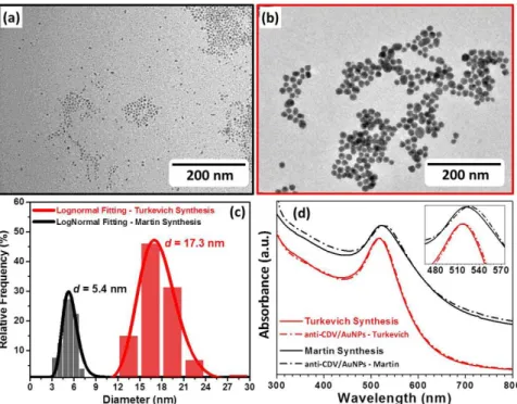
nanoparticles, the solution changed to a red color. Depending on the synthesis method AuNPs with different sizes are obtained. The Turkevich method 20 generated spherical particles with an average diameter of 17.3 nm (Figures 2B, 2C, and 2D), while the Martin method (NaBH4-NaOH)
21
Nascimento, TM et al. 6
Figure 2. (a) TEM micrograph of AuNPs synthesized via Martin method; (b) TEM micrograph of AuNPs synthesized via Turkevich method; (c) Relative frequency counting and lognormal distribution fitting of both AuNPs samples showing their average diameter (d); (f) UV–Vis spectrum of both AuNPs suspension, Martin synthesis (8 ×10⁻⁸ mol L−1) and Turkevich synthesis (1.5 × 10⁻⁹ mol L−1) before and after anti-CDV conjugation.
Microchannel optimization
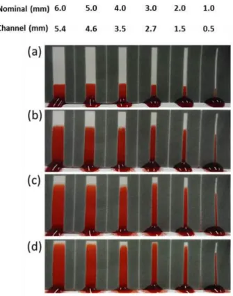
In order to evaluate the design of devices that allows the fastest migration rate of liquids into microchannel, six devices with the thicknesses of the hydrophilic channel ranging from 1 to 6 mm (nominal) were placed in contact with 200 µL of red ink for pen (Waterman, Paris), to assess the migration time of the front of the solvent in each microchannel, as shown in Figure 3A to 3D. After melting, the wax spreads into the original area of channel, resulting in channels with smaller width than the nominal values originally printed 4.
Figure 3. Study of variation of width of the microchannels and fluid migration times. (a) 30 s after the application of the test solution. (b) 1 min 30 s. (c) 2 min 30 s. (d) 3 min 10 s.
After 3:10 min, the channel filling was complete in the platform with 3 mm nominal diameter (2.7 mm channel), and thus this thickness was chosen to perform the lateral flow assay. When the printed microdevices are heated there is a narrowing of the channels due to the lateral spreading of the wax from the hydrophobic barriers, as described by Washburn Equation (Equation 1) 26, characterized by the permeation of melted wax in the porous paper matrix due to capillary flow 4. As long as the Washburn Equation is fluid and porous matrix dependent – the melted wax and the paper, respectively – there is a linear relationship between the width of the barrier and the printed line (Equation 2) and the width of the channel and the space between the hydrophobic barriers (Equation 3) 4, what allows one to predict the final dimensions of the device during designing steps.
(1)
(2)
(3)
L: Distance percolated by the liquid : Liquid viscosity
Nascimento, TM et al. 8
WB: Width of the barrier
WP: Width of the printed line
WC: Width of the hydrophilic channel
WG: Space between the two printed lines
Immunochromatographic assays
The simplest and fastest method of immobilization of biomolecules on cellulose support is through direct adsorption, in which the biomolecule of interest is non-covalently attached to the cellulose by intermolecular interactions such as hydrogen bonding, van der Waals, hydrophobic or electrostatic interactions 27. However, depending on the complexity of the matrix in which the analytes are, the previously immobilized biomolecules can be removed from their original adsorption site what can affect later the detection step 28. The immunocomplex solution (antibodies conjugated with AuNPs and complexed to viral antigens) applied to the paper was eluted with a larger volume of PBS buffer in order to avoid pH variations during the elution and thus preventing migration of the absorbed antibodies.
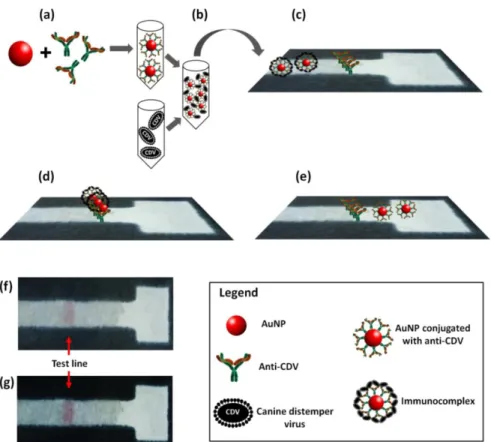
The immunocomplex solution presented a red coloration on paper that was almost completely absent when the diluent buffer was applied. Only when the fluid front containing the whole immunocomplex reached the previously immobilized antibodies on cellulose surface (the test and control lines, respectively), the anti-CDV immobilized on paper recognized the antigen contained on the immunocomplex and interacted with it, thus forming the red line characteristic of a positive result. Non-specific interactions between the immobilized antibodies on cellulose surface and gold nanoparticles could result in a false positive outcome; however, such a problem is avoided by the recovering of the surface of the conjugated AuNPs with BSA, which blocks the surface of the nanoparticles from undesirable interactions 29. If the antigen is absent in the original solution, then the immunocomplex will not interact with the previous immobilized antibodies on the surface of the cellulose and the AuNPs will not concentrate in that test/control region, and there will be no appreciable change in the color of paper to red, thus indicating a negative outcome. The Figure 4 schematizes all stages of a lateral flow assays.
Figure 4. Lateral flow immunoassay on a single layer of paper. (a) Conjugation of gold nanoparticles with anti-CDV. (b) Immunocomplex formation. (c) Anti-CDV antibodies immobilized on cellulose and application of immunocomplex solution at the bottom of microchannel. (d) Recognition of viral antigens contained in the immunocomplex by the immobilized anti-CDV. (e) Anti-CDV antibodies immobilized on cellulose surface and application of gold nanoparticles with antibodies solution (antigens are absent in this case) at the bottom of microchannel, not occurring recognition step between the anti-CDV immobilized with the antibodies in the nanoparticle, and thus there is no change in color in the test line (negative outcome). (f) Photograph of lateral flow positive assay. (e) Photograph of the lateral flow positive assay with an enhanced contrast (digital), for better visualization of the test line.
In the scheme presented in Figure 4 is possible to notice that after the immunocomplex application (Figure 4c) the solution reaches the immobilized antibodies zone on cellulose, and then the bio recognition occurs (Figure 4d) 19, with the consequent development of a red line. The maintenance of the reddish color of the solution suggests the stability of the immunocomplex, corroborating with the spectra presented in Figure 2D. This line indicates that there was a recognition between canine distemper virus and antibodies from the AuNPs, what provides signal intensification and then the red color appears in the test line (Figure 4F; 4G). These results showed that the expected interaction between the immobilized anti-CDV and the immunocomplex from the solution was efficient. Thus, we could monitor the success of the bioconjugation process with gold nanoparticles and the immobilization using bicarbonate/carbonate buffer. conjugation
The next step of this study involves method validation, in order to evaluate the ruggedness of devices, made by the comparison with standard methods (i.e., ELISA)
18
Nascimento, TM et al. 10
CONCLUSION
Immunochromatographic assays using a single layer of chromatographic paper and wax printing arises as a prototype with potential clinical use due to their simplicity and low manufacturing costs. AuNPs synthesis by the Turkevich method resulted in gold nanoparticles with an average diameter of 17.4 nm and a spherical shape ideal for carrying out bioconjugations and for immunological applications. By varying the width of the microchannels we found out that channels with 2.7 mm presented the fastest wetting rates and were chosen as the preferential design for the microdevices. The simplest method of antibody bioconjugation with gold nanoparticles by physical adsorption made it possible to detect canine distemper virus at the test line, indicating the potential use of such devices at home or in veterinary clinics.
ACKNOWLEDGEMENTS
The authors would like to thank the funding agencies FAPESP (Grant N. 2011/13997-8 and 2012/02093-3), CNPq (Grants N. 311323/2011-1, N. 131306/2013-8 and 205453/2014-7) and CAPES for scholarships and financial support, ParteCurae Analysis for the rabbit polyclonal anti-CDV serum production, Biovet for the CDV virus strain, the Laboratory of the Department of Materials Engineering, Federal University of São Carlos and the Institute of Chemistry of São Carlos, University of São Paulo for using their facilities and equipment for AuNPs characterization.
REFERENCES
1- Carrilho E, Phillips ST, Vella SJ, Martinez AW, Whitesides GM. Paper microzone plates. Anal Chem. 2009;81:5990–8.
2- Martinez AW, Phillips ST, Whitesides GM, Carrilho E. Diagnostics for the developing world: microfluidic paper-based analytical devices. Anal Chem. 2010;82:3–10.
3- Martinez AW, Phillips ST, Carrilho E, Thomas SW, Sindi H, Whitesides GM. Simple telemedicine for developing regions: camera phones and paper-based microfluidic devices for real-time, off-site diagnosis. Anal Chem. 2008;80:3699–707.
4- Carrilho E, Martinez AW, Whitesides GM. Understanding wax printing: a simple micropatterning process for paper-based microfluidics. Anal Chem. 2009;81:7091–5. 5- Faraday M. The Bakerian Lecture: Experimental Relations of Gold (and Other Metals) to
Light. Philos Trans R Soc London. 1857;147:145–181.
6- Nanodots S, Link S, El-sayed MA. Spectral Properties and Relaxation Dynamics of Surface Plasmon Electronic Oscillations in. J Phys Chem B 1999;103:8410–8426. 7- Fratila RM, Mitchell SG, Del Pino P, Grazu V, de la Fuente JM. Strategies for the
biofunctionalization of gold and iron oxide nanoparticles. Langmuir. 2014;30:15057– 15071.
8- Elghanian R. Selective Colorimetric Detection of Polynucleotides Based on the Distance-Dependent Optical Properties of Gold Nanoparticles. Science.1997; 277:1078–1081. 9- Mulvaney P. Surface Plasmon Spectroscopy of Nanosized Metal Particles. Langmuir
1996;12:788–800.
10- Nath N, Chilkoti A. A colorimetric gold nanoparticle sensor to interrogate biomolecular interactions in real time on a surface. Anal Chem. 2002;74:504–9.
11- Panda B, Chattopadhyay A. Synthesis of au nanoparticles at “all” pH by H2O2 reduction of HAuCl4. J Nanosci Nanotechnol. 2007;7:1911–5.
12- Ngo YH, Li D, Simon GP, Garnier G. Paper surfaces functionalized by nanoparticles. Adv Colloid Interface Sci. 2011;163:23–38.
13- Zhang C, Zhang Y, Wang S. Development of multianalyte flow-through and lateral-flow assays using gold particles and horseradish peroxidase as tracers for the rapid determination of carbaryl and endosulfan in agricultural products. J Agric Food Chem.
2006;54:2502–7.
14- Linares EM, Kubota LT, Michaelis J, Thalhammer S. Enhancement of the detection limit for lateral flow immunoassays: evaluation and comparison of bioconjugates. J Immunol Methods. 2012;375:264–70.
15- Schoch A, Kettenberger H, Mundigl O, Winter G, Engert J, Heinrich J, et al. Charge-mediated influence of the antibody variable domain on FcRn-dependent pharmacokinetics. Proc Natl Acad Sci. 2015;112:5997–6002.
16- Carvalho OV, Botelho CV, Ferreira CGT, Scherer PO, Soares-Martins JAP, Almeida MR, Silva Júnior A. Immunopathogenic and Neurological Mechanisms of Canine Distemper Virus. Adv Virol 2012;1-10.
17- Fischer CDB, Ikuta N, Canal CW, Makiejczuk A, Allgayer MDC, Cardoso CH, Lehmann FK, Fonseca AS, Lunge VR. Detection and differentiation of field and vaccine strains of canine distemper virus using reverse transcription followed by nested real time PCR (RT-nqPCR) and RFLP analysis. J Virol Methods. 2013;194:39–45.
18- Cho KH, Kim J, Yoo HA, Kim DH, Park SY, Song CS, et al. Use of hydrophilic extra-viral domain of canine distemper virus H protein for enzyme-linked immunosorbent assay development. J Vet Sci. 2014;15:503–9.
19- An DJ, Kim TY, Song DS, Kang BK, Park BK. An immunochromatography assay for rapid antemortem diagnosis of dogs suspected to have canine distemper. J Virol Methods 2008;147:244–9.
20- Turkevich J, Stevenson PC, Hiller J. A Study of the Nucleation and Growth Processes in the Synthesis of Colloidal Gold. Discuss Faraday Soc. 1951;11:55–75. 21- Martin MN, Basham JI, Chando P, Eah S-K. Charged gold nanoparticles in
non-polar solvents: 10-min synthesis and 2D self-assembly. Langmuir. 2010;26:7410–7. 22- Rajukumar K, Tripathi BN, Kurade NP, Parihar NS. An Enzyme-linked Immunosorbent
Assay using Immonoaffinity-purified Antigen in the Diagnosis of Caprine Paratuberculosis and Its Comparison with Conventional ELISAs. Vet Res Commun 2001;25:539–53.
23- Parolo C, Medina-Sánchez M, de la Escosura-Muñiz A, Merkoçi A. Simple paper architecture modifications lead to enhanced sensitivity in nanoparticle based lateral flow immunoassays. Lab Chip.2013;13:386–90.
24- Maffre P, Brandholt S, Nienhaus K, Shang L, Parak WJ, Nienhaus GU. Effects of surface functionalization on the adsorption of human serum albumin onto nanoparticles - a fluorescence correlation spectroscopy study. Beilstein J Nanotechnol. 2014;5:2036– 47.
25- Sperling R a, Parak WJ. Surface modification, functionalization and bioconjugation of colloidal inorganic nanoparticles. Philos Trans A Math Phys Eng Sci 2010;368:1333–83.
26- Washburn EW. The dynamics of capillary flow. Phys Rev 1921;17:273–83. 27- Credou J, Berthelot T. Cellulose: from biocompatible to bioactive material. J
Mater Chem B. 2014;2:4767.
28- Pelton R. Bioactive paper provides a low-cost platform for diagnostics. TrAC Trends Anal Chem. 2009;28:925–42.
29- Sun J, Guo A, Zhang Z, Guo L, Xie J. A Conjugated Aptamer-Gold Nanoparticle Fluorescent Probe for Highly Sensitive Detection of rHuEPO-α. Sensors 2011;11:10490– 501.
30- Swets JA. Measuring the Accuracy of Diagnostic Systems. Science. 1988;240:1285–93.
31- Buck AA, Gart JJ. Comparison of a screening test and a reference test in epidemiologic studies. Am J Epidemiol 1968;83:586–92.