Licenciado em Biologia
Molecular mechanisms of sexual
development in basidiomycetes:
exploring connections with lifestyles
Dissertação para obtenção do Grau de Doutor em
Biologia
Orientador: Doutor José Paulo Sampaio, Prof. Auxiliar, FCT/UNL
Co-orientador: Doutora Paula Gonçalves, Profª. Auxiliar, FCT/UNL
Júri:
Presidente: Profª. Doutora Elvira Júlia da Conceição Matias Coimbra Arguentes: Profª. Doutora Geraldine Butler
Prof. Doutor Alexander Idnurm Vogais: Prof. Doutor Jan Schirawski
Prof. Doutor Manuel António da Silva Santos
Prof. Doutor Álvaro Luís Afonso Moreira Rato Fonseca
Profª. Doutora Paula Maria Theriaga Mendes Bernardo Gonçalves Prof. Doutor José Paulo Nunes de Sousa Sampaio
Inv. Principal Doutora Isabel Antunes Mendes Gordo
i
Marco António Dias Coelho
Molecular mechanisms of sexual
development in basidiomycetes: exploring
connections with lifestyles
“
c
opyright”
by Marco A. Coelho, FCT/UNL
iii
Acknowledgments
I want to express my deeply gratitude to my thesis advisors, Profª. Paula Gonçalves and Prof. José Paulo Sampaio, for their encouragement, enthusiasm and thoughtful guidance along these years.
I also want to thank the members of the thesis committee, Prof. Álvaro Fonseca and Dr. Isabel Gordo, for their interest and suggestions to improve this thesis.
I would like to thank Profª. Regine Kahmann for giving me the opportunity to work at the Department of Organismic Interactions of Max Planck Institute for Terrestrial Microbiology (Marburg, Germany), under supervision of Prof. Jan Schirawski, to whom I am particularly grateful for the helpful suggestions and support.
I am indebted to current and former lab colleagues for providing a friendly lab atmosphere and helpful discussions from which I have learned so much.
I thank my friends for their support and for helping me to stay focused during these years. I greatly value their friendship and I genuinely appreciate their trust in me.
Most importantly, this work would not have been possible without the love, concern, patience and endless support of my family to whom I dedicate this dissertation. A special thank to my dearest and nearest „girl of my life‟ for the immeasurable sacrifices and support and for never failing faith on me.
v
Resumo
Este trabalho procura contribuir para o estudo dos mecanismos moleculares de reprodução sexuada em fungos e para a compreensão da sua relevância na evolução dos diferentes modos de vida dos fungos (parasita vs. saprofítico) e na emergência de linhagens assexuadas.
O desenvolvimento sexual e a patogenicidade estão interligadas em muitos basidiomicetas fitoparasitas, sendo Ustilago maydis o exemplo emblemático de um grupo genericamente
designado por “carvões” ou “morrões”. Estes fungos causam doenças em plantas economicamente
importantes como o milho e a cana do açúcar, e estão classificados no subfilo Ustilaginomycotina (uma das três linhagens principais de basidiomicetas). No entanto, as várias espécies de Ustilago estão filogeneticamente relacionadas com espécies do género Pseudozyma que são consideradas saprófitas e assexuadas. Neste trabalho, o estudo centrado na identificação dos genes que determinam a identidade sexual em fungos (genes MAT ou mating type) mostrou que Pseudozyma prolifica é uma espécie sexuada, fitopatogénica e biologicamente indistinguível de U. maydis. Para as restantes espécies de Pseudozyma, a análise molecular do gene PRF1, que codifica para uma proteína com um papel central na regulação do desenvolvimento sexual em U. maydis, não revelou evidências substanciais de perda de reprodução sexuada neste grupo. No entanto, alguns resultados sugerem que algumas espécies de Pseudozyma poderão estar a adaptar-se a um modo de vida saprofítico.
A linhagem de basidiomicetas que terá divergido primeiro (subfilo Pucciniomycotina) compreende também fitoparasitas muito revelantes (ex. as ferrugens), bem como grupos unicamente constituídos por organismos saprofíticos. Entre os últimos estão incluídas as leveduras pigmentadas (red yeasts) classificadas na ordem Sporidiobolales e que, como modelo de estudo, têm a vantagem de completar o seu ciclo de vida em meio de cultura laboratorial. No entanto, este grupo manteve-se praticamente inexplorado no que diz respeito à caracterização dos sistemas que determinam compatibilidade sexual (sistemas MAT), à identificação de genes MAT e às relações evolutivas entre as várias espécies sexuadas e assexuadas que abrange. Uma análise extensiva de mais de 200 estirpes pertencentes a 32 espécies de Sporidiobolales sugere que as linhagens asexuadas têm origem a partir das linhagens sexuadas, mas que não perduram tempo suficiente para formarem espécies estritamente assexuadas desprovidas de genes MAT. Uma investigação mais aprofundada nas leveduras Rhodosporidium toruloides e Sporidiobolus salmonicolor permitiu, pela primeira vez em Pucciniomycotina, a identificação do conjunto completo de genes MAT. A caracterização mais detalhada e multidisciplinar do sistema MAT em S. salmonicolor produziu resultados inesperados que levaram à proposta de um novo sistema substancialmente diferente dos dois paradigmas anteriormente existentes em fungos (sistemas bipolar e tetrapolar). Dado que o subfilo Pucciniomycotina é basal nos basidiomicetas, este novo sistema designado pseudo-bipolar constitui um contributo significativo para o estudo da evolução dos sistemas MAT em fungos.
vii
Abstract
This work concerns the investigation of the molecular mechanisms of sexual reproduction in fungi and their possible implication for fungal lifestyles (parasitic vs. saprobic) and for the emergence of asexual fungal lineages.
The association between pathogenicity and sexuality is well-known in the basidiomycete plant parasite Ustilago maydis (subphylum Ustilaginomycotina), an economically important smut fungus. However, Ustilago species are phylogenetically interspersed with species of the genus Pseudozyma, which are considered saprobic and asexual. In this work, a study focused on genes involved in determining sexual identity (mating type or MAT genes), showed that Pseudozyma prolifica retains full sexual competence and pathogenicity, being therefore indistinguishable from U. maydis. For other Pseudozyma species, molecular analyses of PRF1, a gene that encodes a master regulator of sexual reproduction in U. maydis, showed no substantial evidence of loss of sexual reproduction. However, some clues were also found suggesting that some Pseudozyma species may be evolving towards a saprobic lifestyle.
The earliest derived lineage of Basidiomycota (subphylum Pucciniomycotina) includes also important plant pathogens (rust and anther smut fungi) as well as lineages composed solely of saprobic organisms. Among the latter, the red yeasts of the order Sporidiobolales have the advantage of completing their life cycle in culture media, but have remained very little explored concerning the characterization of mating systems, the identification of MAT genes and the evolutionary relationships between sexual and asexual species. A comprehensive analysis of more than 200 strains belonging to 32 species of the Sporidiobolales indicated that asexuality seems to originate frequently from sexual lineages, but does not seem to persist long enough to form truly asexual species devoid of MAT genes. A more in-depth investigation of the red yeasts Rhodosporidium toruloides and Sporidiobolus salmonicolor allowed the identification for the first time in the Pucciniomycotina of the complete set of MAT genes. A detailed and multidisciplinary characterization of the mating system in the latter species yielded surprising results. A novel mating system that differs substantially from the two mating paradigms in basidiomycetes, the bipolar and tetrapolar systems, was brought to light. Given the basal phylogenetic position of the Pucciniomycotina within the Basidiomycota, this new system designated pseudo-bipolar, constitutes a significant contribution to the study of the evolution of MAT systems in fungi.
ix
Abbreviations
ABC ATP-binding cassette AIC Akaike information criterion ATP Adenosine triphosphate
bp Base pair
cAMP Cyclic adenosine monophosphate
CBE Chlorazole Black E
cDNA Complementary DNA
cM centimorgan
CT Cycle threshold
CuSO4 Copper (II) sulphate
D1D2 Domains 1 and 2 of the 26S rDNA DAPI 4‟,6-diamidino-2-phenylindole DIC Differential interference contrast
DMSO Dimethyl sulfoxide
dN non-synonymous substitutions per non-synonymous site DNA Deoxyribonucleic acid
DNase Deoxyribonuclease
dpi Days post infection
dS synonymous substitutions per synonymous site
HCl Chloridric acid
HD Homeodomain
HMG High-mobility group
hr Hour(s)
ITS Internal transcribed spacer
IUPAC International Union of Pure and Applied Chemistry JGI Joint Genome Institute
kb Kilobase pair
KOH Potassium hydroxide
LMP Low melting point
LSU Large subunit (ribosome) MAP Mitogen activated protein MAPK Mitogen activated protein kinase
MAT Mating type
Mb Megabase pair
MELs Mannosylerythritol lipids MgCl2 Magnesium chloride
min Minute(s)
ML Maximum likelihood
MnSO4 Manganese (II) sulphate
x
MYA Million years ago
MYP Malt, Yeast extract and Peptone (Soytone) medium NCBI National Center for Biotechnology Information OD640 Optical density at 640 nm
ORF Open reading frame
PAK p21-activated kinase family PCR Polymerase chain reaction PDA Potato dextrose agar medium
PKA Protein kinase A
PP Pseudozyma prolifica
PREs Pheromone response elements
rDNA ribosomal DNA
RFLP Restriction fragment length polymorphism
RHA Rhodotorucine A
RNA Ribonucleic acid
RNase Ribonuclease
rpm Rotations per minute
RT-PCR Reverse transcription polymerase chain reaction
s Second(s)
Tris 2-Amino2-hydroxymethyl-propane-1,3-diol UAS Upstream activating sequence
UV Ultraviolet radiation
v/v Volume per volume
w/v Weight per volume
WGD Whole-genome duplication
YEPS Yeast extract, peptone and sucrose medium
Abbreviations of relevant culture collections
CBS Centraalbureau voor Schimmelcultures, Utrecht, The Netherlands
IAM IAM Culture Collection, Institute of Molecular and Cellular Biosciences, The University of Tokyo, Japan (collection transferred to JCM).
JCM Japan Collection of Microorganisms, Riken Bioresource Center, Saitama, Japan
NRRL Agriculture Research Service (ARS) Culture collection, Peoria, Illinois, USA
PYCC Portuguese Yeast Culture Collection, CREM, Faculdade de Ciências e Tecnologia, Universidade Nova de Lisboa, Caparica, Portugal
xi
Abbreviated species names
C. albicans Candida albicans
C. cinerea Coprinopsis cinerea
C. gattii Cryptococcus gattii
C. heterostrophus Cochliobolus heterostrophus
C. neoformans Cryptococcus neoformans
D. hansenii Debaryomyces hansenii
K. lactis Kluyveromyces lactis
P. anserina Podospora anserina
P. microspora Pholiota microspora
S. cerevisiae Saccharomyces cerevisiae
S. commune Schizophyllum commune
S. pombe Schizosaccharomyces pombe
S. reilianum Sporisorium reilianum
Abbreviations of frequently used generic names
M. Microbotryum
N. Neurospora
P. Pseudozyma
R. Rhodosporidium
Rh. Rhodotorula
S. Sporidiobolus
Sp. Sporobolomyces
xiii
Index
Chapter 1
General Introduction
1.1. The importance of sexual reproduction ... 2
1.1.1. Evolutionary significance of sexual reproduction in eukaryotes ... 2
1.1.2. Sex and lifestyle in Fungi ... 2
1.1.3. Genomic signatures of sex ... 4
1.2. Mating and sexual identity in Fungi ... 6
1.2.1. Mating in Ascomycota ... 6
1.2.1.1. Mating type (MAT) genes of ascomycetes ... 7
1.2.1.2. Cell identity in Saccharomyces and Candida clades (Saccharomycotina) ... 7
1.2.1.3. Organization of MAT locus in filamentous ascomycetes (Pezizomycotina) ... 12
1.2.1.4. Neurospora tetrasperma as a model for evolution of sex chromosomes ... 13
1.2.2. Mating in Basidiomycota ... 14
1.2.2.1. The pheromone / receptor sensing system in basidiomycetes ... 15
1.2.2.2. Homeodomain transcription factors – the second compatibility checkpoint ... 16
1.2.2.3. Tetrapolar mating systems – the smut fungi ... 20
1.2.2.4. Tetrapolar mating systems – the origin of thousands of mating types ... 21
1.2.2.5. Evolution of bipolar systems – multiallelic bipolar basidiomycetes ... 23
1.2.2.6. Evolution of bipolar systems – biallelic bipolar basidiomycetes ... 25
1.2.2.7. Mating systems in Pucciniomycotina –Microbotryum violaceum ... 27
1.2.3. Homothallism in Fungi ... 28
1.2.3.1. Primary homothallism – compatible MAT genes in a single genome ... 28
1.2.3.2. Secondary homothallism – pseudohomothallism ... 29
1.2.3.3. Secondary homothallism – monokaryotic fruiting or same-sex mating ... 29
1.3. Phylogenetic and ecological relationship between parasitic and saprobic basidiomycetous yeasts studied in this thesis ... 31
xiv
Chapter 2
Evolutionary relationship between sexual and asexual species of
Ustilaginales
–
The
Ustilago
/
Pseudozyma
system
2.1. Introduction ... 34
2.2. Material and methods ... 37
2.2.1. Yeast strains, growth conditions and mating tests ... 37
2.2.2. PCR detection of MAT genes in P. prolifica ... 37
2.2.3. Plant pathogenicity tests ... 39
2.2.4. Microscopic observation of fungal growth and proliferation in planta ... 40
2.2.5. Spore germination and segregation analysis ... 40
2.2.6. PCR amplification and sequence of PRF1 homologues ... 41
2.2.7. Sequence data and phylogenetic analysis ... 41
2.2.8. Quantification of the PRF1 expression by Real-Time RT-PCR ... 41
2.3. Results and discussion ... 43
2.3.1. Identification of MAT loci in P. prolifica ... 43
2.3.2. Life cycle and sexual behaviour of P. prolifica ... 44
2.3.3. Evolution of the pheromone response factor (PRF1) in Pseudozyma ... 47
2.3.4. Expression analysis of the PRF1 homologue of P. aphidis ... 49
2.4. Final remarks ... 54
Chapter 3
Identification of mating type (
MAT
) genes in the red yeast
Rhodosporidium
toruloides
3.1. Introduction ... 563.2. Material and methods ... 57
3.2.1. Yeast strains, culture conditions and mating tests ... 57
3.2.2. DNA extraction and common settings for PCR amplification ... 57
3.2.3. Mating type specificity of RHA genes ... 58
3.2.4. Genome walking ... 58
3.2.5. PCR amplification and sequencing of the STE20 alleles ... 58
3.2.6. PCR amplification of putative MAT A1 pheromone receptor (STE3.A1) ... 60
xv
3.3. Results and discussion ... 61
3.3.1. MAT status of R. toruloides strains ... 61
3.3.2. Genomic regions in the vicinity of the pheromone genes in MAT A1 ... 63
3.3.3. Identification of a MAT-specific region in MAT A2 of R. toruloides ... 64
3.3.4. Comparison with syntenic genomic regions from related red yeasts ... 67
3.3.5. Putative MAT locus in Rhodosporidium and related red yeast species ... 72
3.4. Final remarks ... 74
Chapter 4
The pseudo-bipolar
MAT
system of the red yeast
Sporidiobolus salmonicolor
4.1. Introduction ... 764.2. Material and methods ... 77
4.2.1. Strains and mating type designations ... 77
4.2.2. Common settings for PCR amplification ... 77
4.2.3. PCR amplification and sequencing of the MAT A2 pheromone receptor ... 77
4.2.4. PCR amplification and sequencing of the HD1/HD2 region ... 79
4.2.5. Correlation between mating behaviour and the presence of pheromone receptor genes ... 81
4.2.6. Germination of teliospores and microscopic monitoring of meiosis ... 81
4.2.7. Micromanipulation of teliospores and segregation analysis ... 82
4.2.8. Sequence data and phylogenetic analyses... 82
4.2.9. Estimation of the evolution rates of the HD1 gene ... 83
4.2.10. MAT specificity and phylogenetic analysis of genes located at variable distances from the HD1/HD2 and the pheromone receptor regions ... 83
4.2.11. Genotyping of the meiotic progeny ... 83
4.3. Results and discussion ... 84
4.3.1. Identification of MAT genes in both mating types of S. salmonicolor ... 84
4.3.2. Allele number and mating type specificity of HD1/HD2 and pheromone receptor genes in S. salmonicolor ... 86
4.3.3. A pseudo-bipolar mating system ... 92
4.3.4. A model for the evolution of MAT loci in basidiomycetes ... 102
xvi
Chapter 5
Evidence for maintenance of sex determinants but not of sexual stages in red
yeasts
5.1. Introduction ... 108
5.2. Material and methods ... 109
5.2.1. Strains and mating tests ... 109
5.2.2. PCR detection and sequencing of MAT A1 and MAT A2 pheromone receptor genes and the conserved surrounding regions ... 109
5.2.3. PCR amplification and sequencing of the HD1/HD2 region and the RHA2 pheromone precursor gene ... 110
5.2.4. Sequence data and phylogenetic analysis ... 110
5.2.5. Estimation of the evolution rates of 5‟ end and homeodomain regions of the HD1 gene ... 111
5.3. Results and discussion ... 111
5.3.1. Updated phylogeny of the Sporidiobolales ... 111
5.3.2. Pheromone receptor genes as molecular markers of mating type identity ... 113
5.3.3. Pheromone receptor genes in asexual species ... 114
5.3.4. Pheromone receptor genes in self-fertile species ... 116
5.3.5. Phylogeny of the pheromone receptors ... 117
5.3.6. Structure of the MAT A1 and MAT A2 genomic regions encompassing the alternate pheromone receptors ... 117
5.3.7. Diversity and evolution of the homeodomain transcription factors ... 121
5.4. Final remarks ... 125
Chapter 6
Concluding remarks and future directions
6.1. Lifestyle and mode of reproduction in Ustilago/Pseudozyma ... 1286.2. Red yeasts as a novel model for studying the evolution of sex-determining regions in fungi ... 129
xvii
Appendix I
Additional information pertaining to Chapters 2 and 3
I.1. GenBank accession numbers of sequences used in Figure 2.7 ... 152
I.2. RNA isolation and purification from Pseudozyma and Ustilago strains ... 154
I.3. NCBI Trace Archive sequences numbers used to obtain selected genomic regions
of Sporidiobolus salmonicolor (IAM 12258 = CBS 483) ... 155
I.3. GenBank accession numbers of the novel Rhodosporidium toruloides sequences
described in Chapter 3 ... 156
Appendix II
Additional information pertaining to Chapter 4
II.1. NCBI Trace Archive sequences used to assemble the STE3.A2 gene of
Rhodosporidium babjevae (WP1) and the HD1/HD2 region of both Sporidiobolus
salmonicolor (IAM 12258 = CBS 483) and R. babjevae ... 158
II.2. NCBI Trace Archive sequences used to assemble eight genes from the pheromone receptor region and four genes from the HD1/HD2 region in S. salmonicolor (IAM
12258 = CBS 483) ... 159
II.3. NCBI Trace Archive sequences used to assemble nine S. salmonicolor genes that
are located in four different scaffolds of the Sporobolomyces roseus genome ... 160
Appendix III
Additional information pertaining to Chapter 5
III.1. List of species/ strains used in Chapter 5 ... 164
III.2. Primers and specific PCR conditions used in Chapter 5 to amplify several genomic
xix
Figure Index
Chapter 1
Figure 1.1. Schematic representation of the life cycle of the smut fungus U. maydis (on
the left) and of the human pathogen C. neoformans (on the right) ...4
Figure 1.2. Structure and diversity of the MAT loci in the different ascomycete lineages ...8
Figure 1.3. Mating-type regulation pathways in three ascomycete yeasts... 11
Figure 1.4. Structure and diversity of the MAT loci in the different basidiomycete lineages ... 18
Figure 1.5. Life cycle of Coprinopsis cinerea ... 24
Figure 1.6. Mechanisms of homothallism in fungi ... 30
Chapter 2
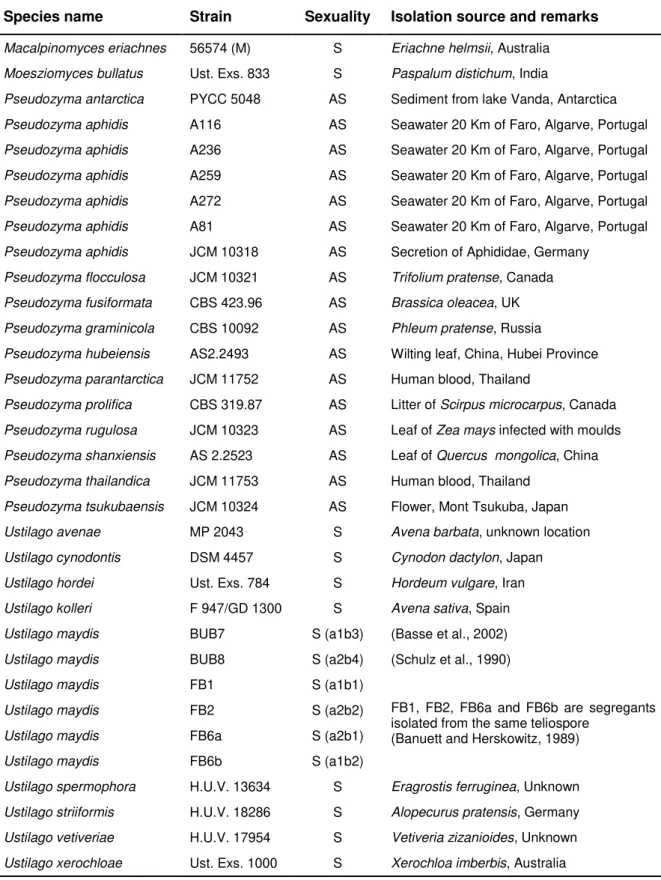
Figure 2.1. PKA and MAPK signalling during mating in Ustilago maydis... 36Figure 2.2. Genomic regions encompassing both MAT loci of Ustilago maydis ... 43
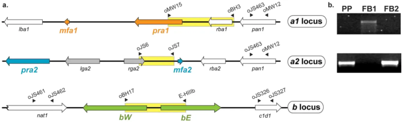
Figure 2.3. Mating tests between P. prolifica (PP) and U. maydis strains on PDA-charcoal plates ... 44
Figure 2.4. Time course microscopic observation of the infection steps of maize seedlings by PP × FB1 ... 45
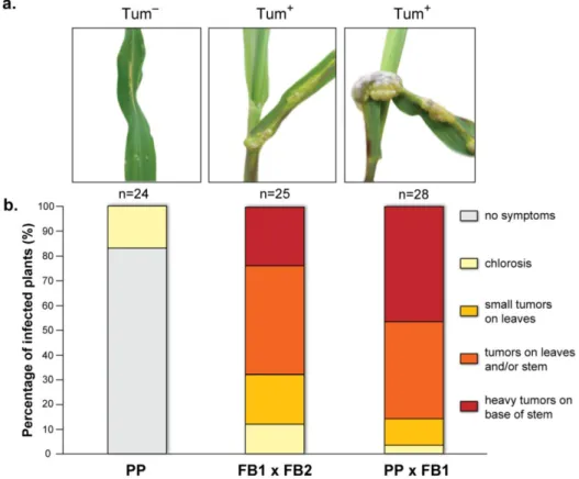
Figure 2.5. Plant pathogenicity resulting from infection with PP × FB1 ... 46
Figure 2.6. Progeny derived from the cross PP × FB1 ... 47
Figure 2.7. Phylogenetic relationships between sexual and asexual species of Ustilaginales ... 48
Figure 2.8. Phylogenetic tree based on the amino acid sequences of Prf1 obtained from the several Ustilaginales species ... 50
Figure 2.9. Conservation of PKA and MAPK phosphorylation sites in Prf1 ... 51
xx
Chapter 3
Figure 3.1. Mating behaviour of Rhodosporidium toruloides ... 62
Figure 3.2. Structure of the STE20 region of the MAT A1 and MAT A2 loci of R. toruloides ... 63
Figure 3.3. Conserved domains of fungal PAK kinases ... 65
Figure 3.4. Nucleotide sequence of the genomic region encoding the RHA2.A2 gene,
showing the predicted pheromone precursor sequence ... 66
Figure 3.5. Mating type-specificity of the RHA2.A2/STE20 region ... 66
Figure 3.6. Structure of a putative MAT locus in Sp. roseus and comparison with
homologous genomic regions of R. toruloides and S. salmonicolor ... 68
Figure 3.7. Phylogenetic tree showing the relationships between PAK kinases from
different fungi, based on alignment of the respective protein sequences ... 69
Figure 3.8. Predicted MAT A1 and MAT A2 pheromone precursor peptides ... 71
Chapter 4
Figure 4.1. Organization of the genomic regions containing the pheromone receptor
(STE3) and the HD1/HD2 genes in S. salmonicolor ... 85
Figure 4.2. Diversity and phylogeny of MAT gene alleles in S. salmonicolor ... 87
Figure 4.3. Phylogeny of HD1/HD2 alleles in the species complex S. salmonicolor/S.
johnsonii ... 89
Figure 4.4. Phylogeny of pheromone receptors in the Basidiomycota ... 90
Figure 4.5. Evolution rate of the HD1 gene in S. salmonicolor... 91
Figure 4.6. Life cycle of Sporidiobolus salmonicolor ... 93
Figure 4.7. Segregation analysis of the STE3 and HD1/HD2 genes after meiosis ... 94
Figure 4.8. Self-fertile phenotype of the diploid strain T8 ... 95
Figure 4.9. Sexual proficiency of T7.1, the new mating type of S. salmonicolor ... 96
Figure 4.10. Analysis of meiotic progeny ... 97
Figure 4.11. Phylogeny of several genes located in the scaffold harbouring the STE3
xxi
Figure 4.12. Phylogeny of genes located in the scaffold harbouring the HD1/HD2 region ... 101Figure 4.13. Phylogeny of HD1/HD2 alleles in Rhodosporidium babjevae ... 103
Figure 4.14. The pseudo-bipolar system and the evolution of MAT loci in Basidiomycota ... 105
Chapter 5
Figure 5.1. Molecular phylogeny of Sporidiobolales based on a concatenated alignment of
the ITS region (ITS1, 5.8S and ITS2) and the D1/D2 domain of the LSU rRNA ... 112
Figure 5.2. Phylogeny of the pheromone receptors of several red yeast species ... 115
Figure 5.3. Synteny of the genomic regions flanking the alternate pheromone receptors in
MAT A1 and MAT A2 strains of several red yeasts species ... 119
Figure 5.4. Synteny between MAT A1 and MAT A2 pheromone receptor loci in different
red yeast species ... 120
Figure 5.5. Diversity and phylogeny of HD1/HD2 alleles in several red yeast species ... 122
Figure 5.6. Pheromone precursors of different red yeast species ... 123
Figure 5.7. Evolution rate of the 5‟ end and homeodomain regions of the HD1 gene in
sexual and asexual red yeast strains ... 124
Appendix III
Figure III.1. Annealing sites of the primers used to amplify the genomic regions under
xxiii
Table Index
Chapter 2
Table 2.1. Strains used in Chapter 2 and relevant information pertaining to them ... 38
Table 2.2. List of primers and specific PCR conditions used in Chapter 2 ... 39
Chapter 3
Table 3.1. List of primers and specific PCR conditions used in Chapter 3 ... 59
Chapter 4
Table 4.1. Strains used in Chapter 4 and relevant information pertaining to them ... 78
Table 4.2. List of primers and specific PCR conditions used in Chapter 4 ... 80
Appendix I
Table I.1. Sequence accession numbers of LSU rDNA (D1/D2 region) and ITS
sequences of species used to construct the phylogenetic tree in Figure 2.7 ... 152
Appendix III
Table III.1. List of species/ strains used in Chapter 5 and relevant information pertaining
to them... 164
Table III.2. List of primers and specific PCR conditions used to amplify the regions under
1. CHAPTER
2
1.1. The importance of sexual reproduction
1.1.1. Evolutionary significance of sexual reproduction in eukaryotes
The origin and maintenance of sex and the reason why most living organisms have adopted a more costly and inefficient (sexual) mode of reproduction is still a mystery for evolutionary biologists. Sexual populations have a two-fold cost compared to asexual populations (Maynard-Smith, 1978). Firstly, a sexual population consists of two sexes and only one is capable of producing offspring. By contrast, in asexual populations, each individual can produce its own progeny, which implies that an asexual population has an inherent capacity to grow more rapidly each generation. Secondly, sexually reproducing diploid organisms will only pass on one-half of their genes to any given offspring because gametes are haploid, whereas in asexual reproduction, the full genome is transmitted from a parental individual to its progeny. Finally, another example of the cost of sex is that mating partners must search for each other to mate (Maynard-Smith, 1978).
Despite its apparent disadvantages, sexual reproduction is an almost universal trend in eukaryotes (Dacks and Roger, 1999; Ramesh et al., 2005), which suggests that it must also confer important benefits. More than a century ago, August Weismann was the first to advocate that the increased genetic variation among the offspring as a product of sex would allow more efficient natural selection and in this way accelerate evolution (Weismann, 1904). Other benefits of sexual reproduction are that sex and recombination serves to purge the genome of deleterious mutations (Muller, 1964) and to generate recombinant progeny better suited in an ever-changing environment (Kondrashov, 1993). In line with this, an increased genetic diversity as a product of recombination is assumed to raise the chance for enhanced fitness, therefore greatly surpassing the efficiency of mitotic reproduction (Barton and Charlesworth, 1998; Goddard et al., 2005). Hence, sex persistence underlines its importance both for individual fitness and long-term survival of species (Hadany and Comeron, 2008).
1.1.2. Sex and lifestyle in Fungi
However, evidence that the cost of sex can be effectively counterbalanced comes from the existence of species that are capable of both sexual and asexual reproduction, as many fungal species. While in fungi, sexual reproduction is usually observed in periods of environmental instability or in sub-optimal ecological conditions, asexual reproduction is preferred when conditions are more favourable. Therefore, the co-existence of sexual and asexual reproduction in fungi might be viewed as an indication that sex offers a selective advantage.
3 most ecosystems (Kirk et al., 2008). Most species are saprobes and efficient decomposers of organic matter, and others evolved symbiotic associations with plants. Furthermore, and of particular interest, while about 32% of fungi are plant pathogens, only about 0.5% are clinically significant human pathogens (Kirk et al., 2008). The majority of known fungi are included in the two sister phyla Ascomycota and Basidiomycota. The Ascomycota outsize the Basidiomycota and comprises most of the plant pathogens and about 90% of the human pathogenic fungi (Morrow and Fraser, 2009). In the Basidiomycota, although the largest clade includes saprobic species (the mushrooms), about one-third of the basidiomycetes (ca. 10 000 species) are plant pathogens (the rust and smut fungi) and only close to 40 species are known to cause infection in humans and animals (Kirk et al., 2008; Morrow and Fraser, 2009).
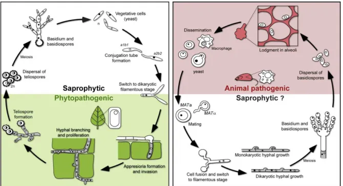
Sexual reproduction and meiosis have been well established for many fungal species and many sexual systems have served as invaluable eukaryotic models of development (Zarnack and Feldbrügge, 2007; Zarnack and Feldbrügge, 2010), transcription regulation (Gustin et al., 1998; Basse and Farfsing, 2006) and signalling pathways (Xue et al., 2008). Research in the field has also been prompted by the fact that for many plant or animal fungal pathogens, sexual reproduction is intimately linked with pathogenicity (Sexton and Howlett, 2006), a feature particularly important in the case of dimorphic pathogenic species. These species alternate between an asexual, single-celled growth form (yeast) and a sexual stage characterized by filamentous growth (mycelia). The asexual (mitosporic) and sexual (meiosporic) forms are also known as the „anamoph‟ and the „teleomorph‟, respectively (Seifert and Gams, 2001). Although dimorphic plant pathogens, such as the smut fungus Ustilago maydis, may hypothetically persist in the asexual (saprophytic) yeast phase, sexual reproduction and concomitant host infection are indispensable for completion of the life cycle (Figure 1.1) (Bakkeren et al., 2008; Nadal et al., 2008). On the other hand, dimorphic pathogens that cause infection in the yeast stage have been also documented, like the human pathogen Cryptococcus neoformans. Infection caused by this organism is thought to be mediated by inhalation of spores produced in the sexual stage (basidiospores), which indicates that a dimorphic transition from a saprophytic filamentous form (sexual stage) to a yeast form (asexual stage) is crucial to initiate the infection process (Heitman, 2006; Nielsen and Heitman, 2007; Heitman, 2010) (Figure 1.1).
4
and asexual spore production (Bell, 1982). These in turn are the conditions when the production of sexual survival spores is important to produce genetically variable offspring to maximize success in face of changed environmental conditions (Bell, 1982; Aanen and Hoekstra, 2007). In light of this, confirmation of strict asexuality is an invaluable information (although often difficult to ascertain), particularly when several lines of evidences suggest that asexuality is a derived character among fungi (LoBuglio et al., 1993).
Figure 1.1. Schematic representation of the life cycle of the smut fungus U. maydis (on the left) and of
the human pathogen C. neoformans (on the right). The yeast form in U. maydis is non-pathogenic and upon mating with a compatible partner switches to an infectious filamentous form that can invade the plant host. In C. neoformans, a similar dimorphic switch originates the potentially infectious propagules (basidiospores) to infect an animal host (adapted from Morrow and Fraser, 2009).
1.1.3. Genomic signatures of sex
5 no sexual stage has ever been observed, implying that sex is either absent, very rare or difficult to observe in nature.
The classical and most unambiguous approach used in looking for evidence of sex is the direct observation of biological mating ability with production of viable and fertile progeny. However, failure to induce sexual reproduction in the laboratory may due to sub-optimal culture conditions rather than inability to mate, which implies that a suitable mating scenario and mating partners must be identified for a successful mating. To circumvent this problem, recent advances in DNA sequencing and genome-wide studies allowed the application of molecular analyses to access the degree of sexual recombination in natural populations as well as to search in genomes for genes involved in sexual reproduction and meiosis. Combining these strategies, convincing evidence exist that asexual fungal species have derived from sexual ancestors (LoBuglio et al., 1993; Anderson and Kohn, 1995), and that the majority of the extant presumed asexual species have phylogenetically close relatives capable of sexual reproduction (Kurtzman and Robnett, 1998; Begerow et al., 2000). Therefore, genuinely and long-standing asexual fungal lineages are indeed rare but, in some cases, asexual reproduction seems to contribute as a significant component to the genetic structure of a species population (Fisher et al., 2005). Nevertheless, even in such clonal populations genetic footprints of recombination may still be detected (Taylor et al., 1999; Fisher et al., 2005).
Of special interest, genomic approaches have been recently used with success to detect genes required for sexual reproduction and indirectly recognize sexual reproduction in some animal pathogenic species (viz. Candida albicans and Aspergillus fumigatus), which so far were believed to propagate exclusively by asexual reproduction (Tzung et al., 2001; Paoletti et al., 2005). These approaches are based on the assumption that if a process has been lost due to a fitness cost to maintain an unused trait, a reduced selective functional constraint for the genes necessary for that function is expected, and they should accumulate deleterious mutations, eventually becoming pseudogenes (Lynch and Conery, 2000; Rouquier et al., 2000). Using this reasoning, the presence of intact, non-degenerate, genes that are specifically involved in mating and sexual reproduction is a good evidence of capacity of sex (Schurko and Logsdon, 2008).
6
1.2. Mating and sexual identity in Fungi
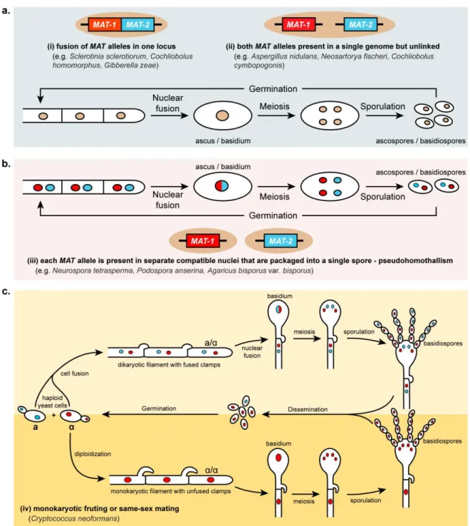
In fungi, sexual identity is always established in the haploid stage (Billiard et al., 2011) and determined by specialized genomic regions called mating type (MAT) loci (Fraser and Heitman, 2005). Results of several studies have revealed that fungal MAT loci vary extensively in length and in genetic content, which is further translated into a remarkable diversity of mating strategies and sex-determining systems (Casselton, 2002; Fraser and Heitman, 2004; Lee et al., 2010b). Most fungal species capable of sexual reproduction are heterothallic (self-sterile) but others are homothallic (self-fertile) (Bakkeren and Kronstad, 1994). In heterothallic species, mating occurs between two sexually compatible haploid individuals that are morphologically identical but are distinguished genetically at the MAT locus. In homothallic species, each individual can mate with itself and several genetic mechanisms have been shown to form the basis for this sexual behaviour (Hicks and Herskowitz, 1976; Lin et al., 2005; Alby et al., 2009; Rodriguez-Carres et al., 2010). The underlying genetic mechanisms of some forms of homothallism are introduced in section 1.2.3.
1.2.1. Mating in Ascomycota
In most species within the phylum Ascomycota, sexual compatibility is determined by short DNA sequences located at a single MAT locus encoding key transcription factors that govern cell type identity and developmental fate (Butler, 2007). This molecular architecture was first determined in the model yeast Saccharomyces cerevisiae, where it was shown that two cell types (a and strains) could be distinguished by different sequence information present at the MAT locus (Astell et al., 1981). These observations were crucial towards the elucidation of the molecular foundations of the so-called bipolar mating behaviour, which is characterized by the existence of two sexes or mating-types. Subsequent analysis of MAT loci in other ascomycetous species led to the identification and definition of the MAT „idiomorphs‟ (instead of „alleles‟) to denote unique sequences unrelated in
7
1.2.1.1. Mating type (
MAT
) genes of ascomycetes
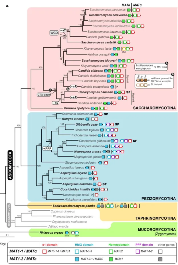
In ascomycetes, MAT loci encode transcription factors belonging to the -domain, homeodomain (HD) or high-mobility group (HMG) families that regulate determinants of sexual identity and development, like peptide pheromones and pheromone receptors belonging to the G protein-coupled receptor family (Fraser and Heitman, 2003). The different sets of MAT genes have been lost or recruited as sex-determinant in different lineages of the ascomycetes and most of the known architectures are summarized in Figure 1.2. In general, the MAT1-1 idiomorph (also designated as
MAT or MTL) is defined by the invariable presence of a gene encoding a protein with a -domain motif (MAT1-1-1 or MAT1 or MTL1), whereas the MAT1-2 idiomorph (also known as MATa or MTLa) is usually established by a gene encoding a transcription factor with a HMG domain (MAT1-2-1, MATa2 or MTLa2). A recent study showed that MAT1 and MATa2 (henceforth designated as
1 and a2) genes are present in opposite mating types of almost all ascomycetes and that 1 genes have most likely evolved from an ancestral HMG domain gene (Martin et al., 2010). These observations further support the hypothesis that alternate HMG proteins were the basis of the ancestral sex-determining system given that they are also found as integral component of MAT in filamentous ascomycetes and in the sex-related locus of basal fungal lineages (e.g. Mucoromycotina and microsporidia) (Idnurm et al., 2008; Gryganskyi et al., 2010; Lee et al., 2010a). Interestingly, the a2 gene, coding for an HMG domain protein, has been recently lost in species included in the clade of S. cerevisiae (Butler et al., 2004), which represents a notable exception to this paradigm (Figure 1.2a). Therefore, the MAT locus of S. cerevisiae is not representative of other ascomycetes and in this clade the role of HMG genes in the determination of sexual identity has been partially replaced by HD transcription factors (a1 and 2). Among ascomycetes, the HD proteins were also identified and are known to play key roles in mating in other species of the Saccharomycotina (Stanton and Hull, 2007) and in S. pombe (matPi) (Kelly et al., 1988; Stanton and Hull, 2007) (Figure 1.2a).
1.2.1.2. Cell identity in
Saccharomyces
and
Candida
clades (Saccharomycotina)
8
9
10
Figure 1.2. Structure and diversity of the MAT locus in the different ascomycete lineages.(a) Distribution
and organization of the different sets of MAT genes in representatives of the three main lineages of the phylum Ascomycota – Saccharomycotina, Pezizomycotina and Taphrinomycotina (shadowed in pink, blue and yellow, respectively) – and in an emblematic species of the Mucoromycotina (shadowed in green). The phylogenetic tree was adapted from (Fitzpatrick et al., 2006). For each species, the gene content of both MAT idiomorphs (MATa and MAT, separated by a vertical line) is represented as given in the key at the bottom. Genes connected by a black horizontal line are genetically linked. In self-fertile species (“SF”), genes are located either at a single genomic region or at different regions. White-rounded boxes in the tree indicate instances where genes were acquired to (+) or loss from (–) the MAT locus. In the CTG clade, the black circle symbolized by “A” in the tree indicate the relative placement of the species Lodderomyces elongisporus for which no MAT homologues were identified even using whole-genome information (Butler et al., 2009); the black circle “B”
indicate the most likely time point when three additional genes, without known functions in mating (OBP, PIK and PAP) were recruited to the MAT locus [their organization is shown for C. albicans in (b)]. In K. lactis an additional gene (3) is present at the MAT idiomorph and codes for a transposase with an essential role in mating type switching from MAT to MATa (Barsoum et al., 2010). For S. pombe it is still uncertain if the mat-Pc gene codes for an HMG or 1 domain protein (Martin et al., 2010). (b) Detailed genomic organization of the MAT idiomorphs of the species highlighted in boldface in the tree. Genes are depicted as arrows indicating the direction of transcription. For each species, alternate MAT idiomorphs are enclosed in white-rounded boxes and MAT genes are coloured as indicated in the key. Additional MAT genes in C. albicans and their non-MAT orthologues in Debaryomyces hansenii are coloured in the same colour. Conserved genes SLA2 and APN2 flanking the MAT idiomorphs are coloured in orange and yellow respectively. All the other flanking genes are shown in white. Orthologous genes or gene clusters (shadowed) are connected by gray or red lines if oriented in the same or opposite direction, respectively. (Based on Galagan et al., 2005; Butler, 2007; Stanton and Hull, 2007; Butler, 2010; Gryganskyi et al., 2010).
respectively (Herskowitz et al., 1992) (Figure 1.3). Mating occurs when a-cells locate -cells via the production of compatible receptors and pheromone ligands, which are under control of the MAT genes but are not mating type-specific (Sprague and Thorner, 1992). For example, in -cells 1 upregulates the a-factor pheromone receptor (STE3) and the -factor pheromone genes (MF1 and MF2), whereas the 2 transcription factor together with the MADS box transcription factor Mcm1, restrict the expression of the a-specific genes including the -factor pheromone receptor (STE2) and the a-factor pheromone genes (MFA1 and MFA2) (Figure 1.3) (Herskowitz et al., 1992; Tsong et al., 2007). Conversely, in cells, specific genes are constitutively activated by Mcm1 and thus the
a-cell is the “default” type (Hwang-Shum et al., 1991). Upon compatible responses between mating partners, cells fuse and subsequent karyogamy occurs to form a diploid cell. In the diploid, a1 and
11
12
Although S. cerevisiae has served as an archetypal fungal mating system for decades, it is remarkable, that in S. cerevisiae and related species of the same clade the a2 gene has been evicted (Figure 1.2 and 1.3). Only recently this question was addressed and it has been suggested that the loss of the a2 gene in this group may be a consequence of the development of an alternative transcriptional pathway for a-specific gene expression, yet leading to an identical result (Tsong et al., 2006; Tsong et al., 2007). This circuit seems to have evolved from an ancestor transcription circuit as found in C. albicans and most likely through a transitional state still observed in Kluyveromyces lactis given that both species have maintained the a2 gene at the MATa locus (Figure 1.3) (Tsong et al., 2006; Tsong et al., 2007). The most striking difference regarding the a-specific gene expression in C. albicans, K. lactis and in S. cerevisiae is that in the first two species, a-specific genes in a-cells are positively regulated by the a2 transcription factor, while in S. cerevisiae, a-specific genes are “on” by default, since activation only requires Mcm1, which is
constitutively expressed. Moreover, to restrict the expression of a-specific genes in -cells, these genes are repressed by 2 (together with Mcm1) in both S. cerevisiae and K. lactis, but not in C. albicans since a-specific genes are “off” by default in -cells. Therefore, K. lactis activates and represses a-specific genes depending on the cell-type identity, which suggests an intermediate situation from those observed in C. albicans and S. cerevisiae. Furthermore, mapping MAT loci and functional evidence in a phylogenetic context indicate that activation of a-specific genes by an a2 protein represents the ancestral mode of regulation as observed in C. albicans (Figure 1.2 and 1.3).
1.2.1.3. Organization of
MAT
locus in filamentous ascomycetes (Pezizomycotina)
13 PPF domain (Figure 1.2). This more complex MAT configuration, the classes of proteins that it contains and their ultimate functions were, nevertheless, expected to be similar to those of C. heterostrophus, where alternate MAT genes (MAT1-1-1 and MAT1-2-1) are required for sexual development (Leubner-Metzger et al., 1997). However, an exception to this rule has been recently found in Sordaria macrospora (a species closely related to P. anserina, Figure 1.2a) where both MAT1-1-1 (-domain) and MAT1-1-3 (HMG) appear not to play a role in vegetative growth or sexual reproduction (Klix et al., 2010). On the other hand, the MAT1-1-2 gene (encoding a protein with a PPF domain) is essential for fruiting body and spore development (Klix et al., 2010). Hence, these
“additional” genes may have important functions during mating and in fertility in species where they
have been recruited to the MAT locus (Ferreira et al., 1998; Turgeon and Debuchy, 2007).
Whereas the MAT locus structure, gene number and arrangement vary in each ascomycete lineage, the position of MAT is unexpectedly well conserved. In general, the MAT locus is situated between homologues of the APN2 (encoding a DNA lyase) and SLA2 (cytoskeleton assembly control) genes (Figure 1.2b). Since both genes are also flanking the MAT locus in Yarrowia lipolytica, and the SLA2 is also found at the right border of MAT in Saccharomyces kluyveri, K. lactis and Pichia angusta, (members of the Saccharomycotina, Figure 1.2b), this may represent the ancestral configuration for all ascomycetes (Butler et al., 2004). However, within the group of yeasts that have undergone whole-genome duplication (WGD) (S. cerevisiae and relatives) and in the Candida lineage (CTG clade), the genes surrounding the MAT locus are different (Figure 1.2) probably due to a marked genome shuffling.
1.2.1.4.
Neurospora tetrasperma
as a model for evolution of sex chromosomes
In fungi, although no straight relationship between the size of the MAT locus and mating ability has been reported so far, a larger MAT locus is hypothesized to assist the suppression of recombination of this unique region. Animal and plants have evolved various sex-determination systems but the most prominent is the acquisition of size-dimorphic sex chromosomes. This system has emerged independently many times by suppression of recombination around the sex-determining genes leading to differentiation and degeneration of the non-recombining regions of the sex chromosomes in successive steps (Charlesworth et al., 2005). There is also evidence from several taxa (mammals, birds and plants) that more than a single event of cessation of recombination can occur between sex chromosomes bringing forth chromosomal regions with different levels of sequence divergence,
14
The MAT locus of the filamentous ascomycete Neurospora tetrasperma displays a similar organization and gene content as the closely related species N. crassa (Figure 1.2b). However, a remarkable feature exists in N. tetrasperma, since the chromosome harbouring the MAT locus fails to recombine over the majority of its length (~7 Mbp, or 75% of the chromosome) during meiosis, but
it is flanked by normally recombining regions, analogous to the “pseudoautosomal” regions of the
human sex chromosomes (Charlesworth et al., 2005; Menkis et al., 2008). Recently, comparative analyses of sequence divergence among alleles of several MAT-linked genes in different lineages of N. tetrasperma (sensu lato) indicate that the large region of suppressed recombination varies in length and gene content (Menkis et al., 2010). Additionally, the events leading to the suppression involved a stepwise cessation of recombination, resulting in two evolutionary strata (Menkis et al., 2010). Although the genetic mechanisms underlying these events are still unknown, they most likely include large genomic inversions and translocations that preclude pairing and crossing over during meiosis. Furthermore, the accumulation of transposable elements in these regions, although not yet explored in N. tetrasperma, was proposed to promote a progressively expansion of the non-homologous regions between MAT chromosomes (Fraser and Heitman, 2004). Finally, several events of gene conversion were detected between MAT alleles during the evolutionary history of this species (Menkis et al., 2010), which might provide an efficient mechanism for gene editing in the absence of recombination (e.g. the removal of deleterious mutations). The forthcoming release of the genome sequencing project of the opposite mating type of N. tetrasperma should help to clarify which factor(s) contribute to constrain recombination in this region.
1.2.2. Mating in Basidiomycota
15 knowledge into the evolutionary advantages or consequences of more than two mating types or sexes.
In a typical heterothallic basidiomycete species, mating of compatible haploid partners –
homokaryotic hyphae or haploid yeast cells – originates a dikaryotic filamentous stage in which the two parental nuclei are replicated in a coordinated fashion without fusion. Karyogamy occurs in the basidia or in other specialized structures (e.g. teliospores), and basidiospores are produced after meiosis (meiospores) to restore the cycle (Hibbett et al., 2007) (Figure 1.1). Although the dikaryotic phase predominates as the vegetative growth form for many basidiomycetes (e.g. the mushrooms), it is of particular importance in pathogenic species like U. maydis in which the dikaryons are the infectious cell type (Banuett, 1995; Brefort et al., 2009).
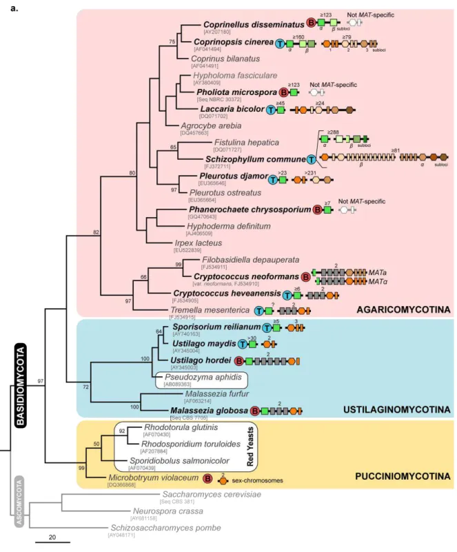
In contrast to the genomic regions that determine the mating type in ascomycetes, the MAT loci of basidiomycetes have evolved a tremendous level of diversity concerning their gene content and organization. Nevertheless, two common and fundamental components regulate two independent molecular mechanisms of self/nonself recognition: one locus encodes pheromones and pheromone
receptors (the “pheromone/ receptor locus”) and the other encodes homeodomain transcription factors (the “homeodomain locus”) (Fraser et al., 2007). For a successful mating and completion of the sexual cycle, cells that conjugate must contain different alleles of both MAT loci (Fraser et al., 2007; Morrow and Fraser, 2009).
1.2.2.1. The pheromone / receptor sensing system in basidiomycetes
16
It is interesting that all basidiomycete mating pheromones so far described, including the pheromones from the human pathogen C. neoformans (Moore and Edman, 1993) and those from the red yeast Rhodosporidium toruloides (Kamiya et al., 1978a), are hydrophobic diffusible lipopetides that contain farnesylation signals equivalent to those found in the a-factor of S. cerevisiae (Bölker and Kahmann, 1993). These active proteins are initially synthesized as larger precursors corresponding to the translated amino acid sequence, which subsequently undergoes posttranslational modifications. Pheromone maturation occurs by sequential events involving the carboxyl-terminal sequence CAAX (in which C is cystein, A is an aliphatic amino acid, and X is any residue) (Bölker and Kahmann, 1993). The mature pheromone is finally exported from the cell via a mechanism that involves an ATP-binding cassette (ABC) transporter (Ste6) that has been identified in both ascomycetes and basidiomycetes (Michaelis et al., 1992; Hsueh and Shen, 2005). In response to the interaction of these ligands with a suitable receptor, mating is initiated through a heterotrimeric G protein located at the plasma membrane that activates the downstream processes including a MAP kinase cascade (MAPK) signalling. This signalling pathway as well as the underlying mechanisms of a-factor processing and secretion were firstly characterized in S. cerevisiae and seem to have been retained in basidiomycetes (Fowler et al., 1999; Hegner et al., 1999; Olesnicky et al., 1999; Hsueh and Shen, 2005). Although the genetic and molecular mechanisms after pheromone perception are best understood in S. cerevisiae, significant advances have been made towards the elucidation of the same processes in basidiomycetes. These mechanisms are known in quite some detail in U. maydis and are described in the Chapter 2.
1.2.2.2. Homeodomain transcription factors
–
the second compatibility checkpoint
17 functional heterodimeric transcription factor is assumed to bind unique target sites within the promoter regions of genes whose function commits cells to a new developmental pathway. Although the homeodomain-regulated pathway is not yet defined in most basidiomycetes, it is indispensable during the latter stages of sexual development (Grandel et al., 2000; Kües et al., 2002; Wahl et al., 2010).
A final requirement of this elaborate system is that HD1 and HD2 proteins from the same cell type must not be allowed to heterodimerize or, otherwise, the homeodomain-regulated pathway would be constitutively active without mating. Several studies have reported that discrimination between self and non-self interactions is conferred by the N-terminal region of both HD1 and HD2 proteins (Yee and Kronstad, 1993; Kües et al., 1994a; Kües et al., 1994b; Yue et al., 1997). Whereas this region is highly variable between different alleles of both HD1 and HD2 genes the homeodomain and C-terminal regions usually display a more conserved pattern. Therefore, these genes seem to evolve
under a pattern called the “divergence-homogenization duality” (Badrane and May, 1999), which is characterized, on one hand, by divergence in the N-terminal region in order to maintain mating compatibility and, on the other hand, a greater conservation in the C-terminal region to preserve the transcriptional regulatory motifs necessary to induce the homeodomain-regulated pathway (Badrane and May, 1999). Therefore, within a population of a certain species, many HD1/HD2 alleles may exist and exhibit a high level of sequence variation. Such variation is consistent with models stating that these loci are subject to balancing selection, being therefore maintained in populations over much longer periods than would be expected for regions under neutral evolution (Wright, 1960; May et al., 1999). The current model of interaction between both HD1 and HD2 protein proposes that dimerization is achieved via a limited number of hydrophobic and polar interactions between the variable regions of the proteins (Kämper et al., 1995; Yee and Kronstad, 1998).
18
19
20
Figure 1.4. Structure and diversity of the MAT locus in the different basidiomycete lineages. (a)
Distribution and organization of the different sets of MAT genes in representatives of the three main lineages of the phylum Basidiomycota – Agaricomycotina, Ustilaginomycotina and Pucciniomycotina (shadowed in pink, blue and yellow, respectively). The phylogenetic tree was inferred using Maximum Parsimony based on the alignment of the nucleotide sequences of the D1/D2 region of the 26S rDNA. Bootstraps values (> 50%) are shown from 1000 replicates. GenBank accession numbers given below species names (for some species, sequences were retrieved from culture collection databases). The gene content of the homeodomain and pheromone/ receptor MAT regions is represented for each species as shown in the key given at the bottom (different colours of each symbol stands for different allelic versions). Genes connected by a black horizontal line are genetically linked. With exception of C. neoformans, for tetrapolar and bipolar species, only one MAT allele is represented with numbers above indicating the number of MAT alleles determined for each species. Species enclosed in white rounded boxes in Ustilaginomycotina and Pucciniomycotina represent members of the two groups of yeasts under study in this thesis. For M. violaceum, only the pheromone receptor genes were so far identified. (b) Detailed genomic organization of the homeodomain and pheromone/ receptor MAT regions of the species highlighted in bold face in the tree. Genes are depicted as arrows indicating the direction of transcription. MAT-specific regions are enclosed in white-rounded boxes and MAT genes coloured as indicated in the key given in (a). When known, conserved genes flanking the MAT loci (coloured in yellow or blue) are shown within each group of species and have the same designation (some of the flanking genes in U. maydis and S. reilianum are included in the MAT locus of U. hordei and M. globosa). In C. cinerea, genes in brackets at the pheromone/ receptor locus refer to the nomenclature used previously for genes as identified by Riquelme et al. (2005). (Based on Bölker et al., 1992; Lengeler et al., 2002; Fraser et al., 2004; Schirawski et al., 2005; Bakkeren et al., 2006; James et al., 2006; James, 2007; Kahmann and Schirawski, 2007; Xu et al., 2007; Bakkeren et al., 2008; Niculita-Hirzel et al., 2008; Morrow and Fraser, 2009; Yi et al., 2009; Metin et al., 2010; Ohm et al., 2010; Stajich et al., 2010; James et al., 2011).
1.2.2.3. Tetrapolar mating systems
–
the smut fungi
21 more complex. In S. reilianum, a detailed analysis of the pheromone/ receptor locus showed that this locus is triallelic and not biallelic as in U. maydis (Schirawski et al., 2005). Furthermore, each of the three alleles encodes a single pheromone receptor and two different pheromones, each of which is recognized by one of the receptors encoded by the other alleles (Figure 1.4b) (Schirawski et al., 2005). In U. maydis, besides the pheromone and receptor genes, the a2 allele harbours two additional genes (lga2 and rga2) and a pheromone pseudogene (Urban et al., 1996). The lga2 and rga2 genes seem to play important roles in mitochondrial inheritance, sexual reproduction and virulence (Bortfeld et al., 2004; Fedler et al., 2009; Mahlert et al., 2009) and are also present in the a2 allele of S. reilianum. However, the presence of a pheromone pseudogene in the a2 allele of U. maydis in a genomic position where a functional pheromone gene exists in S. reilianum supports the hypothesis that the archetype of this locus could in fact be more similar to that of S. reilianum with two functional pheromone genes (Urban et al., 1996; Schirawski et al., 2005).
After fusion of compatible partners, heterozygosity at the homeodomain locus (b locus) is required for dikaryon maintenance and filamentous growth (Gillissen et al., 1992; Kämper et al., 1995). Each homeodomain locus is composed by two divergently transcribed genes encoding homeodomain proteins known as bE (HD1-type) and bW (HD2-type). Their functional activation follows the rules described in the previous section (1.2.2.2). However, in contrast to the biallelic and triallelic pheromone/ receptor locus of U. maydis and S. reilianum, the homeodomain locus of both species is multiallelic: in U. maydis more than 30 allelic versions were found in natural populations, while in S. reilianum five alleles were already identified (Schirawski et al., 2005). Because both pheromone/ receptor and homeodomain loci are genetically unlinked, U. maydis and S. reilianum have a tetrapolar mating system with multiple mating types.
1.2.2.4. Tetrapolar mating systems
–
the origin of thousands of mating types
22
redundant similar genes encoding at least one pheromone receptor and one to three pheromone genes (Figure 1.4). Phylogenetic analysis of the C. cinerea pheromone receptors divided these proteins in two main clusters supporting an early duplication event of the ancestral gene, followed by subsequent sequence diversification (Riquelme et al., 2005).
Interestingly, even in such a complex mating system, compatibility only requires that mating partners bring together different allelic versions of genes at least in one sublocus of both MAT loci (Casselton and Kües, 2007). The organization of the extant MAT subloci is maintained because the DNA sequence that comprises each group of genes (both coding and flanking sequences) is sufficiently different to prevent homologous recombination between allelic versions. Thus, in each sublocus genes remain connected as a single unit such that intralocus recombination leading to compatible gene combinations is prevented (Casselton and Kües, 2007). However, during evolution the different subloci have been randomly mixed suggesting that at some stage they have been flanked by homologous sequences that allowed recombination between subloci (May and Matzke, 1995). Noteworthy, the ~7 kb of homologous sequence between the two first subloci in the homeodomain locus may be a relic of the ancestral configuration. A population level analysis of the homeodomain locus in C. cinerea identified several versions of each sublocus enough to generate 120 genetically different loci (May and Matzke, 1995). Combined with the several subloci versions also found for the pheromone/ receptor locus, more than 12 000 mating types can be generated in C. cinerea (Casselton and Kües, 2007). However, in other fungi (viz. Schizophyllum commune), even more mating types can be generated, since both multiallelic MAT loci are distributed in distinct subloci that can freely recombine (Brown and Casselton, 2001; Fowler et al., 2004) (Figure 1.4b). Taking advantage of the genome sequencing projects, it was possible to examine and compare the MAT loci structure of several other mushroom species. These analyses revealed that synteny (conserved gene order) around the homeodomain locus is maintained across several mushroom species (Figure 1.4b). By contrast, the pheromone/ receptor locus is usually located in a genomic region prone to diversification driven by gene duplications, translocations and transposon insertions (Niculita-Hirzel et al., 2008).
23 clamp cell formation and septation from the subapical cell (Swiezynski and Day, 1960; Raper, 1966; Casselton and Olesnicky, 1998) (Figure 1.5). On the other hand, the genes encoded by the pheromone/ receptor locus initiate nuclear exchange and migration to the apical cell and regulate fusion of the clamp cell with the subapical cell (Casselton and Olesnicky, 1998; Kües, 2000; Kamada, 2002) (Figure 1.5).
Hence, a tetrapolar multiallelic MAT system generates thousands of mating types ensuring that nearly every fusion event produces a compatible interaction. Therefore, this system maintains a high outcrossing frequency while restricting inbreeding. In this context, it is tempting to speculate that in species with a very large number of mating types, the search for suitable mating partners by means of a pheromone-mediated chemoattraction has been abandoned, since any two individuals in a natural population are most likely compatible mates.
1.2.2.5. Evolution of bipolar systems
–
multiallelic bipolar basidiomycetes
Although tetrapolar systems predominate in the phylum Basidiomycota, bipolar species also exist and, most significantly, do not seem to be uniformly distributed. For example, about 25% of mushroom species are estimated to have a bipolar mating system and these occur interspersed with tetrapolar relatives (Raper, 1966). Hypotheses for the foundations of the bipolar mating systems of basidiomycetes have been firstly proposed by John Raper. One such hypothesis assumes that, mutations occurring in either tetrapolar MAT loci leading to self-compatible interactions could turn the tetrapolar system into a bipolar system. Remarkably, mutations leading to self-compatibility were observed in both natural and laboratory strains of the tetrapolar species C. cinerea (Kües et al., 1994b; Olesnicky et al., 2000).