103
MAY/JUNE REV. HOSP. CLÍN. FAC. MED. S.PAULO 54 (3): 103 - 106, 1999
CONGENITAL HYPERTHYROIDISM:
AUTOPSY REPORT
Marcus Aurelho de Lima, Lília Beatriz Oliveira, Neiva Paim and Maria de Fátima Borges
LIMA, M. A. de et al. - Congenital hyperthyroidism: autopsy report . Rev Hosp Clín Fac Med S Paulo, 54(3): 103 - 106 , 1999.
SUMMARY: We report the autopsy of a stillborn fetus with congenital hyperthyroidism born to a mother with untreated Graves’ disease, whose cause of death was congestive heart failure.
The major findings concerned the skull, thyroid, heart, and placenta. The cranial sutures were closed, with overlapping skull bones. The thyroid was increased in volume and had intense blood congestion. Histological examination showed hyperactive follicles. The heart was enlarged and soft-ened, with dilated cavities and hemorrhagic suffusions in the epicardium. The placenta had infarctions that involved at least 20% of its surface, and the vessels of the umbilical cord were fully exposed due to a decrease in Wharton ’s jelly.
Hyperthyroidism was confirmed by the maternal clinical data, the fetal findings of exophthalmia, craniosynostosis, and goiter with signs of folli-cular hyperactivity.
Craniosynostosis is caused by the anabolic action of thyroid hormones in bone formation during the initial stages of development. The delayed initiation of treatment in the present case contributed to the severity of fetal hyperthyroidism and consequent fetal death.
DESCRIPTORS: Congenital hyperthyroidism. Graves’ disease. Thyroid. Craniosynostosis. Autopsy.
Graves’ disease (GD) is charac-terized by hyperthyroidism clinically manifesting as hyperfunctioning dif-fuse hyperplastic goiter, at times accompanied by ophthalmopathy and dermopathy17. In nonendemic goiter areas, GD is the second most com-mon thyroid disease. It occurs at all ages, but is more common during the 3rd and 4th decades of life8. The inci-dence of thyrotoxicosis during preg-nancy is low (0.04 to 1.4%), and only 1 in 70 infants born to thyrotoxic mothers develops signs and symp-toms of hyperthyroidism10,11. Among newborns with hyperthyroidism, 60 % present transitory or self-limiting symptoms, 20 % die as a conse-quence of heart failure, and the remaining 20 % continue to present symptoms for more than 6 months,
requiring more prolonged, though transitory, treatment10.
Having had the opportunity to autopsy a stillborn fetus with congen-ital hyperthyroidism who died of heart failure, we thought it would be justified to describe this case because of its rarity and because of the contri-bution its description may make to a better understanding of the disease.
CASE REPORT
A 27-year old white housewife sought the Gynecology and Obstetrics Outpatient Clinic of the
Faculty of Medicine of Triângulo Mineiro (FMTM) in Uberaba, Minas Gerais State, Brazil for prenatal care during the 16th week of pregnancy. She was in good general condition, with arterial pressure of 130/70 mmHg, and a thyroid enlarged to twice the normal size, attributed to pregnancy. At the gestational age of 32 weeks and 3 days, the patient had exophthalmia and increased thyroid hormone concentrations: free T4, 5.2 ng/dl (normal range: 0.8–2.3 ng/dl), and TSH, 0.03 mIU/ml (normal range = 0.34–4.5 mIU). Weight was 62.8 kg, height 152 cm, body mass index was 27.18 kg/m2, arterial pressure 150 x 90 mmHg, and heart rate 96 systoles/minute. Treatment with propylthiouracil (150 mg every 8 hours) was started. One week later, with a gestational age of 36 weeks
RHCFAP/2971
From “Dep. de Endocrinologia da
and 3 days according to the date of last menstruation and of 37 weeks and 4 days according to ultrasonogra-phy, the patient was admitted to the Teaching Hospital of FMTM, com-plaining of cramp-like pain in the lower abdomen lasting 12 hours. Gynecologic examination showed signs of labor, and the patient gave birth to a stillborn infant.
AUTOPSY
A white male fetus weighing 2750 g, with a vertex-to-heel length of 50.5 cm, vertex-coccyx length of 32 cm, a plantar distance of 7.5 cm, head cumference of 32.5 cm and chest cir-cumference of 30 cm underwent autopsy. The fetus had scarce and fine hair, vernix caseosa in skinfolds, bilateral exophthalmia, and cyanotic extremities.
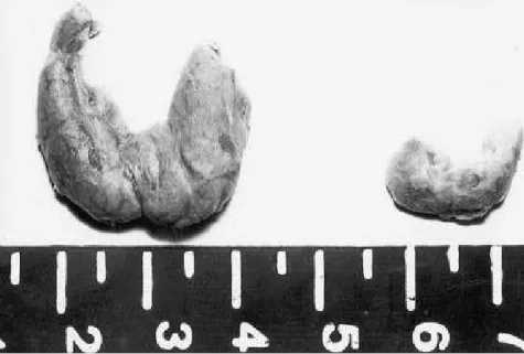
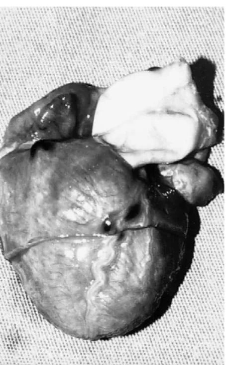
The major findings were observed in the skull, brain, thyroid, spleen, and placenta. The sutures were closed, with overlapping skull bones (Figure 1). The brain weighed 312 g (normal weight = 297 g + 69 g) and had blood congestion and subarachnoid hemor-rhage. The pituitary was investigated; the thyrotrophs were markedly sup-pressed. The thyroid was enlarged (weight, 3 g — normal weight: 1.3 g + 0.9 g) and had intense blood con-gestion (Figure 2). Histologically, the organ had follicles with high colloid epithelium and abundant absorption vacuoles (Figure 3).
The heart was globally enlarged (weight, 26 g — normal weight 19 g ± 5 g), and softened, with dilated cav-ities, had no congenital defects, and
had hemorrhagic suffusions in the epicardium (Figure 4). The pericar-diac fluid was citrine-yellow and slightly increased.
The spleen was enlarged (weight, 23 g — normal weight 9 g ± 4 g), congested and with hematopoietic focal points observed by microscopy. The placenta was normal in vol-ume (weight, 460 g) and had both recent and earlier infarctions involv-ing less than 20 % of its surface. Microscopy showed fibrin deposits and congestion of fetal blood vessels. The umbilical cord vessels were fully exposed due to a decrease in the amount of Wharton‘s jelly. The remaining organs had only polyvis-ceral congestion.
DISCUSSION
We report the autopsy findings for a stillborn infant with congenital hyperthyroidism, whose cause of death appeared to be congestive heart failure. Hyperthyroidism was con-firmed on the basis of maternal clini-cal data, and on the fetal findings of exophthalmia, early closure of cranial sutures (premature craniosynostosis), and goiter with signs of follicular hyperactivity.
Craniosynostosis has been obser-ved as an isolated sign or as part of systemic disease and is one of the complications of congenital or juve-nile hyperthyroidism, being observed more commonly in prolonged thyro-toxicosis7. Thyroid hormones are ana-bolic agents acting on bone formation during the early stages of childhood development, and their excessive con-centrations act as a factor of accelera-tion of skeletal maturaaccelera-tion and of clo-sure of cranial sutures in children1. These hormones increase bone formation directly through nuclear osteoblast receptors18, or indirectly through local growth factors such as insulin-growth factor-I (IGF-I)15,23. The hormones also increase IGF-I protein content14 and IGF-mRNA content22 and bind to the nuclear
104
REV. HOSP. CLÍN. FAC. MED. S.PAULO 54(3):103 - 106, 1999 MAY/JUNE
Figure 1- Skull presenting closed sutures and overlapping bones.
receptors of bone, directly stimulat-ing bone formation13,18.IGF-I is main-ly produced by osteoblasts4 and induces bone formation by stimulat-ing DNA and bone matrix synthesis or by reducing collagen degrada-tion5,16. In view of the fact that IGF-I increases cortical bone formation21, the sagittal suture may be more influ-enced than other sutures. Histological
analysis shows that the suture fissure is filled with mesenchymal tissue similar to the exchange layer of long bone periosteum2. This property of long bones is involved in the general progress of osteogenesis and prolifer-ation of intervening soft tissues24. There are some reports of premature craniosynostosis with changes in thy-roid hormones. Robinson et al.19 reported the first case of a two-month old patient with Graves’ disease with premature total fusion of the sagittal, coronal, and lambdoid sutures. In the case reported here, as illustrated in Figure 1, not only the sagittal suture but also the coronal and lambdoid sutures were prematurely closed, with overlap of the bones at these levels.
Although this was not observed in the present case, the brains of fetuses with hyperthyroidism may present ventriculomegaly represented by in vacuum hydrocephaly secondary to the absence of normal cerebral growth. The delayed brain develop-ment is a result of the premature arrest of cell division and the acceler-ated rate of neuron differentiation12.
After a careful review of the histo-ry of the mother, we considered the diagnosis of Graves’ disease in the eighth month of pregnancy to be late, a fact possibly attributable to the
dif-ficulty in diagnosing hyperthyroidism that starts during pregnancy, especial-ly in its discrete and moderate forms, since the common findings of thyroid hyperfunction are frequently similar to typical changes occurring during pregnancy, i.e., increased cardiac overload, tachycardia, hyperemesis, and pretibial edema. However, the weight loss that characterizes hyper-thyroidism is less evident due to the weight gain typical of pregnancy20.
Disease duration, early diagnosis, and the institution of appropriate treatment are factors that affect fetal prognosis9. The late beginning of treatment in the present case con-tributed to the severity of fetal hyper-thyroidism and to fetal death6. Neonatal thyrotoxicosis is the result of transplacental transfer of immu-noglobulins that stimulate the thyroid, an event that occurs even in mothers treated months or years before preg-nancy. This condition is associated with a mortality rate of 16%10.
Another interesting autopsy find-ings was the exposure of umbilical cord vessels due to the reduction of Wharton‘s jelly. This abnormality may involve the umbilical cord in part or as a whole, possibly leading to ves-sel compression and fetal death. These cords are more frequently observed in fetuses with retarded growth and in preeclampsia, but unknown causes may also to exist3. In the present case, the condition involved the entire cord, and no signs of vessel compression or rupture were observed. On the other hand, we do not know if there is any relationship between this finding and hyperthyroidism.
In view of the rare occurrence of a stillborn fetus with hyperthyroidism, we felt that it would be of extreme importance to study the pathologic changes in a fetus born to a patient with Graves’ disease who was not treated in time to prevent the severity of the situation and the fatal outcome, and for which the immediate cause of death seems to have been heart failure due to volume overload, as shown by the macroscopic findings.
105
MAY/JUNE REV. HOSP. CLÍN. FAC. MED. S.PAULO 54(3): 103- 106 , 1999
Figure 3 - Photomicrograph of the thyroid’s follicles of medium dimensions with intense activity indicated by the large number of reabsorption vacuoles and tall lining epithelium (HE, 100x).
106
REV. HOSP. CLÍN. FAC. MED. S.PAULO 54(3):103 - 106, 1999 MAY/JUNE
LIMA, M. A. de e col. - Hiper-tireoidismo Congenital. Rev Hosp Clín Fac Med S Paulo, 54(3): 103 - 106, 1999.
Relata-se necropsia de natimorto com hipertireoidismo congênito, filho de mãe portadora de doença de Graves não tratada, que teve como causa de óbito insuficiência cardíaca congestiva. Os achados fundamentais foram vistos no crânio, tireóide coração e placenta. As suturas crani-anas encontravam-se fechadas, com acavalgamento dos ossos cranianos.
A tireóide apresentava aumento de volume e congestão sangüinea inten-sa e, histologicamente, os folículos mostravam hiperatividade. O coração estava aumentado de volume, amole-cido, com cavidades dilatadas e sufusões hemorrágicas no epicárdio. A placenta apresentava infartos que acometiam menos de 20% da superfí-cie placentária e os vasos do cordão umbilical encontravam-se completa-mente expostos por diminuição da geléia de Warton.
O hipertireoidismo ficou compro-vado pelos dados clínicos maternos,
os achados fetais de exoftalmia, crani-osinostose prematura e bócio com sinais de hiperatividade folicular.
A craniosinostose é causada pela ação anabólica dos hormônios ti-reoidianos na formação óssea, nos estágios iniciais do desenvolvimen-to. O início tardio do tratamento no presente caso contribuiu para severi-dade do hipertireoidismo fetal e óbito.
DESCRIPTORS: Hipertireoidis-mo congênito. Doença de Graves. Tireóide. Graniosinostose. Necropsia.
RESUMO RHCFAP/2971
REFERENCES
1. AKITA S, NAKAMURA T, HIRANO A et al. - Thyroid hormone-action on rat calvarial sutures. Thyroid1994; 4:99-106. 2 ALBERIUS P & JOHNELL O - Immunohistochemical assessment
of cranial suture development in rats. J Anat 1990; 173:61-8. 3. BENIRSCHKE K & KAUFMANN P - Anatomy and pathology of
the umbilical cord and major fetal vessels. In: BENIRSCHKE K, KAUFFMAN P, eds. - Pathology of the human placenta. 3th ed. New York, Springer, 1995. p. 319.
4. CANALIS E, MCCARTHY T & CENTRELLA M - Isolation and cha-racterization of insulin-like growth factor I (somatomedin-C) from cultures of fetal rat calvariae. Endocrinol 1988; 122: 22-7. 5. CANALIS E - Effect of insulin-like growth factor I on DNA and
pro-tein synthesis in cultured rat calvaria. J Clin Invest1980; 66: 709. 6. DAVIS LE, LUCAS NJ, HANKINS GDV et al. - Thyrotoxicosis complicating pregnancy. Am J Obs and Gynecol 1989;160: 63-70.
7. DUGGAN CA, KEENER EB, GAY BB JR. - Secondary cranios-ynostosis. Am J Radiol 1970; 109:277.
8. FRANSSILA KO - The thyroid. In: LECHAGO J, GOULD VE eds.
Bloodworth’s endocrine pathology. 3th ed. Baltimore, Williams & Wilkins, 1997 p.171-247.
9. GIARDINA S, CONTARINI A & BECCA B - Maternal disea-ses and congenital malformations. Ann Ist Super Sanità1993;
29:69-76.
10. HOLLINGSWORTH DR & MABRY CC - Congenital Graves’ disease: four familial cases with long-term follow-up and pers-pective. Am J Dis Child 1976; 130:148-155.
11 KAPLAN M M Thyroid disease. In: GLEICHER N ed.
-Principles and practice of medical therapy in pregnancy.
Connecticut, Appleton, 1992. p.321-338.
12. KOPELMAN AE - Delayed cerebral development in twins with con-genital hyperthyroidism. Am J Dis Child 1983; 137:842-845. 13. KRIEGER NS, STAPPENBECK TS, & STERN PH
-Characterization of specific thyroid hormone receptors in bone. J Bone Miner Res 1988; 3: 473-8.
14. LAKATOS P, CAPLICE MD, KHANNA V et al. - Thyroid hor-mones increase insulin-like growth factor I content in the medium of rat bone tissue. J Bone Miner Res 1993; 8:1475-81. 15. LINKHART TA & KEFFER MJ - Differential regulation of
insu-lin- like growth factor –I (IGF-I) and IGF- II release from cultu-red neonatal mouse calvaria by parathyroid hormone, transfor-ming growth factor-beta, and 1,25-dihydroxyvitamin D3.
Endocrinol1991;128: 1511-8.
16. MCCARTHY TL, CENTRELLA M & CANALIS E - Parathyroid hormone enhances the transcript and polypeptide levels of insuline-like growth factor I in osteoblast-enriched cultures from fetal rat bone. Endocrinol 1989; 124:1247-53.
17. MUNRO DS, DIRMIKIS SM & HUMPHRIES H - The role of thyroid stimulating immunoglobulins of Graves’s disease in neo-natal thyrotoxicosis. Br J Obst Gynecol 1978; 85:837-843. 18. RIZZOLI R, POSTER J & BURGI U - Nuclear thyroid hormone
receptors in cultured bone cells. Metabolism1986; 35:71-4. 19. ROBINSON DC, HALL R & MUNRO DS - Graves’ disease,
an unusual complication: raised intracranial pressure due to pre-mature fusion of skull sutures. Arch Dis Child1969; 44: 252-7. 20. THOMAS R & REID RL - Thyroid disease and reproductive
dysfunction: A review. Obstet Gynecol1987; 70:789-98. 21.TOBIAS JH, CHOW JW & CHAMBERS TJ - Opposite effects of
insulin-like growth factor-I on the formation of trabecular and cor-tical bone in adult female rats. Endocrinol1992; 131: 2387-92. 22.HOFER K - Thyroid hormones increase insulin-like growth factor
mRNA levels in the clonal osteoblastic cell line MC3T3-E1. FEBS Lett 1994; 345: 67-70.
23. WOLF M, INGBAR SH & MOSES AC - Thyroid hormone and growth hormone interact to regulate insulin-like growth factor-I messenger ribonucleic acid and circulating levels in the rat.
Endocrinol 1989; 125:2905-14.
24. ZIMMERMANN B - Degeneration of osteoblasts involved in intra-membranous ossification of fetal rat calvaria. Cell Tissue Res
1992; 267:75-84, 1992.