J of Evolution of Med and Dent Sci/ eISSN- 2278-4802, pISSN- 2278-4748/ Vol. 4/ Issue 68/ Aug 24, 2015 Page 11785
A STUDY ON ETIOLOGY AND PROFILE OF PLEURAL EFFUSION IN CHRONIC
KIDNEY DISEASE
A. Prem Kumar1, B. M. S. Pathrudu2, N. Usha Rani3, B. Padmaja4, Banavath Durga Prasad Naik5, M. Narayana6, L. Haritha Kumari7, P. Dhilleswarrao8
HOW TO CITE THIS ARTICLE:
A. Prem Kumar, B. M. S. Pathrudu, N. Usha Rani, B. Padmaja, Banavath Durga Prasad Naik, M. Narayana, L. Haritha Kumari, P. Dhilleswarrao. A Study on Etiology and Profile of Pleural Effusion in Chronic Kidney Disease. Journal of Evolution of Medical and Dental Sciences 2015; Vol. 4, Issue 68, August 24;
Page: 11785-11796, DOI: 10.14260/jemds/2015/1700
ABSTRACT: BACKGROUND: Chronic Kidney disease is characterized by decreased glomerular filteration rate. Most pleural effusions occurring in CKD are attributed to renal failure & heart failure and are left alone, but there are other causes responsible for many effusions such as parapneumonic effusion, atelectasis, tuberculosis and other infections and malignancies due to immunosuppression, hence presence of pleural effusion in CKD deserves further evaluation. Our study is conducted to find the etiology and profile of patients with chronic Kidney disease developing pleural effusion.
MATERIALS AND METHODS: Study was conducted among 35 patients with CKD and pleural effusion who attended Government hospital for chest and communicable diseases affiliated to Andhra Medical College, from March 2013 to September 2014. The clinical course of pleural effusions and their biochemical characteristics were studied together with radiographs and other relevant investigations. Study design- hospital based prospective study. OBSERVATIONS AND RESULTS: Of the 35 patients, 57% developed unilateral effusion, 43% bilateral effusion. Among unilateral effusions-minimal effusions were 25%, moderate were 60%, massive were 15%. Patients with transudative effusion were 31%, exudative were 69%. Causes of effusion were as follows: Cardiac failure 31%, Tuberculosis 28%, Malignancy 9%, uremic effusion 14%, parapneumonic 11%, connective tissue disorders 2%. CONCLUSIONS: Apart from cardiac failure, tuberculosis is a major cause of pleural effusion in CKD patients, especially if the effusion is unilateral, exudative in nature, blood tinged, and lymphocyte predominant. ATT produced improvement in clinical and radiological status in these patients.
KEYWORDS: CKD (Chronic Kidney Disease), Pleural effusion, Cardiac failure, Tuberculosis.
INTRODUCTION: Chronic kidney disease is defined as kidney damage for ≥3months, as defined by structural or functional abnormalities of the kidney, with or without decreased GFR, that can lead to decreased GFR, manifests by either: Pathological abnormalities or Markers of kidney damage, including abnormalities in the composition of blood or urine, or abnormalities in imaging tests or Glomerular filtration rate (GFR) <60ml/min/1.73m2 for 3 or more months, with or without kidney damage.1 Kidney failure or end stage renal disease is defined as a GFR of less than 15 ml/minute per 1.73m2 or the need for treatment with dialysis or transplantation.
Chronic kidney disease (CKD), including end-stage renal disease (ESRD), is associated with an increased susceptibility to infection due to advanced age, immune dysfunction, underlying disease, and the dialysis procedure itself leading to increased morbidity and mortality.2
J of Evolution of Med and Dent Sci/ eISSN- 2278-4802, pISSN- 2278-4748/ Vol. 4/ Issue 68/ Aug 24, 2015 Page 11786 Pleural disease is a common problem in patients with chronic renal insufficiency. There are several reasons why pleural disease may be common in patients with chronic kidney disease. These include congestive heart failure, fluid overload, an increased risk of infection (Especially tuberculosis), the presence of diseases associated with renal and pleural manifestations (e.g., systemic lupus erythematosus), uremic pericarditis, an increased risk for certain malignancies and pulmonary embolism. Uremic pleurisy results from an unknown putative agent, and therefore uremic pleuritis is a diagnosis of exclusion, that persists or recurs despite aggressive haemodialysis. Management of TB in these patients raises issues of drug dosing and interactions.
Patients with CKD have immune dysfunction manifested by depressed cell-mediated immunity (CMI). This impairment of CMI makes infection with Mycobacterium tuberculosis more difficult to detect and more likely to progress to TB disease than in immune competent individuals. Increased risk for ischemic heart disease and potential for dilated cardiomyopathy makes these patients especially prone to problems with fluid balance.
In CKD diseases, such as SLE are associated with renal and pleural manifestations, patients with CKD are immune compromised and some studies have suggested that this problem may be at increased risk for certain malignancies, such as non-Hodgkin’s lymphoma and renal, prostate, and uterine Cancer.3 All of these malignancies can involve the pleura. Uraemia per se has been shown to cause a pleuritis by an unknown mechanism. Co-morbid conditions associated with or contributing to CKD may indirectly cause pleural abnormalities.
The presence of unilateral effusion suggests a diagnosis other than heart failure, like tuberculosis or parapneumonic or atelectasis. The reduced humoral and cellular immunity, in addition to delay in diagnosis because of an attenuated clinical response, may explain the high rate of empyema.
Most of the studies looking into the incidence of pleural effusion in patients with CKD are retrospective studies of hospitalized patients, so the present study is done prospectively to know the occurrence, causes, clinical features and management issues of pleural effusion in patients with chronic kidney disease.
AIMS AND OBJECTIVES:
The present study is conducted to find:
1. The demographic characteristics of patients with chronic kidney disease developing pleural effusion.
2. The nature and size of pleural effusion in CKD patients.
3. The clinical presentation, biochemical, pathological, microbiological analysis of pleural fluid in pleural effusion of CKD patients.
4. The etiology of pleural effusion in CKD patients.
J of Evolution of Med and Dent Sci/ eISSN- 2278-4802, pISSN- 2278-4748/ Vol. 4/ Issue 68/ Aug 24, 2015 Page 11787
STUDY CRITERIA: Inclusion Criteria:
1. Patients with an estimated Glomerular filtration rate (GFR) <60ml/min/1.73m2 for 3 or more months, with or without kidney damage1 with Pleural effusion.
GFR was calculated using Cock – Croft - Gualt equation, GFR= (140-age) × (weight in kg) ÷ 72 × Serum creatinine Multiply by 0.85 if female.
2. Age >14 yrs.
Exclusion Criteria:
1. Age <14 yrs. 2. Patients with HIV.
3. Patients with bleeding disorders. 4. Severe co morbidities like recent MI. 5. Patients not willing for thoracocentesis.
Study Procedure: A clinically suspected case of pleural effusion in a chronic kidney disease patient was diagnosed by Chest X-ray and ultrasound chest.
Detailed demographic and clinical parameters including age, sex, smoking history, clinical symptoms with duration (Cough, fever, sputum production, haemoptysis, chest pain, breathlessness) and clinical signs (Pallor, clubbing, enlarged neck nodes, pulse rate, blood pressure) and other systemic examination for the co-morbid illness were evaluated in all patients. In addition to chronic kidney disease, history for other co-morbid illness and habits like smoking and alcoholism were taken.
Co-morbid illnesses were defined as the presence of coexisting cardiac failure, ischemic heart disease, chronic lung disease (COPD), chronic liver disease, malignancies, neurological diseases and diabetes mellitus. All patients were subjected to blood investigations including Complete blood count, ESR, Blood sugar, renal function tests and Liver function tests and urine routine examination. Sputum if present is sent for ZN stain, Gram’s stain, culture and sensitivity.
Chest radiograph is classified according to the size of effusion. The size of the effusion was assessed on the posterior-anterior radiograph by visually estimating the area of the hemi-thorax occupied by pleural fluid. Pleural effusions were deemed to be minimal if it occupied less than one third of hemi-thorax, moderate if it occupied between one third to two third, and massive if it occupied more than two thirds of the hemi-thorax.
Thoracocentesis was performed using a 20 gauze needle syringe and the fluid is studied for the gross appearance, total WBC count, differential count, RBC count, protein, glucose, ADA, LDH, cytology, aerobic and AFB culture.
Pleural fluid was classified as exudative effusion or transudative effusion by applying LIGHTŚ Criteria.
Exudative Pleural Effusions meet at Least one of the following Criteria, whereas Transudative Pleural Effusions meet none (Light's Criteria):
Pleural fluid protein divided by serum protein greater than 0.5 Pleural fluid LDH divided by serum LDH greater than 0.6
J of Evolution of Med and Dent Sci/ eISSN- 2278-4802, pISSN- 2278-4748/ Vol. 4/ Issue 68/ Aug 24, 2015 Page 11788 Pleural biopsy and other relevant investigations such as ultrasound thorax and abdomen, echocardiography, CT thorax were performed if no etiology was obtained from above investigations.
Pleural biopsy was performed with an Abram’s needle.
Complete renal profile including blood urea, serum creatinine, serum total protein, serum albumin, serum cholesterol, serum Na+, K+, Ca++, phosphorus and a complete urine examination was done. Staging of CKD into 5 stages was done based on GFR as proposed by KDOQI guidelines. The etiological diagnosis of pleural effusion was made when the condition had the following features.
Congestive Heart Failure (CHF): Compatible clinical features like syncope, PND, chest pain, dyspnoea on exertion, JVP more than 5cm, and radiologic findings like cardiomegaly, ECG abnormalities like symmetrical T wave inversion, Q waves, remission upon appropriate treatment, and absence of pulmonary infiltrates, and purulent sputum. By 2D-ECHO systolic dysfunction was considered when ejection fraction was less than 50, and diastolic dysfunction was considered by the ratio of early and late diastolic flow which is termed as E: A ratio.
In 2D-ECHO other findings noted were associated left ventricular hypertrophy and regional wall motion abnormalities.
Tuberculous Origin:
1. Sputum for AFB by Ziehl-nelsen method or
2. A positive result on Lowenstein Jensen cultures of pleural fluid, sputum or pleural biopsy specimens or
3. Presence of a caseating granuloma in a pleural biopsy specimen or
4. An exudative lymphocytic effusion with an ADA level of ≥60 U/L, along with a positive tuberculin skin test result.
Parapneumonic Origin: Association with pneumonia, lung abscess, or bronchiectasis; empyema was diagnosed in the presence of purulent fluid or positive culture of parapneumonic effusion.
Malignant Effusion: A pleural effusion was categorized as malignant if pleural fluid cytology, cell block or pleural biopsy findings were positive for malignancy (i.e., true malignant), or if the patient had a known cancer with no other explanation for the effusion (i.e., paramalignant).
Uremic Effusion: Patient with chronic kidney disease with fluid overload in the absence of cardiac failure and pleural fluid analysis showing exudative with glucose levels approximates serum glucose levels, with lymphocyte predominance after ruling out other possible aetiologies and response to dialysis.
Pulmonary Thromboembolism: Angiographic evidence or perfusion gammagraphy showing deficient perfusion.
Connective Tissue Diseases: A pleural effusion was categorised due to connective tissue disease, when serum collagen profile was positive and after ruling out other possible aetiologies.
J of Evolution of Med and Dent Sci/ eISSN- 2278-4802, pISSN- 2278-4748/ Vol. 4/ Issue 68/ Aug 24, 2015 Page 11789
Hepatic Hydrothorax: Transudative, predominant right side pleural effusion with ultrasound abdomen showing features of cirrhosis of liver with portal hypertension.
Obstruction of Brachiocephalic Vein: pleural fluid is border line Transudative, diagnosed by Doppler study of central vein and venography.
Urinothorax: pleural fluid looks and smells like urine, transudative, confirmed when pleural fluid creatinine is greater than serum creatinine.
The results were analysed using standard statistical methods, Mean and Standard D
RESULTS:
Sex Distribution: The study consisted of 35 patients, out of which 28(80%) were males and 7(20%) were females. Sex ratio (Male: Female) is 4:1
Age Distribution: Out of the 35 patients included in the study, patients less than 20 years of age were 1(2.85%), between 21 to 40 years of age were 5(14.28%) and 41 to 60 years of age were 14(40%) and above 60 years were 15(42.85%).
Clinical Presentation of Pleural Effusion in CKD: Of the 35 patients, all of them complained of dyspnoea, 11(31.4%) had chest pain, 15(42.8%) had cough, and 12(34.2%) complained of fever and 10(28.5%) had significant loss of appetite and loss of weight (LOA/LOW). The symptoms of the study group are analysed in table 1.
Clinical Features No. of Cases
Dyspnoea 35 (100%) Chest pain 11 (31.4%)
Cough 15 (42.8%)
Fever 12 (34.2%)
LOW / LOA 10 (28.5%)
Table 1: Clinical Presentation of Pleural Effusion in CKD
Smoking Status: Of the 35 patients, 8(22.8%) were current smokers, 13(37.1%) were former smokers and 14(40%) were never smokers.
Comorbidities and Associated Diseases: In the present study 15(42.8%) had diabetes and 22(62.8%) were hypertensive, 10(28.57%) had both diabetes and hypertension, 11(31.42%) had history of coronary artery disease and 11(31.42%) had history of tuberculosis and 9(25.7%) had other comorbidities like COPD, asthma etc.
J of Evolution of Med and Dent Sci/ eISSN- 2278-4802, pISSN- 2278-4748/ Vol. 4/ Issue 68/ Aug 24, 2015 Page 11790
No. of Pleural Effusion Cases in Relation to Severity of CKD: Of the 35 cases, no cases were seen in stage 1 and 2, 8 (22.85%) cases were seen in stage 3, 11(31.42%) cases were seen in stage 4 and 16(45.71%) cases were seen in stage 5. out of these cases 9(25.71%) were on haemodialysis.
Treatment taken for Kidney Disease: Of 35 cases, 22 were only on medical management, 9 were on hemodialysis and 4 patients were not receiving any treatment.
General Physical Examination Findings: Pallor in 24 cases, clubbing in 12 cases and pedal edema in 19 cases, raised JVP in 10 cases.
Lab Investigation Results: The mean haemoglobin (HB%) value was 9.1±1.43. The mean total leukocyte count (TC) was 10, 845±4, 239.The mean random blood sugar level was 153.8±86, and the mean blood urea level was 120± 55.The mean serum creatinine was 4.0±2.75.
Chest X-Ray Findings: Bilateral effusion were observed in 15 cases, right side effusion in 11 cases, left side effusion in 9 cases. Among right side effusions 6 were minimal, 17were moderate, 1was massive and on left side 5 were minimal, 15 were moderate and 3 were massive.
Type of Effusion: In 11 cases it was transudative effusions and in 24 cases it was exudative effusions.
Appearance of Pleural Fluid: In 7 cases fluid was clear in appearance, 15 were straw colored, 9 were hemorrhagic and 4 were turbid.
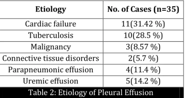
Etiology of Pleural Effusion:
Etiology No. of Cases (n=35)
Cardiac failure 11(31.42 %)
Tuberculosis 10(28.5 %)
Malignancy 3(8.57 %)
Connective tissue disorders 2(5.7 %) Parapneumonic effusion 4(11.4 %)
Uremic effusion 5(14.2 %)
Table 2: Etiology of Pleural Effusion
Pleural Fluid Analysis:
PARAMETER TUBERCULOSIS HEARTFAILURE UREMIA PARA PNEUMONIC
Total cells 2068.3±215 709.09±43.33 1182±113.8 15,265±1651
Protein 3.57±0.29 2.01±0.50 4.2±0.85 3.57±0.28
ADA 69.9±8.25 15.5±3.75 30.2±3.12 53±6.40
Glucose 70±14.19 51.8±18.49 54±13.56 21.75±5.4
J of Evolution of Med and Dent Sci/ eISSN- 2278-4802, pISSN- 2278-4748/ Vol. 4/ Issue 68/ Aug 24, 2015 Page 11791
DISCUSSION: Chronic kidney disease (CKD) is becoming a major global health problem. It is estimated that 1, 00, 000 new patients of end stage renal disease (ESRD) enter renal replacement programs annually in India.4
Given the prevalence of CKD, the frequency (>50%) of haemodialysis in these patients, and the propensity for these patients to have pleural disease, it is not surprising that the constellation of CKD, haemodialysis, and pleural effusion is common.
So the aim of the present study was to look at the various etiologies of pleural effusion occurring in chronic kidney disease patients, and their characteristics. After getting informed consent 35 patients were included in the study of which 28 were males and 7 were females. Mean age of the group was 52.88yrs. Patients above 40 years constituted 82.5%.
Comorbidities: In the present study of the 35 patients, all of them complained of dyspnoea (100%), 11(31.4%) had chest pain, 15(42.8%) had cough, and 12(34.2%) complained of fever, 10 (28.5%) had significant loss of appetite and loss of weight, 19 cases (54.28%) had pedal oedema. The dyspnoea in many patients may also be due to other co morbidities like anaemia and cardiac dysfunction. When the dyspnoea is accompanied by a pleuritic chest pain or fever or cough, it may imply an under lying pleuropulmonary pathology. Hypertension was the common comorbidity (62.8%) followed by diabetes mellitus (42.48%).
Radiology of Pleural Effusion: In the present study bilateral pleural effusion was present in 15 cases (42.8%) and unilateral pleural effusion in 20 cases.
In pleural effusion due to heart failure 11 out of 11 cases were bilateral, in tuberculous pleural effusion right side pleural effusion was present in 6(60%) cases, left side pleural effusion was present in 4(40%) cases. In uremic pleural effusion bilateral pleural effusion was present in 4(80%) cases, in parapneumonic effusion left side pleural effusion was present in 2(50%) cases and right side pleural effusion was present in 2(50%) cases.
Type of Effusion: In the present study transudative pleural effusions were 11(31.42%), all were bilateral, and exudative pleural effusions were 24(68.57%), out of which 7(29.16%) were bi lateral, 17(70.83) were unilateral.
Etiology of Pleural Effusion: In the present study the aetiology of pleural effusion was attributed to cardiac effusion in 11(31.42 %) cases out of 35 cases, tuberculous pleural effusion in 10(28.5 %) cases, uremic pleural effusion in 5(14.2 %) cases, and in 4(11.4 %) cases parapneumonic effusion was present. Malignant pleural effusion was present in 3(8.57 %) cases, pleural effusion due to connective disorder was present in 2(5.71 %) cases.
Pleural Effusion Characteristics in Individual Conditions:
J of Evolution of Med and Dent Sci/ eISSN- 2278-4802, pISSN- 2278-4748/ Vol. 4/ Issue 68/ Aug 24, 2015 Page 11792 In addition to this, impairment of CMI makes infection with mycobacterium tuberculosis more difficult to detect and more likely to progress to TB disease than in immune competent individuals. Smear negative and extra-pulmonary forms are more frequent than in an immunocompetent individual. A high incidence of post-transplant TB has been reported in India, especially miliary TB.
In the present study 28.57% of pleural effusions were due to tuberculosis. The mean age was 53.6±7.90 yrs. Total number of male patients were 8, female patients were 2. Male to female ratio was 4:1. Fever was present in 70% of the patients, cough was present in 70% of the patients, dyspnoea was present in all patients.
Chest pain was complained by 50% of the patients. Pedal oedema was present in 40% of the patients. Radiologically all are unilateral effusions. 40% of patients had given past history of tuberculosis. 50% of patients were on haemo dialysis. In 50% of patients mantoux was positive. Pleural fluid analysis showed a total leukocyte counts of 2068.3±215 cells/mm3, ADA levels of 69.9±8.25, glucose levels of 70±14.19 and total protein levels of 3.57±0.50.
Thus from the above data it is suggested that all patients who are known to have advanced CKD, and those who are on dialysis, and those with a transplantation, should be screened actively for tuberculosis. In patients with exudative effusions, despite failure to establish tuberculosis aetiology, if therapy with common antibiotics fails, a trial of anti-tubercular therapy is justified especially in high tuberculosis endemic country like India.
Uremic Pleural Effusion: Uremic patients undergoing haemodialysis may suffer from uremic pleural effusion. Uremic pleuritis is a fibrinous pleuritis that results from unknown putative agents.6
The characteristic exudative effusion is typically serosanguineous or hemorrhagic with increased lymphocytes. Uremic pleuritis has been reported in 1 to 58%.7,8 of patients with end-stage renal disease. The typical patient with uremic pleural effusion has been undergoing dialysis for one or two years. Patients usually have symptoms at the onset of effusion, with fever, cough, or chest pain. Pleural effusion generally resolves with continued dialysis over several weeks. Some may recur and some patients will progress to fibro thorax.
In the present study, 14.2% of the pleural effusions were due to uraemia. The mean age was 48.6±9.70 yrs. Total number of male patients were 3, female patients were 2. Male to female ratio was 3:2. Fever was present in 60% of the patients, cough was present in 60% of the patients, and dyspnoea was present in all patients. Chest pain was complained by 40% of the patients. Pedal oedema was present in 80% of the patients. Radiologically unilateral effusion was present in one case and bilateral effusion was present in four cases. All the patients were on haemodialysis. On analysis of the pleural fluid, total leukocyte counts were 1182±113.88cells/mm3, of lymphocyte predominance, ADA levels were 30.2±3.12, glucose levels were 54±13.
J of Evolution of Med and Dent Sci/ eISSN- 2278-4802, pISSN- 2278-4748/ Vol. 4/ Issue 68/ Aug 24, 2015 Page 11793 However, in ESRD patients receiving maintenance dialysis, aerobic Gram-positive organisms were the predominant pathogens. S aureus and Enterococcus spp were the most frequently isolated pathogens.
Most stage 4 CKD patients (96%) with culture-positive empyema had underlying comorbidities, most patients were relatively Immunocompromised with either diabetes mellitus (39%), malignancy (15%), or liver cirrhosis (18%). It is well known that Immunocompromised patients are prone to pleural involvement with fungal or aerobic GNB infections.12 The markedly high rate of Gram-negative bacterial infection in the empyema of the stage 4 CKD patients may be associated with the high incidence of underlying disease and poor renal function. However, ESRD patients receiving long-term dialysis had a higher rate of bacteraemia and aerobic Gram-positive empyema despite a similar incidence of underlying disease and a similar frequency of central venous catheter implants.13,14,15 However, a higher incidence of Hickman insertion for maintenance dialysis was noted in ESRD patients.
In the present study 11.4% of patients had parapneumonic effusions. The mean age was 47±8.86. Total number of male patients were 4, there were no female patients. Fever, cough and chest pain were present in 50%, 50% and 25% of the patients respectively. Dyspnoea was present in all patients. Pedal oedema was present in 50% of patients.
Radiologically all were unilateral effusions. Pleural fluid analysis showed a mean total leukocyte counts of 15265±1615.99cells/mm3, of polymorph predominance, ADA levels of 53±6.40, glucose levels of 21.75±5.40 and total protein levels of 3.57±0.28 pleural fluid for culture and sensitivity showed growth of klebsiella in two cases, streptococcus pneumonia in one case and pseudomonas in third case.
Pleural Effusions due To Heart Failure: Congestive heart failure (CHF) is probably the most common cause of pleural effusion in chronic kidney disease.
In the past, it was believed that pleural fluid accumulation in CHF was due to increased pressure in the capillaries in visceral or parietal pleura which resulted in an increased entry of fluid into the pleural space from parietal pleura and a decreased removal of fluid through visceral pleura.
Current theories propose that pleural fluid accumulates in patients with CHF when they have left ventricular failure. The high pressures in pulmonary capillaries lead to increased amounts of fluid in the interstitial spaces.16 which enters the pleural space through the highly permeable visceral pleura.17 Fluid accumulates when the entry of fluid into the pleural space overwhelms the capacity of the lymphatics in the parietal pleura to remove the fluid. Small amounts of fluid may enter the pleural space from the capillaries in either pleural surface. Elevation of systemic venous pressure may decrease the lymphatic clearance from the pleural space.
J of Evolution of Med and Dent Sci/ eISSN- 2278-4802, pISSN- 2278-4748/ Vol. 4/ Issue 68/ Aug 24, 2015 Page 11794 A test useful for establishing diagnosis of CHF is measurement of serum or pleural fluid pro-brain natriuretic peptide (pro-BNP). When the ventricles are subjected to increased pressure or volume, BNP is released.18 Levels below 100pg/mL make CHF unlikely, whereas above 500pg/mL are considered diagnostic of CHF.
In the present study 31.42% of pleural effusions were present due to heart failure. The mean age was 59±3.51. Total number of male patients were 11, with no female patients. All the patients had past history of coronary artery disease. 45.4% patients were on haemodialysis. None of the patients had fever, cough was present in 63.63% of the patients, dyspnoea was present in all patients. Chest pain was complained by 18.18%. Pedal edema was present in 81.81% of the patients.
Radiologically all were bilateral effusions. 88% of patients had cardiomegaly in chest x ray. Pleural fluid analysis showed a mean total leukocyte counts of 709.09±43.33cells/mm3, of lymphocyte predominance, ADA levels were 15.5±3.75, glucose levels were 51.8±18.49 and total protein levels were 2.01±0.50.The mean left ventricular ejection fraction was 42.2±2.4.
Malignant Pleural Effusion: In the present study 3(8.57%) cases were due to malignancy; two of the three cases were paramalignant effusions and in remaining one case the effusion was due to malignant pleural mesothelioma which was proven by pleural biopsy.
Connective Tissue Dissorders: In 2(5.71%) of the 35 cases the effusion was due to systemic lupus erythematosis.
The above findings suggest that though heart failure is the single most common cause of pleural effusion in CKD patients other causes like tuberculosis, uraemic effusions, parapneumonic effusions must be considered and investigated in the appropriate setting.
CONCLUSION: To conclude, pleural involvement is common in patients with chronic renal insufficiency mainly stage 4 and 5. Heart failure was the most common cause among these effusions. Other causes included Tuberculosis, parapneumonic effusions, malignancy and uraemia. The presence of unilateral effusion and absence of Cardiomegaly on chest x-ray, suggests a diagnosis other than heart failure, such as tuberculosis, uraemia and parapneumonic effusions which deserve prompt thoracocentesis, as these cannot be differentiated clinically. Tuberculous effusion must be differentiated from uremic effusion, since management is different.
REFERENCES:
1. Levey A. S, Eckhardt K U, Tsukamoto Y, et al. Definition and classification of chronic kidney disease: a position statement from kidney disease: Improving Global Outcomes (KIDGO). Kidney international. 2005; 67:2089 – 2100.
2. Pesanti EL. Immunologic defects and vaccinaton in patients with chronic renal failure. Infect Dis Clin North Am 2001; 15:813 -832.
3. Maisonneuve P, Agodoa L, Gellert R et al cancer in patients on dialysis for end stage renal disease, An International Collaborative Study. Lancet 1999; 354:93-99.
4. Kher V. End stage renal disease in developing countries. Kidney Int 2002; 62; 350; 62. 5. John GT. Infections after renal transplantation in India. Indian J Nephrol 2003; 13:14.
J of Evolution of Med and Dent Sci/ eISSN- 2278-4802, pISSN- 2278-4748/ Vol. 4/ Issue 68/ Aug 24, 2015 Page 11795 7. Isoda K, Hamamoto Y, uremic pleuritis, clinicopathological analysis of 26 autopsy cases-Bull
Osaka Med Sch 30:73-80, 1984.
8. Sarnak MJ, Jaber BL. Pulmonary infectious mortality among patients with end stage renal disease. Chest 2001; 120: 1883-1887.
9. Richard EB, Christopher JS. Pleural empyema. Clin Infect Dis 1996:22:747-764.
10. Berman SJ, Johnson EW, Nakatsu C, et al., Burden of infection in patients with end stage renal disease requiring long term dialysis. Clin Infect Dis 2004;39:1747-1753
11. Vianna NJ. Nontuberculous bacterial empyema in patients with and without underlying disease. JAMA 1971; 215: 69-75.
12. Kessler M, Hoen B, Mayeux D, et al., Bacteremia in patients on chronic hemodialysis: a multicenter prospective survey. Nephron 1993; 64: 95-100.
13. Hoen B, Kessler M, Hestin D, et al., Risk factors for bacterial infections in chronic hemodialysis patients adult patients; a multicentre prospective survey. Nephrol Dial Transplant 1995; 10:377-381.
14. Hoen B, Paul-Dauphin ADH, Kessler M. Epibacdial: a multicenter prospective study of risk factors for bacteremia in chronic hemodialysis patients. J Am Soc Nephrol 1998; 9: 869-876. 15. Bhattacharya J, Gropper MA, Staub NC. Interstitial fluid pressure gradient measured by
micropuncture in excised dog lung. J Appl Physiol 1984; 56: 271-277.
16. Wiener-Kronish JP, Broaddus VC. Interrelationship of pleural and pulmonary interstitial liquid. Annu Rev Physiol 1993; 55: 209-226.
17. P fister R, Schneider CA. Natriuretic peptides BNP and NT-pro-BNP: established laboratory markers in clinical practice or just perspectives. Clin Chim Acta 2004; 349: 25-38.
J of Evolution of Med and Dent Sci/ eISSN- 2278-4802, pISSN- 2278-4748/ Vol. 4/ Issue 68/ Aug 24, 2015 Page 11796
AUTHORS:
1. A. Prem Kumar 2. B. M. S. Pathrudu 3. N. Usha Rani 4. B. Padmaja
5. Banavath Durga Prasad Naik 6. M. Narayana
7. L. Haritha Kumari 8. P. Dhilleswar Rao
PARTICULARS OF CONTRIBUTORS:
1. Professor, Department of Pulmonary Medicine, Andhra Medical College, Visakhapatnam.
2. Assistant Professor, Department of Pulmonary Medicine, Andhra Medical College, Visakhapatnam.
3. Associate Professor, Department of Pulmonary Medicine, Andhra Medical College, Visakhapatnam.
4. Post Graduate, Department of Pulmonary Medicine, Andhra Medical College, Visakhapatnam.
FINANCIAL OR OTHER
COMPETING INTERESTS: None
5. Post Graduate, Department of Pulmonary Medicine, Andhra Medical College, Visakhapatnam.
6. Post Graduate, Department of Pulmonary Medicine, Andhra Medical College, Visakhapatnam.
7. Post Graduate, Department of Pulmonary Medicine, Andhra Medical College, Visakhapatnam.
8. Post Graduate, Department of Pulmonary Medicine, Andhra Medical College, Visakhapatnam.
NAME ADDRESS EMAIL ID OF THE CORRESPONDING AUTHOR:
Banavath Durga Prasad Naik, Room No. 88, KGH PG Men’s Hostel, Visakhapatnam-530002.
E-mail: [email protected]