‘Mother and Child’ Technique with
a New Catheter: Initial Experience
Fabio Rodrigo Furini
1, Alessandra Teixeira de Oliveira
2, Raic Sergio Bittencourt Francisco
3,
Ismael Arcari Bassani
4, Sara Voltolini
5, Valter Correia de Lima
6ABSTRACT
The our initial experience in this hospital with the GuideLin-erTM, a ‘child’ type rapid exchange guide catheter extension,
designed to facilitate stent and balloon delivery in complex percutaneous coronary interventions, is reported. This guide catheter extension was used in one case of a complex coronary lesion, in another case of complex anatomy and in a third case with dissection of the left internal thoracic artery graft. All of the procedures were performed success-fully. The GuideLinerTM can be used to treat complex artery
lesions and to treat complications during the procedure.
DESCRIPTORS: Coronary artery disease. Angioplasty. Stents.
Myocardial revascularisation.
1 Physician; interventional cardiologist at Hospital São Francisco da
Santa Casa de Misericórdia de Porto Alegre. Porto Alegre, RS, Brazil.
2 Physician; interventional cardiologist at Hospital São Francisco da
Santa Casa de Misericórdia de Porto Alegre. Porto Alegre, RS, Brazil.
3 Physician; vascular surgeon at Santa Casa de Misericórdia de Porto
Alegre. Porto Alegre, RS, Brazil.
4 Resident physician in Haemodynamics and Interventional Cardiology
at Hospital São Francisco da Santa Casa de Misericórdia de Porto Alegre. Porto Alegre, RS, Brazil.
5 Resident physician in Haemodynamics and Interventional Cardiology
at Hospital São Francisco da Santa Casa de Misericórdia de Porto Alegre. Porto Alegre, RS, Brazil.
Case Report
RESUMO
Técnica “Mother and Child” com um Novo Cateter: Experiência Inicial
Relatamos nossa experiência inicial com o GuideLinerTM,
uma extensão de cateter-guia de rápida troca tipo “child”, desenvolvido para facilitar a entrega de stents e balões em intervenções arteriais percutâneas complexas. Descrevemos sua utilização em um caso com lesão coronária complexa, em outro com anatomia complexa, e em um terceiro caso com dissecção do enxerto de artéria torácica interna esquerda. Todos os procedimentos foram realizados com sucesso. O GuideLinerTM pode auxiliar no tratamento de lesões arteriais
complexas e de complicações durante o procedimento.
DESCRITORES: Doença da artéria coronariana. Angioplastia.
Stents. Revascularização miocárdica.
© 2012 Elsevier Editora Ltda. and Sociedade Brasileira de Hemodinâmica e Cardiologia Intervencionista. All rights reserved.
T
he GuideLinerTM (Vascular Solutions – Minneapolis, KS, USA) is a modiied ‘child’ type rapid exchange guide catheter extension that provides excellent sup-port, selective intubation, and coaxial alignment. It has a highly lexible 20-cm distal extremity, a polytetraluo-roethylene (PTFE) internal coating, a metal coil structure in the centre, and an outer coating of Pebax® (Arkema – Colombes, France) with silicone lubrication. The catheter is 145 cm long, with a stainless steel strut connected to the distal tube by a metal collar. The distal tube has a radiopaque marker at a distance of 2.7 mm from thedistal tip and two external markings: a single mark that is 95 cm from the proximal extremity and a pair of marks 105 cm from the proximal extremity. The GuideLinerTM decreases the diameter of the ‘mother’ guide catheter by 1 F (the inner diameter of the GuideLinerTM 6 F is equal to 5 F = 0.056 inch/1.42 mm) and is available in 6 F, 7 F, and 8 F diameters. The catheter can be intro-duced through the ‘Y’ haemostatic valve and must be advanced over the 0.014 inch guidewire, thus working as a rapid-exchange balloon catheter. The use of the GuideLinerTM in three procedures is described.
6 Physician; interventional cardiologist and Head of the Interventional
Cardiology Service at Hospital São Francisco da Santa Casa de Mise-ricórdia de Porto Alegre. Porto Alegre, RS, Brazil.
Correspondence to: Fabio Rodrigo Furini. Serviço de Cardiologia In-tervencionista. Rua Prof. Annes Dias, 295 – Centro Histórico – Porto Alegre, RS, Brazil – CEP 90020090
E-mail: furini.fabio@cardiol.br
CASE REPORT
Case 1
A 72-year-old male patient was admitted with post-myocardial infarction of the inferior wall angina. Coronary angiography via the right radial artery re-vealed a calciied and tortuous right coronary artery with 90% stenosis and a negative image suggestive of a thrombus in the proximal third of the artery. The circumlex artery was occluded and the anterior de-scending artery had 40% stenosis. The right coronary artery was cannulated with an AL1 6 F guide catheter (Medtronic – Minneapolis, MI, USA), through which an intracoronary infusion of abciximab was administered. The stenosis was corrected with a 0.014 inch BHW guidewire (Abbott Vascular – Santa Clara, CA, USA). It was not possible to advance the ExportTM 6 F thrombus aspiration catheter (Medtronic – Minneapolis, MI, USA), even with the aid of a second 0.014 inch BHW guide-wire (buddy guide-wire). The extensive manipulation caused kinking in the ExportTM catheter proximal to the target stenosis. Stenosis pre-dilation was then performed with a 2.5/12 mm TrekTM balloon (Abbott Vascular – Santa Clara, CA, USA). During the attempt to cross the irst curve with the 3.5/20 mm Pro-KineticTM Energy stent (Biotronik – Bulach, Switzerland), the stent broke free in the proximal third of the right coronary artery. It was decided to crush this stent against the vessel wall in the proximal third of the right coronary artery with a 3.5 x 9 mm MaverickTM balloon (Boston Scientiic Co. – Natick, MA, USA).
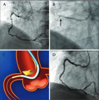
It was not possible to position a new 3.0/20 mm Pro-KineticTM Energy stent at the stenosis. It was there-fore decided to use the GuideLinerTM 6 F catheter for selective intubation of the right coronary artery, which was positioned beside the crushed stent in the proximal third, allowing the release of the 3.0/20 mm Pro-KineticTM stent in the proximal third stenosis after the curve with a good inal angiographic result (Figure 1).
Case 2
A 75-year-old male patient who had a deinitive pacemaker implanted for a complete atrioventricular block was hospitalised with class 3 stable angina ac-cording to the Canadian Cardiovascular Society (CCS) criteria. Coronary angiography showed a long left main coronary artery with 40% focal stenosis (a minimal luminal area of 5.7 mm2 on intracoronary ultrasound), an anterior descending artery showing dissection and 70% stenosis at the ostium, as well as an eccentric segmental stenosis of 70% in the middle third (a minimum luminal area of 3.5 mm2 on intracoronary ultrasound), a tortuous circumlex artery with 90% ste-nosis in the middle third of the marginal branch, and a right coronary artery without signiicant lesions. The
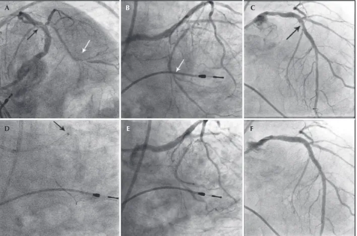
patient refused to undergo surgery despite a SYNTAX score of 32 points. Percutaneous coronary intervention was performed via the femoral artery (Figure 2). The left main coronary artery was cannulated with an XB4 7 F guide catheter (Cordis Co. – New Jersey, USA). Due to the marked proximal tortuosity of the circumlex artery, it was impossible to cross its ostium with a Mini TrekTM 2.0/15 mm balloon (Abbott Vascular – Santa Clara, CA, USA) over a 0.014 inch Whisper guide wire (Abbott Vascular – Santa Clara, CA, USA). At that moment, a GuideLinerTM 6 F catheter was positioned in the ostium of the circumlex artery, and the Mini TrekTM 2.0/15 mm balloon was navigated without dificulty to the stenosis of the marginal branch of the circumlex artery, which was treated conventionally with the balloon, with a good inal angiographic result. A Whisper guidewire was then placed distally in the left anterior descending artery. Two XienceTM Prime stents (Abbott Vascular – Santa Clara, CA, USA) were successfully implanted with an overlay of 2 mm in the stenosis in the middle third of the left anterior descending artery (3.0/15 and 3.0/33 mm). Two other XienceTM Prime stents were successfully implanted, also with a 2-mm overlap in the anterior descending artery-left main coronary artery segment and in the left main coronary artery (4.0/33 mm and 4.0/12 mm, respectively), without using the GuideLinerTM.
Figure 1 – In A, transradial coronary angiography showing criti-cal stenosis and thrombus in the proximal right coronary artery. In B, 3.5/20 mm Pro-KineticTM stent (white arrow) crushed in the proximal
third of the right coronary artery and ‘mother’ (AL1 6 F) and ‘child’ (the radiopaque spot of the GuideLinerTM 6 F is identiied by the black
arrow) catheters. In C, schematic illustration representing image B. In D, the inal angiographic control after 3.0/20 mm Pro-KineticTM stent
implanting. Source: Figure 1C – A section of the catheter GuideLinerTM brochure. Available at http://www.vascularsolutions.com
A
C D
Case 3
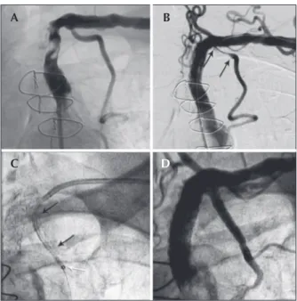
A 75-year-old male patient with a history of coronary artery bypass grafting (left internal thoracic artery-left anterior descending artery, a saphenous vein graft to the right coronary artery, and a sequential saphenous graft to the diagonal and marginal branches) was hospitalised due to acute coronary syndrome without ST-segment el-evation. The coronary angiography via the femoral vein showed 95% stenosis in the left subclavian artery, with a slow distal low to the LITA-LAD graft. It was decided to catheterise the left subclavian artery and the left internal thoracic artery-anterior descending artery graft via the left radial artery. A 90% stenosis was observed in the left anterior descending artery distal to the anastomosis, and the team decided to treat the left subclavian artery via the femoral artery with a DynamicTM 7.0/25 mm stent implant (Biotronik – Bulach, Switzerland). The control angiography showed dissection at the ostium of the left internal thoracic artery, with a slow distal low (thrombolysis in myocardial infarction [TIMI] 1). A Pro KineticTM Energy 3.0/22 mm
stent was successfully implanted at the ostium of the left internal thoracic artery via the left radial artery. During an attempt to go past the stent at the ostium of the left internal thoracic artery with a Mini VisionTM 2.0/23 mm stent (Abbott Vascular – Santa Clara, CA, USA) stent for the treatment of the anterior descending artery, the stent was displaced. At moment, the ostium of the left internal thoracic artery was passed with a GuideLinerTM 6 F cath-eter, which allowed for the passage of the Mini VisionTM stent to the native bed of the left anterior descending artery without dificulty. The procedure was completed with the placement of a second Pro-KineticTM 3.0/13 mm stent in the ostium of the left internal thoracic artery after withdrawal of the GuideLinerTM (Figure 3).
DISCUSSION
The treatment of severe stenoses in tortuous, calciied coronary arteries, sometimes with chronic occlusions and a complex anatomy, is a technical challenge for the release of stents in approximately 5% of procedures.1
Figure 2 – In A, B, and C, coronary angiography via the femoral approach shows a left coronary artery with a long trunk and 40% stenosis in the body. The anterior descending artery dissection (black arrow) and ostial stenosis of 70% and tortuous circumlex artery with marginal branch shows the 90% proximal stenosis (white arrow). A permanent pacemaker electrode in the right ventricle. In D, at the luoroscopy, the ‘mother and child’ technique with the distal extremity of the GuideLinerTM (the black arrow identiies the radiopaque markers) at the origin of the circumlex artery
and the angioplasty guidewire in the marginal branch. In E and F, the inal angiographic result after balloon angioplasty in the marginal branch and implantation of four drug-eluting stents.
A
D E F
Different strategies, such as parallel guide wires (buddy wires), guidewires with more support and weight on the tip, balloons as anchors, atherectomy, and deep intubation with a guide catheter have been designed to overcome this challenge. However, deep intubation is limited by the possibility of dissection, low obstruction or the incapacity to perform the procedure with the guide catheter. Although useful, these strategies are not universally effective. To improve support for coronary interventions, longer, lexible, and lower-proile (‘child’) guide catheters were developed, which are capable of being introduced into the conventional guide catheters (‘mother’) and entering the vascular beds – the so-called ‘mother and child’ technique.
The GuideLinerTM is a modiied ‘child’ type rapid exchange guide catheter extension that allows both greater support and selective intubation with coaxial alignment. This study reports the initial experience with this cath-eter in three patients: in two patients, to help manage complications, and in one patient with anatomic varia-tions of the left main coronary artery. In the irst case, using the GuideLinerTM, it was possible to go past the stent that had detached from the balloon and was crushed against the arterial wall in the proximal third of the vessel, in order to implant another stent for the
treatment of a critical and calciied lesion in the right coronary artery. In the second case, the positioning of the GuideLinerTM in the bifurcation of a very long left main coronary artery allowed for the crossing of a critical lesion in the circumlex artery and the po-sitioning of the angioplasty balloon. In the last case, the cathe terisation of the left internal thoracic artery with the GuideLinerTM through the stent allowed it to be advanced into the native bed without dificulty. This technique has been described by other surgeons1,2 and deies conventional medical practice by treating irst the distal lesions and then the proximal lesions. The ‘child’ catheter was used to overcome dificulties and to assist in the management of complications.
The introduction of the GuideLinerTM into arteries with functional calibres < 2.5 mm, cerebral arteries, and the venous system is not recommended by the manufacturer. There are, however, descriptions of its successful use in venous grafts.2,3 Furthermore, it is recommended, as a precaution, not to introduce the catheter more than 10 cm beyond the tip, as the GuideLinerTM can get stuck in the guide catheter (generally in the second curvature of the catheter). An intubation beyond 20 cm completely externalises the tube with the metal collar to the vessel, causing severe damage.
Recently, in new uses for the GuideLinerTM, Cunning-ton and Egred4 reported success in removing a trapped RotablatorTM olive-shaped burr (Boston Scientiic Co. – Natick, MA, USA) with the aid of the GuideLinerTM, using counter-traction. Tunuguntla et al.5 described the reduced volume of contrast required for intervention in the obtuse marginal branch with the subselective intubation of the circumlex artery. Similarly, Pershad et al.1 reported a signiicant reduction in the volume of contrast required for the visualisation of a right coronary artery occlusion distal to a large aneurysm of the proximal third.
Luna et al.6 reported dissection in the proximal anterior left descending artery during intubation with a GuideLinerTM 7 F. Murphy and Spence7 described their irst complication with the use of the GuideLin-erTM during primary coronary angioplasty of the ostium of the right coronary artery while withdrawing of the GuideLinerTM over the stent for its release: a perforation of the balloon catheter system occurred, caused by the metal collar and the incapacity to appropriately inlate the stent balloon.
Some recommendations for the use of the Guide-LinerTM are a) to introduce it only over a 0.014 inch guidewire, preferably over a balloon (considering the anchor balloon technique), and to proceed carefully, while watching the pressure curve; b) to advance the stent only over the irst guidewire because if there is a second guidewire, it may have passed externally to the catheter; c) to be careful with the passage of Figure 3 – In A, severe stenosis of the left subclavian artery before
the origin of the left internal thoracic artery. In B, dissection of the ostium of the left internal thoracic artery (the limits of the dissec-tion are between the black arrows) after a DynamicTM 7.0/25 mm
stent implant in the left subclavian artery. In C, the GuideLinerTM in
the proximal third of the left internal thoracic artery, with a distal radiopaque marker (white arrow) after the Pro-KineticTM 3.0/22 mm
first stent, which was displaced in the ostium (the stent limits are between the black arrows). In D, the final result after implantation of a second Pro KineticTM 3.0/13 mm stent in the ostium of the left
internal thoracic artery.
A
C
B
higher-proile stents through the metal collar, especially drug-eluting stents with diameters ≥ 4 mm, as they may be damaged; d) to use a ‘child’ catheter that has the same proile as the ‘mother’ catheter because it slips less and decreases the chance that other materials will pass between the catheters; and e) to not make rotations when moving the GuideLinerTM, lessening the chances of the wire twisting around the metal rod.
The authors believe this new catheter may help to treat complex coronary lesions and to manage com-plications and possibly reduce the contrast volume. A probable and unexplored site for its use is the treatment of lesions in visceral vessels by a radial approach, as it can provide more support and range to perform these procedures.
CONFLICTS OF INTEREST
The authors declare no conlicts of interest.
REFERENCES
1. Pershad A, Sein V, Laufer N. GuideLiner catheter facilitated PCI: a novel device with multiple applications. J Invasive Cardiol. 2011;23(11):E254-9.
2. Mamas MA, Fath-Ordoubadi F, Fraser DG. Distal stent delivery with Guideliner catheter: irst in man experience. Catheter Cardiovasc Interv. 2010;76(1):102-11.
3. Wiper A, Mamas M, El-Omar M. Use of the GuideLiner cathe-ter in facilitating coronary and graft incathe-tervention. Cardiovasc Revasc Med. 2011;12(1):68.e5-7.
4. Cunnington M, Egred M. GuideLiner, a child-in-a-mother catheter for successful retrieval of an entrapped rotablator burr. Catheter Cardiovasc Interv. 2012;79(2):271-3.
5. Tunuguntla A, Daneault B, Kirtane A. Novel use of the Guide-Liner catheter to minimize contrast use during PCI in a patient with chronic kidney disease. Catheter Cardiovasc Interv. 2011 Nov 22. [Epub ahead of print]
6. Luna M, Papayannis A, Holper EM, Banerjee A, Brilakis ES. Transfemoral use of the GuideLiner catheter in complex coronary and bypass graft interventions. Catheter Cardiovasc Interv. 2011 Jul 29. [Epub ahead of print]