© 2013 Sociedade Brasileira de Hemodinâmica e Cardiologia Intervencionista. Published by Elsevier Editora Ltda. All rights reserved.
Acute Myocardial Infarction Due to Spontaneous
Coronary Artery Dissection – A Series of Five Cases
Roberto Ramos Barbosa
1, Fábio Salerno Rinaldi
2, J. Ribamar Costa Jr.
3, Fausto Feres
4,
Alexandre Abizaid
5, Amanda G. M. R. Sousa
6, J. Eduardo Sousa
7RESUMO
Infarto Agudo do Miocárdio por Dissecção Espontânea de Artérias Coronárias – Série de Cinco Casos
Relatamos série de 5 pacientes internados por síndrome coronária aguda, entre 2008 e 2010, nos quais a cinecoro-nariograia evidenciou dissecção coronária espontânea. A dis-secção coronária espontânea é uma causa rara de infarto do miocárdio, mais comumente observada em indivíduos jovens e sem histórico de doenças cardiovasculares. O manejo ideal é incerto, principalmente pela limitada experiência clínica com essa entidade. A ultrassonograia intracoronária é uma ferramenta útil tanto para o diagnóstico da dissecção coronária espontânea como para guiar o tratamento percutâneo.
DESCRITORES: Síndrome coronariana aguda. Infarto do
mio-cárdio. Vasos coronários. Angioplastia. Stents.
ABSTRACT
We report a series of ive patients hospitalized due to acute coronary syndrome, from 2008 to 2010, in whom coronary an-giography showed spontaneous coronary dissection. Spontaneous coronary artery dissection is a rare cause of acute myocardial infarction, most commonly seen in young individuals with no history of cardiovascular disease. The optimal management is uncertain, especially due to the limited clinical experience with this entity. Intravascular ultrasound is a useful tool to diagnose spontaneous coronary artery dissection as well as to guide percutaneous therapy.
DESCRIPTORS: Acute coronary syndrome. Myocardial
infarc-tion. Coronary vessels. Angioplasty. Stents.
1 Mastership. Interventionist cardiologist physician of the Instituto de
Cardiologia do Espírito Santo. Vitória, ES, Brazil.
2 Interventionist Cardiologis Physician of the Santa Casa de Marília.
Marília, SP, Brazil.
3 Doctor. Interventionist cardiologist physician of the Interventional
Cardiology Services of the Instituto Dante Pazzanese de Cardiologia. São Paulo, SP, Brazil.
4 Doctor. Interventionist cardiologist physician of the Interventional
Cardiology Services of the Instituto Dante Pazzanese de Cardiologia. São Paulo, SP, Brazil.
5 Full professor. Director of the Interventional Cardiology Service of
the Instituto Dante Pazzanese de Cardiologia. São Paulo, SP, Brazil.
6 Full professor. Director general of the Instituto Dante Pazzanese de
Cardiologia. São Paulo, SP, Brazil.
7 Full professor. Director of the Center of Interventions in Structual
Heart Diseases of the Instituto Dante Pazzanese de Cardiologia. São Paulo, SP, Brazil.
Correspondence to: Roberto Ramos Barbosa. Av. Marechal Campos, 1.579 – Santa Cecília – Vitória, ES, Brazil – CEP 29043-260 E-mail:[email protected]
Received on: 01/29/2013 • Accepted on: 04/27/2013
Case Report
S
pontaneous coronary dissection is a rare entity,usually found in the literature as isolated case reports, which generally affects young and/or female patients.1 In most cases, classical risk factors
for coronary atherosclerotic disease (CAD) are absent. Since it is an infrequently studied disease, its etiology remains largely unknown. It can cause death in up to 40% of cases, usually in the irst days of the clinical picture.2 The prognosis and the therapeutic approach
remain uncertain.
Although it usually occurs in arteries without obstructive atherosclerotic disease at the angiography,
spontaneous coronary dissection may be related with the inlammatory reaction in the medial layer of ves-sels, mainly initiated by autoantibodies produced dur-ing pregnancy or in the irst weeks after childbirth,3
as a result of antiphospholipid antibody syndrome or changes in the extracellular matrix metabolism.4 Thus,
an endothelial dysfunction appears to be the physio-pathological basis of spontaneous coronary dissection.
de Cardiologia between January of 2008 and February of 2011, in whom the coronary angiography showed or was suggestive of spontaneous coronary dissection.
CASE REPORTS
Case 1
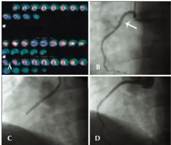
Male patient, 41 years old, with no CAD risk factors, sought medical care due to typical angina chest pain at rest. Electrocardiogram (ECG) at admission showed ST elevation < 1 mm in the inferior wall. Subsequent ECGs did not evidence of alterations in relation to the admission ECG. There was mild elevation of myocardial necrosis markers. Myocardial perfusion scintigraphy was performed in the presence of chest pain and evidenced hypoperfusion, suggesting inferior wall ischemia (Figure 1A). The patient was submitted to invasive stratiication six hours after hospital admission. Coronary angiography showed spontaneous dissection in the proximal third of the right coronary artery, with 70% stenosis at its most critical point (Figure 1B). The other coronary arteries showed no lesions. After contrast injection and image acquisition for the right coronary artery, the patient went into cardiorespiratory arrest in ventricular ibrillation, which was promptly reversed with biphasic deibrilla-tion at 200 J. There was slowing of the right coronary artery low. It was decided to perform a percutaneous coronary intervention (PCI) with a bare-metal stent to the right coronary artery (Figure 1C), which was suc-cessful (Figure 1D). Follow-up of 34 months showed clinical evolution without adverse cardiac events. The
patient is currently using acetylsalicylic acid (ASA) and simvastatin.
Case 2
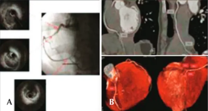
Female patient, 37 years old, in postpartum period, without CAD risk factors, sought medical care present-ing with typical chest pain, mild, which persisted hours after an episode of severe pain. Optimized clinical treatment was established, with complete pain relief. She was transferred to the Instituto Dante Pazzanese de Cardiologia with a diagnostic hypothesis of NSTEMI. She remained hemodynamically stable. The ECG showed a pathological Q wave in the inferior wall, with no ST elevation (30 hours after the episode of severe chest pain). A coronary angiography was performed three days later and showed an image suggestive of spontaneous dissection in the middle third of the right coronary ar-tery, without signiicant obstructive lesion, with normal distal low (Figure 2A). The other coronaries showed no lesions; left ventriculography showed inferior-mid-basal wall akinesia. An intravascular ultrasound (IVUS) of the right coronary artery was performed, which conirmed the coronary dissection, showing an intimal lap (Figure 2B) and vessel wall hematoma (Figure 2C). Due to the patient’s clinical stability, the prolonged evolution of the event (STEMI), and akinesia of the ventricular segment involved with no signs of viability, it was decided not to perform the PCI, maintaining the clinical treatment. Follow-up of 29 months has shown evolution with no adverse cardiac events. The patient is currently using ASA, atenolol, and atorvastatin.
Case 3
Male patient, 34 years of age, with no risk factors for CAD, was admitted to another service presenting mild dyspnoea, approximately 24 hours after an episode of poorly characterized chest pain. He was transferred to the Instituto Dante Pazzanese de Cardiologia with a diagnosis of STEMI in anterior wall, with late presen-tation. He remained hemodynamically stable, without chest pain. ECG showed pathological Q wave in the extensive anterior wall, with ST elevation of 2 mm (six days after the episode of chest pain). Coronary angiography was performed eight days after the start of the clinical picture and presented image suggestive of
Figure 1 – In A, myocardial perfusion scintigraphy, performed in the presence of chest pain, shows hypoperfusion in the inferior wall. In B, coronary angiography shows spontaneous dissection in the proxi-mal third of the right coronary artery (arrow). In C, performance of percutaneous coronary intervention in the right coronary artery. In D, optimal angiographic result after percutaneous coronary intervention with stenting.
Figure 2 – In A, coronary angiography shows image suggestive of spontaneous dissection in the middle third of the right coronary artery (arrow), with normal distal low. In B, intravascular ultrasound demonstrates intima lap (arrow). In C, intravascular ultrasound shows evidence of vessel wall hematoma (arrow).
A
C
B
D
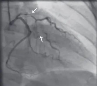
spontaneous dissection in the proximal third of the left anterior descending artery with moderate reduction in calibre in this segment and normal distal low (Figure 3A). The other coronary arteries did not present any lesions. Left ventriculography demonstrated anterior wall akinesia and apical dyskinesia (Figure 3B). IVUS was not performed. Due to the patient’s clinical stabil-ity, prolonged evolution of the event, and akinesia in the ventricular segment involved, it was decided not to perform PCI, maintaining the clinical treatment. Clinical follow-up of 18 months showed no adverse cardiac events. Echocardiogram showed akinesia of the anterolateral wall and apical dyskinesia of the left ventricle, with left ventricular ejection fraction of 35%. The patient is in functional class II and has not required hospitalisation for heart failure during this period. He is currently using carvedilol, enalapril, spironolactone, ASA, and simvastatin.
Case 4
Female patient, 24 years old, with no CAD risk factors, was admitted to the emergency room with typical chest pain, of strong intensity, accompanied by dizziness and nausea, radiating to the left arm, with duration of 2 hours. ECG showed ST segment elevation in the inferior wall. The patient was then referred to the catheterisation lab for possible primary PCI. Coronary angiography evidenced sudden and regular narrowing in the transition from the proximal third to the middle third of the right coronary artery, and the distal seg-ment of the coronary artery had reduced calibre, but maintained the epicardial low (Figure 4A). There was no obstructive disease in the left coronary artery. At the end of the diagnostic procedure, the patient experienced signiicant pain relief. IVUS was performed for the right coronary artery, showing dual lumen of long extension, with evidence of hematoma in the false lumen (Figure 4A). Due to the patient’s clinical stability and pain relief
at that moment, as well as the dificulty to deine the extension of the coronary dissection, it was decided not to perform PCI, and clinical treatment was maintained. Admission ECG at the coronary unit demonstrated that the ST segment returned to baseline levels and clinical outcome was satisfactory. One month after the event, the patient underwent computed tomography of the coronary arteries, which conirmed the resolution of the dissection and recovery of the lumen diameter through-out the right coronary artery (Figure 4B). Follow-up of 14 months has shown no adverse cardiac events, and the patient is currently using ASA.
Case 5
Female patient, 43 years old, smoker, was admit-ted with chest pain lasting for eight hours. ECG on admission showed inverted T wave in the anterior wall, as well as an increase of myocardial necrosis markers. Coronary angiography showed a clear image of spontaneous dissection in the left main coronary artery with extension of the dissection to the middle third of the left circumlex artery, with slowing of distal low in this vessel. After contrast injection and image acquisition of left coronary artery, there was extension of the dissection to the left anterior descending artery, with 90% stenosis in its most critical point (Figure 5). There were no signiicant lesions in the right coronary artery. The patient remained hemodynamically stable, with mild chest pain. Intravenous nitroglycerin was immediately administered, and the cardiac surgical team was called. Emergency coronary artery bypass graft (CABG) surgery was performed on the same day, with placement of internal mammary artery graft to the left anterior descending artery and saphenous vein graft to the irst marginal branch of the left circumlex artery. In the immediate postoperative period, there was bleeding increased by the mediastinal and left pleural drains, and surgical rapprochement was necessary to
Figure 3. In A, coronary angiography shows spontaneous dissection in the proximal third of the left anterior descending artery (arrow). In B, Left ventriculography shows akinesis of the anterior wall and apical dyskinesia.
Figure 4 – In A, coronary angiography shows spontaneous dissection in the right coronary artery – sudden narrowing of the vessel in the transition of its proximal segment to medium. Intravascular ultrasound (IVUS) shows long extension dual lumen, with the presence of large hematoma in the false lumen. In B, coronary CT performed one month after the acute episode shows complete healing of the hematoma and restoration of the vascular lumen.
A B
control the bleeding. The patient remained in critical condition after reoperation, with dificulty weaning off of vasoactive drugs and worsening of shock, with ST elevation in the anterior wall; death occurred on the second postoperative day.
DISCUSSION
When treating a young patient with a diagnosis of AMI without risk factors for CAD and coronary angio-graphy with no signs of severe obstructive lesions, it is imperative to consider the possibility of non-atherosclerotic coronary disease, such as coronary vasospasm, Takotsubo syndrome, and spontaneous coronary dissection. The latter, in turn, in spite of its little known causes, may be associated with pregnancy and postpartum autoimmune phenomena, antiphospholipid antibody syndrome, and
ibromuscular dysplasia.5 However, since many cases
lack the clinical identiication of any of these factors, it is presumed that there is some endothelial dysfunction mechanism that is not fully understood, which leads to spontaneous coronary dissection.
The ive cases described here illustrate the clinical presentation and diagnostic uncertainty involving spon-taneous coronary dissection. The clinical presentation often occurs later, due to scepticism of both patients and family members, as well as of physicians, regarding the diagnosis of acute coronary syndrome (ACS). The low clinical risk of patients, who are generally young and do not have the classical risk factors for CAD, tends to delay the initial treatment and invasive treatments, such as coronary angiography.
Case 2, who was a postpartum patient, was sub-mitted to antiphospholipid antibody syndrome test after
hospital discharge, which was not conirmed, leaving a presumptive diagnosis of autoimmune phenomenon related to pregnancy and the postpartum period. Case 4 disclosed the use of anovulatory drug (oral contracep-tive), which may or may not be related to the cardiac event; however, it was not possible to accurately establish a causal association between the medication and the spontaneous coronary dissection. In case 5, it is assumed that smoking contributed to endothelial dysfunction, culminating in spontaneous coronary dissection; how-ever, in the absence of corroborative methods for this association, this hypothesis remains speculative. In the other cases, no disease or factor was clearly identiied as a determinant of spontaneous coronary dissection in the acute phase of the event or during the subsequent clinical investigation. Thus, as commonly observed in these cases, no aetiology was conirmed by laboratory tests and associated with spontaneous coronary dissec-tion in the reported cases, which would be of interest for therapeutic follow-up and for future studies.
All spectra of ACS may be present in the clinical presentation of spontaneous coronary artery dissection, including sudden death (up to 50% of cases, usually involving the left coronary trunk), in which the diagnosis in most cases is attained at the necropsy, with limited angiographic documentation.6 The clinical and
angio-graphic diagnosis of this entity may be underestimated, since most cases do not undergo assessment through valuable diagnostic tools such as IVUS or optical co-herence tomography, but instead undergo intervention in stenotic coronary lesions or simply have CAD ruled out in the absence of these lesions, without searching for an underlying cause for the presenting clinical pic-ture. The angiographic appearance, despite suggestive, may not clearly disclose the coronary artery dissection, which must be considered when there is (together with the described clinical criteria) sudden and signiicant reduction in calibre, obstruction with smooth edges, and no spect of atherosclerotic disease in the coronary arteries or line of dissection with or without false lumen.
A condition most commonly found in women, spontaneous coronary dissection has a female to male ratio of 3:1; 25% to 31% of cases are observed during pregnancy or the puerperal period.2 In females, dissections
occur predominantly in the left coronary artery (87%), generally affecting young women without traditional risk factors. Men, in turn, tend to be affected at an older age and, sometimes, with the presence of CAD risk factors, with involvement of the right coronary artery in 67% of cases. Generally, the left anterior descending artery is affected in 75% of cases; the right coronary artery in 20%, the left circumlex artery in 4%; and the left main coronary artery in less than 1% of cases.7
A strong association is observed between spontaneous coronary artery dissection and pregnancy, postpartum period, and oral contraceptive use.8 During pregnancy,
pathological changes may occur in the arterial wall
due to fragmentation of reticular ibres, hypertrophy of smooth muscle cells, and changes in the content of mucopolysaccharides and protein composition, leading to weakening of the vessel wall and, to its rupture dur-ing labor or afterwards.6
The vascular plane where the spontaneous dissection occurs most frequently is the outer third of the tunica media or between the media and adventitia.6 Histological
indings in postmortem studies have shown inlammatory iniltrate, mainly eosinophilic, in the adventitial layer of vessels in 25% to 40% of patients who died from spontaneous coronary dissection. Proteases released by eosinophils may promote proteolytic reactions in the vascular walls, and may contribute to dissection.
Due to its rarity, the best treatment for spontaneous coronary dissection has not been determined, and may vary according to the clinical presentation, consider-ing the persistence or relief of ischemic symptoms, the patient’s hemodynamic status, coronary anatomy and dissection extension, the number of involved vessels, and the distal coronary low. Although there is no ro-bust scientiic evidence, IVUS can be a valuable tool in cases of spontaneous coronary dissection, conirming diagnosis and measuring its length.9
In clinically-stable patients, especially when coronary blood low is restored, a conservative medical approach may be considered, taking into account the high in-cidence of spontaneous healing of the dissection and low incidence of long-term adverse events. The use of nitrates and calcium-channel blockers can reduce local spasm and dissection progression; beta-blockers may be used to minimize hemodynamic forces, and antiplatelet drugs can prevent platelet aggregation and subsequent intramural thrombus formation. The use of ibrinolytics is controversial in this scenario – although the dissolu-tion of intramural thrombus may appear, initially, as an attractive beneit, the use of these drugs can cause the propagation of the dissection.10 Buys et al.11 reported the
case of a 31-year-old woman admitted with ACS caused by spontaneous coronary dissection, who showed, after the use of thrombolytic therapy, worsening of bleeding in the false lumen and extension of the dissection. The currently available data are not suficient to deine the role of antithrombotic agents and glycoprotein IIb/IIIa inhibitors in these cases.
In clinically unstable patients or those with persistent ischemic symptoms, a more invasive approach should be considered, possibly with a reperfusion strategy, which is likely to provide better survival probability. PCI should be the irst option in dissections involving a single vessel, especially when there is involvement of its entire length. It is often necessary to implant more than one stent to treat the dissection, and there is no proven beneit of drug-eluting stents when compared to bare-metal stents in such circumstances. Stent implanta-tion is intended to seal the intimal lap to re-expand the
true lumen. Special care should be used to minimize the risk of obstructing intramural hematomas present in the false lumen, since the elimination of the latter can compress the hematoma, dislocating it to the proximal or distal border of the stent, perpetuating the dissec-tion.10 Furthermore, the possibility of having the guide in
the false lumen should be completely discarded before expanding the stent. IVUS can be useful to clarify the situation, and its use is recommended to evaluate stent expansion and strut apposition in relation to the vessel walls, and also to prevent entry into the false lumen. 9
CABG has high mortality in this scenario (up to 33%), and should be reserved for dissections involving the left coronary trunk or multiple vessels. However, CABG can be technically dificult, as the dissection may extend further distally than shown by the angiography, complicating the decision regarding placement of the graft. Furthermore, the dissection can subsequently extend to the distal parts of the revascularized ves-sels, compromising the surgical outcome;6 this is the
likely adverse event the patient in case 5 experienced, culminating with her death in the postoperative period.
Despite the recommendation to individualize the therapeutic strategy, the long-term results for survivors of spontaneous coronary dissection are generally encour-aging, with low rates of event recurrence (either in the same vessel or another). However, the optimal method of follow-up for these patients is yet to be established, as is the best conduct to prevent further episodes. In general, it is concluded that spontaneous coronary dissection is an infrequent cause of ACS, which usually affects young individuals, and whose ideal management is uncertain, partially due to the limited experience with this clinical entity. IVUS is a useful diagnostic tool, especially in cases in which the dissection is not clearly visible at the coronary angiography, and can also guide and optimize percutaneous treatment.12 When there is hemodynamic
or electrical instability, or persistent chest pain, a CABG strategy should be considered, given the severity of the usually observed clinical presentation.
CONFLICTS OF INTEREST
The authors declare no conlicts of interest.
REFERENCES
1. Kamineni R, Sadhu A, Alpert JS. Spontaneous coronary artery dissection: report of two cases and a 50-year review of the literature. Cardiol Rev. 2002;10(5):279-84.
2. Koller PT, Cliffe CM, Ridley DJ. Immunosuppressive therapy for peripartum-type spontaneous coronary artery dissection: case report and review. Clin Cardiol. 1998;21(1):40-6. 3. Bonnet J, Aumailley M, Thomas D, Grosgogeat Y, Broustet JP,
Bricaud H. Spontaneous coronary artery dissection: case report and evidence for a defect in collagen metabolism. Eur Heart J. 1986;7(10):904-9.
5. Saw J, Poulter R, Fung A, Wood D, Hamburger J, Buller C. Spontaneous coronary artery dissection in patients with i-bromuscular dysplasia: a case series. Circ Cardiovasc Interv. 2012;5(1):134-7.
6. Kar S, Shah PK. Acute coronary syndrome caused by coro-nary artery dissection mimicking acute plaque rupture. Ver Cardiovasc Med. 2001;2(4):215-9.
7. Virmani R, Forman MB, Robinowitz M, McAllister HA Jr. Coronary artery dissections. Cardiol Clin. 1984;2(4):633-46. 8. Azam MN, Roberts DH, Logan WF. Spontaneous coronary
artery dissection associated with oral contraceptive use. Int J Cardiol. 1995;48(2):195-8.
9. Arnold JR, West NE, van Gaal WJ, Karamitsos TD, Banning AP. The role of intravascular ultrasound in the management of spontaneous coronary artery dissection. Cardiovasc Ultra-sound. 2008;6:24.
10. Butler R, Webster MW, Davies G, Kerr A, Bass N, Armstrong G, et al. Spontaneous dissection of native coronary arteries. Heart. 2005;91(2):223-4.
11. Buys EM, Suttorp MJ, Morshuis WJ, Plokker HW. Extension of a spontaneous coronary artery dissection due to thrombolytic therapy. Cathet Cardiovasc Diagn. 1994;33(2):157-60. 12. Silva EOA, Furini FR, Mota FM, Lima VC. Dissecção coronária