839 Rev Soc Bras Med Trop 50(6):839-842, November-December, 2017 doi: 10.1590/0037-8682-0512-2016
Short Communication
Corresponding author: Dr. Ricardo Alejandre Aguilar.
e-mail: [email protected]
Received 7 December 2016
Accepted 10 August 2017
Seroprevalence of human
Trypanosoma cruzi
infection
in the North of Estado de Mexico
Saúl González-Guzmán
[1],[2], Sergio Pichardo-Ávila
[3], Eulalia Mimbrera-Rodríguez
[3],
José Antonio Crescencio-Trujillo
[4], María de Lourdes Gasca-Leyva
[5],
Fernando Martínez-Hernández
[6], Nancy Rivas
[7]and
Ricardo Alejandre-Aguilar
[7][1]. Laboratorio del Banco Central de Sangre, Centro Médico Nacional “La Raza”, Instituto Mexicano del Seguro Social, Ciudad de Mexico, Mexico. [2]. Unidad de Enseñanza e Investigación, Hospital Regional de Alta Especialidad de Zumpango, Estado de Mexico, Mexico. [3]. Departamento de
Epidemiología, Jurisdicción Sanitaria Zumpango, Estado de Mexico, Mexico. [4]. Departamento de Medicina Preventiva, del Hospital Regional de Alta Especialidad de Zumpango, Estado de Mexico, Mexico. [5]. Dirección del Banco Central de Sangre, Centro Médico Nacional “La Raza”, IMSS, Ciudad de Mexico, Mexico. [6]. Departamento de Ecología de Agentes Patógenos, Hospital General “Dr. Manuel Gea González”, Ciudad de Mexico, Mexico.
[7]. Departamento de Parasitología, Escuela Nacional de Ciencias Biológicas, Instituto Politécnico Nacional. Ciudad de Mexico, Mexico.
Abstract
Introduction: Chagas disease is a neglected public health problem in Mexico; however, detailed studies to determine the
seroprevalence in some states have not been performed. Methods: A total 1,504 human serum from thirteen communities in Estado de Mexico, were analyzed with three diagnostics techniques. Results: The overall seroprevalence was 9.1%, with high prevalence among people aged 51-60 years, while people aged 0-29 years were seronegative against T. cruzi. Conclusions: Our data demonstrated the seroprevalence of T. cruzi in the North of the Estado de Mexico, an area considered as non-endemic; however, epidemiological conditions necessary for natural transmission were found.
Keywords:Trypanosoma cruzi. Diagnostic. Seroprevalence.
Chagas disease is caused by the protozoan parasite Trypanosoma cruzi, which has infected at least 6-7 million people in Latin America alone1. This infection is primarily transmitted to humans through feces contaminated with the parasite from the insect subfamily Triatominae. However, there are other mechanisms for acquiring the parasite, such as blood transfusions, considered as the second most important mode of T. cruzi transmission2. Infection rates of T. cruzi in the blood banks in some cities in America range from 3 to 53%, while a Mexican study found a seroprevalence of 1.5% in 1992 and 0.4% in 20103,4. However, in Mexico, the infection rates are heterogeneous among the states, e.g., Veracruz State and Puebla had seroprevalence of 0.9% and 7.7%, respectively5,6. However, detailed studies to determine the seroprevalence by state have not been established.
Although some programs have been established to control Chagas disease in endemic areas, such as in Puebla, Colima, Jalisco, Chiapas, Veracruz, Yucatan, Guerrero, and Oaxaca; in contrast, in other states, such as Hidalgo and the Estado de Mexico, there is limited knowledge of this parasitosis and the associated insect vectors7.
Velasco-Castrejon3 conducted the fi rst study in the Estado de Mexico and found T. cruzi prevalence of 0.2%, Estrada-Franco et al.8 documented specifi c antibodies to T. cruzi in 7.1% of humans and 21% of dogs from Tejupilco, a rural area of the Estado de Mexico, concluding that dogs may be domestic reservoirs and may contribute to the human transmission of T. cruzi in this area. Additionally reported are the following triatomine vector species:Meccus pallidipennis, T. dimidiata, and T. barberi. Furthermore, Barbabosa-Pliego et al.9 also determined the prevalence of T. cruzi infection in 24% of dogs and 35% of triatomines collected in the sanitary region located in the Southern part of the Estado de Mexico. In the Toluca Valley, Estado de Mexico, Quijano-Hernández et al.10 performed an epidemiological study in non-domiciliary and domiciliary dogs and found no evidence of T. cruzi in local domiciliary dogs or triatomines. In summary, the Estado de Mexico is not considered an endemic area for Chagas disease, hence, few serological and vector studies have been performed in this state, and such studies have only been conducted in the Southern region10.
840
González-Guzmán S et al. -T. cruzi serology in Estado de Mexico
Municipality Community
Total Positive
Rate* n % n %
Hueypoxtla 1,183 78.7 104 75.9 8.7
Ajoloapan 229 15.2 26 19.0 11.4 Casa Blanca 5 0.3 1 1.0 20.0 Guadalupe Nopala 46 3.2 10 7.3 21.7 Hueypoxtla 585 38.9 27 19.7 4.6 Jilotzingo 82 5.5 12 8.8 14.6 San José Bata 20 1.3 2 1.5 10 Tezontlalpan 102 6.8 7 5.1 6.9 Tianguistongo 28 1.9 6 4.4 21.4 Zacacalco 84 5.6 13 9.5 15.5
Tequixquiac 321 21.3 33 24.1 10.3
San José 91 6.0 14 10.2 15.4 San Mateo 99 6.6 9 6.6 9.1 Tlapanaloya 131 8.7 10 7.3 7.6
Total 1,504 137 9.1
TABLE 1: The positivity percentage in the Hueypoxtla and Tequixquiac municipalities, State of Mexico.
*Rate per each hundred inhabitants
Chagas disease in the Estado de Mexico, Mexico, we analyzed 1,504 samples from thirteen villages distributed in the North of the state, using three diagnostic serological techniques.
An open population study was conducted in thirteen villages of the Hueypoxtla and Tequixquiac municipalities in the Estado de Mexico. Each participant was informed about the protocol by health personnel in each community. All participants signed the letter of informed consent, and they also completed a survey (clinic-based epidemiological data) on personal habits and customs, housing types, clinical manifestations, and recognition of triatomines. Venous blood samples were collected, and centrifuged for 20 minutes each to subsequently obtain serum which was maintained at -20°C until processing. Serological screening was performed using an enzyme-linked immunosorbent assay (ELISA) coupled with chemiluminescence (CHLIA), using the commercially available equipment Architect model I 2000 SR from Abbott according to the manufacturer’s instructions and was conducted at the Central Blood Bank of the National Medical Center, La Raza. Absorbance was measured spectrophotometrically at 492nm. Optical densities with values greater than that of the negative controls, and generally higher than 0.90 of absorbance were considered as positive; and each positive sample was run in triplicate. Seropositive samples were confi rmed by ELISA and indirect hemagglutination (IHA) techniques, performed in Laboratorio Estatal de Salud Pública of Estado de Mexico according to NORMA Ofi cial Mexicana NOM-032-SSA2-2002 and NORMA Ofi cial Mexicana NOM-253-SSA1-2012.
The main occupations of the participants were agriculture and livestock production. Six villages studied correspond to urban areas while the rest were rural zones. A total of 137 samples were found with positive antibody to T. cruzi; with a prevalence of 9.1% which differed by gender. Higher prevalence among the 80 (58.2%) women and 57 (41.8%) men who tested positive were shown, higher than the prevalence reported in 2010 for the country (0.4%) or (0.2%) in 1992 by Velazco-Castrejón et al.4; while for this State, Estrada-Franco et al. reported 7.1%8.Compared with other states, the Northern part of the Estado de Mexico had a prevalence ten times higher than that reported for the State of Puebla (0.9%) or Querétaro (0.6%) in 20095,12.
The differences in the prevalence in the study area compared with other countries such as Brazil, in which there were blood bank reports of only 0.2% should be noted13. In a study of women and children attending a primary health care center in Buenos Aires, Argentina, they showed a prevalence of 4.0%14; the previous data therefore suggest the need to modify the status of the State of Mexico as an endemic area for Chagas disease with the implementation of health programs focused on the control of this parasitosis.
841
Rev Soc Bras Med Trop 50(6):839-842, November-December, 2017
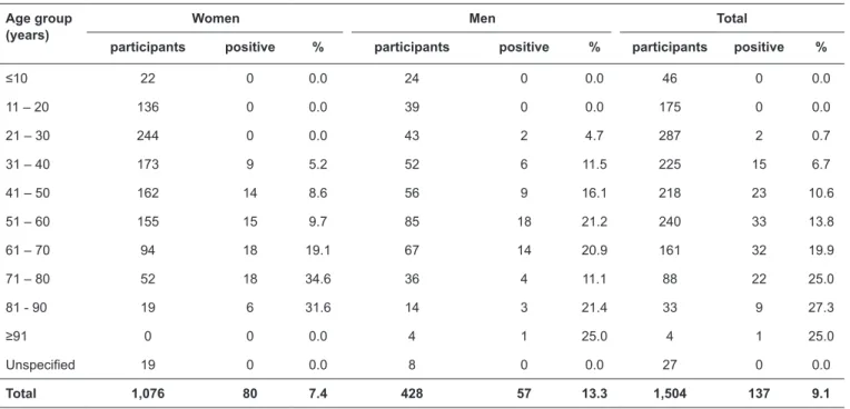
Age group (years)
Women Men Total
participants positive % participants positive % participants positive %
≤10 22 0 0.0 24 0 0.0 46 0 0.0
11 – 20 136 0 0.0 39 0 0.0 175 0 0.0 21 – 30 244 0 0.0 43 2 4.7 287 2 0.7 31 – 40 173 9 5.2 52 6 11.5 225 15 6.7 41 – 50 162 14 8.6 56 9 16.1 218 23 10.6 51 – 60 155 15 9.7 85 18 21.2 240 33 13.8 61 – 70 94 18 19.1 67 14 20.9 161 32 19.9 71 – 80 52 18 34.6 36 4 11.1 88 22 25.0 81 - 90 19 6 31.6 14 3 21.4 33 9 27.3
≥91 0 0 0.0 4 1 25.0 4 1 25.0
Unspecifi ed 19 0 0.0 8 0 0.0 27 0 0.0
Total 1,076 80 7.4 428 57 13.3 1,504 137 9.1
TABLE 2: Seropositivity by age and gender of the participant from the thirteen communities in the North of Estado de Mexico.
San José community had the highest prevalence of 14 (15.4%) positives.
In this study, three different techniques were used to determine the seroprevalence: the positive samples were analyzed with ELISA and IHA screening tests with 107 and 105 samples, respectively. Although, discrepancies were detected between the two techniques, both have acceptable sensitivity and specifi city values. All positive patients were notifi ed of their results, and administered with appropriate treatment under medical supervision; periodic studies were also conducted to determine changes in seropositivity.
Generally, the participant’s ages ranged from zero to 98 years, with the majority aged between 20 and 60 years. Positive T. cruzi serology was found among people aged 29-91 years and was especially prevalent in individuals aged 40 to 80 years (Table 2). Higher rates were observed among people aged 81-90 years, which might suggest a possible correlation between the prevalence and age. It is also important to emphasize that no seropositive cases were identifi ed among people aged 0-28 years (34.3%). This trend could be explained by the improvement in living conditions and the change in housing conditions due to the use of better construction materials and more suitable topcoats, thereby limiting intra- and peridomiciliary vector transmission. However, this does not eliminate the possibility of infection since the conditions favoring natural transmission such as cohabitation with the parasite, insects’ vector and potential parasite reservoirs (i.e., cows, goats, pigs, sheep, horses, poultry and other domestic and wild animals) still exist.
A high percentage of participants reported not having traveled to endemic areas, never left their community, and not having received blood transfusions; therefore, further studies are needed to identify the main risk factors.
Unfortunately, no epidemiological data was obtained regarding infestation and infection indexes, since the area was not considered endemic by the health authorities; however, in those places where volunteers were permitted to conduct a search for the bugs, the vector T. barberi was located in the barns. In the Northern part of the state, there were no data on the presence of T. barberi and there were no records in the literature about the presence of vectors in this area. This specie is mainly characterized by its location in domiciled and peri-domiciled areas with altitudes of up to 2,400 meters above sea level while in neighboring states (surrounding the study region, such as Hidalgo and Mexico City)15, it has also been reported. Interestingly, the seronegativity found in patients aged 0 to 28 years does not rule out the active vectorial transmission, as the people reported an awareness of the presence of vector in the area, although it requires further studies to determine the forms of infection.
It is important to note that with the existence of transmitter in the area, and because a higher T. cruzi seroprevalence rate was identifi ed in the population, there are undoubtedly latent infections among the population throughout this region.
Our fi ndings demonstrated the seroprevalence of T. cruzi in Hueypoxtla and Tequixquiac municipalities, in Estado de Mexico.
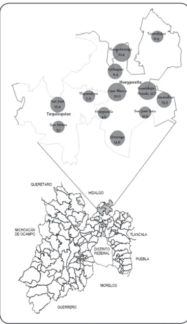
The North of Estado de Mexico was previously considered non-endemic, although we found epidemiological conditions (such as the presence of infected vectors, seropositive patients, and potential reservoirs) necessary for the natural transmission of T. cruzi (Figure 1).
842
FIGURE 1 - Location of the area of study in the North of Estado de Mexico. The circles on the map indicate the location of the communities where cases occurred; the incidence rates from each community are presented.
Ethical consideration:
The protocol was approved by the Committee of Research Ethics of the “Hospital Regional de Alta Especialidad” from Zumpango, Estado de Mexico, Mexico.
Acknowledgments
We offer our deepest thanks to all of the technicians in the Primary Health Care programs of the various Centros of Salud municipalities of Hueypoxtla, Tequixquiac, and Zumpango for their help in locating patients. We are grateful to the laboratory of the Hospital General of Hueypoxtla for sample collection and storage. We are grateful to Martha Eloisa Velázquez Germán, Jefa Jurisdiccional of Zumpango, for her support in the project facilities. We are grateful to Cesar Florencio Martínez, the Chief of the Hospital General of Hueypoxtla Laboratory.
Confl ict of interest
The authors declare that there is no confl ict of interest.
REFERENCES
1. World Health Organization (WHO). Chagas disease (American trypanosomiasis). Fact sheet No. 340. Geneva WHO; 2016. Updated March 2016. Accessed 2016 Apr. [cited 2016 Sep 28]. Available at: http://www.who.int/mediacentre/factsheets/fs340/en/
2. Kirchhoff LV, Paredes P, Lomelí-Guerrero A, Paredes-Espinoza M, Ron-Guerrero CS, Delgado-Mejía M, et al. Transfusion-associated Chagas disease (American trypanosomiasis) in Mexico: implications for transfusion medicine in the United States. Transfusion. 2006;46(2):298-304.
3. Velasco-Castrejón O, Valdespino JL, Tapia-Conyer R, Salvatierra B, Guzmán-Bracho C, Magos C, et al. Seroepidemiología de la enfermedad de Chagas en Mexico. Salud Publica Mex. 1992;34(2):186-96.
4. Novelo-Garza BA, Benítez-Arvizu A, Peña-Benítez J, Galván-Cervantes A, Morales-Rojas A. Detección de Trypanosoma cruzi en donantes de sangre. Rev Med Inst Mex Seguro Soc. 2010;48(2):139-44. 5. Sánchez-Guillén MC, Barnabé C, Guégan JF, Tibayrenc M, Velásquez-Rojas M, Martínez-Munguía J, et al. High prevalence anti-Trypanosoma
cruzi antibodies, among blood donors in the state of Puebla, a
non-endemic area of Mexico. Mem Inst Oswaldo Cruz. 2002;97(7):947-52. 6. Salazar-Schettino PM, Rojas G, Bucio M, Cabrera M, García G,
Ruíz A, et al. Trypanosoma cruzi y su asociación con factores de riesgo en menores de 18 años de Veracruz, Mexico. Rev Panam Salud Publica. 2007;22(2):75-82.
7. Becerril-Flores M, Rangel-Flores E, Imbert-Palafox JL, Gómez-Gómez JV, Figueroa-Gutiérrez AH. Human infection and risk of transmission of Chagas disease in Hidalgo State, Mexico. Am J Trop Hyg. 2007;76(2):318-23.
8. Estrada-Franco JG, Bhatia V, Diaz-Albiter H, Ochoa-García L, Barbabosa A, Vázquez-Chagoyán JC, et al. Human Trypanosoma
cruzi infection and seropositivity in dogs, Mexico. J Emerg Infect
Dis. 2006;12(4):624-30.
9. Barbabosa-Pliego A, Gil PC, Hernández DO, Aparício-Burgos JE, Montes de Oca-Jiménez R, Martínez-Castañeda JS, et al. Prevalence
of Trypanosoma cruzi in dogs (Canis familiaris) and triatomines
during 2008 in a sanitary region of the State of Mexico, Mexico. Vector Borne Zoonotic Dis. 2011;11(2):151-6.
10. Quijano-Hernández IA, Castro-Barcena A, Barbabosa-Pliego A, Ochoa-García L, Del Ángel-Caraza J, Vazquez-Chagoyan J. Seroprevalence Survey of American Trypanosomiasis in Central Valley of Toluca. Sci World J. 2012;(2012):1-3. Article ID 450619. 11. Cooley G, Etheridge RD, Boehlke C, Bundy B, Weatherly
DB, Minning T, et al. High throughput selection of effective serodiagnostics for Trypanosoma cruzi infection. PLoS Negl Trop Dis. 2008;2(10):e316.
12. Serrano-Machuca JJ, Villarreal-Ríos E, Galicia-Rodríguez L, Vargas-Daza ER, Martínez González, Mejía Damián AF. Detección de anticuerpos circulantes en donantes de sangre en Mexico. Rev Panam Salud Publica. 2009;26(4):355-9.
13. Lopes PS, Ramos PLP, Gómez-Hernández C, Ferreira GLS, Rezende-Oliveira K. Prevalence of Chagas disease among blood donor candidates in Triangulo Mineiro, Minas Gerais State, Brazil. Rev Inst Med Trop Sao Paulo. 2015;57(6):461-5.
14. Moscatelli G, Berenstein A, Tarlovsky A, Siniawski S, Biancardi M, Ballering G, et al.. Urban Chagas disease in children and women in primary care centres in Buenos Aires, Argentina. Mem Inst Oswaldo Cruz. 2015;110(5):644-8.
15. Salazar-Schettino PM, de Haro-Arteaga I, Cabrera-Bravo M. Tres especies de triatominos y su importancia como vectores de
Trypanosoma cruzi en Mexico. Medicina (B Aires). 2005;65(1):63-9.