Major Article
Corresponding author: Dra. Vanete Thomaz-Soccol e-mail: [email protected]
Received 3 April 2017 Accepted 12 December 2017
New strategy to improve quality control of Montenegro
skin test at the production level
Deborah Carbonera Guedes
[1], João Carlos Minozzo
[1],[2],
Aline Kuhn Sbruzzi Pasquali
[1], Craig Faulds
[3], Carlos Ricardo Soccol
[1]and Vanete Thomaz-Soccol
[1][1]. Programa de Pós-Graduação Strictu Sensu em Engenharia de Bioprocessos e Biotecnologia, Universidade Federal do Paraná, Curitiba, PR, Brasil. [2]. Centro de Produção e Pesquisa de Imunobiológicos, Secretaria da Saúde do Estado do Paraná, Piraquara, PR, Brasil.
[3]. Department of Biotechnology, Université Aix Marseille, Marseille, France.
Abstract
Introduction: The production of the Montenegro antigen for skin test poses dificulties regarding quality control. Here, we
propose that certain animal models reproducing a similar immune response to humans may be used in the quality control of Montenegro antigen production. Methods: Fifteen Cavia porcellus (guinea pigs)were immunized with Leishmania amazonensis
or Leishmania braziliensis, and, after 30 days, they were skin tested with standard Montenegro antigen. To validate C. porcellus
as an animal model for skin tests, eighteen Mesocricetus auratus (hamsters) were infected with L.amazonensis or L. braziliensis, and, after 45 days, they were skin tested with standard Montenegro antigen. Results: Cavia porcellus immunized with L. amazonensis or L. braziliensis, and hamsters infected with the same species presented induration reactions when skin tested with
standard Montenegro antigen 48-72h after the test. Conclusions: The comparison between immunization methods and immune
response from the two animal species validated C. porcellus as a good model for Montenegro skin test, and the model showed
strong potential as an in vivo model in the quality control of the production of Montenegro antigen.
Keywords: Cutaneous leishmaniasis diagnosis. Montenegro skin test. Cavia porcellus.
INTRODUCTION
Cutaneous leishmaniasis (CL) is a serious social and public health problem, because it can result in sequels1. Early diagnosis
and a suitable treatment are the best tools to control the disease1.
Immunological methods have been largely used as a screening tool for diagnosis1,2. One method widely used in Brazil is
the Montenegro skin test (MST) based on the delayed-type hypersensitivity reaction (DTH)2. The in vivo manifestation of
cellular immune response is an induration that can be measured and semi-quantiied by skin tests3. The method has been widely
used as a complementary leishmaniasis diagnosis because of its high sensitivity and speciicity. Furthermore, MST is an easy method to perform, low cost, does not require sophisticated equipment, and can be performed in loco4.
The Montenegro antigen available in Brazil is provided by the Centro de Produção e Pesquisa de Imunobiológicos (CPPI) in Paraná State, Southern Brazil. The production is authorized and inspected by the Agência Nacional de Vigilância Sanitária
(ANVISA) that establishes the standard evaluation of internal testing for antigen production (RDC 59/2000; RDC 167/2004)5-7.
During the production process, methods for the qualitative and quantitative control of the produced antigens are necessary to evaluate the antigen eficiency and to validate the lots of antigen5-7. Currently, this analysis is performed in in vivo
systems that are exposed to the antigen in order to evaluate the biological response to exposure5-7. This control is performed in
CL human patients5. Such approach demands clinical cases of
CL and poses ethical questions, making the quality control of antigen a complicated process. This study aimed to address these issues in order to establish an experimental model capable to replace the current in vivo model in the quality control process of Montenegro antigen production.
METHODS
This study was divided in two stages. To evaluate the immune response of Cavia porcellus to standard Montenegro antigen in order to establish this species as an experimental model capable of replacing the current in vivo process used in the quality control of Montenegro antigen production. Secondly, to validate C. porcellus as a suitable animal model, Mesocricetus auratus, which is considered a susceptible bio model for
Animal model selection
Antigen preparation • Leishmaniasp. cultivation ; • Antigen obtainment .
Immunized / infection with Leishmania amazonensisorL.
braziliensis .
Period of cellular immunity development
Skin test – Standard MST antigen (CPPI)
Induration reaction readouts (24, 48 and 72 h) Establishment of positive
criteria
Antigen formulation • Protein extraction + IFA • 107parasites/0.1 mL saline
solution
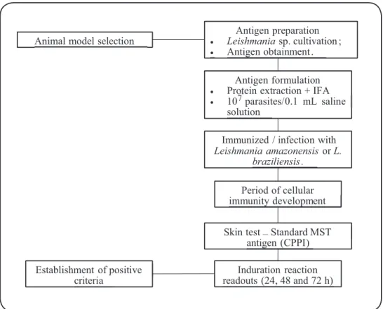
FIGURE 1 – Flowchart outlining the methodology steps. IFA: Incomplete Freund’s Adjuvant. MST: Montenegro skin test.CPPI: Centro de Produção e Pesquisa de Imnunobiológicos do Paraná.
1. Evaluation of Cavia porcellus immune response to skin test
Fifteen adult male guinea pigs, albino strain, weighing 250-350g, were maintained in the animal facility, housed in polypropylene cages, and fed a balanced diet with water and food ad libitum. The animals were maintained in groups of
ive animals. The animal number for this study was calculated as follow: N = (Zα/2 ? (δ/E)) 2 where Z
α/2: 1.96; E: standard error (5%); δ: standard deviation = amplitude/4. The standard deviation was 9.5. Our aim was to minimize the number of animal used in the experiment while maintaining statistically relevant data (according to the 3 Rs concept)10.
The experiments were conducted in four steps (Figure 1). The irst step consisted in the preparation of Leishmania antigen to immunize C. porcellus. Promastigotes forms of L. amazonensis
(MHOM/BR/73/M2269) and L. braziliensis (MHOM/BR/94/
M2903) from the Centre de Ressources Biologiques des
Leishmania, Montpellier, France, were cultivated until a concentration of 107 parasites/mL was reached. After washing
three times with saline solution at 0.9, 0.3, and 0.9%, the pellet was stored at -20°C until further use. The soluble Leishmania
antigens were prepared as described by Szargiki et al.11. Briely,
the promastigotes were defrosted and centrifuged at 4°C for 15 min at 800 x g. The pellet was diluted in sterile distilled
water in a volume equal to half of the pellet volume. The cell
suspension was disrupted by heat shock, alternating between -80°C and 50°C for 5 min, and further sonicated six times at 30% of potency at intervals of 1 min in an ice bath. The extracted solution was centrifuged at 14,000 x g at 4°C for 60
min and lyophilized. The antigen emulsions (L. amazonensis
and L. braziliensis) were prepared by diluting 20.5mg of each
lyophilized antigen in 5.1mL of sterile water, and 3.3mL of this solution was added to 3.3mL of incomplete Freund’s adjuvant (IFA). The inal concentration of the antigen emulsion was 4mg/ mL. The guinea pigs were divided into three groups with ive animals in each. The groups were as follows: G1: immunized with L. amazonensis antigen emulsion; G2: immunized with
L. braziliensis antigen emulsion; and G3: inoculated with IFA
emulsion and saline solution (negative control group). The guinea pigs were intramuscularly inoculated with 1mL (4mg/ mL) of antigen emulsion or IFA. The inoculum (0.5mL) was intramuscularly injected in two sites of guinea pig chest. After 30 days, C. porcellus was skin tested with standard Montenegro
unspeciic reaction or cross reactivity to it. The Montenegro antigen was inoculated at a concentration of 4µg/0.1mL. The readouts of the reaction were performed at 24, 48, and 72h after the intradermal inoculation by measuring the diameter of the indurations using a caliper.
2. Validation of Cavia porcellus as an animal model for skin test
To validate the proposed animal model, the same experimental design was applied to gold hamster (M. auratus), capable of developing the disease through infection with
L. amazonensis or L. braziliensis. Eighteen male, weighing
140-160 g and 6-8 weeks old, were used. The animals were housed in polypropylene cages and fed with water and food ad libitum. The animals were maintained in groups of six. The animal number for this study was calculated as previously mentioned. For experimental infection, promastigotes forms of
L. amazonensis and L. braziliensis were separately cultivated
in biphasic brain heart infusion culture medium and incubated at 24ºC. Promastigotes cultured until stationary phase (5 days) were used to infect the hamster groups. Briely, the parasites were harvested, diluted in ultrapure water (5x), counted, and adjusted to a concentration of 1 x 10⁷ parasites in 0.1mL of
saline solution for inoculation. Three groups with six animals each were formed as follows: GH1: infected with 1 x 10⁷
L. amazonensis; GH2: infected with 1 x 10⁷L. braziliensis; and
GH3: saline solution (negative control group). The hamsters were intradermally inoculated in the right hind paw with 0.1mL of saline solution containing 1 x 10⁷ parasites. After 45 days,
when initial lesions were observed, hamsters were skin tested with standard Montenegro antigen. First, they were depilated, and the inoculation was performed as described above. The skin test solutions were intradermally inoculated using an antigen concentration of 4µg/0.1mL. The readouts of the reaction were performed at 24, 48, and 72h after measuring the diameter of the indurations using a caliper.
Statistical analysis
The homogeneity of variance analysis and the Kolmogorov-Smirnov test for the normal condition of variable evaluation were performed using the Statistica 7 software. A Kruskal-Wallis one-way analysis with a Dunn post-test was performed to analyze the signiicant difference among groups using GraphPad Prism 6 software, assuming a signiicance level of 5%.
Ethical considerations
This study was approved by the Research Ethics Committee of the Federal University of Paraná (Process n. 101328/2015-69).
RESULTS
The results showed that Cavia porcellus immunized with Leishmania amazonensis presented an induration reaction with 0.1 and 0.2mL of antigen (Figure 2A). With 0.1mL of Montenegro antigen, the average induration diameters ranged from 10.4 (24h) to 4.8mm (72h). After 48h, the induration diameter was 4.9mm. With 0.2mL of Montenegro antigen, the
average induration diameters ranged from 9.4 (24h) to 4.4mm (72h). After 48h, the induration diameter was 5.1mm (Table 1).
In C. porcellus immunized with L. braziliensis,the results showed that both volumes (0.1 and 0.2mL) promoted induration reactions (Figure 2B). Inoculation with 0.1mL of Montenegro
antigen resulted in indurations with average diameters ranging from 7.6 (24h) to 4.5mm (72h). After 48h, the induration diameter was 5.0mm. With 0.2mL of Montenegro antigen, the average induration diameters varied from 6.7 (24h) to 4.5mm (72h). After 48h, the induration diameter reached 4.8mm (Table 1).
In C. porcellus immunized with IFA and saline solution
(negative control group), induration reactions were not observed. Statistical analysis showed that there was no signiicant difference between the induration reactions with inoculation of 0.1 and 0.2mL of Montenegro antigen in the guinea pigs immunized with L. amazonensis or L. braziliensis
(p-value < 0.01) after 24, 48, and 72h.
The skin test in M. auratus showed that the six animals
infected with L. amazonensis presented induration reaction with
both 0.1 and 0.2mL of Montenegro antigen (Figure 3A). The average induration diameters ranged from 3.25 (24h) to 3.42mm (72h). After 48h, the induration diameter was 3.75mm. With 0.2mL of Montenegro antigen, the average induration diameters ranged from 5.6 (24h) to 5.4mm (72h). After 48h, the induration diameter was 6.2mm (Table 2). In M. auratus infected with L. braziliensis, all animals presented induration reactions in both inoculation sites with 0.1 and 0.2mL of Montenegro antigen (Figure 3B). The average induration diameters of inoculation site with 0.1mL Montenegro antigen ranged from 3.67 (24h) to 3.58mm (72h). After 48h, the induration diameter was 4.83mm. At inoculation site with 0.2mL of Montenegro antigen, the average induration diameters ranged from 5.25 (24h) to 3.92mm (72h). After 48h, the induration diameter was 5.58mm (Table 2).
In hamsters infected with saline solution (negative control group), induration reactions were not observed. Statistical analysis showed that, in the group infected with L. amazonensis, there was a considerable difference between induration reactions resulting from inoculation with 0.1 and 0.2mL of Montenegro antigen at 24 and 48h. After 72h, there was no signiicant difference between induration reactions with 0.1 and 0.2mL of Montenegro antigen (p-value = 0.01). For the group infected with L. braziliensis, there was no signiicant difference between
induration reactions with 0.1 and 0.2mL of Montenegro antigen after 24, 48, and 72h (p-value = 0.04).
DISCUSSION
A volume of 0.1mL of Montenegro antigen is the standard amount used to inject patients when performing the skin test for cutaneous leishmaniasis12. In this study, the higher level of
FIGURE 2- A. Montenegro skin test in Cavia porcellus previously immunized with Leishmania amazonensis. a: Induration reactions against standard Montenegro antigen with an inoculation of 0.1mL. b: Induration reactions against Montenegro antigen with an inoculation of 0.2mL. B. Reactionto Montenegro antigen in Cavia porcellus previously immunized with Leishmania braziliensis. c:
Induration reactions against standard Montenegro antigen with an inoculation of 0.1mL. d: Induration reactions against standard Montenegro antigen with an inoculation of 0.2mL. The diameter of indurations was measured 24, 48, and 72h after the antigen
inoculation. Each value corresponds to individual induration reaction of the ive animals in each group. MST: Montenegro skin test.
A
B
Leishmania amazonensis Leishmania braziliensis
Group Readout Mean (SD) mm 95% CI Mean (SD) mm 95% CI
MST antigen
0.1mL
24h 10.40 (0.54) 9.72-11.08 7.60 (0.54) 6.92-8.28
48h 4.98 (0.04) 4.92-5.03 5.06 (0.13) 4.89-5.22
72h 4.80 (0.44) 4.24-5.35 4.50 (0.50) 3.87-5.12
MST antigen
0.2mL
24h 9.40 (1.14) 7.98-10.82 6.70 (1.71) 4.56-8.83
48h 5.18 (0.84) 4.13-6.22 4.86 (1.93) 2.45-7.26
72h 4.40 (0.54) 3.72-5.08 4.50 (1.50) 2.63-6.36
TABLE 1: Diameter in mm of induration after Montenegro skin in Cavia porcellus immunized with Leishmania amazonensis or Leishmania braziliensis.
Leishmania amazonensis Leishmania braziliensis
Group Readout Mean (SD) mm 95% CI Mean (SD) mm 95% CI
MST antigen 0.1mL
24h 3.25 (1.72) 1.44-5.06 3.66 (1.21) 2.39-4.93
48h 3.75 (1.40) 2.27-5.22 4.83 (1.25) 3.52-.14
72h 3.41 (1.71) 1.61-5.21 3.58 (0.80) 2.74-4.42
MST antigen 0.2mL
24h 5.66 (1.63) 3.95-7.38 5.25 (1.47) 3.70-6.79
48h 6.25 (1.60) 4.56-7.93 5.58 (1.31) 4.19-6.96
72h 5.41 (1.24) 4.11-6.72 3.91 (1.15) 2.70-5.13
TABLE 2: Diameter of induration after reaction against Montenegro antigen in Mesocricetus auratus infected with Leishmania amazonensis or Leishmania braziliensis.
SD: standard deviation. 95% CI: 95% conidence interval; MST: Montenegro skin test.
FIGURE 3-A. Montenegro skin test in Mesocricetus auratus infected with Leishmania amazonensis. a: Induration reactions against standard Montenegro antigen with an inoculation of 0.1mL. b: Induration reactions against standard Montenegro antigen with an inoculation of 0.2mL. B. MST in Mesocricetus auratus infected with Leishmania braziliensis. c: Induration reactions against standard Montenegro antigen with an inoculation of 0.1mL. d: Induration reactions against standard Montenegro antigen with an inoculation of 0.2mL. The diameter of indurations was measured 24, 48, and 72h after skin test. Each value corresponds to individual induration reaction of the six animals in the group. MST: Montenegro skin test.
A
compared to the group immunized with L. braziliensis. These results showed that 0.1-mL Montenegro antigen inoculation is the best volume of antigen for skin test in the C. porcellus model.
The positivity criteria established for skin test in guinea pigs in the present study were induration reactions ≥ 0.5cm and readouts at 48-72h after the intradermal test. Some studies are in accordance with the positivity criteria established in the present study13-15. The readouts after 24 h were not considered
in the test evaluation, because 24h characterized the initial stage of the immunology response in DTH reactions, representing the cell migration to the injection site3. The inlux of T cells reaches
the maximum levels at 48-72h, indicating the time to determine the result of skin test3.
The animal model chosen for this study was C. porcellus, because it is a well-established animal model to study DTH in different types of diseases and is also used in the investigation of several infectious diseases16-20. Furthermore, some studies on
the genetics of guinea pig have shown immunological analogies between these species21. For instance, guinea pig genes that
encode for major histocompatibility complex (MHC) proteins are homologous to human proteins, and genetic expression pattern of interferon-γ (IFN-γ) and interleukin 12 (IL-12) are similar in the two species21.
The skin test in hamsters showed that they developed induration reaction for Montenegro antigen in both infected groups (L. amazonensis and L. braziliensis), irrespective of the inoculum volume. The skin test volume of 0.2mL promoted higher induration reaction in hamsters than volume 0.1mL in both the infected groups, but induration responses were not statistically different. The induration response difference was considerable just in hamsters in the L. amazionensis
group after 48h. However, this result was in accordance with induration reaction developed in guinea pigs injected with the same Montenegro antigen volumes. As in guinea pigs, there was no considerable difference between induration reactions promoted by 0.1 or 0.2mL of Montenegro antigen. These results in hamsters validated the experimental model proposed in this work.
The guinea pigs proposed as a model for quality control have many advantages compared to hamsters. Firstly, in all infected groups (L. amazonensis, L. braziliensis,and negative control saline), hamsters presented lesions resulting from the phenol in the vehicle solution, which were not observed in guinea pigs. This could be a result of the more sensitive skin of the hamsters. Furthermore, a sensitive skin makes the intradermal injection dificult, because the needle can easily punch the skin or produce lesions, leading to dificult readouts. Secondly, a volume of 0.2mL of Montenegro antigen was required for the skin test in hamsters in order to observe induration reactions. Thirdly, hamster fur grows faster than that of guinea pigs, causing dificulties in performing the readouts after 48 and 72h. Fourthly, induration reaction response in hamsters was heterogeneous between the animals, while in guinea pigs this response was homogeneous, reducing the standard deviation and making the test more reliable. Fifthly, hamsters have strait dorsal compared to guinea pigs, making it dificult to organize
the skin test inoculation sites. Furthermore, guinea pigs need be immunized with dead parasites (parasite proteic extract) and do not develop the disease, while hamsters need to be infected (live parasites) to respond to skin test8,9,14. Once immunized, the risk
of contamination is reduced, and the time needed to develop an immune response is faster than that of hamsters (guinea pigs = 30 days; hamsters = minimum of 45 days). Thus, guinea pigs are better animal models to be used in an industrial qualitative control process. Moreover, they are already used as bio models for skin test antigen evaluation in tuberculosis19, leprosy22, and immunodeiciency
virus type 1 infection20. Additionally, the process of experimental
infection in hamsters is time consuming and requires well-trained professionals8,9. Observations made during experimental tests with
hamsters also conirm that guinea pigs are a suitable animal model for Montenegro antigen quality control process.
We conclude that the experimental tests performed conirm that this experimental model may be used to replace the current human in vivo model in quality control process of Montenegro antigen production. The induration reaction was observed in all animals in both immunized groups. The establishment of this experimental model for skin tests represents a good alternative for qualitative and quantitative control process of antigen production.
Acknowledgments
We thank the Centro de Produção e Pesquisa de Imunobiológicos of Paraná State for the technical support and for providing the Montenegro antigen for this study.
Financial support
We thank inancial support from Conselho Nacional de Desenvolvimento
Cientíico e Tecnológico [(CNPq); Grant No. 307387/2011-9 and 480292/2012-4], Fundação Araucária [Grant No. 122/2010 (Protocol 17401)], and
Programa Nacional de Pós-Doutorado-Coordenação de Aperfeiçoamento de Pessoal de Nível Superior [(PNPD-CAPES); Protocol 2847/2011].
Conlict of interest
The authors declare that there is no conlict of interest.
REFERENCES
1. Gomes CM, Morais OO, Roselino AM, De Paula NA, Soares KA, Sampaio RNR. Complementary exams in the diagnosis of American
tegumentar leishmaniasis. An Bras Dermatol. 2014;89(5):701-9.
2. de Paiva-Cavalcanti M, De Morais RCS, Pessoa-E-Silva R,
Trajano-Silva LAM, Gonçalves-de-Albuquerque SC, Tavares DHC, et al. Leishmaniasis diagnosis: an up date on the use of immunological and molecular tools. Cell Biosc. 2015;5:31.
3. Abbas AK, Lichtman AH, Pillai S. Imunologia Celular e Molecular.
7ª edição. Rio de Janeiro: Elsevier; 2012.
4. Antonio LF, Fagundes A, Oliveira RVC, Pinto PG, Bedoya-Pacheco SJ, Vasconcellos ECF, et al. Montenegro skin test and age of skin lesion as predictors of treatment failure in cutaneous leishmaniasis.
Rev Inst Med Trop S Paulo. 2014;56(5):375-80.
5. Agência Nacional de Vigilância Sanitária (ANVISA). Brasília:
6. Agência Nacional de Vigilância Sanitária (ANVISA).
Resolução RDC nº59. Brasília: ANVISA; 2000. Acessado em outubro 2017. Disponível em: http://portal.anvisa.gov.br/
documents/33836/2814380/RDC+59+2014+Nomes+comerciais. pdf/507c42ee-0309-493d-96ad-02b7344256a2>.
7. Agência Nacional de Vigilância Sanitária (ANVISA).
Resolução RDC nº 167. Brasília: ANVISA; 2004. Acessado em outubro 2017. Disponível em: <http://portal.anvisa.gov.br/ documents/33832/259143/RDC+n%C2%BA+167%2C+de+24+de+j
ulho+de+2017.pdf/ee73f6c6-1969-4d2a-ad1b-7f76cb16d3fc> 8. Hommel M, Jaffe CL, Travi B, Milon G. Experimental models for
leishmaniasis and for testing anti-leishmanial vaccines. Ann Trop
Med Parasitol. 1995;89(Suppl 1):55-73.
9. Robledo SM, Carrillo LM, Daza A, Restrepo AM, Muñoz DL, Tobón J, et al. Cutaneous leishmaniasis in the dorsal skin of
hamsters: a useful model for the screening of antileishmanial drugs. J Vis Exp. 2012;62:3533.
10. Zurlo J, Rudacille D, Goldberg AM. The three Rs: the way forward.
Environ Health Perspect. 1996:104(8):878-80.
11. Szargiki R, Castro EA, Luz E, Kowalthuk W, Machado AM, Thomaz-Soccol V. Comparison of serological and parasitological methods for cutaneous leishmaniasis diagnosis in the state of
Parana, Brazil. Braz J Infect Dis. 2009;13(1):47-52.
12. Skraba CM, de Mello TFP, Pedroso RB, Ferreira EC, Demarchi IG, Aristides SMA, et al. Evaluation of the reference value for the
Montenegro skin test. Rev Soc Bras Med Trop. 2015;48(4):437-444.
13. Briand EW, Ruble GR, Stiteler J, Harris LD, Burge JR, Soranaka ET,
et al. Comparison of adjuvants with Leishmania antigens in a guinea pig model to induce delayed-type hypersensitivity responses. Lab
Anim Sci. 1999;49(5):519-21.
14. Krabiri AR, Bagheri F, Assmar M. Leishmania major: species
speciic delayed hypersensitivity reaction induced by exogenous
secreted antigen in the guinea pig. Exp Parasitol. 2006;112(3):
184-6.
15. Krabiri AR, Bagheri F, Assmar M. Leishmania major: common
antigen responsible for induction of delayed-type hypersensitivity
response in guinea pigs. Parasitol Res. 2007;100(3):629-32.
16. Kim YJ. Eficiency of recombinant bacilli calmette-guérin in inducing humoral and cell mediated immunities against human
immunodeiciency virus type 1 third variable domain in immunized mice. Yonsei Med J. 2011;52(1):173-80.
17. Komori T, Nakamura T, Matsunaga I, Morita D, Hattori Y,
Kuwata H, et al. A microbial glycolipid functions as a new class
of target antigen for delayed-type hypersensitivity. J Biol Chem.
2011;286(19):16800-6.
18. Kukharenko AE, Babaev AA, Shchelchkova NA, Lapshin RD, Vedunova MV, Gravel IV, et al. Estimation of general toxicity and immunological safety of a novel therapeutic vaccine against human
papilomavirus- associated diseases. Sov Technol Med. 2015;7:
92-96.
19. Mallaghini M, Thomaz-Soccol V, Probst CM, Krieger MA, Preti
H, Kritski A, et al. Recombinant antigen production for assays of
intradermoreaction for diagnosis and surveillance of tuberculosis.
J Biotechnol. 2011;156(1):56-8.
20. Moradi J, Mosavari N, Ebrahimi M, Arefpajohi R, Tebianian M. Evaluation of Mycobacterium tuberculosis early secreted antigenic target 6 recombinant protein as a diagnostic marker in skin test.
Osong Public Health Res Perspect. 2015;6(1):34-8.
21. Padilla-Cardin DJ, Mcmurray DN, Hickey AJ. The guinea pig as a
model of infectious disease. Comp Med. 2008;58(4):324-40.
22. Alban SM, de Moura JF, Minozzo JC, Mira MT, Thomaz Soccol V.
Identiication of mimotopes of Mycobacterium leprae as potential