266
Revista da Sociedade Brasileira de Medicina Tropical 45(2):266-268, mar-abr, 2012
Case Report/Relato de Caso
INTRODUCTION
CASE REPORT
1. Faculdade de Medicina,Universidade Federal de Goiás, Goiânia, GO. 2. Departamento de Cardiologia, Hospital Anis Rassi, Goiânia, GO.
Address to: Dr. Anis Rassi. Hospital Anis Rassi. Av. José Alves 453, Setor Oeste, 74110-020 Goiânia, GO, Brasil.
Phone: 55 62 3227-9000; Fax: 55 62 3227-9121 e-mail: arassijr@terra.com.br, anisrassi@arh.com.br Received in 18/11/2010
Accepted in 28/07/2011
Advanced megaesophagus (Group III) secondary to vector-borne
Chagas disease in a 20-month-old infant
Lactente de um ano e oito meses de idade com megaesôfago (Grupo III) por doença de Chagas
adquirida vetorialmente
Anis Rassi
1,2,Jofre Marcondes de Rezende
1and Anis Rassi Junior
2ABSTACT
he authors report the case of a female infant with Group III (or Grade III) megaesophagus secondary to vector-borne Chagas disease, resulting in severe malnutrition that reversed ater surgery (Heller technique). he infant was then treated with the antiparasitic drug benznidazole, and the infection was cured, as demonstrated serologically and parasitologically. Ater follow-up of several years without evidence of disease, with satisfactory weight and height development, the patient had her irst child at age 23, in whom serological tests for Chagas disease yielded negative results. hirty years ater the initial examination, the patient’s electrocardiogram, echocardiogram, and chest radiography remained normal.
Keywords: Infant. Megaesophagus (Grade III). Vector-borne Chagas disease.
RESUMO
Os autores relatam um caso de lactente com megaesôfago do Grupo III por doença de Chagas vetorialmente adquirida, responsável por acentuada desnutrição, da qual se recuperou com a cirurgia de Heller. Submetida em seguida a tratamento da infecção chagásica com benzonidazol, logrou cura, parasito e sorologicamente demonstrada. Seguiram-se anos sem qualquer incidente, com desenvolvimento pondo-estatural normal. Teve seu primeiro ilho aos 23 anos, em quem as provas sorológicas para doença de Chagas resultaram negativas. Decorridos 30 anos após o atendimento inicial, continuava com eletrocardiograma e ecocardiograma e/ou exame radiológico do coração normais.
Palavras-chaves: Lactente. Megaesôfago (Grupo III). Doença de Chagas vetorial.
Commonly, megaesophagus is a relatively late manifestation of digestive tract involvement in Chagas disease. Its occurrence in children is rare. Among 1,761 cases of megaesophagus treated at the Hospital of the School of Medicine of the Federal University of Goiás
during a period of 21 years, only 9 (0.5%) cases of children aged 1 to 10 years have been reported1.
Santos et al.2 reported 40 cases in children aged up to 12 years, of which only ive were 5 years old or less; the youngest was aged 2 years and 6 months.
Exceptionally, the onset of megaesophagus is even earlier in infants, for example, since it may appear in the congenital form of Chagas disease3-5 and in the vector-borne form of the disease when the diagnosis of the initial period of infection is known6-8.
he precocity of the involvement of the digestive tract can be explained by Koeberle9, who demonstrated that denervation of the Auerbach's and the Meissner's plexuses already occurs in the acute phase of the infection and that a 90% denervation results in motor impairment of the organ at the onset of megaesophagus. He expressed this viewpoint in a phrase that has become well known: “he fate of the chagasic patient is deined in the acute phase.”
he case we report is noteworthy for describing the youngest child in the known literature on megaesophagus secondary to the vector-borne form of Chagas disease, as well as for its evolution.
he patient was aged one year and eight months, had Group III megaesophagus, according to the radiological classiication of Rezende et al.10, with important manifestations of the condition, and was accompanied by one of the authors (Rassi A) for thirty years.
Female patient (registration number 350/77), white, born on February 9, 1976 in rural Angical (BA) and a resident thereof, atended to on October 20, 1977.
267
Rassi A et al - Infant megaesophagus secondary to vector-borne Chagas disease
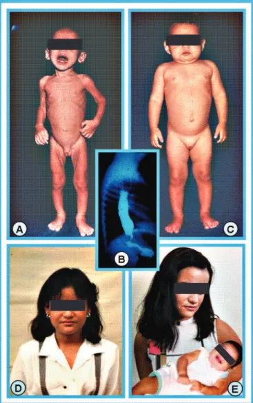
they were all apparently healthy. he mother underwent an indirect immunoluorescence test for Chagas disease on March 2, 1978, and indirect immunoluorescence, indirect hemagglutination, and ELISA tests for Chagas disease on May 8, 2007, both with negative results. he patient appeared to be chronically sufering, was dehydrated and malnourished, in poor nutritional status (Figure 1A), and weighed 7.5kg (15% deicit). he height was not recorded, and the axillary temperature was 36°C. Her heart rhythm was regular, with a rate persistently above 140 per minute, and her BP was 70/50mmHg. he adipose tissue was greatly reduced, and the skeletal muscles were hypotrophic. he esophagus radiograph showed Grade III megaesophagus (Figure 1B). he chest radiograph showed a normal cardiac area and pleuropulmonary ields. he electrocardiogram showed sinus tachycardia (187bpm).
A: During the iniial care consultaion (October 20, 1977). B: Esophagus radiograph at the iniial examinaion showing Grade III megaesophagus. C: Ater Heller surgery for the treatment of megaesophagus (May 17, 1978). D: The paient as an adolescent. E: The
paient and her irstborn. She was cured of Trypanosoma cruzi infecion, and her child is therefore free from the possibility of having congenital Chagas disease.
The disclosure of the photographs was authorized by the paient and approved by the Ethics Commitee of Anis Rassi Hospital.
FIGURE 1 – Montage of photos of the patient taken at diferent times.
Indirect immunoluorescence and complement ixation tests for Chagas disease were positive. Xenodiagnoses performed with four boxes, each containing 10 triatomines, were all positive.
The patient was admitted to the Hospital of the School of Medicine of the Federal University of Goiás (Goiânia, GO) on October 20, 1977. After receiving parenteral nutrition for correction of nutritional deicits, she underwent surgery (Heller’s extramucosal cardiomyotomy) on April 3, 1978. Ater surgery, she was able to swallow food normally, showing excessive appetite and gaining weight. By May 17, 1978, she already weighed 10kg, with normal adipose tissue and skeletal muscles (Figure 1C). hen she underwent some exams, which showed the following results: the chest radiograph and electrocardiogram were normal, the esophagus radiograph showed a size reduction when compared to the exam conducted prior to the surgery, and the barium enema revealed a slightly elongated sigmoid.
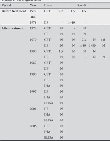
Aiming at the speciic treatment of Chagas disease, we outlined the parasite-serological proile of the patient by xenodiagnosis and serological tests (complement fixation and indirect fluorescent antibody tests) (Tables 1 and 2, which also include the same tests conducted at the irst consultation).
She was treated with benznidazole at a dose of 10mg/kg/ day, twice daily (12/12h) for 60 days. As a side efect, she had generalized urticaria, predominantly on the trunk, which lasted for only three days and was treated with a commercial product made of dexchlorpheniramine and betamethasone.
he patient was discharged on July 1, 1978. She remained in
Goiânia until December 15, 1980, to undergo repeated control examinations after specific treatment, including xenodiagnosis,
TABLE 1 - Xenodiagnosis.
Triatomines (n)
Period Year Exams (n) Boxes (n) examined dead
Before treatment 1977/78 2 6 140 20
4 16
Ater treatment 1978 0 0 179 21
5 20
1979 0 0 478 162
16 64
1980 0 0 567 113
17 68
1982 0 0 178 22
5 20
1987 0 0 147 13
4 16
1990 0 0 262 18
7 28
1991 0 0 252 28
7 28
1997 0 0 30 10
1 4
Ater treatment 0 0 2,093 387
62 248
268
Rev Soc Bras Med Trop 45(2):266-268, mar-abr, 2012
DISCUSSION
REFERENCES TABLE 2 - Serological tests.
Period Year Exam Result
Before treatment 1977 CFT 2.2 1.5 1.5
and
1978 IIF - 1/40 -
Ater treatment 1978 CFT N - N
IIF N N N
1979 CFT N N 2.3 N 1.6
IIF N N 1/40 1/80 N
1980 CFT 1.5 N N N
IIF N N - N N
1987 CFT N
IIF N
1990 CFT N
IIF N
IHA N
1997 IIF N
IHA N
ELISA N
2001 IIF N
IHA N
ELISA N
2006 IIF N
IHA N
ELISA N
CFT: complement ixation test; IIF: indirect immunoluorescence; IHA: indirect hemagglutination; N: negative; ELISA: Enzyme-Linked Immunoabsorbent Assay.
conducted fortnightly, and serological tests, conducted quarterly
(Tables 1 and 2). She returned to her hometown on December 16, 1980, to reside in a house located in an urban area and not infested by triatomines.
She was followed up for several years without incidents, with normal height-weight development (Figure 1D). She had her menarche in 1988. She married in 1995 and, in 1999, had her irst child, who weighed 2,350g and whose serological tests for Chagas disease (indirect immunoluorescence, indirect hemagglutination, and ELISA), conducted in 2007, were negative (Figure 1E). Later reassessments of the patient, a total of 5 in the period between 1987 and 2006, showed continued negative results of xenodiagnosis
(Table 1) and serological tests, now with the addition of indirect hemagglutination and ELISA tests but excluding the complement ixation test (Table 2). In these reassessments, the electrocardiogram and echocardiogram and/or heart radiological exams were consistently normal.
Although extremely rare, and therefore worthy of disclosure, there are cases of Grade III megaesophagus secondary to Chagas disease in early childhood, either in the congenital transmission form or in the vector-borne transmission form.
In this case, the possibility of a congenital form was excluded by the negative results of the maternal serological tests on two occasions, indicating a vector-borne transmission; the initial period of infection was not diagnosed, which is usual in the natural history of Chagas disease11.
his case demonstrated ive important facts: I) the ignorance of the parents, which caused the delay in bringing the child to medical treatment; II) the evolution of megaesophagus to Grade III within a short period of time; III) the importance of the surgical treatment, which allowed the fast recovery of the patient’s nutritional status; IV) the value of the etiological treatment, which resulted in the cure of Chagas disease; and V) the interruption of the Chagas disease epidemiological cycle by the elimination of T. cruzi from a human host.
1. Rezende JM, Moreira H. Forma digestiva da doença de Chagas. In: BrenerZ, Andrade ZA, Barral-Neto M, editors. Trypanosomacruzi e Doença de Chagas, 2ª ed. Rio de Janeiro (RJ): Guanabara Koogan; 2000. p. 297-343.
2. Santos AM, Jorge J, Santana E, Rosa H, Teixeira AS, Rezende JM. Megaesôfago chagásico na infância. Aspectos clínicos. Rev Goiana Med 1974; 20:171-190. 3. Atias A, Almonte C. Megaesófago en un lactante con Enfermedad de Chagas
probablemente congénita. Bol Chil Parasitol 1962; 17:46-48.
4. Tafuri WL, Lopes ER, Nunan B. Doença de Chagas congênita. Estudo clínico-patológico de um caso com sobrevida de seis meses. Rev Inst Med Trop São Paulo 1973; 15:322-330.
5. Bitencourt ACL. Doença de Chagas congênita na Bahia. Rev Baiana Saude Publ 1984; 11:165-208.
6. Koeberle F. Enteropatias e enteromegalias. Simpósio Internacional sobre Enfermedad de Chagas. Buenos Aires: Sociedad Argentina Parasitologia; 1972. p. 77-84.
7. Rassi A, Rezende JM, Doles J. Caso de doença de Chagas observado desde o período inicial da infecção, com aparecimento precoce de megaesôfago e megacólon. Rev Soc Bras Med Trop 1968; 2:303-315.
8. Dias JCP. Doença de Chagas em Bambuí, Minas Gerais, Brasil. Estudo clínico - epidemiológico a partir da fase aguda, entre1940 e 1982. [Tese]. Faculdade de Medicina da Universidade Federal de Minas Gerais. Belo Horizonte (MG); 1982. 376 p.
9. Koeberle F. Patogenia da moléstia de Chagas. Estudo dos órgãos musculares ôcos. Rev Goiana Med 1957; 3:155-180.
10. Rezende JM, Lauar KM, Oliveira AR. Aspectos clínicos e radiológicos da aperistalsis do esôfago. Rev Bras Gastroenterol 1960; 12:247-262.