INTRODUCTION
Article/Artigo
1. Graduate Course in Health Science: Infectology and Tropical Medicine, Faculty of Medicine, Federal University of Minas Gerais, Brazil.
Address to:. Dr. José Roberto Lambertucci. Deptº de Clínica Médica/FM/UFMG Av. Alfredo Balena 190, 30130-100 Belo Horizonte, MG, Brazil.
Phone: 55 31 3337-7781 e-mail: [email protected]
Received in 14/07/2010
Accepted in 16/09/2010
Glomerulonephritis in schistosomiasis mansoni: a time to reappraise
Glomerulonefrite na esquistossomose mansônica: um tempo para reavaliar
Valério Ladeira Rodrigues
1, Alba Otoni
1, Izabela Voieta
1, Carlos Maurício de Figueiredo Antunes
1and
José Roberto Lambertucci
1ABSTACT
Introduction: he current prevalence of glomerulonephritis in patients with hepatosplenic schistosomiasis mansoni in Brazil was evaluated. Methods: Sixty three patients (mean age 45.5±11 years) atending the outpatient infectious disease clinic of a University Hospital in Belo Horizonte, Brazil, from 2007 to 2009, were consecutively examined and enrolled in the present investigation. Diagnosis of hepatosplenic schistosomiasis was based on epidemiological, clinical and parasitological data and imaging techniques. Eight patients, who presented >30mg/day albuminuria, were submited to percutaneous ultrasound guided renal biopsy. Kidney tissue fragments were examined under light, direct immunoluorescence and electron microscopy.
Results: All patients showed mesangial enlargement. In ive, mesangial hypercellularity was observed and four presented duplication of the glomerular basement membrane. Areas of glomerular sclerosis were diagnosed in four. Deposits of immunoglobulin M and C3 were present in six samples; deposits of IgG in four, IgA in three and C1q in two samples. In all patients, immunoglobulin A was reported in the lumen of renal tubules. Deposits of kappa
and lambda were observed in six samples. Electron microscopy revealed dense deposits in the glomerular tissue of three patients. Arterial hypertension, small esophageal varices, slight increases in serum creatinine and decreases in serum albumin were associated with glomerular disease. Conclusions: Renal disease associated with hepatosplenic schistosomiasis was veriied in 12.7% of patients and type I membranoproliferative glomerulonephritis was observed in 50% of them. Schistosomal glomerulopathy still is an important problem in patients with hepatosplenic schistosomiasis in Brazil.
Key-words: Schistosomiasis. Schistosomal glomerulopathy. Glomerulonephritis. Histopathology.
RESUMO
Introdução: Avaliou-se a frequência de glomerulonefrite em pacientes com esquistossomose hepatosplênica no Brasil. Métodos: Selecionou-se para o estudo, 63 pacientes (idade média de 45,5±11 anos) avaliados consecutivamente no ambulatório de doenças infecciosas de um hospital universitário de Belo Horizonte, Brasil, no período de 2007 a 2009. O diagnóstico da esquistossomose foi baseado em dados epidemiológicos, clínicos, parasitológicos e de imagem. Os oito pacientes que apresentaram albuminúria acima de 30mg em 24 horas submeteram-se a biópsia renal percutânea dirigida por ultrassonograia. As amostras de tecido renal foram analisadas à microscopia óptica, eletrônica e de luorescência direta. Resultados: Havia expansão do mesângio em todos. Em cinco, houve proliferação de células mesangiais e em quatro observou-se duplicação da membrana basal glomerular. Áreas de esclerose glomerular foram diagnosticadas em quatro. Depósitos de imunglobulinas M e C3 foram patentes em seis amostras; IgG em quatro, IgA em três e C1q em duas. Em todos os pacientes relatou-se luorescência para IgA dentro dos túbulos renais. Depósitos de kappa e lambda foram vistos em seis amostras. A microscopia eletrônica demonstrou depósitos eletrondensos em tecido glomerular. A presença de hipertensão arterial, varizes do esôfago de pequeno calibre, pequenos aumentos de creatinina e diminuição de albumina sérica associaram-se à ocorrência de dano renal. Conclusões: A frequência de lesão renal foi de 12,7%, no presente estudo, e a glomerulonefrite membranoproliferativa do tipo I foi encontrada em 50%. A lesão renal associada à esquistossomose permanece um problema importante no Brasil.
Palavras-chaves: Esquistossomose. Nefropatia esquistossomótica. Glomerulonefrite. Histopatologia.
It has been estimated that 3.5% of the world’s population (200 million people) is infected with the 3 main species of Schistosoma (mansoni, haematobium and japonicum). he disease causes 200 thousand deaths in sub-Saharan Africa each year. In Brazil, 42 million individuals live in endemic areas and 6 million are infected with Schistosoma mansoni1,2. Renal involvement in schistosomiasis is an important cause of end-stage renal disease and increases the socioeconomic impact of schistosomiasis3-5.
No consensus has been achieved regarding the prevalence of renal disease in schistosomiasis. Studies in Brazil show that up to 15% of patients with hepatosplenic schistosomiasis also present renal disease6-14. In Egypt, Barsoum15,16 reported that in 10% of patients on chronic hemodialysis, schistosomiasis was the primary cause of their renal failure. In Brazil, in the late sixties, Andrade and Queiroz6 detected glomerular lesions in 9 (45%) out of 20 patients with hepatosplenic schistosomiasis in an autopsy series. Another study in Brazil revealed a prevalence of glomerulonephritis associated with hepatosplenic schistosomiasis of 15%8 in a nephrology reference center. In ield-works, using proteinuria as a marker of renal disease, the prevalence varied from 6% to15%7,9,11.
In a more recent and instigating review, Andrade17 suggested that renal disease associated with schistosomiasis was decreasing in Brazil ater mass chemotherapy for schistosomiasis was initiated in the late seventies. Since then, over 13 million treatments/ re-treatments were undertaken in the country4,18-22.
The present study was devised to update the current prevalence of renal disease in patients with hepatosplenic schistosomiasis who atended a reference center for schistosomiasis of an University Hospital of the Southeast of Brazil, from 2007 to 2009.
METHODS
Patients
Federal University of Minas Gerais, in Brazil, from February 2007 to January 2009, were consecutively selected for this investigation. he study was approved by the Human Research Ethical Board of the School of Medicine of the Federal University of Minas Gerais. Sixty-six patients provided writen informed consent to participate and were examined; however, three were excluded during the initial diagnostic procedures (one coinfected with hepatitis C virus) or during follow-up (one refused to continue the required tests and one moved to another city). herefore, 63 patients, 46 (73%) male, in the age range of 34 to 56 years-old (mean of 45.5±11), were followed until the completion of the laboratory tests.
In Figure 1, the origins of the 63 patients with hepatosplenic schistosomiasis are depicted. Most of them came from the State of Minas Gerais.
Diagnosis of hepatosplenic schistosomiasis
Diagnosis was based on: epidemiological evidence, contact with stream water from endemic areas; clinical evidence, hepato and splenomegaly; portal hypertension, esophageal varices diagnosed during upper digestive endoscopy; imaging techniques, showing signiicant portal vein collaterals, S. mansoni eggs in the stools; and ultrasound or magnetic resonance imaging, showing the characteristic Symmers’ ibrosis of the liver21-24. All patients had three negative
parasitological stool examinations by the Kato-Katz technique and they also reported previous treatment for schistosomiasis ranging from 2 to 5 years before being enrolled in the present investigation.
Clinical examination
Clinical examination was performed by one author (VLR). Arterial blood pressure was measured on 2 diferent occasions (one week apart) by the same physician following the protocol established by the Brazilian Consensus on Systemic Arterial Hypertension25.
Patients were then instructed on the correct way of collecting a 24h urine specimen. In the second visit to the outpatient clinic, 10ml of venous blood was obtained and stored for laboratory tests.
Laboratory studies
Routine laboratory tests were performed, including a hemogram (counter Sysmex, E 2100 D), coagulation tests, liver function (AST, ALT, alkaline phosphatase, bilirubin, serum albumin) and renal function tests (BUN, serum creatinine). Antibodies against hepatitis C (anti-HCV) and B (HbsAg and anti-Hbc) were tested. Autoimmune diseases were also investigated: antinuclear antibodies (ANA), protoplasmic-staining antineutrophil cytoplasmic antibodies (p-ANCA) and antineutrophil cytoplasmic antibodies (c-ANCA) were measured by an indirect immunofluorescent method and anti-thyroglobulin antibodies were determined. Fasting glycemia and glycohemoglobin were measured. Venereal disease research laboratory (VDRL) was measured by a locculation test. Serum ASTO and rheumatoid factor were also investigated.
Renal function
Ater 12h of oral liquid restriction, an early morning urine sample was collected in a sterile recipient. Part of the urine was seeded in a culture medium for microbiological study. Using another sample, ater centrifugation, abnormal elements were investigated in the urine under light microscopy (ampliication 400X). One blood cell in male centrifuged urine was considered abnormal and 3 or more in the female samples. he presence of acanthocytes and codocytes (G1 cells) defined the presence of erythrocyte dysmorphism. Dysmorphic cells were investigated by phase-contrast microscopy.
Proteinuria was measured in a 24h urine sample. Whenever the qualitative test was positive, the sulfosalicylic acid technique was used or an immunoenzymatic test was performed. Albuminuria >30mg/ day was considered abnormal.
Renal function was evaluated by calculating the glomerular iltration rate (GFR) according to the MDRD (Modiication of Diet in Renal Disease) formula: GFR = 170 x serum creatinine-0.999 x age-0.176 x [0.762 if female] x BUN-0.170 x albumin+0.318. he serum creatinine used in the formula was measured by a colorimetric method.
Serum complement, cryoglobulins and immunoglobulins
FIGURE 1 - Origin of the patients with hepatosplenic schistosomiasis. One red ball may represent more than one patient. here are cases from Bahia (BA) and Espírito Santo (ES).
Serum C3 and C4 levels were quantiied by nephelometry. he presence of cryoglobulins was investigated by placing the serum sample in 4ºC for 72h and observing the formation of a precipitate which disappeared ater heating. Serum immunoglobulin A was measured by nephelometry ater the addition of speciic anti-sera and the results expressed in mg/dL.
Renal biopsy
All patients with evidence of renal involvement were submited to percutaneous ultrasound guided kidney biopsy, following a routine proposed by Quintino de Lima e Barros26. The following findings were considered evidence of renal disease: albuminuria >30mg/day, hematuria with G1 cells and/or erythrocyte casts.
Exclusion from the study
RESULTS
TABLE 1 - Clinical data of 63 patients with hepatosplenic schistosomiasis.
Patients (n = 63)
Clinical data number percentage
Spleen
palpable 48 77.2
splenectomized 15 22.8
Esophageal varices*
small 13 20.6
medium 25 39.7
large 26 39.7
Hepatomegaly
let lobe increase 19 30.2
difuse increase 22 34.9
non palpable 22 34.9
Digestive bleeding
yes 41 65.1
no 22 34.9
Systemic arterial blood pressure (mmHg)
mean** SBP=127.6±20.7 DBP=78.8±11.5 *diagnosed by upper digestive endoscopy, **mean of 63 patients, SBP: systolic blood pressure, DBP: diastolic blood pressure.
Histopathology of kidney fragments
hree fragments were obtained by kidney biopsy. One was stored in 10% formaldehyde for light microscopy. Another was frozen in liquid nitrogen for immunoluorescence and a third fragment was stored in 2.5% glutaraldehyde for electron microscopy. All the samples were sent to the Department of Pathology of the Triângulo Mineiro Faculty of Medicine for processing and examination.
For light microscopy the fragments were embedded in parain and 2μm thicksectionswerecuton a microtome and stained with hematoxylin and eosin, Masson's trichrome, silver methenamine and picrosirius. For immunoluorescence, fragments embedded in tissue tech were cut to 2μm thicknesson a cryostat and investigated for deposits of IgA, IgG and IgM, kappa and lambda light chains and complement C1q and C3 and ibrinogen. For electron microscopy the tissue was ixed in Trump's ixative then routinely processed into resin embedded blocks; ultrathin sections were stained with uranyl acetate and lead citrate and examined with a Philips CM100 transmission electron microscope.
Statistical analysis
Data from questionnaires were transferred to a databank using EpiData 3.1 (EpiData Association, Odense, Denmark) and analyzed by the Statistical Package for Social Sciences (SPSS) 12.0 (SPSS inc., IBM Company, Chicago, Illinois). As a irst step, descriptive analysis (frequency tables and exploratory data analysis for qualitative and quantitative variables, respectively) was performed. Cases and controls were compared using the Chi square or Fisher exact (qualitative variables) and Mann-Whitney (quantitative variables) tests. Logistic regression models were performed to identify risk factors associated with renal disease. To enter variables in the model, a p value of 0.2 was used. To remain in the inal model, a p value of 0.05 was adopted. Odds ratios (OR) and 95% conidence intervals (95%CI) were estimated. Model adjustment was tested by goodness of it using the Hosmer & Lemeshowtechnique27.
Ethical aspects
he present investigation was approved by the Human Research Ethical Board of the School of Medicine of the Federal University of Minas Gerais.
Sociodemographic and clinical characteristics
Forty-six (73.0%) out of 63 patients were male, with a mean age of 45.5±11 years-old. he mean weight was 65.7kg (±11.9) and the mean height was 164.9cm (±8.5). he mean BMI was 24.1kg/m2
(±3.5) and mean body surface area was 1.7m2 (±0.2). Clinical data
for the 63 patients are shown in Table 1. he mean systemic blood pressure was signiicantly higher in patients with renal disease than in controls (p=0.03). A patient with hypertension was 8 times more likely to present renal disease. Small sized esophageal varices were more common in the group with renal disease (p=0.02).
Laboratory tests
Antinuclear antibodies (ANA) were detected in 6 (9.5%) out of 63 patients at titers <1:160. Rheumatoid factor was present in 16 (25.4%), cryoglobulinemia in 3 (4.8%), p-Anca in 3 (4.8%), C3 levels were consumed in 21 (33.3%) and C4 in 29 (46%). Serum protein electrophoresis revealed polyclonal hypergamma-globulinemia (>1.5g/dL) in 40 (63.5%) patients. No signiicant diferences were observed between the groups with and without renal disease for the above tests.
Findings in urine
None of the patients presented glycosuria, or bilirubinuria and culture of urine specimens for bacteria were negative. Seven (11.1%) patients presented hematuria and 8 (12.7%) presented proteinuria. Patients with increased proteinuria were considered to have renal disease. In the 24h urine specimens, the measured creatinine and the expected creatinine were both calculated using the modiied
Cockcrot-Gault formula: [(140-age) x ideal weight x 0.2] x 0.85 for women, and was always superior to 0.85, validating other urinary solutes measured.
Kidney histology
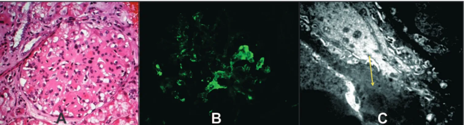
Mesangial injury was reported in all 8 biopsy specimens examined. In 2 cases, only mesangial expansion was observed and in 6, mesangial expansion and cell proliferation were observed. Duplication of the glomerular basement membrane of the capillary walls was revealed in 4 cases. In 3 fragments, segmental glomerular sclerosis was reported and in one, the sclerosis afected more the 50% of the glomeruli (difuse involvement). Deposits of immunoglobulins were observed in most fragments (87.5%); IgG in 50%, IgM in 63% and IgA in 38%. he C3 fraction of complement was reported in 75% and C1q in 2 samples. In renal tubules, around tubular casts, deposits of IgA were observed in all cases and kappa and lambda chains in 75%. Electron-dense deposits were observed in the glomerular structure of 3 patients; all with type I membranoproliferative glomerulonephritis
(Figure 2).
FIGURE 2 - A). Optic microscopy of a patient with type I membranoproliferative glomerulonephritis; B). Immunoluorescence showing IgG deposits in the glomerular structure of a glomerulus; C). Electron microscopy showing electron dense deposits (arrow) in the basement membrane of a glomerulus.
DISCUSSION
Eight (12.7%) out of 63 patients presented evidence of glomerular involvement. In 4, a diagnosis of type I membranoproliferative glomerulonephritis was conirmed. hese indings are similar to those previously described and do not support a decrease in the frequency of renal disease in hepatosplenic schistosomiasis reported by others17.
he most common clinical evidence of renal involvement in the present cases were systemic arterial hypertension, edema, proteinuria and glomerular hematuria. Nephrotic syndrome (proteinuria over 3.5g/day) was reported in 4 (50%) out of 8 patients with renal disease and, in all, hematuria and low serum levels of C3 were present. In one patient, renal biopsy was performed because he had proteinuria of 73.6mg/day and a very low serum level of the C3 component of complement; renal disease was conirmed by kidney biopsy. herefore, even minor alterations in the urine of patients with hepatosplenic schistosomiasis, should be investigated by the atending physician.
A curious finding was the association of esophageal varices of small caliber and the presence of renal disease. We postulated that patients with large esophageal varices died earlier (digestive bleeding) and, consequently, older people (those with small varices) would have survived longer and had a greater chance of developing renal injury. However, no age diference was determined between groups with diferent varix sizes. Another hypothesis was that patients with smaller varices had large spontaneous portal shunts (porta-cava
communication) deviating blood from the varices and redirecting blood antigens to the kidney (still to be evaluated). he present indings must be conirmed by others. It is important to emphasize that the size of varices is based on the subjective evaluation of the endoscopy examiner.
Nearly one third of the patients presented a decrease in the serum levels of the C3 and C4 components of complement and the consumption of complement was not related to kidney damage. he subject has been documented by others and its pathogenesis and importance in glomerular disease is disputed28. Madwar et al29 were the irst to demonstrate persistent low levels of complement and its association with immune-complexes in schistosomiasis. Since only a small number of patients develop glomerulonephritis in the presence of circulating immune-complexes and complement consumption (70% of the cases), it is been suggested that some patients present what has been called nephritogenic immune-complexes30. More recently, Skelly31 revealed the existence of receptors for C2, C3, C8, C9 and Fc on the surface of the schistosome; the worm blocking the complement cascade would be avoiding its own destruction by the host's immune system.
In 16 (25.4%) patients, rheumatoid factor was increased in serum (>20IU/mL), but it was not higher in those with glomerulonephritis. Some authors have reported lower serum levels of rheumatoid factor in patients with schistosomal glomerulonephritis32-34. he results obtained here do not support previous reports. he presence of autoantibodies in this study may be explained by the nonspeciic increase in gamma globulin observed and reported in chronic diseases, such as hepatosplenic schistosomiasis and visceral leishmaniasis; there is no evidence that autoantibodies, usually in low titers, have any inluence on the pathogenesis of these diseases.
he immunoluorescence study on kidney fragments of the patients did not show any singular patern, probably relecting diferent stages of glomerular damage. he suggestion by Egyptian researchers35 that immunoglobulin A plays an important role in the aggravation of renal lesion in schistosomiasis, mediated by IL-6 and IL-10, was not conirmed in here. Serum levels of IgA were within normal range in all patients. In Egypt, the association of schistosomiasis with hepatitis C has been reported and the inluence of the viral infection in the pathogenesis of the glomerulonephritis cannot be underestimated.
IgA was present in the glomerular structure of 2 patients. In one, mesangial expansion was also present, which might suggest a diagnosis of Berger's nephropathy (primary IgA nephropathy).
However, deposits of C1q, a product of the classic complement pathway that does not occur in IgA nephropathy, were also veriied. In the second case, together with IgA, deposits of IgM, IgG and C3 were also observed. IgM deposits are not expected in Berger's nephropathy (also known as IgG-IgA nephropathy)36. Glomerular deposition of IgA is also common in patients with liver cirrhosis and it has been explained by impairment in the depuration of immunoglobulins by Kupfer cells37. More recently, it has been shown that the IgA deposited in Berger's nephropathy is structurally abnormal (sub-galactosylated molecules) and that it is possible to investigate the structure of its molecule.
Interestingly, in all the patients, IgA was identified in renal tubules around tubular casts. Since this is not a common inding, we hypothesized that an increase in the synthesis of this immunoglobulin was occurring due to chronic inflammatory stimulation of the intestinal mucosa caused by schistosomiasis. hus, this macromolecule (160kd) would cross a damaged glomerular capillary and ind its way into the renal tubules, without having any part in the schistosomal glomerulopathy.
he authors declare that there is no conlict of interest. CONFLICT OF INTEREST
FINANCIAL SUPPORT
REFERENCES
surrounded by IgA were observed in all cases. he importance of IgA in the pathogenesis of this nephropathy is unknown and awaits further reined studies of the molecular structure of IgAs deposited in the glomeruli.
Conselho Nacional de Desenvolvimento Científico e Tecnológico.
1. World Health Organization. [internet] - 2008; [cited July 2009]. Available at: htp://www.who.int/schistosomiasis/en/.
2. Lambertucci JR, Rocha RS, Carvalho OS, Katz N. A esquistossomose mansoni em Minas Gerais. Rev Soc Bras Med Trop 1987; 20:47-52.
3. Lambertucci JR, Godoy P, Neves J, Bambirra EA, Ferreira MD. Glomerulonephritis in Salmonella-Schistosoma mansoni association. Am J Trop Med Hyg 1988; 38:97-102.
4. Lambertucci JR, Godoy P, Bambirra EA, Neves J, Tafuri WL, Leite VH. Renal involvement in Salmonella-Schistosoma mansoni association. Rev Soc Bras Med Trop 1987; 20:83-90.
5. Lambertucci JR. Schistosoma mansoni: pathological and clinical aspects. In: Jordan P, Webbe G, editors. Human Schistosomiasis. Wallingford (UK): Cab International; 1993. p.195-235.
6. Andrade ZA, de Queiroz AC. Renal lesions in hepatosplenic schistosomiasis. Rev Inst Med Trop Sao Paulo 1968; 10:36-40.
7. Bina JC. A ield study of proteinuria in individuals infected with Schistosoma mansoni. Rev Soc Bras Med Trop 1985; 18:7-10.
8. Brito T. Schistosoma mansoni associated glomerulopathy. Rev Inst Med Trop S Paulo 1999; 41:269-272.
9. Lehman Jr JS, Mot KE, De Souza CA, Leboreiro O, Muniz TM. he association of schistosomiasis mansoni and proteinuria in an endemic area. A preliminary report. Am J Trop Med Hyg. 1975; 24:616-618.
10. Martinelli R, Rocha H. Revisão/atualização em nefrologia clínica: envolvimento glomerular na esquistossomose mansônica. J Bras Nefrol 1996; 3:279-289. 11. Rabello AL, Lambertucci JR, Freire MH, Garcia MM, Amorim MN, Katz N.
Evaluation of proteinuria in an area of Brazil endemic for schistosomiasis using a single urine sample. Trans R Soc Trop Med Hyg 1993; 87:187-189. 12. Rocha H, Cruz T, Brito E, Susin M. Renal involvement in patients with
hepatosplenic schistosomiasis mansoni. Am J Trop Med Hyg 1976; 25: 108-115.
13. Sobh MA, Moustafa FE, Sally SM, Foda MA, Deelder AM, Ghoneim MA. A prospective, randomized therapeutic trial for schistosomal specific nephropathy. Kidney Int 1989; 36:904-907.
14. Zatz R, Romão Jr JE, Noronha IL. Nephrology in Latin America, with special emphasis on Brazil. Kidney Int 2003; 83:S131-134.
15. Barsoum RS. Schistosomal glomerulopathies. Kidney Int 1993; 44:1-12. 16. Barsoum RS. he changing face of schistosomal glomerulopathy. Kidney Int
2004; 66:2472-2484.
17. Andrade ZA. he situation of hepatosplenic schistosomiasis in Brazil today. Mem Inst Oswaldo Cruz 1998; 93 (supl 1):313-316.
18. Machado PA. he Brazilian program for schistosomiasis control, 1975-1979. Am J Trop Med Hyg 1982; 31:76-86.
19. Amaral RS, Tauil PL, Lima DD, Engels D. An analysis of the impact of the Schistosomiasis Control Programme in Brazil. Mem Inst Oswaldo Cruz 2006; 101 (supl 1):79-85.
20. Drummond SC, Silva LC, Amaral RS, Sousa-Pereira SR, Antunes CM, Lambertucci JR. Morbidity of schistosomiasis mansoni in the state of Minas Gerais, Brazil. Mem Inst Oswaldo Cruz 2006; 101(suppl 1):37-44.
21. Lambertucci JR, Cota GF, Pinto-Silva RA, Serufo JC, Gerspacher-Lara R, Costa Drummond S, et al. Hepatosplenic schistosomiasis in ield-based studies: a combined clinical and sonographic deinition. Mem Inst Oswaldo Cruz 2001; 96 (supl):147-150.
22. Lambertucci JR, Serufo JC, Gerspacher-Lara R, Rayes AA, Teixeira R, Nobre V, et al. Schistosoma mansoni: assessment of morbidity before and ater control. Acta Trop 2000; 77:101-109.
23. Lambertucci JR , dos Santos Silva LC, Andrade LM, de Queiroz LC, Carvalho VT, Voieta I, et al. Imaging techniques in the evaluation of morbidity in schistosomiasis mansoni. Acta Trop 2008; 108:209-217.
24. Pinto-Silva A, Abrantes WL, Antunes CM, Lambertucci JR. Sonographic features of portal hypertension in schistosomiasis mansoni. Rev Inst Med Trop Sao Paulo. 1994; 36:355-361.
25. Sociedade Brasileira de Cardiologia [Internet]. III Consenso Brasileiro de Hipertensão Arterial. Campos do Jordão: Sociedade Brasileira de Cardiologia; 1998. Disponível em: http://departamentos.cardiol.br/dha/publicacoes/ consenso3/consen.asp/.
26. Quintino-de-Lima E, Barros RT. Biópsia renal. In: Dantas M, Alves MAR, Barros RT, editors. Glomerulopatias: patogenia, clínica e tratamento. 2ª ed. São Paulo: Editora Sarvier; 2006. p. 25-35.
27. Hosmer DW, Lemeshow S. Applied Survival Analysis: Regression Modeling of Time to Event Data. John Wyley Editor; New York, 1999.
28. Hussein MM, Ahmed MM, Fayed M. Factors affecting C3 in intestinal schistosomiasis. J Egypt Soc Parasitol 1993; 3:769-773.
29. Madwar MA, O'Shea JM, Skelton JA, Soothill JF. Complement components and immunoglobulins in patients with schistosomiasis. Clin Exp Immunol 1978; 34:354-358.
30. Santoro F, Prata A, Silva AE, Capron A. Correlation between circulating antigens detected by the radioimmunoprecipitation-polyethylene glycol assay (RIPEGA) and C1q-binding immune complexes in human schistosomiasis mansoni. Am J Trop Med Hyg 1981; 30:1020-1025.
31. Skelly PJ. Intravascular schistosomes and complement. Trends Parasitol 2004; 20:370-374.
32. Carvalho EM, Andrews BS, Martinelli R, Dutra M, Rocha H. Circulating immune complexes and rheumatoid factor in schistosomiasis and visceral leishmaniasis. Am J Trop Med Hyg 1983; 32:61-68.
33. Harboe M. Rheumatoid factors in leprosy and parasitic diseases. Scand J Rheumatol 1988;75 (suppl):309-313.
34. Abbas MM, Abdel-Kader S. A study of autoimmunity in schistosomiasis. J Egypt Soc Parasitol 1993; 23:289-296.
35. Homeida M, Ahmed S, Dafalla A, Suliman S, Eltom I, Nash T, et al. Morbidity associated with Schistosoma mansoni infection as determined by ultrasound: a study in Gezira, Sudan. Am J Trop Med Hyg 1988; 39:196-201.
36. Kawasaki Y. he pathogenesis and treatment of IgA nephropathy. Fukushima J Med Sci 2008; 54:43-60.