Renal Function in Hepatosplenic
Schistosomiasis – An Assessment of Renal
Tubular Disorders
Daniella Bezerra Duarte1,2, Lucas Alexandre Vanderlei1, Raianne Kı´via de Azeveˆdo Bispo1, Maria Eliete Pinheiro1, Geraldo Bezerra da Silva Junior3, Alice Maria Costa Martins4, Gdayllon Cavalcante Meneses4, Elizabeth De Francesco Daher2*
1.Department of Internal Medicine, School of Medicine, Federal University of Alagoas, Maceio´, AL, Brazil,2.
Department of Internal Medicine, Post-Graduation Program in Medical Sciences, School of Medicine, Federal University of Ceara´, Fortaleza, CE, Brazil,3.School of Medicine, Post-Graduation Program in Collective Health, Health Sciences Center, University of Fortaleza, Fortaleza, CE, Brazil,4.Department of Pharmacy and Clinical Analysis, Federal University of Ceara´, Fortaleza, CE, Brazil
Abstract
Background:Renal involvement in Schistosoma mansoniinfection is not well studied. The aim of this study is to investigate the occurrence of renal abnormalities in patients with hepatosplenic schistosomiasis (HSS), especially renal tubular disorders.
Methods:This is a cross-sectional study with 20 consecutive patients with HSS followed in a medical center in Maceio´, Alagoas, Brazil. Urinary acidification and concentration tests were performed using calcium chloride (CaCl2) after a 12-h
period of water and food deprivation. The biomarker monocyte chemoattractant protein 1 (MCP-1) was quantified in urine. Fractional excretion of sodium (FENa+),
transtubular potassium gradient (TTKG) and solute-free water reabsorption
(TcH2O) were calculated. The HSS group was compared to a group of 17 healthy
volunteers.
Results: Patients’ mean age and gender were similar to controls. Urinary acidification deficit was found in 45% of HSS patients. Urinary osmolality was
significantly lower in HSS patients (588¡112 vs. 764¡165 mOsm/kg, p50,001)
after a 12-h period of water deprivation. TcH2O was lower in HSS patients
(0.72¡0.5 vs. 1.1¡0.3, p50.04). Urinary concentration deficit was found in 85% of HSS patients. The values of MCP-1 were higher in HSS group than in control group
(122¡134 vs. 40¡28 pg/mg-Cr, p50.01) and positively correlated with the values
of microalbuminuria and proteinuria.
OPEN ACCESS
Citation:Duarte DB, Vanderlei LA, Bispo RKdA, Pinheiro ME, Silva Junior GBd, et al. (2014) Renal Function in Hepatosplenic Schistosomiasis – An Assessment of Renal Tubular Disorders. PLoS ONE 9(12): e115197. doi:10.1371/journal.pone. 0115197
Editor:Giovanna Valenti, University of Bari Aldo Moro, Italy
Received:August 15, 2014
Accepted:November 19, 2014
Published:December 22, 2014
Copyright:ß2014 Duarte et al. This is an open-access article distributed under the terms of the
Creative Commons Attribution License, which permits unrestricted use, distribution, and repro-duction in any medium, provided the original author and source are credited.
Data Availability:The authors confirm that all data underlying the findings are fully available without restriction. All relevant data are within the paper.
Funding:This study was supported by the Brazilian Research Council (CNPq - Conselho Nacional de Desenvolvimento Cientı´fico e Tecnolo´gico). The funders had no role in study design, data collection and analysis, decision to publish, or preparation of the manuscript.
Conclusions: HSS is associated with important kidney dysfunction. The main abnormalities found were urinary concentrating ability and incomplete distal acidification defect, demonstrating the occurrence of tubular dysfunction. There was also an increase in urinary MCP-1, which appears to be a more sensitive marker of renal damage than urinary albumin excretion rate.
Introduction
Schistosoma mansoniinfection remains an important public health problem. This
chronic parasitic disease affects about 200 million people worldwide [1]. Renal involvement in schistosomiasis is described mainly by glomerular involvement [2,3,4,5]. Schistosomal glomerulopathy is associated with the hepatosplenic form of the disease; however, it has also been observed in the hepatointestinal form [6,7]. Its incidence varies between 5–6%, increasing to 15% when considering only patients with the hepatosplenic form [8]. The immunological nature of glomerular involvement in schistosomiasis is well established [5,9,10]. The most frequently reported histological types are chronic membranoproliferative
glomerulonephritis and focal segmental glomerulosclerosis, usually with nephro-tic syndrome [11,12,13,14]. There are other important factors, in addition to parasite antigens, which seem to contribute to the pathogenesis of glomerular disease in schistosomiasis, such as the collateral circulation of the portal system due to the degree of hepatic involvement, the inefficiency of the hepatic
macrophage system, the severity and duration of infestation, racial and genetic factors [13,15].
The search for new renal biomarkers is extremely important, as they may provide early diagnosis of renal disorders, allowing the adoption of measures to prevent progression to end-stage renal disease. The Monocyte Chemotactic Protein-1 (MCP-1) is one of the recently studied new biomarkers. This protein is expressed in injury and inflammation sites and directs the recruitment of macrophages, which bind to chemokine receptors to promote macrophage adhesion and chemotaxis [16]. MCP-1 has been correlated with urinary levels of albuminuria in diabetic nephropathy patients and also in other primary and secondary glomerulopathies [17,18]. Recent Brazilian study found high levels of the urinary biomarker MCP-1 in patients with asymptomatic schistosomiasis [19].
Materials and Methods
Patients
This is a cross-sectional study of 20 consecutive patients with clinical and laboratory diagnosis of HSS, undergoing infectious disease consultation in public health centers in the Northeast of Brazil, from August 2012 to August 2013. Exclusion criteria were age less than 18 or greater than 65 years, hypertension
(systolic blood pressure $140 mmHg and/or diastolic blood pressure
$90 mmHg), diabetes mellitus (fasting glucose levels greater than 100 mg/dL
and/or treatment for diabetes), heart failure, diuretic use within the last 15 days, systemic lupus erythematosus or another collagenosis, recurrent urinary tract infection or history of previous renal disease (glomerular filtration rate #60 mL/ min, nephrolithiasis and use of nephrotoxic drugs). Liver disease of different etiologies (viral or alcoholism) and decompensated schistosomiasis within the last 6 months, with ascites or gastrointestinal bleeding were also excluded. Serological tests for hepatitis B and C were performed in all patients. All patients had been treated at least once in their lifetimes with a specific antiparasitic drug and the time interval between treatment and study enrollment was at least one year. The drugs used were praziquantel (40–50 mg/kg single dose) or oxaminiquine (15 mg/kg single dose). The patients were compared to a control group that consisted of 17 healthy volunteers, all blood donors. They all were tested for schistosomiasis.
Ethics Statement
The study protocol was approved by the Ethical Committee of the Institution (Federal University of Alagoas, protocol 004228-2011-81). Patients were included in the study only after signing the informed consent form.
Hepatosplenic schistosomiasis diagnosis
Schistosomiasis diagnosis was suspected based on epidemiological and clinical data and confirmed by detection of Schistosoma mansoni eggs in feces, rectal or
liver biopsy. Clinical diagnosis of HSS was based on the presence of hepatomegaly and splenomegaly in physical or ultrasonographic examination, periportal fibrosis, increased portal vein diameter, previous history of gastrointestinal bleeding, presence of esophageal varices at endoscopy or history of splenectomy. The mean time of diagnosis was determined from medical records.
Clinical and laboratorial parameters
evaluated: complete blood count, fasting blood glucose, plasma creatinine (Pcr),
urea (PUrea), arterial pH, bicarbonate (Bic), sodium (PNa+), potassium (PK+), calcium (PCa2+), phosphorus (PP), magnesium (PMg2+) and chloride (PCl-), serum
albumin and globulin, aspartate aminotransferase (AST), alanine aminotransfer-ase (ALT), gamma-glutamyl transferaminotransfer-ase (GGT), total bilirubin and fractions, alkaline phosphatase (ALP), international normalized ratio (INR), complement (laboratory C3 and C4), 24-h urinary creatinine, sodium, potassium, magnesium, chloride, proteinuria (UprotV), urinalysis, urinary MCP-1 and microalbuminuria.
The Child-Pugh score was used to assess liver function of patients [20,21].
Renal function evaluation
Glomerular Filtration Rate (GFR) was measured in 24-hour urine collection and was considered abnormal when#60 ml/min/1.73 m2. All patients were submitted to a 12-h water and food deprivation. Fractional excretion of sodium (FENa+), magnesium (FEMg++) and potassium (FEK+), transtubular potassium gradient (TTKG) and solute-free water reabsorption (TcH2O) were calculated by standard
formulae. Microalbuminuria was determined by 24-h urine collection and
abnormal values were .30 mg/day. MCP-1 was determined in the first urine
sample (T0) and the values corrected by urinary creatinine.
Urinary concentrating ability was evaluated by the ratio between urinary and plasma osmolality (U/Posm) after a 12-h water and food deprivation. A baseline
sample (T0) was collected before the administration of an intranasal spray of
DDAVP [22] (desmopressin acetate, 20 mcg) and a second sample was collected
4 h (T4) later.
Urinary acidification was evaluated by measuring urinary pH (UpH) at baseline
(T0) and 4 h (T4) after ingestion of CaCl2, 2 mEq//kg of body weight [23].
Metabolic acidosis induced by calcium chloride (CaCl2) load was documented by
a decrease in serum HCO3– concentrations.3 mmol/1 and a pH ,7.35. Failure
to decrease urinary pH to ,5.5 after CaCl2 load was considered consistent with
some form of distal renal tubular acidosis (RTA). All tubular function tests were also performed in the control group.
Analytical methods
was assessed by freezing-point depression. Blood pH and bicarbonate were determined in a pH blood gas system (AVL compact-1, Medical Instruments). Urinary pH was measured with a pH-meter (pG1800, GEHAKA). C3 and C4 complement fractions were measured by immunoturbidimetry. Total serum albumin and globulin were measured by protein electrophoresis. Urinary MCP-1 measurement was performed through ELISA using the R&D Systems kit, Inc (Minneapolis, MN, USA) and the values corrected by urinary creatinine.
Statistical methods
HSS patients were compared with the control group. The chi-square, Pearson, likelihood ratio and Fisher’s exact test were applied to measures of association and homogeneity in the distribution of categorical data. To verify the normality of distribution of continuous variables, the Kolmogorov-Smirnov test was used. The Levene test was used to compare variability. In the condition of data normality,
the comparison between two means was made by Student’s t test. In the case of
non-normal data, the Mann-Whitney test was applied. Pearson’s correlation test was used to compare two numeric variables. Pearson’ or Spearman’ correlation coefficients, when appropriate, were used to test the association between
continuous variables. Data were expressed as mean¡SD. P,0.05 was considered statistically significant. SPSS software for Windows, release 16.0 (SPSS Inc. Chicago, USA) was used in all the analyses.
Results
Of the 53 patients followed in the outpatient clinics with a diagnosis of hepatosplenic schistosomiasis from August 2012 to August 2013, 11 were excluded due to age older than 65 years, 5 due to diabetes mellitus type 2, 4 due to previous systemic arterial hypertension, 3 due to previous nephrolithiasis, 4 due to chronic viral hepatitis, 3 due to previous chronic kidney disease and 3 due to alcoholic liver cirrhosis. Twenty patients who agreed to participate in the study were included. The mean age of the patients was 42.2¡9.2 years (range 32–57), 50% of each gender. All patients had epidemiological criteria for schistosomiasis. The mean educational level was 3.9¡3.0 years of study. Mean time of diagnosis was 15.5¡11.5 years. History ofSchistosoma mansonieggs in feces was identified
in 75% and diagnosis by liver biopsy was carried out in 25% of patients. All patients had splenomegaly and 70% had undergone splenectomy within a time
period of 10.5¡10 years. Hepatomegaly, however, was present in only 20% of
cases. All patients had evidence of periportal fibrosis at ultrasonography. Intestinal symptoms were found in 30% of patients. Esophageal varices were present in 95% of patients and 55% of them had experienced previous episodes of gastrointestinal bleeding. Other demographic and clinical characteristics are shown in Table 1.
lower in the HSS group compared with the control group (195,520¡107,290 vs.
270,660¡34,092/mm3,P50.01). Hypergammaglobulinemia was present in 65%
of patients and hypocomplementemia in 10 (50%). There was no correlation between complement levels and renal function parameters. The results of general laboratory parameters are summarized in Table 2.
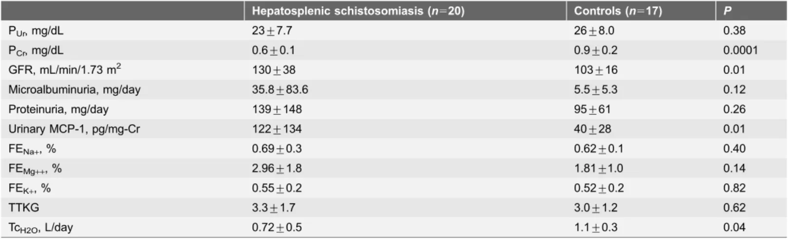
Hepatosplenic schistosomiasis patients have higher GFR and
altered tubular function in comparison with the controls
The comparison between the HSS patients and the control group showed no differences in age, gender, body mass index, systolic and diastolic blood pressure (Table 1). As seen in Tables 3 and4, HSS patients had higher GFR (130¡38 vs.
103¡16 mL/min/1.73 m2, P50.01). Glomerular hyperfiltration (GFR.120 mL/
min/1.73 m2) was observed in 8 (40%) HSS patients. GFR#60 ml/min/1.73 m2
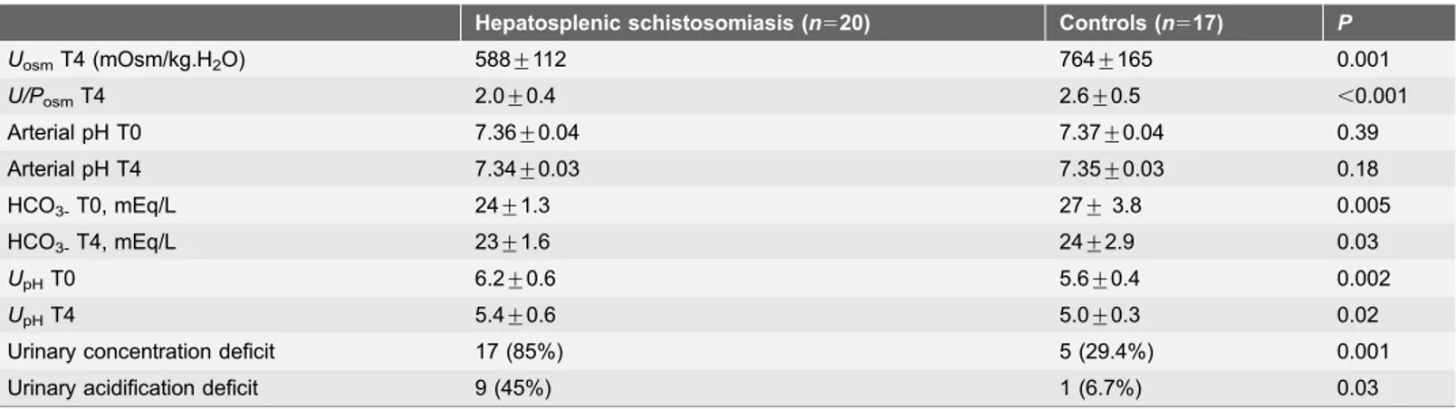
was not observed in any patient. Urinary concentrating ability defect, as
demonstrated by a lower UOsmafter desmopressin (DDAVP) administration
(588¡112vs 764¡165 mOsm/Kg, P50.001) and a lower U/POsm ratio
(2.05¡0.40vs.2.66¡0.55,P,0.001) was observed when comparing HSS patients
and controls. No increase of UOsm after DDAVP was observed in 17 (85%)
patients. Urinary pH did not decrease to 5.5 or less in response to acid-loading with CaCl2in 9/20 (45%) of patients. Levels of serum bicarbonate (HCO3-) were
lower in HSS patients. Arterial pH was similar in the two groups before and after the acid-loading with CaCl2. There was no difference between the HSS group and
the control group regarding FENa+, FEMg++, FEK+and TTKG. The levels of TcH2O
were lower in HSS patients than in the control group (0.72¡0.5vs. 1.1¡0.3,
P50.04).
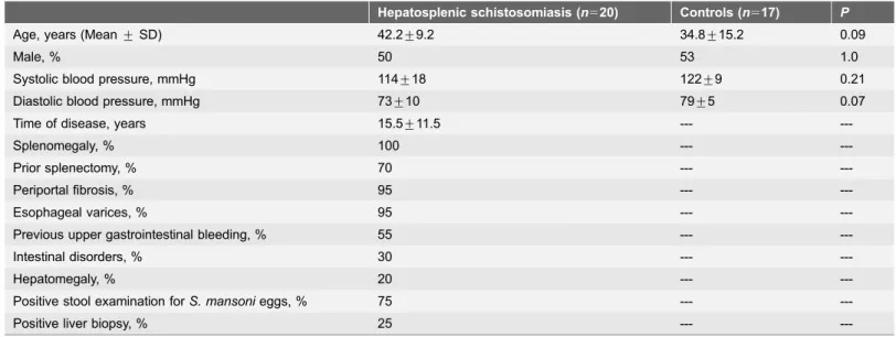
Table 1.Demographic and clinical characteristics of the study population.
Hepatosplenic schistosomiasis (n520) Controls (n517) P
Age, years (Mean¡SD) 42.2¡9.2 34.8¡15.2 0.09
Male, % 50 53 1.0
Systolic blood pressure, mmHg 114¡18 122¡9 0.21
Diastolic blood pressure, mmHg 73¡10 79¡5 0.07
Time of disease, years 15.5¡11.5 ---
---Splenomegaly, % 100 ---
---Prior splenectomy, % 70 ---
---Periportal fibrosis, % 95 ---
---Esophageal varices, % 95 ---
---Previous upper gastrointestinal bleeding, % 55 ---
---Intestinal disorders, % 30 ---
---Hepatomegaly, % 20 ---
---Positive stool examination forS. mansonieggs, % 75 ---
---Positive liver biopsy, % 25 ---
---Data are shown as mean¡SD or percentages. Significant P,0.05vs.control by Student’tand Fisher’s exact tests.
Table 2.General laboratory parameters of hepatosplenic schistosomiasis patients and controls.
Hepatosplenic schistosomiasis (n520) Controls (n517) P
Hemoglobin,g/dL 13.9¡1.4 14.1¡1.3 0.80
Hematocrit,% 42.4¡4.2 42.4¡4.5 1.0
Leukocytes,/mm3 6,098
¡2,760 6,446¡479 0.60
Platelets,/mm3 195,520¡107,290 270,660¡34,092 0.01
PNa,mEq/L 139¡3.0 138¡3.9 0.15
PK,mEq/L 4.4¡0.4 4.3¡0.4 0.41
PCa,mg/dL 9.7¡0.6 9.2¡0.3 0.05
PP,mg/dL 3.2¡0.5 3.6¡0.6 0.15
PCl,mEq/L 103¡3.2 107¡7.6 0.43
PMg,mg/dL 1.9¡0.2 1.9¡0.2 0.52
C3,mg/dL 88¡28 ---
---C4,mg/dL 19¡9 ---
---AST,UI/L 41¡13 ---
---ALT,UI/L 37¡12 ---
---Alkaline phosphatase,UI/L 120¡86 ---
---GGT,UI/L 104¡82 ---
---Total bilirubin,mg/dL 0.94¡0.46 ---
---Direct bilirubin,mg/dL 0.23¡0.25 ---
---Indirect bilirubin,mg/dL 0.71¡0.25 ---
---Total protein,g/dL 8.1¡0.4 ---
---Albumin,g/dL 4.3¡0.4 ---
---Globulin,g/dL 3.8¡0.55 ---
---INR 1.28¡0.18 ---
---Values are expressed as mean¡SD. Significant P,0.05vscontrol by Studentttest. PNa+, plasma sodium; PK+, plasma potassium; PCa2+, plasma calcium;
PP, plasma phosphorus; PCl-, plasma chloride; PMg2+, plasma magnesium AST, aspartate aminotransferase; ALT, alanine aminotransferase; GGT, gamma
glutamyl transferase; INR, international normalized ratio.
doi:10.1371/journal.pone.0115197.t002
Table 3.Comparison of urinary concentration and acidification tests in hepatosplenic schistosomiasis patients and controls.
Hepatosplenic schistosomiasis (n520) Controls (n517) P
UosmT4 (mOsm/kg.H2O) 588¡112 764¡165 0.001
U/PosmT4 2.0¡0.4 2.6¡0.5 ,0.001
Arterial pH T0 7.36¡0.04 7.37¡0.04 0.39
Arterial pH T4 7.34¡0.03 7.35¡0.03 0.18
HCO3-T0, mEq/L 24¡1.3 27¡3.8 0.005
HCO3-T4, mEq/L 23¡1.6 24¡2.9 0.03
UpHT0 6.2¡0.6 5.6¡0.4 0.002
UpHT4 5.4¡0.6 5.0¡0.3 0.02
Urinary concentration deficit 17 (85%) 5 (29.4%) 0.001
Urinary acidification deficit 9 (45%) 1 (6.7%) 0.03
Values are expressed as mean¡SD. Significant P,0.05vscontrol by Studentttest.Uosm, urinary osmolality after DDAVP;U/Posm, urinary to plasma
osmolality ratio; HCO3-, serum bicarbonate;UpH, urinary pH.
Hepatosplenic schistosomiasis patients have higher urinary
MCP-1 in comparison with the controls
Levels of microalbuminuria and proteinuria did not differ between HSS patients and the control group. The levels of MCP-1 were higher in the HSS group than in controls (122¡134vs 40¡28 pg/mg-Cr,P50.01) (Table 4). Abnormalities in
urinary sediment were present in 20% of the 20 patients - hematuria was observed
in one case (5%) and leukocyturia in 3 (15%). Microalbuminuria .30 mg/day
was found in 3 cases (15%), macroalbuminuria .300 mg/day in 1 (5%) and
proteinuria .150 mg/day in 5 (25%). Nephrotic-range proteinuria was not
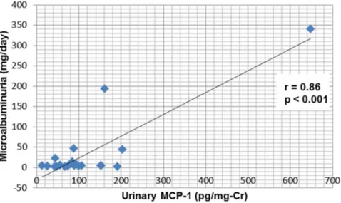
observed in any patient. MCP-1 correlated positively with microalbuminuria and proteinuria (Figs. 1 and2).
Discussion
The results of this cross-sectional analysis of renal function in hepatosplenic schistosomiasis clarify important aspects of tubular dysfunction in these patients and demonstrate a positive correlation between a new biomarker (MCP-1) and microalbuminuria.
The most common clinical finding related to schistosomiasis was portal hypertension. All patients had the hepatosplenic schistosomiasis form, without parasitic activity. A comparison of general laboratory tests between schistoso-miasis patients and the control group did not identify significant differences, except for platelet levels, which were lower in schistosomiasis patients. This stems from the fact that although almost all patients had undergone previous
splenectomy, the ones that had not undergone surgery had hypersplenism -associated thrombocytopenia.
Table 4.Comparison of renal function in hepatosplenic schistosomiasis patients and controls.
Hepatosplenic schistosomiasis (n520) Controls (n517) P
PUr, mg/dL 23¡7.7 26¡8.0 0.38
PCr, mg/dL 0.6¡0.1 0.9¡0.2 0.0001
GFR, mL/min/1.73 m2 130
¡38 103¡16 0.01
Microalbuminuria, mg/day 35.8¡83.6 5.5¡5.3 0.12
Proteinuria, mg/day 139¡148 95¡61 0.26
Urinary MCP-1, pg/mg-Cr 122¡134 40¡28 0.01
FENa+, % 0.69¡0.3 0.62¡0.1 0.40
FEMg++, % 2.96¡1.8 1.81¡1.0 0.14
FEK+, % 0.55¡0.2 0.52¡0.2 0.82
TTKG 3.3¡1.7 3.0¡1.2 0.62
TcH2O, L/day 0.72¡0.5 1.1¡0.3 0.04
Values are expressed as mean¡SD. Significant P,0.05vs.control by Studentttest. PCr, plasma creatinine; PUr, plasma urea; GFR, glomerular filtration
rate; MCP-1, Monocyte Chemotactic Protein-1 FENa+, fractional excretion of sodium; FEMg++, fractional excretion of magnesium; FEK+, fractional excretion of
potassium; TTKG, transtubular potassium transport; TcH2O, reabsorption of free water solute.
The Child-Pugh and MELD (Model for End-Stage Liver Disease) scores were used to assess the severity and prognosis of chronic liver disease of the patients [20,21,24]. All patients had normal liver function tests and were classified as Child-Pugh score A. The MELD score could not be calculated because all patients had values of creatinine, INR and bilirubin within normal limits. The minimum amount required for the calculation is 1 for all three markers [24].
Fig. 1. Correlation between urinary MCP-1 and albuminuria in schistosomiasis patients.
doi:10.1371/journal.pone.0115197.g001
Fig. 2. Correlation between urinary MCP-1 and proteinuria in schistosomiasis patients.
The hepatorenal syndrome is characterized by renal failure due to severe vasoconstriction of the renal circulation. It occurs in up to 10 percent of patients with advanced cirrhosis and ascites [25,26]. As hepatocellular synthetic function is preserved in schistosomiasis until the onset of very advanced stages of the disease, lobular architecture is preserved and nodular regenerative hyperplasia does not occur and thus, hepatorenal syndrome is not observed. Hepatic failure in schistosomiasis is transient. Hepatic decompensation usually occurs after an episode of gastrointestinal bleeding [11,27,28,29]. Although the patients had esophageal varices, we excluded patients who had had a bleeding episode in the last six months.
In our study, hypergammaglobulinemia was present in 65% of patients, which is higher than that described in literature [5] and hypocomplementemia was observed in 50%. In schistosomal nephropathy, glomerular injury may initially be asymptomatic or manifest only by hypocomplementemia. The behavior of complement is variable and may be decreased in 45 to 55% of cases [5]. Martinelli
et al. [30] studied the serum complement in patients with histological diagnosis of
schistosomal glomerulonephritis and found hypocomplementemia in 83% of patients. In the present study, there was no correlation between complement levels and renal function parameters, especially proteinuria and microalbuminuria, corroborating the idea that the hypocomplementemia may be an initial manifestation of schistosomal nephropathy.
The glomerular filtration rate was higher in HSS group compared with the control group. No patient was malnourished, which could explain this finding. Thus, there were a large number of HSS patients with glomerular hyperfiltration (40%). The main long-term consequence of glomerular hyperfiltration is the reduction of GFR due to glomerulosclerosis. No patient had reduced GFR below 60 mL/min/1.73 m2. The prevalence of chronic kidney disease (CKD) in patients with schistosomiasis is unclear. Martinelli et al. [31], in a study of renal biopsies
from 21 patients with hepatosplenic schistosomiasis, found that the progressive nature of kidney disease was not influenced by therapy with anti-parasitic drugs or immunosuppressants.
For the first time, we have shown that schistosomiasis infection may present with renal tubular dysfunction demonstrated by the loss of the ability of urinary acidification and concentration. Urinary concentrating ability defect was observed in 85% of our patients. Unlike other parasitic diseases, there is no study in literature about schistosomiasis patients’ inability to concentrate urine under water deprivation conditions, although it has been investigated in other infectious diseases. Oliveiraet al. [32] studied the urinary concentrating ability in 37 patients
with American Cutaneous Leishmaniasis (ACL) before and after treatment with pentavalent antimonial and found a prevalence of 77% of urinary concentrating defect, not reversed after treatment. Chughet al. [33] found urinary concentrating
defect and an impaired acidifying mechanism in 9 of 36 leprosy patients (25%), after an 18-h period of water deprivation. Ponce et al. [34] found urinary
lower levels of TCH2O (solute free water reabsorption) in HSS patients, showing a
deficit in water reabsorption, which is associated with the urinary concentration defect.
In the present study, the mean values of UpH were higher in HSS patients than controls. UpH T4.5.5 was present in 45% of cases, suggesting distal tubular acidosis. There are no reports in the literature on defects in urinary acidification in schistosomiasis. This abnormality has been described in other parasitic diseases, such as cutaneous leishmaniasis, in which urinary acidification defect was found in 40% of patients before treatment and 16% after treatment, suggesting a significant improvement in acidification ability after specific treatment [32]. In visceral leishmaniasis (kala-azar), urinary acidification defect was reported in 64% of cases after treatment [35]. Drutz and Gutman [36] studied 49 leprosy patients and found that urine pH did not decrease below 5.5 in response to NH4Cl in 20% of cases. Other studies also found urinary acidification defect in leprosy patients, with variable incidence [34,37].
Proteinuria is a common characteristic in schistosomiasis. Sobhet al[38] found
proteinuria by dipstick in 20% of 240 ambulatory asymptomatic patients with schistosomiasis. In another study, proteinuria was detected in 24.7% of 89 patients with hepatosplenic schistosomiasis and in only 4.6% of 86 patients with the hepatointestinal form of the disease [39]. In the present study, only
hepatosplenic patients were included, and proteinuria levels higher than 150 mg/ day was found in 25% of cases. No patient had nephrotic-range proteinuria. Hematuria was observed in only one case (5%) and pyuria in 3 (15%).
Microalbuminuria is a known early predictor of glomerular lesion in patients with diabetes and cardiovascular diseases [40–42]. This change has been described in infectious diseases, but it is not yet a well-defined marker of glomerular dysfunction [43]. Elnojomiet al [44] detected abnormal microalbuminuria levels
in 40% of patients with leishmaniasis without glomerular dysfunction. In another study of American cutaneous leishmaniasis, Oliveira et al [32] found abnormal
microalbuminuria in 35% of patients before treatment and in only 8% after treatment, suggesting that glomerular injury can be caused by the parasitic disease itself. In leprosy, Oliveiraet al[45] identified the presence of microalbuminuria in
8.5% of multibacillary patients. A higher prevalence of microalbuminuria was found in another study involving patients with leprosy, which identified
microalbuminuria higher than 20 mg/l in 15.8% of 96 patients with leprosy [46].
In the present study, microalbuminuria .30 mg/day was found in 15%, and
macroalbuminuria .300 mg/day in 5% of patients and there was no correlation
between microalbuminuria and GFR. To date, only one study evaluated
microalbuminuria in schistosomiasis. This recent study compared microalbumi-nuria levels between treated and untreated patients infected byS. mansoni with a
agent [19]. In the present study, MCP-1 levels were higher in schistosomiasis patients than in the control group. Moreover, we found a positive correlation between MCP-1 levels and microalbuminuria and 24-h proteinuria in patients with schistosomiasis, suggesting a role of MCP-1 in the early detection of renal damage associated with schistosomiasis.
In other kidney disorders of metabolic (diabetes mellitus), immunological (lupus nephritis) and genetic origin (autosomal dominant polycystic kidney disease) urinary MCP-1 has been correlated with urinary albumin excretion rate, glomerular filtration rate reduction and other features of kidney injury.
Hanemann et al. [19] made the first report of an infectious disease causing an
increase in urinary MCP-1, including patients with subclinical schistosomiasis. They found increased levels of urinary MCP-1 and a positive correlation between
MCP-1 and microalbuminuria [19]. Our study is the second to demonstrate this
association, also with schistosomiasis, but with hepatosplenic patients. Whether this finding is limited to schistosomiasis or may be initiated by any chronic infectious state remains unknown, but it is possible to speculate that any chronic infectious disease can induce renal inflammation and, consequently, urinary MCP-1 increase [18].
A limitation of our study is that we evaluated only hepatosplenic patients and it was not possible to determine whether portal hypertension could influence the results. It was not possible to compare liver function tests between the two groups, as the control group did not undertake them. However, this does not seem to affect the results, as all patients had test results within the normal range. A comparison between patients with different clinical forms of schistosomiasis could help determine whether the findings are related to the infection itself or if other factors related to portal hypertension could be influencing it.
In conclusion, the frequency of mild glomerular dysfunction, as well as tubular dysfunction, was considerably high in our cohort of HSS patients, even without clinical manifestations. It is important to evaluate renal function in patients with schistosomiasis for early detection and treatment of complications, mainly because it is a disease that predominates in younger individuals who are at higher risk of renal function loss and should benefit from measures to slow kidney disease progression. More studies are needed to evaluate the usefulness of MCP-1 as an early biomarker for schistosomal nephropathy.
Acknowledgments
Author Contributions
Conceived and designed the experiments: DBD GBSJ EFD. Performed the experiments: DBD LAV RKAB MEP AMCM GCM. Analyzed the data: DBD GBSJ EFD. Contributed reagents/materials/analysis tools: AMCM GCM. Wrote the paper: DBD GBSJ EFD.
References
1. Chitsulo L, Loverde P, Engels D(2004) Disease Watch: Schistosomiasis. TDR Nat Rev Microbiol 2: 12–13.
2. Barsoum R, Nabil M, Saady G, Genin C, Saleh E, et al. (1996) Immunoglobulin-A and the pathogenesis of schistosomal glomerulopathy. Kidney Int 50: 920–928.
3. Nussenzveig I, De Brito T, Carneiro CR, Silva AM(2002) Human Schistosoma mansoni-associated glomerulopathy in Brazil. Nephrol Dial Transplant 17: 4–7.
4. Barsoum R(2004) The changing face of schistosomal glomerulopathy. Kidney Int 66: 2472–2484.
5. Martinelli R, Rocha H (1996) Review/Update in Clinical Nephrology: glomerular involvement in schistosomiasis. J Bras Nefrol 18: 279–282.
6. Sobh MA, Moustafa FE, Sally SM, Deelder AM, Ghoniem MA (1988) Characterization of kidney lesions in early schistosomal-specific nephropathy. Nephrol Dial Transplant 3: 392–398.
7. Abensur H, Nussenzveig I, Saldanha LB, Pestalozzi MS, Barros MT, et al. (1992) Nephrotic syndrome associated with hepatointestinal schistosomiasis. Rev Inst Med Trop Sao Paulo 34: 273–276.
8. Van Velthuysen MLF(1996) Glomerulopathy associated with parasitic infections. Parasitol Today 12: 102–107.
9. Van Marck EAE (1983) The glomerulopathy associated with Schistosoma mansoni infection. An experimental study in the mouse. Acta Leiden 50: 1–123.
10. Barsoum RS(1993) Schistosomal glomerulopathies. Kidney Int 44: 1–12.
11. Andrade ZA, Van Marck EAE(1984) Schistosomal glomerular disease. Mem Inst Oswaldo Cruz 79: 499–506.
12. Andrade ZA, Van Marck E(1987) Schistosomal glomerular disease. Mem Inst Osvaldo Cruz 82: 25–29.
13. Rodrigues VL, Otoni A, Voieta I, Antunes CM, Lambertucci JR (2010) Glomerulonephritis in schistosomiasis mansoni: a time to reappraise. Rev Soc Bras Med Trop 43: 638–642.
14. Dos Santos WLC, Sweet GMM, Bahiense-Oliveira M, Rocha PN(2011) Schistosomal glomerulopathy and changes in the distribution of histological patterns of glomerular diseases in Bahia, Brazil. Mem Inst Oswaldo Cruz 106: 901–904.
15. Barsoum RS(1987) Schistosomal glomerulopathy: selection factors. Nephrol Dial Transplant 2: 488– 497.
16. Grandaliano G, Gesualdo L, Ranieri E, Monno R, Montinaro V, et al.(1996) Monocyte chemotactic peptide-1 expression in acute and chronic human nephritides: a pathogenetic role in interstitial monocytes recruitment. J Am Soc Nephrol 7: 906–913.
17. Banba N, Nakamura T, Matsumura M, Kuroda H, Hattori Y, et al. (2000) Possible relationship of monocyte chemoattractant protein-1 with diabetic nephropathy. Kidney Int 58: 684–690.
18. Dantas M, Roma˜o EA, Costa RS, dos Reis MA, Vieira Neto OM, et al.(2007) Urinary excretion of monocyte chemoattractant protein-1: a biomarker of active tubulointerstitial damage in patients with glomerulopathies. Kidney Blood Press Res 30: 306–313.
20. Child CG, Turcotte JG(1964) Surgery and portal hypertension. In: Child CG, editor. The liver and portal hypertension. Philadelphia: Saunders. Pp. 50–64.
21. Pugh RN, Murray-Lyon IM, Dawson JL, Pietroni MC, Williams R (1973) Transection of the oesophagus for bleeding oesophageal varices. Br J Surg 60: 646–649.
22. Tryding N, Sterner G, Berg B, Harris A (1987) Subcutaneous and intranasal administration of 1-deamino-8-d-arginine vasopressin in the assessment of renal concentration capacity. Nephron 45: 27– 30.
23. Oster JR, Hotchkiss JL, Carbon M, Farmer M, Vaamonde CA (1975) A short duration renal acidification test using calcium chloride. Nephron 14: 281–289.
24. Kamath PS, Wiesner RH, Malinchoc M, Kremers W, Therneau TM, et al.(2001) A model to predict survival in patients with end-stage liver disease. Hepatology 33: 464–470.
25. Gine`s P, Cardenas A, Arroyo V, Rodes J(2004) Management of Cirrhosis and Ascites. N Engl J Med 361: 1646–1654.
26. Gine`s P, Schrier RW(2009) Renal failure in cirrhosis. N Engl J Med 361: 1279–1290.
27. Rebouc¸as G (1975) Clinical Aspects of Hepatosplenic Schistosomiasis: A Contrast with Cirrhosis. Yale J Biol Med 48: 369–376.
28. De Cock KM(1986) Hepatosplenic schistosomiasis: a clinical review. Gut 27: 734–745.
29. Ross AG, Bartley PB, Sleigh AC, Olds GR, Li Y, et al.(2002) Schistosomiasis. N Engl J Med 346: 1212–1220.
30. Martinelli R, Brito E, Rocha H(1980) Value of beta 1C/1A globulin serum levels as an early index of glomerular involvement in Schistosoma mansoni infection. Am J Trop Med Hyg 29: 882–995.
31. Martinelli R, Noblat ACB, Brito E, Rocha H(1989) Schistosoma mansoni-induced mesangiocapillary glomerulonephrites: Influence of therapy. Kidney Int 35: 1227–1233.
32. Oliveira RA, Lima CG, Mota RMS, Martins AM, Sanches TR, et al.(2012) Renal function evaluation in patients with American Cutaneous Leishmaniasis after specific treatment with pentavalent antimonial. BMC Nephrol 13: 44–49.
33. Chugh KS, Damle PB, Kaur S, Sharma BK, Kumar B, et al.(1983) Renal lesions in leprosy amongst north India patients. Postgrad Med J 59: 707–711.
34. Ponce P, Ramos A, Ferreira ML, Pinto G, Lacerda MH(1989) Renal involvement in leprosy. Nephrol Dial Transplant 4: 81–84.
35. Lima Verde EM, Lima Verde FAA, Lima Verde FA, Silva Junior GB, Daher EF(2007) Evaluation of renal function in human visceral leishmaniasis (kala-azar): a prospective study on 50 patients from Brazil. J Nephrol 20: 432–438.
36. Drutz D, Gutman R(1971) Renal tubular acidosis in leprosy. Ann Intern Med 75: 475.
37. Sritharan V, Venkatesan K, Bharadwaj VP, Girdhar BK(1981) Renal functions in lepromatous leprosy patients. Lepr India 53: 437–442.
38. Sobh M, Moustafa F, El-Arbagy A, Shebab EL, Din M, et al.(1990) Nephropathy in asymptomatic patients with active Schistosoma mansoni infection. Int Urol Nephrol 22: 37–43.
39. Bina JC(1981) Influeˆncia da terapeˆutica especı´fica na evoluc¸a˜o da esquistossomose mansoni. Rev Patol Trop 10: 221–267.
40. Russo LM, Bakris GL, Comper WD(2002) Renal handling of lbumin: a critical review of basic concepts and perspective. Am J Kidney Dis 39: 899–919.
41. Gerstein HC, Mann JF, Yi Q, Zinman B, Dinneen SF, et al. (2001) Albuminuria and risk of cardiovascular events, death and heart failure in diabetic and non-diabetic individuals. JAMA 286: 421– 426.
42. Wachtell K, Ibsen H, Olsen MH, Borch-Johnsen K, Lindholm LH, et al. (2003) Albuminuria and cardiovascular risk in hypertensive patients with left ventricular hypertrophy: the LIFE study. Ann Intern Med 139: 901–906.
44. Elnojomi N, Musa AM, Younis BM, Elfaki M, El-Hassan AM, et al.(2010) Surrogate markers of subtle renal injury in patients with visceral leishmaniasis. Saudi J Kidney Dis Transpl 21: 872–875.
45. Oliveira RA, Silva GB Jr, Souza CJ, Vieira EF, Mota RM, et al.(2008) Evaluation of renal function in leprosy: a study of 59 consecutive patients. Nephrol Dial Transplant 23: 256–262.