(CRISP) with Promising Activity against Trypanosomes
and
Leishmania
Camila M. Adade1,2, Ana Lu´cia O. Carvalho2,3, Marcelo A. Tomaz4, Tatiana F. R. Costa5, Joseane L. Godinho2,5, Paulo A. Melo4, Ana Paula C. A. Lima5, Juliany C. F. Rodrigues2,4,5,6,7, Russolina B. Zingali2,3, Thaı¨s Souto-Padro´n1,2*
1Instituto de Microbiologia Paulo de Go´es, Universidade Federal do Rio de Janeiro, Rio de Janeiro, Brazil,2Instituto Nacional de Cieˆncia e Tecnologia de Biologia Estrutural e Bioimagem, Rio de Janeiro, Brazil,3Instituto de Biquı´mica Me´dica, Universidade Federal do Rio de Janeiro, Rio de Janeiro, Brazil,4Instituto de Cieˆncias Biome´dicas, Universidade Federal do Rio de Janeiro, Rio de Janeiro, Brazil,5Instituto de Biofı´sica Carlos Chagas Filho, Universidade Federal do Rio de Janeiro, Rio de Janeiro, Brazil,6Instituto Nacional de Metrologia, Qualidade e Tecnologia, Inmetro, Rio de Janeiro, Brazil,7Nu´cleo Multidisciplinar de Pesquisa em Biologia (NUMPEX-BIO), Polo Avanc¸ado de Xere´m, Universidade Federal do Rio de Janeiro, Duque de Caxias, Brazil
Abstract
Background: The neglected human diseases caused by trypanosomatids are currently treated with toxic therapy with limited efficacy. In search for novel anti-trypanosomatid agents, we showed previously that theCrotalus viridis viridis(Cvv) snake venom was active against infective forms ofTrypanosoma cruzi. Here, we describe the purification of crovirin, a cysteine-rich secretory protein (CRISP) from Cvv venom with promising activity against trypanosomes andLeishmania.
Methodology/Principal Findings:Crude venom extract was loaded onto a reverse phase analytical (C8) column using a high performance liquid chromatographer. A linear gradient of water/acetonitrile with 0.1% trifluoroacetic acid was used. The peak containing the isolated protein (confirmed by SDS-PAGE and mass spectrometry) was collected and its protein content was measured.T. cruzitrypomastigotes and amastigotes, L. amazonensispromastigotes and amastigotes andT. brucei rhodesiense procyclic and bloodstream trypomastigotes were challenged with crovirin, whose toxicity was tested against LLC-MK2cells, peritoneal macrophages and isolated murineextensor digitorum longusmuscle. We purified a single protein from Cvv venom corresponding, according to Nano-LC MS/MS sequencing, to a CRISP of 24,893.64 Da, henceforth referred to as crovirin. Human infective trypanosomatid forms, including intracellular amastigotes, were sensitive to crovirin, with low IC50or LD50values (1.10–2.38mg/ml). A considerably higher concentration (20mg/ml) of crovirin was required to elicit only limited toxicity on mammalian cells.
Conclusions:This is the first report of CRISP anti-protozoal activity, and suggests that other members of this family might have potential as drugs or drug leads for the development of novel agents against trypanosomatid-borne neglected diseases.
Citation:Adade CM, Carvalho ALO, Tomaz MA, Costa TFR, Godinho JL, et al. (2014) Crovirin, a Snake Venom Cysteine-Rich Secretory Protein (CRISP) with Promising Activity against Trypanosomes andLeishmania. PLoS Negl Trop Dis 8(10): e3252. doi:10.1371/journal.pntd.0003252
Editor:Michael P. Pollastri, Northeastern University, United States of America
ReceivedMay 16, 2014;AcceptedSeptember 8, 2014;PublishedOctober 16, 2014
Copyright:ß2014 Adade et al. This is an open-access article distributed under the terms of the Creative Commons Attribution License, which permits unrestricted use, distribution, and reproduction in any medium, provided the original author and source are credited.
Data Availability:The authors confirm that all data underlying the findings are fully available without restriction. All relevant data are within the paper and its Supporting Information files.
Funding:This work was supported by Conselho Nacional de Desenvolvimento Cientı´fico e Tecnolo´gico (CNPq) (grant numbers 560931/2010-7; 306967/2011-1), Fundac¸a˜o Carlos Chagas Filho de Amparo a Pesquisa do Estado do Rio de Janeiro (FAPERJ) (grant numbers E-26/102.581/2010; E-26/102.874/2012; E-26/110.621/ 2012) and Coordenac¸a˜o de Aperfeic¸oamento de Pessoal de Nı´vel Superior (CAPES). The funders had no role in study design, data collection and analysis, decision to publish, or preparation of the manuscript.
Competing Interests:The authors have declared that no competing interests exist.
* Email: souto.padron@micro.ufrj.br
Introduction
The pathogenic trypanosomatids from the genera Leish-mania and Trypanosoma infect over 20 million people worldwide, with an annual incidence of ,3 million new
infections in at least 88 countries. An additional 400 million people are at risk of infection by exposure to insect vectors harboring parasites [1–3]. Leishmania and trypanosome infections predominate in poorer nations, and are considered
neglected diseases that have ‘‘fallen below the radar of modern drug discovery’’ [4].
meglumine antimoniate (Glucantime) and sodium stibogluconate (Pentostan). Amphotericin B and pentamidine are used as second-line drugs in patients resistant to first-second-line therapy [1,6]. Recently, miltefosine has been used in India as part of combination therapy regimens to treat VL, and the largest increase in miltefosine activity was seen in combination with amphotericin B [7,8].
There are two forms of HAT (also known as sleeping sickness), caused by two subspecies ofT. bruceiparasites (T. b. gambienseor
T. b. rhodesiense). Both HAT forms culminate in parasite invasion of the central nervous system, with gradual nervous system damage if untreated. The currently used antiHAT drugs -melarsoprol, eflornithine, pentamidine, and suramin - are highly toxic and have lost efficacy in several regions. Also, treatment is difficult to administer in resource-limiting conditions, and often unsuccessful [9,10].
Chagas’ disease, caused byT. cruzi, affects the cardiovascular, gastrointestinal, and nervous systems of human hosts and has become, in recent decades, a worldwide public health problem due to travelers and migratory flow [2,11]. Chagas’ disease chemo-therapy is based on the use of nifurtimox and benznidazole, two very toxic nitroheterocyclic compounds with modest efficacy (especially against late stage chronic disease), and ‘plagued’ by the emergence of drug resistance [12].
Given the high toxicity and limited efficacy of current treatments for leishmaniasis, Chagas’ disease and HAT, the development of novel chemotherapeutics against these neglected diseases is essential. Animal venoms and poisons are natural libraries of bioactive compounds with potential to yield novel drugs or drug leads for pharmacotherapeutics [13]. In particular, snake venoms have proven to be interesting sources of potential novel agents against neglected diseases, including Chagas’ disease [14–17] and leishmaniasis [18–23].
Cysteine-rich secretory proteins (CRISPs) are single chain bioactive polypeptides with molecular masses of ,20–30 kDa
found in snake venom, reptilian venom ducts [24–26] and also in the salivary glands, pancreatic tissues, reproductive tracts [27–31]. In mammals, CRISPs are also expressed at low levels in non-reproductive tissues and organs, including skeletal muscle, spleen
and thymus [32]. CRISPs belong to the CAP (Crisp, antigen 5, and pathogenesis-related) superfamily of proteins [33].
CRISP amino acid sequences have high degree of sequence identity and similarity, and include a highly conserved pattern of 16 cysteine residues which form 8 disulfide bonds [34]. Ten of these cysteine residues form an integral part of a well-conserved cysteine-rich domain at the C-terminus, although CRISP N-terminal sequences are overall more conserved than other regions of these proteins [33–35]. Snake venom CRISPs belong to the CRISP-3 subfamily [36], one of four subgroups of CRISPs, according to amino acid sequence homology. Most biological targets of snake venom CRISPs described to date are ion channels [37–43], although the functions and the molecular targets of most snake venom CRISPs remain to be determined. Some snake venom CRISPs had their biological activities tested on crickets and cockroaches [35]. Snake venom CRISPs have been shown to block the activity of L-type Ca2+
and/or K+
-channels and also of cyclic nucleotide-gated (CNG) ion channels, thereby preventing the contraction of smooth muscle cells [26,37,40–43]. The CRISPs catrin, piscivorin and ophanin, from the snake Crotalus atrox, caused moderate blockage of L-type calcium channels, partially inhibiting the contraction of smooth fibers from mouse caudal arteries [26]. ThePhilodryas patagoniensis(green snake) CRISP patagonin was capable of generating myotoxicity when injected into the gastrocnemius muscle, but did not induce edema formation, haemorrhage or inhibition on platelet aggregation [44]. Despite their myotoxicity, there are no reports of CRISP protein lethality to mice, in concentrations of up to 4.5 mg/kg [35,45], and patagonin did not induce systemic alterations in mice, or histological changes in tissues from the cerebellum, brain, heart, liver and spleen [44].
In a previous publication, we showed that crude venom from the rattlesnakeCrotalus viridis viridishad anti-parasitic activity against all forms ofT. cruzi, and could be a valuable source of molecules for the development of new drugs against Chagas’ disease [46]. In search for the molecular source of the anti-parasitic activity found in Cvv crude venom, we purified a Cvv CRISP that will be henceforth referred to as ‘crovirin’. Here, we describe the purification, biochemical characterization and biological activity of crovirin against pathogenic trypanosomatids parasites and mammalian cells, showing that crovirin is active against infective developmental forms of trypanosomes andLeishmania, at doses that elicit no or minimal toxic effects on human cells.
Methods
Venom samples, compounds and reagents
Crude venom from the rattlesnakeCrotalus viridis viridis(Cvv) and adjuvants such as parasites growth media, were purchased from Sigma–Aldrich Chemical Co (St. Louis, MO, USA). Benznidazole (Bz) (Laborato´rio Farmaceˆutico do Estado de Pernambuco [LAFEPE], Brazil), diminazene aceturate (Berenil [Ber], Hoechst Veterina˜r GmbH, Mu¨nchen, Germany), and Amphotericin B (Amp-B) (Sigma) were used as a reference drugs for Chagas disease, sleeping sickness and leishmaniasis treatment, respectively. The material and reagents used in SDS-PAGE were from Bio-Rad Laboratories, Inc. Molecular weigh markers LMW were from Fermentas Life Sciences. Mass spectrometry grade Trypsin Gold was from Promega. All other reagents and chemicals were from Merck (Darmstadt, Germany), Tedia Company and Eurofarma Laborato´rios SA.
Purification of Crovirin
Lyophilized Cvv venom (10 mg) was dissolved in 1 ml of 20 mM Tris–HCl, 150 mM NaCl, pH 8.8 and centrifuged at
Author Summary
The pathogenic trypanosomatid parasites of the genera
5,000 gfor 2 min. The supernatant was applied onto a reverse phase analytical C8 column (5mm, 25064.6 mm) (Kromasil,
Sweeden), previously equilibrated with the same buffer. Venom proteins were separated by reverse phase HPLC (Shimadzu, Japan). Fractions (0.7 ml/tube) were collected at a 1 ml/h flowrate. A linear gradient of water/acetonitrile containing 0.1% trifluoroacetic acid (TFA) was used. The elution profile was monitored by absorption at 280 nm, and the molecular homoge-neity of the relevant fractions was verified by SDS-PAGE. Fractions containing protein peaks were dried in a Speed-Vac (Savant, Thermo Scientific, USA) and resuspended in distilled water prior to protein quantification by the Bradford method. Molecular mass determination was performed by MALDI-TOF and by electrospray ionization (ESI) mass spectrometry using a Voyager-DE Pro and a QTrap 2000 (both from Applied Biosystems), respectively.
In-gel digestion
Protein bands were excised from Coomassie Brilliant Blue-stained SDS-PAGE gels and cut into smaller pieces, which were destained with 25 mM NH4HCO3in 50% acetonitrile for 12 h.
The pieces obtained from the non-reducing gels were reduced in a solution of 10 mM dithiothreitol and 25 mM NH4HCO3for 1 h
at 56uC, and then alkylated in a solution of 55 mM iodoacetamide and 25 mM NH4HCO3, for 45 min in the dark. The solution was
removed, the gel pieces were washed with 25 mM NH4HCO3in
50% acetonitrile, and then dehydrated in 100% acetonitrile. Finally, all pieces from reducing and non-reducing gels were air-dried, rehydrated in a solution of 25 mM NH4HCO3containing
100 ng of trypsin, and digested overnight at 37uC. Tryptic peptides were then recovered in 10ml of 0.1% TFA in 50%
acetonitrile.
Nano LC-MS/MS mass spectrometry
The peptides extracted from gel pieces were loaded into a Waters Nano Acquity system (Waters, MA, USA) and desalted on-line using a Waters Symmetry C18 180mm620 mm, 5mm trap column. The typical sample injection volume was 7.5ml, and liquid chromatography (LC) was performed by using a BEH 130 C18 100mm6100 mm, 1.7mm column (Waters, MA, USA) and
eluting (0.5ml/min) with a linear gradient of 10–40% acetonitrile, containing 0.1% formic acid. Electrospray tandem mass spectra were performed in a Q-Tof quadrupole/orthogonal acceleration time-of-flight spectrometer (Waters, Milford, MA) linked to a nano ACQUITY system (Waters) capillary chromatograph. The ESI voltage was set at 3300 V, the source temperature was 80uC and the cone voltage was 30 V. The instrument control and data acquisition were conducted by a MassLynx data system (Version 4.1, Waters), and experiments were performed by scanning from a mass-to-charge ratio (m/z) of 400–2000 using a scan time of 1 s, applied during the whole chromatographic process. The mass spectra corresponding to each signal from the total ion current (TIC) chromatogram were averaged, allowing for accurate molecular mass measurements. The exact mass was determined automatically using Q-Tof’s LockSpray (Waters, MA, USA). Data-dependent MS/MS acquisitions were performed on precursors with charge states of 2, 3 or 4 over a range of 50–2000m/z, and under a 2m/zwindow. A maximum of three ions were selected for MS/MS from a single MS survey. Collision-induced dissociation (CID) MS/MS spectra were obtained using argon as the collision gas at a pressure of 40 psi, and the collision voltage varied between 18 and 90 V, depending on the mass and charge of the precursor. The scan rate was 1 scan/s. All data were processed using the ProteinLynx Global server (version 2.5, Waters). The processing
automatically lock mass calibrated them/zscale of both the MS and the MS/MS data utilizing a lock spray reference ion. The MS/MS data were also charge-state deconvoluted and deisotoped with the maximum entropy algorithm MaxEnt 3 (Waters, MA, USA).
Mass spectrometry data analysis
Proteins corresponding to the tryptic peptides from peak 3 were identified by correlation of tandem mass spectra and the NCBInr database of proteins (Version 050623), using the MASCOT software (Matrix Science, version 2.1). Settings allowed for one missed cleavage per peptide, and an initial mass tolerance of 0.2 Da was used in all searches. Cysteines were assumed to be carbamidomethylated, and a variable modification of methionine (oxidation) was allowed. Identification was considered positive when at least two peptides matched the protein sequence with a mass accuracy of less than 0.2 Da.
Parasites
T. cruzitissue culture trypomastigotes (CL-Brener clone) were obtained from the supernatants of 5 to 6-day-old infected LLC-MK2 cells maintained in RPMI-1640 medium (Sigma)
supple-mented with 2% FCS for 5–6 days at 37uC in a humidified 5% CO2. Theses trypomastigotes were also used to obtain intracellular
amastigotes in macrophage cultures.
The MHOM/BR/75/Josefa strain ofL. amazonensis, isolated from a patient with DCL by C. A. Cuba-Cuba (Universidade de Brasilia, Brazil), was used in the present study. Amastigote forms were maintained by hamster footpad inoculation, while promas-tigotes were cultured axenically in Warren’s medium [47] supplemented with 10% fetal bovine serum (FBS) at 25uC. Infective promastigotes were used to obtain intracellular amasti-gotes in macrophage cultures, as described previously [48]. Bloodstream form (BSF) T. brucei rhodesiense (strain IL1852) were cultivated in HMI-9 medium (Invitrogen) supplemented with 10% inactivated FBS (Biosera-South America) and 10% of serum plus supplement (SAFC Bioscience, USA), at 37uC in a humidified 5% CO2 incubator [49]. Procyclic-form (PCF) T. brucei rhodesiense (strain 457) were grown in SDM-79 medium (LGC Biotecnologia) supplemented with 10% heat-inactivated FBS, at 28uC [50].
Ethics statement
In this study, we used 5-week-old female CF1 mice as sources of peritoneal macrophages and of muscle sample forex vivoassays (described below). All animal experimentation protocols received the approval by the Commission to Evaluate the Use of Research Animals (CAUAP, from the Carlos Chagas Filho Biophysics Institute - IBCCF), and by the Ethics Committee for Animal Experimentation (Health Sciences Center, Federal University of Rio de Janeiro – UFRJ) (Protocol no. IBCCF 096/097/106), in agreement with Brazilian federal law (11.794/2008, Decreto nu
6.899/2009). We followed institutional guidelines on animal manipulation, adhering to the ‘‘Principles of Laboratory Animal Care’’ (National Society for Medical Research, USA) and the ‘‘Guide for the Care and Use of Laboratory Animals’’ (National Academy of Sciences, USA).
Parasite cytotoxicity assays
sulfoxide (DMSO), and the final concentration of the solvent never exceeded 0.5%, which is not toxic for parasites and mammalian cells. Ber stock solution (0.188 mg/ml) was prepared in pyrogen-free water.
Axenically grown parasite forms were treated with crovirin for up to 72 h in the same culture conditions used for growth (described above). The following crovirin concentrations were used to treat axenic forms: 1.2–4.8mg/ml (L. amazonensis
promasti-gotes) and 0.6–4.8mg/ml crovirin (T. brucei rhodesienseBSF and
PCF). IC50 values were calculated based on daily counting of
formalin-fixed parasites using a hemocytometer. Positive controls were run in parallel with 4.7mg/ml Amp-B [51] and 39.8 ng/ml
Ber [52], respectively.
T. cruzi tissue culture trypomastigotes were treated with crovirin (0.45–4.8mg/ml) at a density of 16106 cells/ml, for
24 h at 37uC (in RPMI media containing 10% FCS). LD50
(50% trypomastigote lysis) values were determined based on direct counting of formalin-fixed parasites using a hemocy-tometer. Bz was used as reference drug, in a 3.39mg/ml
concentration [53].
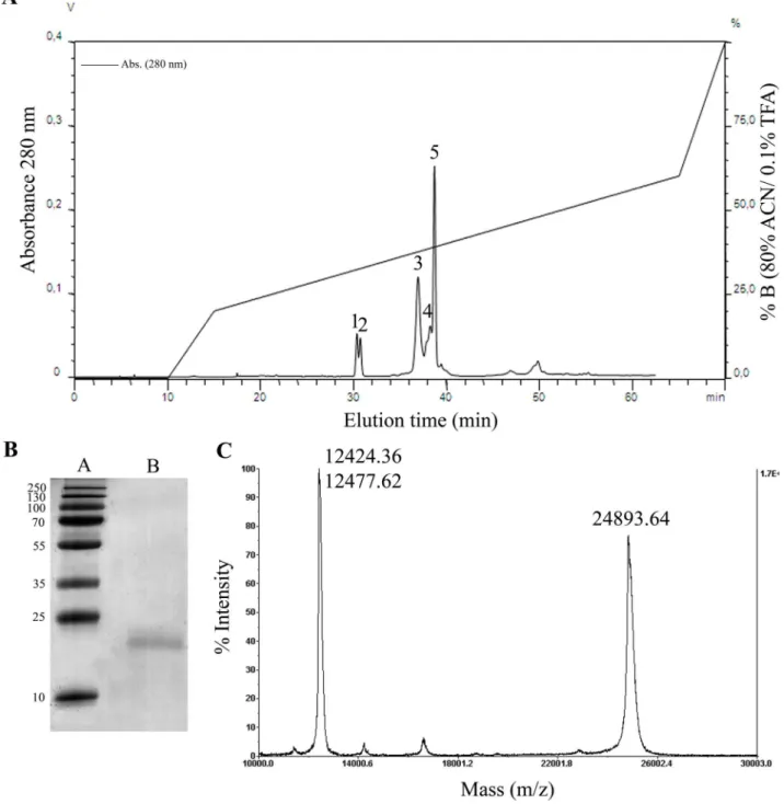
Figure 1.(A)Crovirin purification from Cvv venom using a reverse phase analytical C8column, where the protein was eluted as
peak 3.(B) SDS-PAGE analysis of peak 3 (lane B) containing the purified crovirin protein under reducing conditions. The gel was stained with Coomassie blue. Lane A, molecular weight markers. (C) MALDI-TOF mass spectrometry analyses of the intact protein yielded a molecular mass of 24,893.64 Da. The peaks of 12,424.36 and 12,477.62 Da correspond to doubly-charged (z = 2) cationic forms of crovirin.
To evaluate the effects of crovirin on T. cruzi and L. amazonensis intracellular amastigotes, peritoneal macrophages from CF1 mice were harvested by washing with RPMI medium (Sigma), and plated in 24-well tissue culture chamber slides, allowing them to adhere to the slides for 24 h at 37uC in 5% CO2.
Adherent macrophages were infected with tissue cultureT. cruzi
trypomastigotes (at 37uC) orL. amazonensis metacyclic promas-tigotes (at 35uC) at a macrophage-to-parasite ratio of 1:10, for 2 h. After this period, non-internalized parasites were removed by washing, cultures were incubated for 24 h in RPMI with 10% FCS, and fresh medium with crovirin (0.45–3.6mg/ml for T. cruzi, and 0.6–9.6mg/ml forL. amazonensis) was added daily for
72 h. At different time-points (24, 48 and 72 h) cultures were fixed with 4% paraformaldehyde in PBS (pH 7.2) and stained with Giemsa for 15 min. The percentage of infected cells and the number of parasites per 100 cells were determined by light microscopy examination. Positive controls of T. cruzi and L. amazonensis amastigotes infected cells were run in parallel with cultures treated with 0.73mg/ml Bz [53] and 0.07mg/ml Amp-B
[54], respectively.
Mammalian cell cytotoxicity assays
LLC-MK2 cells were maintained in RPMI medium
supple-mented with 10% FCS. Prior to treatment with crovirin, cells were seeded in 24-well plates containing glass coverslips and incubated in RPMI medium supplemented with 10% FCS for 24 h at 37uC. Cells were then treated with 4.8, 10 and 20mg/ml crovirin at 37uC for 72 h. LC50values (concentrations that reduces by 50%
the cellular viability) for crovirin were calculated from daily counts of the number of viable cells, using trypan blue as an exclusion dye. At least 500 cells were examined per well, on a Zeiss Axiovert light microscope (Oberkochen, Germany).
In addition, mouse peritoneal macrophages were seeded on 96-well plates, incubated in RPMI medium with 10% FCS for 24 h at 37uC and treated with 4.8, 10 and 20mg/ml crovirin at 37uC, for 72 h. After this period, cells were washed with PBS (pH 7.2), and the wells were filled with RPMI medium without phenol red containing 10 mM glucose and 20ml of a solution of 2 mg/ml MTS (3-(4,5-
dimethylthiazol-2-yl)-5-(3-carboxymethoxyphenyl)-2-(4-sulfophenyl)-2H-tetrazolium salt) and 0.92 mg/ml PMS (phenazine methosulfate), prepared according to the manufacturer’s instructions (Promega, Madison, WI, USA). Following 3 h of incubation at 37uC, formation of a soluble formazan product by viable cells was measured using a plate reader, by absorbance at 490 nm. All cytotoxicity experiments were carried out in triplicates.
Ex vivomytotoxicity assay
The myotoxicity of crovirin was studiedex vivousing a muscle creatin kinase (CK) activity assay [55]. The analysis consisted of monitoring the rate of CK release from isolated mouseextensor digitorum longus (EDL) muscle bathed in a solution containing crovirin (10mg/ml). Adult male and female Swiss mice (25.065.0 g) were anesthetized with ethyl ether and killed by cervical dislocation. EDL muscles were collected, freed from fat and tendons, dried and weighed. Muscle samples were then homogenized in 2 ml saline/0.1% albumin and their CK content was determined using a commercial diagnostic kit (Bioclin, Brazil). Four EDL muscles were mounted vertically on a cylindrical chamber and superfused continuously with Ringer’s solution equilibrated with 95% O2/5% CO2. At 30 to 90-min intervals, the
perfusing solution was collected and replaced with fresh solution. The collected EDL samples were used for the measurement of CK activity as described above. Muscles were weighed at the end of the experiment (2 h later). Enzyme activity is reported as international units corrected for muscle mass.
Statistical analysis
Mean value comparisons between control and treated groups were performed using the Kruskal-Wallis test in the BioEstat 2.0 program for Windows. Differences with p#0.05 were considered statistically significant.
Results
Purification of crovirin, a CRISP from the snake venom of C. viridis viridis
In a previous study, we showed that the Cvv venom had anti-parasitic activity againstT. cruzi[46]. Preliminary analysis of Cvv
Figure 2. Alignment of crovirin peptide sequences (identified by MALDI-TOF-TOF) and homologous sequences fromCalloselasma rhodostoma(gi:190195317),Crotalus viridis viridis(gi:190195319) andGloydius blomhoffi(ablomin protein- gi:48428846) deposited in GenBank.Bold letters highlight identical residues in the same position of all protein sequences.
venom fractions by reverse-phase chromatography (not shown) indicated that the activity eluted with fractions containing peak 3 of the chromatographic profile (Fig. 1A). Thus, we analyzed the main chromatographic fraction corresponding to peak 3 by SDS-PAGE and MALDI-TOF mass spectrometry (Fig. 1B–C). SDS-PAGE analysis of peak 3 showed a single polypeptide, with a relative molecular mass of 24 kDa (Fig. 1B) and 28 kDa (data not shown), under reducing and non-reducing conditions, respectively. We will refer to this protein henceforth as crovirin. MALDI-TOF analysis of the intact protein showed a molecular mass of 24,893.64 Da (Fig. 1C). The peaks of 12,424.36 and 12,477.62 Da in the MS profile correspond to doubly-charged (z = 2) cationic forms of the protein. The amino acid sequence of tryptic crovirin peptides (produced by Nano LC-MS/MS mass spectrometry analysis) is nearly identical to a partial sequence of a Cvv CRISP (GenBank gi:190195319) (Fig. 2). The MS/MS-derived sequences are also nearly identical to those of a CRISP protein from Calloselasma rhodostoma (GenBank gi:190195317) and have high degree of sequence similarity to several other snake venom CRISPs, including
ablomin (Fig. 2). The MS/MS spectrum of the fragmented peptide ions was matched by MASCOT displayed a coverage of 48% of identical peptides, with a p$355 indicating extensive homology to the CRISP fromC. rhodostoma. The MS results strongly suggested that a CRISP from Cvv snake venom had been purified, and corresponded to crovirin.
Crovirin has significant anti-parasitic activity against infective forms of trypanosomatids, with minimal toxicity
First of all, we investigated crovirin citotoxicity over mammalian host cells before proceeding with our analysis of the anti-parasitic activity of this venom protein.
LLC-MK2 cells were treated with crovirin for 72 h and
examined for viability using a trypan blue exclusion assay (Fig. 3A). None of the tested crovirin concentrations (4.8, 10 or 20mg/ml) were capable of inducing significant loss of cell viability, even after 72 h of treatment. In addition, we tested the activity of crovirin against murine peritoneal macrophages to investigate its cytotoxicity towards primary host cells. Treated cells were examined using an MTS assay, and no significant toxicity (p#
0.05) was observed in any treatment conditions (Fig. 3B). Creatine kinase (CK) activity was measured before and two hours afterextensor digitorum longus(EDL) muscle exposure to 10mg/ ml crovirin. We did not observed significant CK release from treated EDL muscles compared to control (saline) after 2 hours of incubation with crovirin, indicating that this protein did not generated appreciable myotoxicity at the concentration tested.
After establishing that crovirin had only minimal cytotoxic effects towards mammalian cells at concentrations of up to 20mg/
ml, we tested the anti-parasitic activity of purified crovirin against relevant developmental forms of three different species of pathogenic trypanosomatid parasites, namelyL. amazonensis, T. cruziandT. brucei rhodesiense.
We tested crovirin activity against the two infective T. cruzi
forms, trypomastigotes and amastigotes. Trypomastigote forms do not multiply and do not remain viable after several days in culture media at 37uC. Therefore, the effect of crovirin towardsT. cruzi
trypomastigotes was evaluated as the ability of the protein to lyse cells after 24 h of treatment (Fig. 4A). The calculated LD50 of
crovirin for trypomastigotes was 1.1060.13mg/ml (Table 1).
This concentration displayed the second higher selectivity index (18.2) (Table 1) among all crovirin treatments.
The treatment with 3.39mg/ml Bz exhibited a 65.8% of parasites lysis at same conditions.T. cruziamastigotes multiply in the intracellular environment. Crovirin inhibited the growth of amastigotes inside peritoneal macrophages in a dose-dependent manner (Fig. 4B), with an IC50 of 1.8460.53mg/ml when cells
were treated with crovirin for 72 h (Table 1). Crovirin presented a discret superior trypanocidal activity against the intracellular forms as compared with Bz (Fig. 4B).
Crovirin activity was also tested against infective promastigote and amastigote forms of L. amazonensis, one of the species responsible for CL. None of the crovirin concentrations tested inhibited significantly the proliferation ofL. amazonensis promas-tigotes in axenic media, unlike Amp-B treatment, which resulted in a reduction of a little over 80% in the number of parasites after 72 h of treatment. In contrast, crovirin inhibited the proliferation of intracellular amastigotes ofL. amazonensisin a concentration-dependent manner (Fig. 4C–D). The effect of crovirin on amastigote proliferation was evident as early as 24 h after the start of treatment, and the IC50for crovirin after 72 h of treatment
was 1.2160.89mg/ml (Table 1). After 48 h incubation, the IC50
of 1.05mg/ml also resulted in the highest selectivity index (19.1),
being less toxic treatment to mammalian host cells. However, no
Figure 3. Cytotoxicity analyses of crovirin on LLC-MK2cells (A)
tested concentration of crovirin had superior leishmanicidal activity against amastigotes forms as compared with Amp-B (Fig. 4D).
Both developmental forms ofT. brucei rhodesiensetested here (PCF and BSF) were sensitive to crovirin treatment. A different profile of growth inhibition in the presence of crovirin was observed for PCF (Fig. 4E) and BSF (Fig. 4F) parasites, with IC50 values of 1.1360.31 and 2.0660.12mg/ml, respectively,
after 72 h of treatment. The 39.8 ng/ml Ber treatment resulted in a remarkable growth inhibition of both BCF and PCF than crovirin treatment (Fig. 4E–F).
Discussion
There is an urgent need for the development of novel compounds for the treatment of trypanosomatid-borne diseases,
Figure 4. Crovirin effects onT. cruzitrypomastigotes (A) and intracellular amastigotes (B),L. amazonensispromastigotes (C) and intracellular amastigotes (D), and T. brucei rhodesiense PCF (procyclic form, in E) and BSF (bloodstream forms, in F).T. cruzi
trypomastigotes were treated with crovirin in RPMI medium with 10% FCS for 24 h only, since they do not survive in growth medium for longer and do not divide.T. bruceiBSF and PCF andL. amazonensispromastigotes were treated with crovirin in complete growth media, for up to 72 h. Amastigotes ofT. cruziandLeishmaniawere treated with crovirin as dividing intracellular forms (infecting peritoneal mouse macrophages). Error bars represent standard deviation the mean of 3 independent experiments.
currently treated with ‘dated’ chemotherapeutic agents with high toxicity and limited efficacy, partly due to the emergence of drug resistance. Animal venoms and toxins, including snake venoms, can provide compounds directly useful as drugs, or with potential as drug leads for the synthesis of novel therapeutic agents [22]. Previously, our group showed that Cvv crude venom displayed anti-parasitic activity against different T. cruzi developmental forms [46]. We have now extended this research with the purification of crovirin, a CRISP from Cvv venom with promising activity against key infective stages of the life cycle ofT. cruzi,T. brucei rhodesienseandL. amazonensis. Furthermore, we show that crovirin has low toxicity towards host cells and mouse muscle, in agreement with the low or absent toxicity reported for most CRISPs proteins [35,44–45].
CRISPs proteins are often given names that refer to the organism from which they were isolated. The first CRISP described in reptiles was isolated from the skin secretion of the lizard Heloderma horridum, and was named helodermin [56]. Examples of proteins isolated from snake venoms are patagonin, isolated from Philodryas patagonensis [44], latisemin, isolated from sea snake Laticauda semifasciata, tigrin isolated from
Rhabdophis tigrinus tigrinus [41], and ablomin, isolated from
Gloydius blomhoffi [41]. CRISPs sequences have also been identified in transcriptome analysis of venom glands [57–58] or are deposited at databanks but were not purified or studied. A partial CRISP sequence from Crotalus viridis viridis (GenBank gi:190195319) likely corresponding to central and C-terminal regions of crovirin was identified by transcriptome analysis of venom gland tissue. However, this is the first report on the purification and study of crovirin.
One of the most important findings of the present study was the activity of crovirin against the intracellular proliferation of trypanosomatids. Amastigotes are key developmental forms during
the development and maintenance of infections by Leishmania
and T. cruzi, representing the replicative intracellular stages of these protozoan parasites. Substantial inhibition of bothT. cruzi
and L. amazonensis intracellular amastigote proliferation was observed at crovirin concentrations significantly lower than those required to cause damage to host cells, including mouse EDL muscles. These results are particularly important because the currently available drugs to treat leishmaniasis and Chagas’ disease are known to have lower anti-amastigote activity [1,6].
The effects of crovirin over both the procyclic and the bloodstream form ofT. brucei rhodesiense are also encouraging, suggesting that crovirin might be useful in the development of new anti-HAT chemotherapeutics. In conclusion, our results demon-strate that crovirin has promising trypanocidal and leishmanicidal effects, and represents a potential avenue for drug development against leishmaniasis, Chagas’ disease and HAT, since its anti-parasitic effects are matched by low toxicity to host cells and muscles. Further studies are now required to extend our knowledge on the potential use of crovirin as an alternative compound to improve the effectiveness of treatment of trypano-somatid-borne neglected diseases.
Acknowledgments
We thank Tarcı´sio Correˆa for valuable technical assistance.
Author Contributions
Conceived and designed the experiments: CMA ALOC PAM JCFR APCAL RBZ TSP. Performed the experiments: CMA ALOC MAT TFRC JLG. Analyzed the data: CMA ALOC RBZ TSP. Contributed reagents/materials/analysis tools: CMA ALOC MAT TFRC JLG PAM JCFR RBZ TSP APCAL. Wrote the paper: CMA ALOC RBZ TSP.
References
1. Alvar J, Croft S, Olliaro P (2006) Chemotherapy in the treatment and control of leishmaniasis. Adv Parasitol 61: 223–274.
2. Schmunis GA, Yadon ZE (2010) Chagas disease: A Latin American health problem becoming a world health problem. Acta Trop 115: 15–21. 3. Utzinger J, Becker SL, Knopp S, Blum J, Neumayr AL, et al. (2012) Neglected
tropical diseases: diagnosis, clinical management, treatment and control. Swiss Medical Weekly 142: w13727.
4. Don R, Chatelain E (2009) Drug Discovery for Neglected Diseases: View of A Public-Private Partnership. In: Selzer PM, editor. Antiparasitic and Antibacterial Drug Discovery: From Molecular Targets to Drug Candidates. pp. 33–43.
5. Rodrigues JC, Godinho JL, de Souza W (2014) Biology of human pathogenic trypanosomatids: epidemiology, lifecycle and ultrastructure. Subcell Biochem 74: 1–42.
6. Croft SL, Barrett MP, Urbina JA (2005) Chemotherapy of trypanosomiases and leishmaniasis. Trends Parasitol 21: 508–12.
7. Sindermann H, Croft SL, Engel KR, Bommer W, Eibl HJ et al. (2004) Miltefosine (Impavido): the first oral treatment against leishmaniasis. Med Microbiol Immunol 193: 173–80.
8. Van Griensven J, Balasegaram M, Meheus F, Alvar J, Lynen L, et al. (2010) Combination therapy for visceral leishmaniasis. Lancet Infect Dis 10: 184–94.
Table 1.Crovirin activity towards medically important trypanosomes andLeishmania.
Species (clone or strain) Developmental form treated IC50aor LD50a(SI)b
24 hc 48 hc 72 hc
T. cruzi(CLBrener) Trypomastigote 1.1060.13 (18.2) nd nd
Amastigoted 1.64
60.44 (12.2) 2.0160.37 (9.9) 1.8460.53 (10.9)
L. amazonensis(MHOM/BR/75/Josefa) Promastigote .4.8 .4.8 .4.8
Amastigoted 2.38
60.62 (8.4) 1.0560.17 (19.1) 1.2160.89 (16.5)
T. brucei rhodesiense(427 - PCF; IL1852 - BSF) PCF 2.1860.50 (9.2) 1.7060.13 (11.8) 1.1360.31(17.7)
BSF 2.1860.39 (9.2) 2.2460.56 (9.0) 2.0660.12 (9.7)
aThe IC
50and LD50values are expressed inmg/ml.
bSelectivity Index (SI)ewas determined according to the ratio between the LC
50e/IC50or LD50values. SI values, where LC50.20mg/ml.
cTreatment period. dparasite/100 cells.
nd: Not determined.
9. Barrett MP, Boykin DW, Brun R, Tidwell RR (2007) Human African trypanosomiasis: pharmacological re-engagement with a neglected disease. Br J Pharmacol 152: 1155–71.
10. Jacobs RT, Nare B, Phillips MA (2011) State of the Art in African Trypanosome Drug Discovery. Curr Top Med Chem 11: 1255–74.
11. Schmunis GA (2007) The globalization of Chagas disease. ISBT Sci Ser 2: 6–11. 12. Dias JCP (2009) Elimination of Chagas disease transmission: perspectives for Chagas disease control in Latin America: a review. Mem Inst Oswaldo Cruz 97: 603–12.
13. Fox JW, Serrano SM (2007) Approaching the golden age of natural product pharmaceuticals from venom libraries: an overview of toxins and toxin-derivatives currently involved in therapeutic or diagnostic applications. Curr Pharm Des 13: 2927–34.
14. Adade CM, Chagas GS, Souto-Padro´n T (2012)Apis melliferavenom induces different cell death pathways inTrypanosoma cruzi. Parasitol 139: 1444–61. 15. Ciscotto P, Machado de Avila RA, Coelho EAF, Oliveira J, Diniz CG, et al.
(2009) Antigenic, microbicidal and antiparasitic properties of an L -amino acid oxidase isolated fromBothrops jararacasnake venom. Toxicon 53: 330–41. 16. Gonc¸alves AR, Soares MJ, De Souza W, DaMatta RA, Alves EW (2002)
Ultrastructural alterations and growth inhibition ofTrypanosoma cruzi and Leishmania majorinduced byBothrops jararacavenom. Parasitol Res 88: 598– 602.
17. Tempone AG, Andrade HF, Spencer PJ, Lourenc¸o CO, Rogero JR, et al. (2001) Bothrops moojenivenom killsLeishmaniaspp. with hydrogen peroxide generated by its L-amino acid oxidase. Biochem Biophys Res Commun 280: 620–4. 18. Brand GD, Leite JR, De Sa Mandel SM, Mesquita DA, Silva LP, et al. (2006)
Novel dermaseptins fromPhyllomedusa hypochondrialis(Amphibia). Biochem Biophys Res Commun 347: 739–46.
19. Fernandez-Gomez R, Zerrouk H, Sebti F, Loyens M, Benslimane A, et al. (1994) Growth inhibition ofTrypanosoma cruziandLeishmania donovaniinfantum by different snake venoms: Preliminary identification of proteins fromCerastes cerastesvenom which interacts with the parasites. Toxicon 32: 875–82. 20. Passero LFD, Tomokane TY, Corbett CEP, Laurenti MD, Toyama MH (2007)
Comparative studies of the anti-leishmanial activity of threeCrotalus durissus ssp. venoms. Parasitol Res 101: 1365–71.
21. Peichoto ME, Tavares FL, Dekrey G, Mackessy SP (2011) A comparative study of the effects of venoms from five rear-fanged snake species on the growth of Leishmania major: identification of a protein with inhibitory activity against the parasite. Toxicon 58: 28–34.
22. Tempone AG, Sartorelli P, Mady C, Fernandes F (2007) Natural products to anti-trypanosomal drugs: an overview of new drug prototypes for American Trypanosomiasis. Cardiovasc Hematol Agents Med Chem 5: 222–35. 23. Toyama MH, Toyama D deO, Passero LF, Laurenti MD, Corbett CE, et al.
(2006) Isolation of a new L-amino acid oxidase fromCrotalus durissus cascavella venom. Toxicon 47: 47–57.
24. Fry BG, Vidal N, Norman JA Vonk FJ, Scheib H, et al. (2006) Early evolution of the venom system in lizards and snakes. Nature439: 584–8.
25. Hill RE, Mackessy SP (2000) Characterization of venom (Duvernoy’s secretion) from twelve species of colubrid snakes and partial sequence of four venom proteins. Toxicon 38: 1663–87.
26. Yamazaki Y, Hyodo F, Morita T (2003) Wide distribution of cysteine-rich secretory proteins in snake venoms: isolation and cloning of novel snake venom cysteine-rich secretory proteins. Arch Biochem Biophys 412: 133–41. 27. Haendler B, Kratzschmar J, Theuring F, Schleuning WD (1993) Transcripts for
cysteine-rich secretory protein-1 (CRISP-1; DE/AEG) and the novel related CRISP-3 are expressed under androgen control in the mouse salivary gland. Endocrinol 133: 192-8.
28. Ito N, Mita M, Takahashi Y, Matsushima A, Watanabe YG, et al. (2007) Novel cysteine-rich secretory protein in the buccal gland secretion of the parasitic lamprey,Lethenteron japonicum. Biochem Biophys Res Commun 358: 35–40. 29. Kierszenbaum AL, Lea O, Petrusz P, French FS, Tres LL (1981) Isolation,
culture, and immunocytochemical characterization of epididymal epithelial cells from pubertal and adult rats. Proc Natl Acad Sci USA 78: 1675-9. 30. Koppers AJ, Reddy T, O’Bryan MK (2011) The role of cysteine-rich secretory
proteins in male fertility. Asian J Androl 13: 111–7.
31. Roberts K, Johnston DS, Nolan MA, Wooters JL, Waxmonsky NC, et al. (2007) Structure and function of epididymal protein cysteine-rich secretory protein-1. Asian J Androl 9: 508–14.
32. Reddy T, Gibbs GM, Merriner DJ, Kerr JB, O’Bryan MK (2008) Cysteine-rich secretory proteins are not exclusively expressed in the male reproductive tract. Dev Dyn 237: 3313–23.
33. Sunagar K, Johnson WE, O’Brien SJ, Vasconcelos V, Antunes A (2012) Evolution of CRISPs associated with toxicoferan-reptilian venom and mammalian reproduction. Mol Biol Evol 29: 1807–22.
34. Yamazaki Y, Morita T (2004) Structure and function of snake venom cysteine-rich secretory proteins. Toxicon 44: 227–31.
35. Osipov AV, Levashov MY, Tsetlin VI, Utkin YN (2005) Cobra venom contains a pool of cysteine-rich secretory proteins. Biochem Biophys Res Commun 328: 177–82.
36. Wang YL, Kuo JH, Lee SC, Liu JS, Hsieh YC, et al. (2010) Cobra CRISP functions as an inflammatory modulator via a novel Zn2+- and heparan sulfate-dependent transcriptional regulation of endothelial cell adhesion molecules. J Biol Chem 285: 37872–83.
37. Brown RL, Haley TL, West KA, Crabb JW (1999) Pseudechetoxin: a peptide blocker of cyclic nucleotide-gated ion channels. Proc Natl Acad Sci USA 96: 754–59.
38. Brown RL, Lynch LL, Haley TL, Arsanjani R (2003) Pseudechetoxin binds to the pore turret of cyclic nucleotide-gated ion channels. J Gen Physiol 122: 749– 60.
39. Shikamoto Y, Suto K, Yamazaki Y, Morita T, Mizuno H (2005) Crystal Structure of a CRISP Family Ca2+
-channel Blocker Derived from Snake Venom. J Mol Biol 350: 735–43.
40. Yamazaki Y, Brown RL, Morita T (2002a) Purification and cloning of toxins from elapid venoms that target cyclic nucleotide-gated ion channels. Biochem 41: 11331–7.
41. Yamazaki Y, Koike H, Sugiyama Y, Motoyoshi K, Wada T, et al. (2002b) Cloning and characterization of novel snake venom proteins that block smooth muscle contraction. Eur J Biochem 269: 2708–15.
42. Wang J, Shen B, Guo M, Lou X, Duan Y, et al. (2005) Blocking effect and crystal structure of natrin toxin, a cysteine-rich secretory protein fromNaja atra venom that targets the BKCa channel. Biochem 44: 10145–52.
43. Wang F, Li H, Liu MN, Song H, Han HM, et al. (2006) Structural and functional analysis of natrin, a venom protein that targets various ion channels. Biochem Biophys Res Commun 351: 443–8.
44. Peichoto ME, Mackessy SP, Teibler P, Tavares FL, Burckhardt PL, et al. (2009) Purification and characterization of a cysteine-rich secretory protein from Philodryas patagoniensis snake venom. Comp Biochem Physiol C Toxicol Pharmacol 150: 79–84.
45. Utkin YN, Osipov AV (2007) Non-lethal polypeptide components in cobra venom. Curr Pharm Des 13: 2906–15.
46. Adade CM, Cons BL, Melo PA, Souto-Padro´n T (2011) Effect ofCrotalus viridis viridis snake venom on the ultrastructure and intracellular survival of Trypanosoma cruzi. Parasitol 138: 46–58.
47. Warren LG (1960) Metabolism ofSchizotrypanum cruzi, Chagas. I. Effect of culture age and substrate concentration on respiratory rate. J Parasitol 46: 529– 39.
48. Macedo-Silva ST, de Oliveira Silva TL, Urbina JA, de Souza W, Rodrigues JC (2011) Antiproliferative, Ultrastructural, and Physiological Effects of Amioda-rone on Promastigote and Amastigote Forms ofLeishmania amazonensis. Mol Biol Int 1–12.
49. Costa TF, Reis FC, Lima AP (2012) Substrate inhibition and allosteric regulation by heparan sulfate of Trypanosoma brucei cathepsin L. Biochim Biophys Acta 1824: 493–501.
50. Rocha GM, Miranda K, Weissmu¨ller G, Bisch PM, de Souza W (2010) Visualization of the flagellar surface of protists by atomic force microscopy. Micron 41: 939–44.
51. Maciel-Rezende CM, de Almeida L, Costa E´ D, Pires FR, Alves KF, et al. (2013) Synthesis and biological evaluation againstLeishmania amazonensisof a series of alkyl-substituted benzophenones. Bioorg Med Chem 21: 3114–9.
52. Kibona SN1, Matemba L, Kaboya JS, Lubega GW (2006) Drug-resistance of Trypanosoma b. rhodesienseisolates from Tanzania. Trop Med Int Health 11: 144–55.
53. Timm BL, Da Silva PB, Batista MM, Farahat AA, Kumar A, et al. (2014)In vitroinvestigation of the efficacy of novel diamidines againstTrypanosoma cruzi. Parasitol 141:1272–6.
54. De Morais-Teixeira E, Gallupo MK, Rodrigues LF, Romanha AJ, Rabello A (2014)In vitrointeraction between paromomycin sulphate and four drugs with leishmanicidal activity against three New WorldLeishmaniaspecies. J Antimi-crob Chemother 69: 150–4.
55. Saturnino-Oliveira J, Tomaz MA, Fonseca TF, Gaban GA, Monteiro-Machado M, et al. (2012) Pulsed ultrasound therapy accelerates the recovery of skeletal muscle damage induced byBothrops jararacussuvenom. Braz J Med Biol Res 45: 488–96.
56. Mochca-Morales J, Martin BM, Possani LD (1990) Isolation and characteriza-tion of helothermine, a novel toxin from Heloderma horridum horridum (Mexican beaded lizard) venom. Toxicon 28: 299–309.
57. Cidade DA, Sima˜o TA, Da´vila AM, Wagner G, Junqueira-de-Azevedo IL, et al. (2006) Bothrops jararaca venom gland transcriptome: analysis of the gene expression pattern. Toxicon 48: 437–61.