UNIVERSIDADE DE SÃO PAULO
HOSPITAL DE REABILITAÇÃO DE ANOMALIAS CRANIOFACIAIS
PAULA KARINE JORGE
Evaluation of the dental arch in children with cleft lip and palate by
means of 3D digital models
PAULA KARINE JORGE
Evaluation of the dental arch in children with cleft lip and palate by
means of 3D digital models
Dissertação apresentada ao Hospital de Reabilitação de Anomalias Craniofaciais da Universidade de São Paulo para obtenção do título de MESTRE em Ciências da Reabilitação.
Área de concentração: Fissuras Orofaciais e Anomalias Relacionadas
Orientadora: Profª. Drª. Thais Marchini de Oliveira Valarelli
BAURU
2014
HOSPITAL DE REABILITAÇÃO DE ANOMALIAS CRANIOFACIAIS
Rua Silvio Marchione, 3-20. Caixa Postal: 1501
17012-900 Bauru/SP - Brasil (14) 3235-8000
Prof. Dr. Marco Antônio Zago - Reitor da USP
Dr.ª Regina Célia Bortoleto Amantini - Superintendente HRAC/USP
Autorizo, exclusivamente, para fins acadêmicos e científicos, a reprodução total ou parcial deste trabalho.
_____________________________ Paula Karine Jorge
Bauru,___ de __________de _____.
Jorge, Paula Karine
J82e Evaluation of the dental arch in children with cleft lip and palate by means of 3D digital models/ Paula Karine Jorge. Bauru, 2014.
68 p.: il.; 30 cm
Dissertação (Mestrado – Área de Concentração:
Fissuras Orofaciais e Anomalias Relacionadas) – Hospital de Reabilitação de Anomalias Craniofaciais, Universidade de São Paulo
Orientadora: Prof.ª Dr.ª Thais Marchini de OliveiraValarelli
Descriptors: 1. Cleft Lip 2. Cleft Palate 3. Orthopedic/ methods 4. Dental Models 5. Maxilla /growth &
FOLHA DE APROVAÇÃO
Dissertação apresentada e defendida por Paula Karine Jorge e aprovada pela Comissão Julgadora em ___/___/___.
______________________________________________________________ Prof.(a) Dr.(a)
Instituição
______________________________________________________________ Prof.(a) Dr.(a)
Instituição
______________________________________________________________ Profa. Dra. Thais Marchini de Oliveira Valarelli - Orientadora
Hospital de Reabilitação e Anomalias Craniofaciais - USP
______________________________________________________________ Profa. Dra. Daniela Gamba Garib Carreira
Presidente da Comissão de Pós-Graduação do HRAC/USP
D
edico este trabalho...A
GRADECIMENTOSAgradeço à Deus, por ter me dado o dom da vida.
À minha amada família José Paulo e Thina, Thiago e Nani, Vanessa e Conrado, por apoiar minhas escolhas, entender minha ausência, pelos conselhos carinhosos e pelo amor incondicional, por todas as vezes que discordaram da minha opinião e mesmo assim me amaram e me acolheram, por estenderem a mão, quando eu não tinha mais forças, por lutarem comigo na realização dos meus sonhos, por apoiarem em todas as etapas da minha vida, por ajudarem a construir quem eu sou hoje.
À minha queria orientadora Prof.ª Drª. Thais Marchini de Oliveira, professora que muito ensinou e ainda me ensina, por não ter medo de ousar, por ser uma orientadora brilhante, por se aprimorar a cada conquista, por seu entusiasmo, amor e dedicação resultarem num trabalho leve e prazeroso, pelo sorriso que renova, mesmo quando tudo parece fora de ordem, com a confiança de quem trilha seu próprio caminho.
Ao grupo da Pós-Graduação: Luciana, Tati, Adriana, Karine, Nádia, Bianca, Aninha, Natal e Fernanda, pelos estudos em conjunto, pela equipe construída, pelo companheirismo e ajuda à pesquisa e à clínica.
À querida amiga Karine Vaz Laskos Sakoda, pela presença em todos os momentos auxiliando na constituição desta dissertação, pelo companheirismo, capacidade, eficiência e eficácia que torna tudo mais simples, meu profundo agradecimento!
Às amadas professoras Marcinha, Bia, Gisele e Cleide, por toda dedicação pela profissão, por todo o carinho, paciência e amor ao ensinar, por demonstrarem o amor à Odontopediatria, mas acima de tudo, obrigada por vocês me acolherem e me ensinarem a ser dentista.
Á querida Professora Dr. Cleide Felício de Carvalho Carrara, que me deu a oportunidade dividindo seus conhecimentos clínicos e acadêmicos, sempre disposta a explicar mil vezes se necessário fosse.
Ao professor Dr. Fabricio Pinelli Valarelli, pelo auxílio no delineamento estatístico e sua didática natural ao ensinar, as quais tornou possível este trabalho.
À querida Professora Dr. Sandra Rahal, por ter sido minha primeira referência em Odontologia e me mostrar o quão apaixonante é esta profissão.
Ao Sávio, pelos momentos vividos, conselhos, abraços e pelas conversas sobre a vida, pelo sorriso, afago, carinho e amor, pela sinceridade que transborda como “os olhos de uma criança”, para sempre no meu coração!
Aos meus amores Vivi, Vivizinha e Aninha, por me deixarem sempre à vontade numa amizade sincera e franca, por me mostrarem que a Odontologia é linda e apaixonante, e pelo momento em que cada uma com suas peculiaridades entraram na minha existência. Aos meus amados amigos de infância, que tornam a minha existência mais linda e cheia de vida, e por mostrarem o significado da palavra amizade.
Aos amigos do PTS, por todos os momentos juntos, pois dividimos e somamos, com o intuito de conhecermos o desconhecido, por ‘laboratoriarmos’ que há algo além do físico, além da aparência, pelo amor incondicional que nos orienta para o mais bravo e sereno dos mundos, o interior.
Ao querido Guido Pedroli, por me ajudar em todos os momentos sem hesitação e bom humor, que serenamente repetia:“Calma! Conseguiremos!”
À querida Rosmarie Gilbert, por me ajudar nas dificuldades, nos meus estudos e permitir que me sentisse em casa.
À Dr. Wanda Maria Gnoinski, por mostrar-me a importância do orientador caminhar junto ao aluno, por compreender meus erros aceitando-me como estudante, e assim tornar o meu sonho possível.
À minhas amadas Juliane e Tina, por me mostrar que a amizade transcende idioma, fronteira e cultura.
Aos funcionários da Pós-graduação do HRAC, Tatiana, Andréia, Rogério e Mazé, que facilitam a realização dos procedimentos burocráticos, pelo eterno sorriso que acalma o dia a dia do aluno.
A
GRADECIMENTOS ADMINISTRATIVOSAo Hospital de Reabilitação de Anomalias Craniofaciais – USP, na pessoa da senhora superintendente Dra. Regina Amantini Bortolotto, que tornou possível a realização desta pesquisa, toda a minha gratidão.
Ao Programa de Pós-Graduação em Ciências da Reabilitação do Hospital de Reabilitação de Anomalias Craniofaciais-USP, na pessoa da senhora coordenadora Profa. Dra. Daniela Gamba Garib Carrera.
À Profa. Dra. Maria Aparecida de Andrade Moreira Machado, Diretora da Faculdade de Odontologia de Bauru-USP pelo incansável esforço para integração deste campus.
À Profa. Dra. Daniela Rios, chefe do Departamento de Odontopediatria, Ortodontia e Saúde Coletiva da Faculdade de Odontologia de Bauru-USP pelo acolhimento durante o curso de disciplinas e clínicas no referido departamento.
À Profa. Dra. Salete Moura Bonifácio da Silva, responsável pela Disciplina de Odontopediatria do Departamento de Odontopediatria, Ortodontia e Saúde Coletiva da Faculdade de Odontologia de Bauru-USP por tão gentilmente me aceitar no Programa de Aperfeiçoamento de Ensino.
À FAPESP pelo auxílio financeiro para elaboração desta dissertação. Pela oportunidade de fazer o mestrado que eu sempre sonhei.
À Universidade de Zurique, por ter me recebido e pela oportunidade de expandir meus horizontes por meio de diferentes procedimentos, técnicas, protocolos, e o mais incrível, a cultura.
“Procuro partes tão distantes Pedaços soltos do que fui e estou Mas só você me traz
O que não posso ver”
ABSTRACT
Evaluation of the dental arch in children with cleft lip and palate by means of 3D digital models [dissertation]. Bauru: Hospital de Reabilitação de Anomalias Craniofaciais, Universidade de São Paulo; 2014.
The aim of this study was to evaluate the dimensional alterations of dental arches of cleft lip and palate children after cheiloplasty at two rehabilitation centers. The sample was composed of 94 digital models and divided in two groups: Group I – 23 children, assisted at University of Zurich – with presurgical orthopedic intervention (Hotz plate); Group II – 24 children, assisted at Hospital for Rehabilitation of Craniofacial Anomalies of the University of São Paulo – without presurgical orthopedic intervention. The three dimensional images of dental study models were used to evaluate before lip repair (stage 1) and approximately 1 year old (stage 2). The obtained measures were: intercanine distance, intertuberosity distance, anteroposterior arch distance, anteroposterior cleft length, anterior and posterior cleft width. The comparison between stages 1 and 2 was evaluated in group I, group II and between group I and II. The differences between groups were verified by Independent t, Wilcoxon and Mann-Whitney tests. In Group I, the comparisons between stages 1 and 2 showed that the intertuberosity and anteroposterior arch distance increased and the intercanine, anterior and posterior cleft distances decreased. In Group II, the comparisons between stages 1 and 2 showed that intertuberosity distance increased and intercanine, anteroposterior cleft length distances, anterior and posterior cleft widths decreased. The comparison of the dimensional alterations of dental arches between the two rehabilitation centers exhibited differences: in the stage 1, the intercanine distance was smaller in Group II than in Group I; in the stage 2, the anterior cleft width, the intercanine distance and the anteroposterior cleft length were smaller in Group II than in Group I, suggesting the superposition of maxillary segments; in Group II, there was a greater narrowing of the anterior and posterior cleft widths, suggesting that Hotz plate induced a more evenly and equidistant approximation of maxillary segments in Group I.It is worth emphasizing that more long term studies should be conduct to reduce the variability in treatment protocols for cleft lip and palate patients, thus assuring the best evidence-based treatment.
RESUMO
Avaliação dos arcos dentários de crianças com fissura labiopalatina por meio de modelos digitais tridimensionais [dissertação]. Bauru: Hospital de Reabilitação de Anomalias Craniofaciais, Universidade de São Paulo; 2014.
O propósito deste estudo foi avaliar as alterações dimensionais dos arcos dentários de crianças com fissura de lábio e palato após a queiloplastia emdois centros de reabilitação.A amostra foi composta por 94 imagens digitais de modelos de gesso, e dividida em dois grupos: Grupo I – 23 crianças, tratadas na Universidade de Zurique, com intervenção ortopédica pré-cirúrgica (placa de Hotz); Grupo II – 24 crianças tratadas no Hospital de Reabilitação de Anomalias Craniofaciais da Universidade de São Paulo, sem intervenção ortopédica pré-cirúrgica. As imagens tridimensionais dos modelos de gesso foram avaliados antes da queiloplastia (estágio 1) e com aproximadamente 1 ano de idade (estágio 2). As seguintes dimensões foram obtidas: comprimento anteroposterior da fissura, amplitude anterior da fissura, amplitude posterior da fissura, comprimento anteroposterior do arco,distância intercaninos e distância intertuberosidades. As comparações foram realizadas entre os estágios 1 e 2 e entre os grupos I e II. As diferenças entre os grupos foram verificadas pelo teste t independente, os testes de Wilcoxon e Mann-Whitney foram usados. No Grupo I, entre os estágios 1 e 2, as distâncias anteroposterior do arco e intertuberosidade aumentaram e as distância intercaninos, amplitude anterior e posterior da fissura diminuíram.No Grupo II, entre os estágios 1 e 2, a distância intertuberosidade aumentou e as distância intercaninos, distância anteroposterior da fissura, amplitude anterior e posterior da fissura diminuíram. A comparação entre as alterações dimensionais dos arcos dentários entre os dois centros de reabilitação apresentaram diferenças: no estágio 1, a distância intercaninos foi menor no Grupo II que no Grupo I; e no estágio 2, a amplitude anterior da fissura, a distância intercaninos e o comprimento anteroposterior da fissura foram menores no Grupo II que no Grupo I, sugerindo sobreposição dos segmentos maxilares. No Grupo II, houve maior estreitamento nas amplitudes anterior e posterior da fissura, sugerindo que a placa de Hotz no Grupo I induziu a aproximação dos segmentos maxilares de forma mais uniforme e equidistante.É importante salientar que mais estudos longitudinais precisam ser conduzidos a fim de reduzir a variabilidade de protocolos de tratamento em pacientes com fissura lábio e palato, garantindo o melhor tratamento baseado em evidências científicas.
LIST OF ILLUSTRATION
LIST OF TABLES
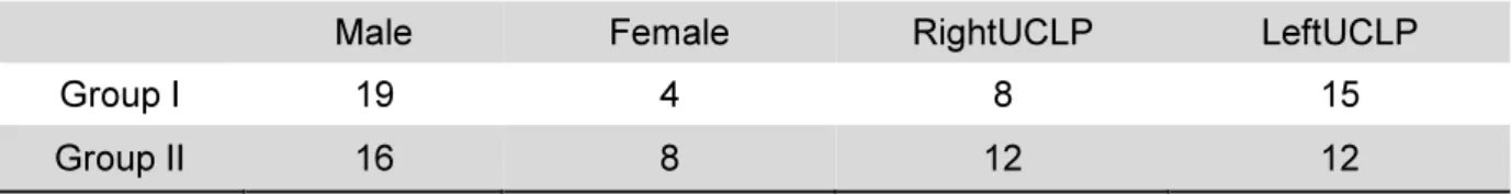
Table1 - Sample distribution by gender and cleft side ... 24
Table2 - Depended t test, Wilcoxon test and Dahlberg’s formula
to assess the intraexaminer reproducibility ... 29
Table 3 - Independent t test and Mann-Whitney test to assess the
mean age ... 29
Table 4 - Sample distribution by gender and UCLP side ... 30
Table5 - Maxillary dimensional alterations (mm) between stages
1 and 2 in group I, dependent t test and Wilcoxon test... 30
Table6 - Maxillary dimensional alterations (mm) between stages
1 and 2 in group II, dependent t test ... 31
Table 7 - Comparison of maxillary dimensional (mm) between
groups I and II at stage 1 – independent t test ... 31
Table 8 - Comparison of maxillary dimensional (mm) between
groups I and II at stage 2- independent t test and
Mann-Whitney ... 32
Table 9 - Comparison of maxillary dimensional (mm) at stage 2 –
stage 1 (S2-S1) between groups I and II – independent
LIST OF ABBREVIATION AND ACRONYMS
S1 Stage 1
S2 Stage 2
GI Group I
GII Group II
HRAC/USP Hospital for the Rehabilitation of Craniofacial Anomalies,
University of São Paulo
SD Standard Deviation
IQD Inter quartil deviation
DF Degree of freedom
ʎ2 chi-square value
SUMMARY
1. INTRODUCTION ... 15 2. OBJECTIVE... 19 2.1. GENERAL OBJECTIVE ... 19 2.2. SPECIFIC OBJECTIVE ... 19 3. MATERIAL AND METHODS ... 23
3.1. SAMPLE SELECTION ... 23 3.2. OBTAINMENT OF DENTAL STUDY MODELS ... 24 3.3. OBTAINMENT OF THE MEASUREMENTS ... 24 3.4. STATISTICAL ANALYSIS ... 26 3.4.1 Error method ... 26 3.4.2. Comparative analysis ... 26 4. RESULTS ... 29
4.1. MEASUREMENT ERROR ... 29 4.2. DIMENSIONAL ALTERATIONS IN GROUP I ... 30 4.3 DIMENSIONAL ALTERATIONS IN GROUP II ... 30 4.4. DIMENSIONAL ALTERATIONS BETWEEN GROUP I AND II ... 31 5. DISCUSSION ... 35 6. CONCLUSION ... 41 7. REFERENCES ... 45 APPENDIX A ... 53 REVISÃO DE LITERATURA ... 53
Introduction 15
1. INTRODUCTION
The cleft lip and palate is one of the most prevalent congenital alterations in the human being and have been recognized by the World Health Organization as a relevant health problem (WHO, 2004). It is a facial alteration of embryologic origin due to deficiency or failure during the fusion of one frontonasal, two maxillary and two mandibular processes. The anatomic and functional alterations of cleft lip and palate individuals show several requirements related to medical, dental and speech care (FREITAS et al, 2012a).
The fact that the professional is aware of the dimensional alterations of the dental arches occurring in cleft lip and palate subjects is of extreme therapeutic interest, once it can influence on the stability of the results obtained in the rehabilitation (FREITAS et al, 2012b).
The rehabilitative treatment of these individuals is a constant challenge and it should be started at birth (FREITAS et al, 2012a). It is important to emphasize that the dental records enable clinical evaluation of treatment planning, elucidation of legal processes and outcome comparisons among treatment centers (VEGTER; HAGE, 2000; NOLLET et al, 2004; AHMAD, 2009).
The clinical documentation of children at early childhood to evaluate the measurements of dental arches prior to primary surgeries is a fundamental aspect for the rehabilitative process because the result obtained during every phase can demonstrate the treatment progress (WUTZL et al, 2009). The measurements and assessments of the cleft width and the dimensions of the maxillary dental arches play an important role in the determination of the most adequate treatment planning suitable for each cleft severity and type (BRAUMANN et al, 2002). In the long term, the documentation protocol enables the evaluation of both the alterations and growth of dental arches, which consequently leads to further studies favoring the treatment of cleft lip and palate patients (HEIDBUCHEL; KUIJPERS-JAGTMAN, 1997).
16 Introduction
the best treatment, which is difficult to find because of the large variability of malformations and the individual response of each patient to therapy (FARRONATO et al, 2014). Therefore, there is a need for more multicenter studies, to reduce the protocol modalities to ensure an evidence-based clinical practice for clef lip and palate treatment (DE LADEIRA; ALONSO, 2012).
Nowadays, studies have been conducted with three dimensional (3D) images of dental study models (LO et al, 2003; DIAH et al, 2007; LATIEF et al, 2012). The literature has stated that there is accuracy between dental study models and 3D images (ZILBERMAN; HUGGARE; PARIKAKIS, 2003; DA SILVEIRA et al, 2004; LEIFERT et al, 2009), which is an ideal tool to evaluate morphological alteration of edentulous maxilla of individuals with oral clefts (BRAUMANN et al, 2002).
Objective 19
2. OBJECTIVE
2.1. GENERAL OBJECTIVE
The aim of this study was to evaluate the dimensional alterations of dental arches of cleft lip and palate children after cheiloplasty at two rehabilitation centers, comparing the protocols with and without presurgical orthopedics.
2.2. SPECIFIC OBJECTIVE
1) To compare the dimensions of dental arches at different treatment stages and to evaluate the dimensional alterations in the patients treated at the University of Zurich.
2) To compare the dimensions of dental arches at different treatment stages and to evaluate the dimensional alterations in the patients treated at the Hospital for Rehabilitation of Craniofacial Anomalies of the University of Sao Paulo. 3) To evaluate and compare the dimensional alterations of dental arches
Material and Methods 23
3. MATERIAL AND METHODS
3.1. SAMPLE SELECTION
This study was approved by the Ethics in Research Committee of HRAC/USP under protocol no. #167.992 (Annex A). The patients were selected from the archives of the rehabilitation centers.
This study inclusion criteria were children with unilateral cleft lip and palate (UCLP), both genders,aged from 0.27 to 1.50 years-old, and regularly enrolled at both HRAC/USP and UZH. The exclusion criteria comprised children with Simonart’s bands, associated syndrome or malformation, incomplete documentation and with previous cheiloplasty surgery.
The sample size was calculated so that the number of selected children met the representative rating to conduct the study. Considering a prior study of Prahl et al (2001), with significance level of 5%, test power of 80% and difference to be detected of 1.44, the minimum sample size was calculated to be 23 individuals per group.
The study was conducted on 47 children with UCLP. The sample was composed of 94 digital models and divided into two groups, as follow:
• Group I – 23 children, born between 1970 and 1980, and assisted at UZH – with presurgical orthopedic intervention (Hotz plate).
• Group II – 24 children, born between 2010 and 2012, and assisted at HRAC/USP – without presurgical orthopedic intervention.
The following lip repair procedures were executed:
• Group I – the children worn Hotz plate since birthup to12 – 18months, on average. The lip repair was performed by modified Millard technique at 0.48 years-oldon average.
24 Material and Methods
The assessment was carried outthrough digital models of the maxillary arch into two stages in both groups:
• Stage 1 (S1) – Before lip repair.
• Stage 2 (S2) – approximately1 yearold.
Table 1 displays the gender distribution and UCLP side in groups I and II.
Table 1 – Sample distribution by gender and cleft side
Male Female RightUCLP LeftUCLP
Group I 19 4 8 15
Group II 16 8 12 12
3.2. OBTAINMENT OF DENTAL STUDY MODELS
The evaluation of dental arches was carried out by using dental study models. The size of the tray was verified prior to the impression procedure according to dental arch size. The impression material was handled following the manufacturer’s instructions, placed onto the tray and pressed against the maxillary arch and kept for some seconds until its setting. The material used in HRAC/USP was condensation silicon (Perfil, Vigodent), whereas alginate (Tetrachrom – Kaniedenta) was used in UZH. After the impression, the dental models were obtained and digitized to measure the dental arches with the aid of a scanner (Scanner 3Shape R700TM Scanner, Copenhagen, Denmark).
3.3. OBTAINMENT OF THE MEASUREMENTS
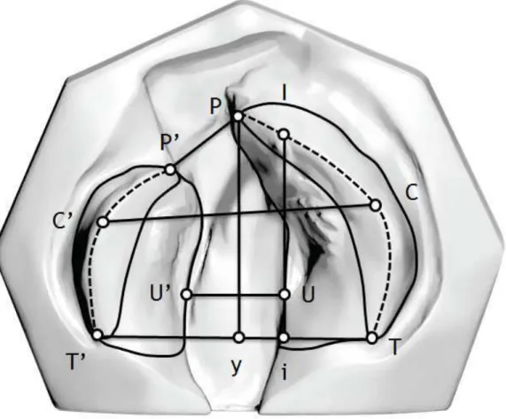
The 3D OrthoAnalyzerTM (Software, Copenhagen, Denmark) was used to evaluate the measurements and to define the landmarks (STÖCKLI, 1969; BOLTER, 1979; SECKEL et al, 1995) (Figure 1):
• C-C’ – Intercanine distance, from the lateral sulcus of the alveolar ridge crest of one side tothe lateral sulcus of the alveolar ridge crest of the other side.
Material and Methods 25
• I-i – Anteroposterior arch distance, from the interincisal point projected towards a line crossing the alveolar ridge crest to a point projected towards T-T’.
• P-y – Anteroposterior cleft length, from P projection to a point projected towards T-T’.
• P-P’ – Anterior cleft width, from the cleft edges of one side to the cleft edges of the other side.
• U-U’ – Posterior cleft width, pointed at the maximum posterior gap distance.
26 Material and Methods
3.4. STATISTICAL ANALYSIS
The statistical tests was performed by Sigma Plot software (Sigma Plot for Windows – Version 12.0), adopting the significance level of 5%.
3.4.1 Error method
For the analysis of intraexaminer error, models of 1/3 of patients were randomly measured again 15 days after the first measurement. Paired t-test was used to calculate the systematic error, with a significance level of 5%.
3.4.2. Comparative analysis
Results 29
4. RESULTS
4.1. MEASUREMENT ERROR
The assessment of normal distribution in groups I and II was verified by Shapiro-Wilk test (Annex B).
Table 2 shows the intraexaminer reproducibility for the different measurements, indicating that there was no statistically significant difference in the repeated measurements. All the measurements proved to be sufficiently reproducible (P≤0.05).
Table 2 – Dependent t test, Wilcoxon test and Dahlberg’s formula to assess the intraexaminer
reproducibility.
1st measurement 2nd measurement
Variables Mean (Median) SD (IQD) Mean (Median) SD (IQD) Dalberg P
P-P' (S1) 6.963 2.877 6.971 2.718 0.3963 0.958
Py (S1) 28.353 2.596 28.051 2.713 0.6441 0.210
I-i (S1) 27.434 1.914 27.231 2.309 0.5957 0.368
C-C' (S1) 31.211 2.69 30.963 2.676 0.3542 0.05
U-U’ (S1) 11.093 1.508 11.234 1.506 0.3468 0.281
T-T' (S1) 33.042 2.575 33.193 2.529 0.3925 0.307
P-P' (S2) 0.858 2.954 0.899 2.99 0.2702 0.691
Py (S2) 29.18# 2.66# 29.39# 2.285# 0.3153 0.151
I-i (S2) 29.205 2.0602 29.231 2.619 0.2823 0.811
C-C' (S2) 29.485 2.827 29.389 3.06 0.6516 0.701
U-U’ (S2) 9.425 2.537 9.261 2.29 0.3798 0.250
T-T' (S2) 34.421 2.744 34.698 2.53 0.8379 0.383
# median and IQD (interquartile deviation) of Wilcoxon test.
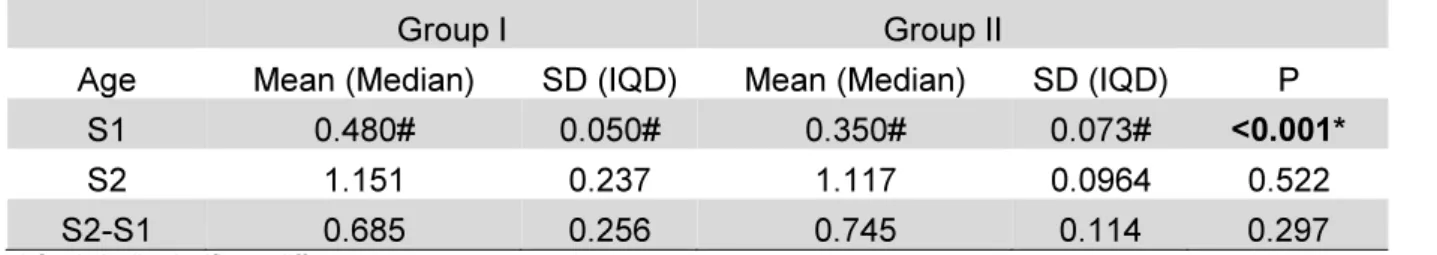
Statistically significant difference was found in S1 mean age (years), between groups I and II (Table 3).
Table 3– Independent t test and Mann-Whitney test to assess the mean age
Group I Group II Age Mean (Median) SD (IQD) Mean (Median) SD (IQD) P
S1 0.480# 0.050# 0.350# 0.073# <0.001*
S2 1.151 0.237 1.117 0.0964 0.522 S2-S1 0.685 0.256 0.745 0.114 0.297
* Statistically significant difference
30 Results
There was no significant difference in gender and cleft sidedistribution in both groups (Table 4).
Table 4 – Sample distribution by gender and UCLP side
GI GII DF x2 P
Male 19 16
1 1,5699 0,2102
Female 4 8
Right Side 8 12
1 1,11 0,2915
Left Side 15 12
DF- degree of freedom; x2 – chi-square value
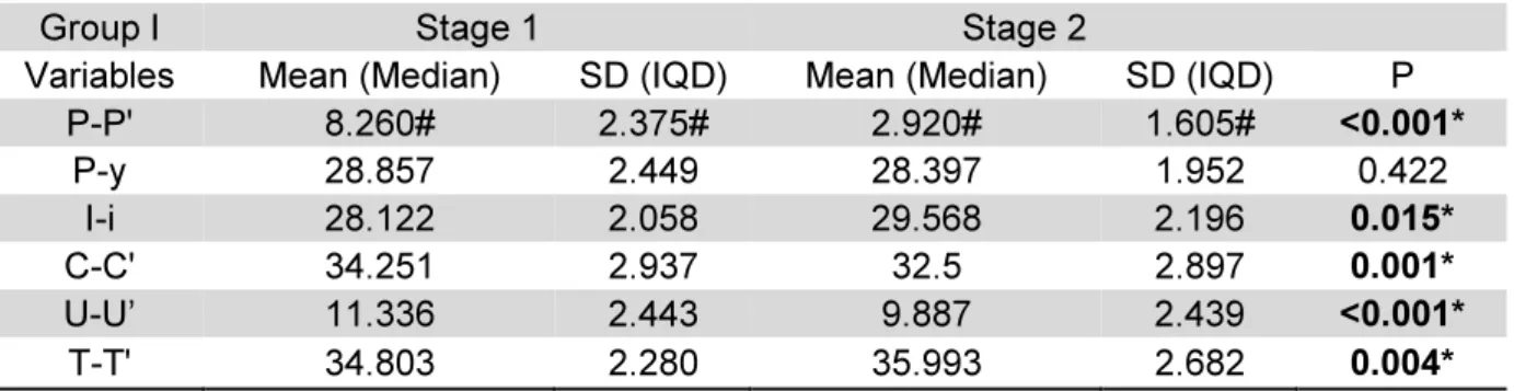
4.2. DIMENSIONAL ALTERATIONS IN GROUP I
In Group I, the dimensional alteration of dental arches for the anterior cleft width (P-P’), intercanine distance (C-C’) and posterior cleft width (U-U’) measurements decreased. However, anteroposterior arch distance (I-i) and intertuberosity distance (T-T’) measurements increased immediately after lip repair. There was no statistically significant difference in anteroposterior arch length (P-y) measurement (Table 5).
Table 5 - Maxillary dimensional alterations (mm) between stages 1 and 2 in group I, dependent t test
and Wilcoxon test
Group I Stage 1 Stage 2 Variables Mean (Median) SD (IQD) Mean (Median) SD (IQD) P
P-P' 8.260# 2.375# 2.920# 1.605# <0.001*
P-y 28.857 2.449 28.397 1.952 0.422 I-i 28.122 2.058 29.568 2.196 0.015*
C-C' 34.251 2.937 32.5 2.897 0.001*
U-U’ 11.336 2.443 9.887 2.439 <0.001*
T-T' 34.803 2.280 35.993 2.682 0.004* * Statistically significant difference
# median and IQD (interquartile deviation) of Wilcoxon test.
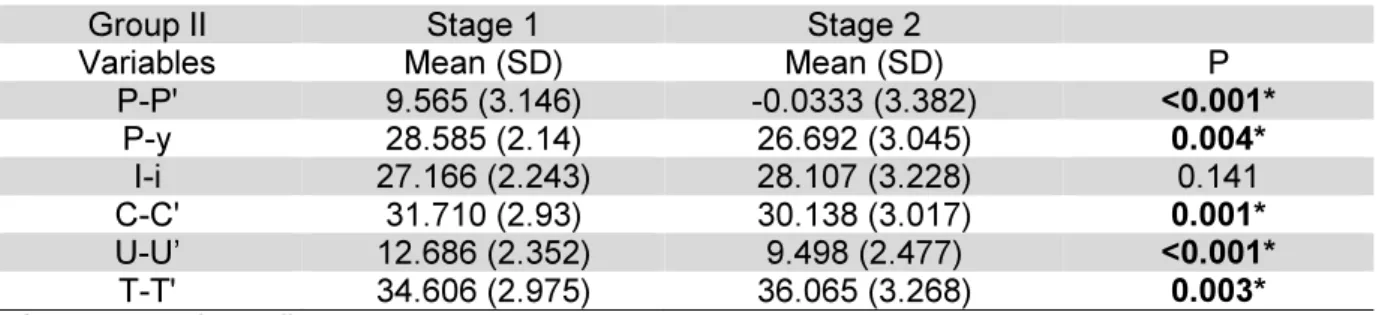
4.3 DIMENSIONAL ALTERATIONS IN GROUP II
Results 31
posterior cleft width (U-U’) measurements decreased. However, intertuberosity distance (T-T’) measurement increased immediately after lip repair. There was no statistically significant difference inanteroposterior arch distance(I-i) measurement (Table 6).
Table 6 - Maxillary dimensional alterations (mm) between stages 1 and 2 in group II, dependent t test.
Group II Stage 1 Stage 2
Variables Mean (SD) Mean (SD) P P-P' 9.565 (3.146) -0.0333 (3.382) <0.001*
P-y 28.585 (2.14) 26.692 (3.045) 0.004*
I-i 27.166 (2.243) 28.107 (3.228) 0.141 C-C' 31.710 (2.93) 30.138 (3.017) 0.001*
U-U’ 12.686 (2.352) 9.498 (2.477) <0.001*
T-T' 34.606 (2.975) 36.065 (3.268) 0.003* * Statistically significant difference
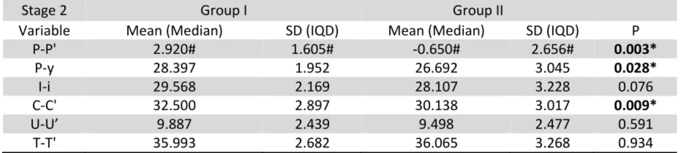
4.4. DIMENSIONAL ALTERATIONS BETWEEN GROUP I AND II
There were statistically significant differences between groups in stages S1 (Table 7), S2 (Table 8) and S2-S1 (Table 9).
Table 7 shows the statistically significant intercanine distance (C-C’) measurement at S1. Table 8 displays the statistically significant differences in anterior cleft width (P-P’), anteroposterior arch length (P-y) and intercanine distance (C-C’) measurements between groups at S2. The alterations in S2-S1 were statistically significant in anterior cleft width (P-P’) and posterior cleft width (U-U’) measurements, in groups I and II (Table 9).
Table 7 – Comparison of maxillary dimensional (mm) between groups I and II at stage 1 –
independent t test
Stage 1 Group I Group II
Variable Mean (SD) Mean (SD) P
P-P' 8.253 (2.908) 9.565 (3.146) 0.145 P-y 28.857 (2.449) 28.585 (2.14) 0.686 I-i 28.122 (2.058) 27.166 (2.243) 0.135 C-C' 34.251 (2.937) 31.71 (2.93) 0.005* U-U’ 11.336 (2.443) 12.626 (2.352) 0.060 T-T' 34.803 (2.28) 34.606 (2.975) 0.801
32 Results
Table 8: Comparison of maxillary dimensional (mm) between groups I and II at stage 2 - independent t
test and Mann-Whitney.
Stage 2 Group I Group II
Variable Mean (Median) SD (IQD) Mean (Median) SD (IQD) P
P-P' 2.920# 1.605# -0.650# 2.656# 0.003*
P-y 28.397 1.952 26.692 3.045 0.028*
I-i 29.568 2.169 28.107 3.228 0.076
C-C' 32.500 2.897 30.138 3.017 0.009*
U-U’ 9.887 2.439 9.498 2.477 0.591
T-T' 35.993 2.682 36.065 3.268 0.934
* Statistically significant difference
# median and IQD (interquartile deviation) of Mann-Whitney test.
Table 9: Comparison of maxillary dimensional alterations (mm) at stage 2 - stage 1 (S2-S1) between groups I and II – independent t test and Mann-Whitney.
* Statistically significant difference
# median and IQD (interquartile deviation) of Mann-Whitney test.
S2-S1 Group I Group II
Variable Mean (Median) SD (IQD) Mean (Median) SD (IQD) P P-P' -4.730# 1.54# -9.375# 2.029# <0.001*
P-y -0.461 2.7 -1.893 2.911 0.088
I-i 1.446 2.633 0.941 3.023 0.545
Discussion 35
5. DISCUSSION
This present study evaluated two cleft lip and palate rehabilitation centers with different treatment protocols. At University of Zurich (Group I), presurgical orthopedics (Hotz plate) is used after birth, at the first days of life. Cheiloplasty is performed at 6 months of age through modified Millard, Veau-Grob or Čelešnik technique. At 18 months of age, prior to soft palate repair through Widmaier-Perko technique, Hotz plate is removed. Between 4 and 5 years-old, hard palate is repaired by Perko technique. At University of Zurich, primary surgeries have been performed by plastic and oral and maxillofacial surgeons (HOTZ; GNOINSKI, 1979; HOTZ; GNOINSKI, 1982; GNOINSKI; RUTZ, 2009).
At the Hospital for Rehabilitation of Craniofacial Anomalies of the University of Sao Paulo (Group II), at 3 months of age, cheiloplasty is achieved by Millard or Spina technique. At 12 month-old, palataplosty is carried out by Von Langenbeck, Wardill or Furlow technique. Plastic surgeons performed all primary surgeries and presurgical orthopedics is not used (BERTIER, 2007; FREITAS et al, 2012a).
There was difference statistically significant in stage 1 between the groups I and II in relation of age. Liao and Mars 2005 suggested that early lip repair was better than later lip repair, then it produced a major bone remodeling, however, this comparison was performed with children among 6 months, 14.8 and 17 years old. Nevertheless, the literature is limited regarding researches which assess the interference of lip repair on growth and development of the maxilla, especially regarding the age of the groups of the present study.
36 Discussion
the pressure on dental arch and consequently the superposition of the maxillary segments (HOTZ; GNOINSKI, 1979).
The anteroposterior arch distance increased, but the anteroposterior cleft length did not show significant alteration in Group I. Accordingly, the anteroposterior maxillary growth in this group can be explained by the use of Hotz plate, which act as a retainer because it evenly spreads the forces that the repaired lip muscles impose throughout the alveolar ridge surface (HOTZ; GNOINSKI, 1979). Thus, Hotz plate provides a spontaneous growth at the anteroposterior length of the maxillary segments, which assume a more regular position closer to the normal patterns (HOTZ; GNOINSKI, 1976; HOTZ et al, 1978; HOTZ; GNOINSKI, 1979).
Grabowski et al. (2006) employed presurgical orthopedics and did not observe an anteroposterior maxillary growth in cleft lip and palate patients from birth to the eruption of permanent incisors.Braumann et al (2002) showed a significant increase of the anteroposterior length of the arch with the use of Hotz plate from birth to 12 months of age. Gnoinski and Rutz (2009) observed that presurgical orthopedics reduced maxillary growth deficiency in children with cleft lip and palate children. These studies show that presurgical orthopedics enables the anteroposterior arch growth, as seen in this present study. On the other hand, Prahl et al. (2001) found a presurgical orthopedic effect on anterior, medial, and posterior widths only up to soft palate repair. In 2003, Prahl et al. observed that presurgical orthopedics did not prevent the collapse of the maxillary segments from birth to 1 and half year of age.
In Group II, there was a decrease both in the anterior and posterior cleft width and in the anteroposterior cleft length. There were no differences between stages in the anteroposterior arch distance. Studies have indicated the influence of cheiloplasty by decreasing the anterior and posterior cleft width (MAZAHERI; HARDING; NANDA, 1967; HONDA et al, 1995; HUANG et al, 2002; REISER; SKOOG; ANDLIN-SOBOCKI, 2013).
Discussion 37
Concerning to Groups I and II, there was a significant increasing after lip repair regarding intertuberosity distance. Other studies have confirmed this result, thus validating the assumption that cheiloplasty does not interfere on intertuberosity distance growth (MAZAHERI; HARDING; NANDA, 1967; BRAUMANN et al, 2002; HUANG et al, 2002; REISER; SKOOG; ANDLIN-SOBOCKI, 2013).
The comparisons between the two cleft lip and palate rehabilitation centers aimed to analyze dental arch alterations according to the different treatment protocols. At stages 1 and 2, Group II intercanine distance was smaller than Group I, indicating that there was a molding effect on dental widht, which can lead to the superposition of maxillary segments. This occurred when there was a significant decrease in the anterior cleft arch, which agrees with other studies (HAN; SUZUKI; TASHIRO, 1995; HONDA et al, 1995; REISER; SKOOG; ANDLIN-SOBOCKI, 2013). At stage 2, in Group II anteroposterior arch distance and anterior cleft width were smaller than in Group I. Studies have affirmed that cheiloplasty decreases the anterior cleft width and acts as a permanent retrusive force (HUANG et al, 2002; REISER; SKOOG; ANDLIN-SOBOCKI, 2013), thus suggesting that lip repair can be an inhibiting factor of maxillary growth at vertical direction (GNOINSKI, 1982; LIAO; MARS, 2005; LI et al, 2006).
There were statistically significant differences in relation to the measurements of the anterior and posterior cleft width in the comparison of the changes between groups at stage 2 - stage 1 (S2-S1). The rationale behind this result is due to the use of Hotz plate once it guides the maxillary segments to more evenly approximation (MISHIMA et al., 1996; YAMADA et al, 2003), thus improving the arch morphology and creating conditions for the normalization of tongue positioning (GNOINSKI, 1990; YAMADA et al, 2003). It has been suggested that presurgical orthopedics maintains the normal functions and stimulates bone growth (GRABOWSKI et al, 2006).
38 Discussion
Conclusion 41
6. CONCLUSION
Based on the results obtained from the sample of this study, according to the methodology proposed, it was possible to conclude that:
1) In Group I, the comparisons between stages 1 and 2 showed that the intertuberosity and anteroposterior arch distance increased and the intercanine, anterior and posterior cleft distances decreased.
2) In Group II, the comparisons between stages 1 and 2 showed that, intertuberosity distance increased; and intercanine, anteroposterior archlength, anterior and posterior cleft widths decreased.
3) The comparison of the dimensional alterations of dental arches between the two rehabilitation centers exhibited differences:
o at stage 1, the intercanine distance was smaller in Group II than in Group I; and at stage 2, the anterior cleft width, the intercanine distance and the anteroposterior cleft length were smaller in Group II in Group I, suggesting the superposition of maxillary segments;
Reference 45
7. REFERENCES
AHMAD, I. Digital dental photography. Part 2: Purposes and uses. Br Dent J, v. 206, n. 9, p. 459-464, May 9 2009.
BERTIER, C.E., TRINDADE, I.E.K., SILVA FILHO, O.G. Cirurgias Primárias de Lábio e Palato. In: TRINDADE, I. E. K. S. F., O.G. Fissuras Labiopalatinas: uma abordagem interdisciplinar. São Paulo: Livraria Santos Editora Ltda; 2007. p. 73 - 85.
BOLTER, H. Oberkiefer-Alveolarbogen-masse bei LKG-Spalträgern nach Geburt und mit 5 Jahren.: Eine Standortbestimmung der primären Behandlung in Zürich. Med. Diss. , v., n., p., 1979.
BRAUMANN, B.; KEILIG, L.; BOURAUEL, C.; JAGER, A. Three-dimensional analysis of morphological changes in the maxilla of patients with cleft lip and palate. Cleft Palate Craniofac J, v. 39, n. 1, p. 1-11, Jan 2002.
CARRARA, C.F.C. Avaliação do tamanho das lâminas palatinas no resultado do crescimento dos arcos dentários e relação interarcos na dentadura decídua ou mista precoce. 2011. 106 f. - Universidade de São Paulo, Bauru, 2011.
CHIU, Y.T.; LIAO, Y.F.; CHEN, P.K. Initial cleft severity and maxillary growth in patients with complete unilateral cleft lip and palate. Am J Orthod Dentofacial Orthop, v. 140, n. 2, p. 189-195, Aug 2011.
DA SILVEIRA, A.C.; MARTINEZ, O.; DA SILVEIRA, D.; DAW, J.L., JR.; COHEN, M. Three-dimensional technology for documentation and record keeping for patients with facial clefts. Clin Plast Surg, v. 31, n. 2, p. 141-148, Apr 2004.
DE LADEIRA, P.R.; ALONSO, N. Protocols in cleft lip and palate treatment: systematic review. Plast Surg Int, v. 2012, n., p. 562892, 2012.
DIAH, E.; LO, L.J.; HUANG, C.S.; SUDJATMIKO, G.; SUSANTO, I.; CHEN, Y.R. Maxillary growth of adult patients with unoperated cleft: answers to the debates. J Plast Reconstr Aesthet Surg, v. 60, n. 4, p. 407-413, 2007.
46 Reference
FREITAS, J.A.; DAS NEVES, L.T.; DE ALMEIDA, A.L.; GARIB, D.G.; TRINDADE-SUEDAM, I.K.; YAEDU, R.Y. et al. Rehabilitative treatment of cleft lip and palate: experience of the Hospital for Rehabilitation of Craniofacial Anomalies/USP (HRAC/USP)--Part 1: overall aspects. J Appl Oral Sci, v. 20, n. 1, p. 9-15, Feb 2012a.
FREITAS, J.A.; GARIB, D.G.; OLIVEIRA, M.; LAURIS RDE, C.; ALMEIDA, A.L.; NEVES, L.T. et al. Rehabilitative treatment of cleft lip and palate: experience of the Hospital for Rehabilitation of Craniofacial Anomalies-USP (HRAC-USP)--part 2: pediatric dentistry and orthodontics. J Appl Oral Sci, v. 20, n. 2, p. 268-281, Mar-Apr 2012b.
GNOINSKI, W. Infant orthopedic and later orthodontic monitoring for unilateral cleft lip and palate patients in Zurich. In: BARDACH, J. M., HL. EDS. Multidisciplinary management of cleft lip and palate. Philadelphia: W.B. Saunders; 1990. p. 578 - 585.
GNOINSKI, W.M. Early maxillary orthopaedics as a supplement to conventional primary surgery in complete cleft lip and palate cases - long-term results. J Maxillofac Surg, v. 10, n. 3, p. 165-172, Aug 1982.
GNOINSKI, W.M.; RUTZ, G. A longitudinal cephalometric study from age 5 to 18 years on individuals with complete bilateral cleft lip and palate. J Craniofac Surg, v. 20 Suppl 2, n., p. 1672-1682, Sep 2009.
GRABOWSKI, R.; KOPP, H.; STAHL, F.; GUNDLACH, K.K. Presurgical orthopaedic treatment of newborns with clefts--functional treatment with long-term effects. J Craniomaxillofac Surg, v. 34 Suppl 2, n., p. 34-44, Sep 2006.
HAN, B.J.; SUZUKI, A.; TASHIRO, H. Longitudinal study of craniofacial growth in subjects with cleft lip and palate: from cheiloplasty to 8 years of age. Cleft Palate Craniofac J, v. 32, n. 2, p. 156-166, Mar 1995.
HEIDBUCHEL, K.L.; KUIJPERS-JAGTMAN, A.M. Maxillary and mandibular dental-arch dimensions and occlusion in bilateral cleft lip and palate patients form 3 to 17 years of age. Cleft Palate Craniofac J, v. 34, n. 1, p. 21-26, Jan 1997.
HONDA, Y.; SUZUKI, A.; NAKAMURA, N.; OHISHI, M. Relationship between primary palatal form and maxillofacial growth in Japanese children with unilateral cleft lip and palate: infancy to adolescence. Cleft Palate Craniofac J, v. 39, n. 5, p. 527-534, Sep 2002.
Reference 47
HOTZ, M.; GNOINSKI, W. Clefts of the secondary palate associated with the "Pierre Robin syndrome". Management by early maxillary orthopaedics. Swed Dent J Suppl, v. 15, n., p. 89-98, 1982.
HOTZ, M.; GNOINSKI, W. Comprehensive care of cleft lip and palate children at Zurich university: a preliminary report. Am J Orthod, v. 70, n. 5, p. 481-504, Nov 1976.
HOTZ, M.M.; GNOINSKI, W.M. Effects of early maxillary orthopaedics in coordination with delayed surgery for cleft lip and palate. J Maxillofac Surg, v. 7, n. 3, p. 201-210, Aug 1979.
HOTZ, M.M.; GNOINSKI, W.M.; NUSSBAUMER, H.; KISTLER, E. Early maxillary orthopedics in CLP cases: guidelines for surgery. Cleft Palate J, v. 15, n. 4, p. 405-411, Oct 1978.
HUANG, C.S.; WANG, W.I.; LIOU, E.J.; CHEN, Y.R.; CHEN, P.K.; NOORDHOFF, M.S. Effects of cheiloplasty on maxillary dental arch development in infants with unilateral complete cleft lip and palate. Cleft Palate Craniofac J, v. 39, n. 5, p. 513-516, Sep 2002.
LATIEF, B.S.; LEKKAS, K.C.; SCHOLS, J.G.; FUDALEJ, P.S.; KUIJPERS, M.A. Width and elevation of the palatal shelves in unoperated unilateral and bilateral cleft lip and palate patients in the permanent dentition. J Anat, v. 220, n. 3, p. 263-270, Mar 2012.
LEIFERT, M.F.; LEIFERT, M.M.; EFSTRATIADIS, S.S.; CANGIALOSI, T.J. Comparison of space analysis evaluations with digital models and plaster dental casts. Am J Orthod Dentofacial Orthop, v. 136, n. 1, p. 16 e11-14; discussion 16, Jul 2009.
LI, Y.; SHI, B.; SONG, Q.G.; ZUO, H.; ZHENG, Q. Effects of lip repair on maxillary growth and facial soft tissue development in patients with a complete unilateral cleft of lip, alveolus and palate. J Craniomaxillofac Surg, v. 34, n. 6, p. 355-361, Sep 2006.
LIAO, Y.F.; MARS, M. Long-term effects of lip repair on dentofacial morphology in patients with unilateral cleft lip and palate. Cleft Palate Craniofac J, v. 42, n. 5, p. 526-532, Sep 2005.
48 Reference
MAZAHERI, M.; ATHANASIOU, A.E.; LONG, R.E., JR.; KOLOKITHA, O.G. Evaluation of maxillary dental arch form in unilateral clefts of lip, alveolus, and palate from one month to four years. Cleft Palate Craniofac J, v. 30, n. 1, p. 90-93, Jan 1993.
MAZAHERI, M.; HARDING, R.L.; NANDA, S. The effect of surgery on maxillary growth and cleft width. Plast Reconstr Surg, v. 40, n. 1, p. 22-30, Jul 1967.
MEAZZINI, M.C.; TORTORA, C.; MORABITO, A.; GARATTINI, G.; BRUSATI, R. Factors that affect variability in impairment of maxillary growth in patients with cleft lip and palate treated using the same surgical protocol. J Plast Surg Hand Surg, v. 45, n. 4-5, p. 188-193, Sep 2011.
MISHIMA, K.; SUGAHARA, T.; MORI, Y.; SAKUDA, M. Three-dimensional comparison between the palatal forms in complete unilateral cleft lip and palate with and without Hotz plate from cheiloplasty to palatoplasty. Cleft Palate Craniofac J, v. 33, n. 4, p. 312-317, Jul 1996.
NOLLET, P.J.; KATSAROS, C.; VAN 'T HOF, M.A.; BONGAARTS, C.A.; SEMB, G.; SHAW, W.C. et al. Photographs of study casts: an alternative medium for rating dental arch relationships in unilateral cleft lip and palate. Cleft Palate Craniofac J, v. 41, n. 6, p. 646-650, Nov 2004.
PRAHL, C.; KUIJPERS-JAGTMAN, A.M.; VAN'T HOF, M.A.; PRAHL-ANDERSEN, B. A randomised prospective clinical trial into the effect of infant orthopaedics on maxillary arch dimensions in unilateral cleft lip and palate (Dutchcleft). Eur J Oral Sci, v. 109, n. 5, p. 297-305, Oct 2001.
PRAHL, C.; KUIJPERS-JAGTMAN, A.M.; VAN 'T HOF, M.A.; PRAHL-ANDERSEN, B. A randomized prospective clinical trial of the effect of infant orthopedics in unilateral cleft lip and palate: prevention of collapse of the alveolar segments (Dutchcleft). Cleft Palate Craniofac J, v. 40, n. 4, p. 337-342, Jul 2003.
REISER, E.; SKOOG, V.; ANDLIN-SOBOCKI, A. Early dimensional changes in maxillary cleft size and arch dimensions of children with cleft lip and palate and cleft palate. Cleft Palate Craniofac J, v. 50, n. 4, p. 481-490, Jul 2013.
ROSS, R.B. Treatment variables affecting facial growth in complete unilateral cleft lip and palate. Cleft Palate J, v. 24, n. 1, p. 5-77, Jan 1987.
Reference 49
SEMB, G.; BRATTSTROM, V.; MOLSTED, K.; PRAHL-ANDERSEN, B.; SHAW, W.C. The Eurocleft study: intercenter study of treatment outcome in patients with complete cleft lip and palate. Part 1: introduction and treatment experience. Cleft Palate Craniofac J, v. 42, n. 1, p. 64-68, Jan 2005a.
SEMB, G.; BRATTSTROM, V.; MOLSTED, K.; PRAHL-ANDERSEN, B.; ZUURBIER, P.; RUMSEY, N. et al. The Eurocleft study: intercenter study of treatment outcome in patients with complete cleft lip and palate. Part 4: relationship among treatment outcome, patient/parent satisfaction, and the burden of care. Cleft Palate Craniofac J, v. 42, n. 1, p. 83-92, Jan 2005b.
SHAO, Q.; CHEN, Z.; YANG, Y. Effects of Lip Repair on Maxillofacial Morphology in Patients With Unilateral Cleft Lip With or Without Cleft Palate. Cleft Palate Craniofac J, v., n., p., Jan 17 2014.
SHAW, W.C.; DAHL, E.; ASHER-MCDADE, C.; BRATTSTROM, V.; MARS, M.; MCWILLIAM, J. et al. A six-center international study of treatment outcome in patients with clefts of the lip and palate: Part 5. General discussion and conclusions. Cleft Palate Craniofac J, v. 29, n. 5, p. 413-418, Sep 1992.
STÖCKLI, P.W. A method for evaluation of maxillary arch form in complete unilateral cleft lip and palate cases. 1969f. - Northwestern University, Chicago, Illnois, 1969.
VEGTER, F.; HAGE, J.J. Standardized facial photography of cleft patients: just fit the grid? Cleft Palate Craniofac J, v. 37, n. 5, p. 435-440, Sep 2000.
WHO. Global strategies to reduce the health care burden of craniofacial anomalies: report of WHO meetings on international collaborative research on craniofacial anomalies. Cleft Palate Craniofac J, v. 41, n. 3, p. 238-243, May 2004.
WUTZL, A.; SINKO, K.; SHENGELIA, N.; BROZEK, W.; WATZINGER, F.; SCHICHO, K. et al. Examination of dental casts in newborns with bilateral complete cleft lip and palate. Int J Oral Maxillofac Surg, v. 38, n. 10, p. 1025-1029, Oct 2009.
YAMADA, T.; MORI, Y.; MISHIMA, K.; SUGAHARA, T. Nasolabial and alveolar morphology following presurgical orthopaedic treatment in complete unilateral clefts of lip, alveolus and palate. J Craniomaxillofac Surg, v. 31, n. 6, p. 343-347, Dec 2003.
Appendix 53
APPENDIX A
REVISÃO DE LITERATURA
FISSURA LABIOPALATINA
A fissura de lábio e palato é uma alteração congênita craniofacial que ocorre entre a 4ª e a 12ª semana de vida intrauterina, período o qual está ocorrendo o desenvolvimento embrionário da face e do palato (SILVA-FILHO; FREITAS, 2007; GARIB, 2010; FREITAS et al, 2012a). A fissura labiopalatina tem etiologia multifatorial, incluindo fatores genéticos e ambientais, os quais estão envolvidos na ação da migração, diferenciação e proliferação das células que formarão a face (WHO 2004; GARIB, 2010; FREITAS et al, 2012a).
No mundo, nasce uma criança com fissura a cada 2 minutos e meio (WHO, 2004). No Brasil, registram-se 5.800 nascimentos por ano (WHO, 2004; SILVA-FILHO; FREITAS, 2007; AAPD 2008). As fissuras podem envolver os lábios, rebordo alveolar e o palato (SILVA-FILHO; FREITAS, 2007; GARIB, 2010; FREITAS et al, 2012b). Por caracterizarem-se por diversos tipos, as fissuras labiopalatinas podem gerar diferentes sequelas. Nos casos de indivíduos com fissura pré-forame, as áreas envolvidas restringem-se somente ao esboço externo da face, ou seja, nariz, lábio e rebordo alveolar superior. As fissuras isoladas de palato têm o menor envolvimento estético e dentário, a implicação para esses indivíduos é funcional e as sequelas mais comuns são o regurgitamento de líquidos pela cavidade nasal, voz anasalada e deficiência auditiva, dependendo do grau de envolvimento anatômico. Os piores prognósticos estão ligados à fissura transforame incisivo por haver uma maior alteração morfológica, pois não houve fusão entre os palatos primário e secundário. A morfologia facial do paciente com fissura labiopalatina é fortemente influenciada pelos fatores genéticos, tipo e amplitude da fissura e pelas cirurgias primárias (SILVA FILHO, 2007).
REABILITAÇÃO DAS FISSURAS LABIOPALATINAS
54 Appendix
pacientes, englobando aspectos estéticos, funcionais e psicossociais (GRACIANO; TAVANO; BACHEGA, 2007), o qual é definido por fala inteligível, audição com padrão normativo, bom perfil facial, simetria labial e nasal adequadas, sorriso atrativo, boa oclusão, saúde das estruturas orais e autoestima favorável (VARGERVIK; OBEROI; HOFFMAN, 2009). Sendo assim, é importante deter o conhecimento detalhado de padrão morfológico característico das fissuras e a influência exata que as cirurgias primárias exercem sobre o crescimento do complexo craniofacial (SILVA FILHO, 2007).
Alguns estudos sugerem que as cirurgias reparadoras exercem um papel importante podendo diminuir todas as dimensões dos arcos dentários (ATHANASIOU; MAZAHERI; ZARRINNIA, 1987), havendo a constatação de um perfil achatado (ROSS, 1987), e uma maior severidade na deformação do arco maxilar (NAKAMURA et al, 2005). A severidade inicial da fissura e uma quantidade intrínseca de tecido das lâminas palatinas são considerados elementos que afetam o crescimento facial (CHIU; LIAO; CHEN, 2011; SHAO; CHEN; YANG, 2014).
Mazaheri et al. (1967) analisaram cefalometrias de 12 crianças com fissura transforame unilateral e sugeriram que a redução do tamanho da fissura palatina após o reparo de lábio envolve dois mecanismos (1) o reestabelecimento do complexo muscular labial e (2) o crescimento das lâminas palatinas. Os autores afirmaram que o reestabelecimento do musculo labial exerceu uma força modeladora, a qual resultou em subsequente diminuição da amplitude da fissura, e que o crescimento das lâminas palatinas também contribuiu para a diminuição da amplitude da fissura.
Appendix 55
que o reparo tardio, e a variável cirurgião não apresentou um impacto estatisticamente significante na morfologia dento-facial.
Shetye e Evans (2006) realizaram um estudo retrospectivo para investigar a deficiência maxilar observada em pacientes com fissura. Para isto, examinaram as radiografias cefalométricas laterais de pacientes não operados e de pacientes com fissura de lábio e palato. Os autores procuraram elucidar se a deficiência maxilar é um defeito intrínseco da maxila ou da intervenção cirúrgica em pacientes tratados com idade precoce. Deste modo analisaram 30 pacientes com fissura transforame unilateral, sem síndrome, e sem cirurgias prévias e compararam com 30 pacientes sem fissura. Conclui-se que o potencial para o crescimento normal da maxila existiu nos pacientes que apresentam a fissura. Sugeriu-se que os distúrbios do crescimento maxilar em pacientes cirurgicamente operados estava relacionado primariamente à intervenção cirúrgica.
Foi realizado um estudo por Chiu et al. (2011), que propuseram estabelecer se há uma relação entre a severidade da fissura ao nascimento e o crescimento maxilar. Para isso, foram analisadas medidas em modelos de gesso de 29 crianças sem síndromes com fissura transforame unilateral. Os resultados encontrados foram que há uma relação entre a área da fissura e a protusão maxilar, e também há uma relação entre a área do palato e a amplitude maxilar. Sugeriu-se que há uma relação significativa entre a severidade inicial da fissura e o crescimento maxilar.
56 Appendix
tratamento dental, fonoaudiologia e intervenções psicossociais. Conclui-se que o tratamento “Gold standard” é difícil de ser encontrado, por causa da grande variabilidade das malformações e a reposta subjetiva de cada paciente em relação ao tratamento reabilitador.
Shao et al. (2014) avaliaram em um estudo retrospectivo sobre os efeitos do reparo de lábio no desenvolvimento maxilofacial de pacientes com fissura unilateral de lábio com ou sem fissura de palato. Foram avaliados 38 pacientes com fissura unilateral completa de lábio e alvéolo operados, e 37 pacientes com fissura unilateral completa de lábio e palato que tiveram somente o lábio operado, os quais foram comparados com um grupo controle. Analisaram cefalogramas antes e após o pico da puberdade, os dados deste estudo mostraram que os pacientes com fissura completa unilateral de lábio e palato demostraram um crescimento maxilar próximo ao do grupo controle, um menor comprimento maxilar e incisivos superiores retroinclinados, e a mandíbula rotacionada no sentido horário. Os autores sugerem que existe um crescimento próximo do padrão de normalidade nos pacientes com fissura de lábio com ou sem fissura de palato que realizaram somente o fechamento do lábio, indicando que o reparo de lábio pode não ter um impacto negativo do desenvolvimento maxilofacial, pois somente influencia na inclinação dos incisivos superiores. E que as alterações do crescimento da maxila e mandíbula podem ser uma consequência inerente à fissura.
ORTOPEDIA PRÉ-CIRÚRGICA
McNeil (1954) introduziu o tratamento ortopédico pré-cirúrgico em pacientes com fissura labiopalatina. A confecção de aparelhos objetivava restaurar o contorno do arco maxilar normal a partir da mobilização controlada das lâminas palatinas e consequentemente houve diminuição da amplitude da fissura. Após o alinhamento do arco maxilar o paciente era submetido à cirurgia.
Appendix 57
De acordo com Rocha (1987) o uso das placas patina proporcionam uma alimentação adequada,levam e/ou mantem as lâminas palatinas a uma posição anatômica mais próxima da normalidade, diminui problemas das vias aéreas e respiratórios, auditivos, orientar o posicionamento da língua, evitar hábitos de sucção, estimular o crescimento ósseo da extremidade dos segmentos maxilares e ainda ser um apoio psicológico para os responsáveis.
Hotz (1990) empregou o uso da placa passiva com a finalidade de orientar o crescimento durante um período prolongado, a qual permite que ocorra o máximo de crescimento sem nenhum tipo de restrição antes das cirurgias primárias foram realizadas aos 6 meses (reparo de lábio), 18 meses (reparo do palato molde). Prahl et al. (2003) avaliaram o efeito da ortopedia pré-cirúrgica em relação à forma do arco e posição dos segmentos maxilares em crianças com fissura unilateral completa de lábio e palato, sem nenhuma outra malformação, com e sem o uso de placas passivas. Modelos de estudo foram avaliados e observou-se a sobreposição dos segmentos maxilares da 15ª até a 78ª semana em ambos os grupos. Nenhuma diferença significativa foi encontrada na presença e no grau de severidade da sobreposição dos segmentos maxilares. Concluindo-se que a ortopedia precoce deve ser abandonada como um recurso para correção do arco maxilar em pacientes com fissura labiopalatina.
Bongaarts et al (2006) analisaram o efeito da ortopedia pré-cirúrgica em pacientes com fissura unilateral de lábio e palato, através de um estudo prospectivo randomizado com a participação de 3 centros de reabilitação labiopalatina. Esses pacientes foram divididos em dois grupos, sem e com ortopedia pré-cirúrgica até o fechamento do palato mole com 53 semanas de idade. As dimensões dos arcos maxilares: amplitude, profundidade, comprimento, forma do arco, posição vertical do segmento menor, contato e sobreposição dos segmentos foram analisadas por modelos de estudo aos 4 e 6 anos de idade. Nenhuma diferença esteticamente significante foi encontrada. Concluindo que a ortopedia pré-cirúrgica não possui nenhum efeito satisfatório que evidencie o seu uso.
58 Appendix
palatina e na dentadura decídua completa o arco maxilar. A combinação da ortopedia pré-cirúrgica, que estimula o crescimento e a função, com cirurgias reparadoras minimamente invasivas permitem eu efeito longitudinal positivo ao longo do desenvolvimento da dentição.
DOCUMENTAÇÃO ODONTOLÓGICA
Para uma adequada análise do processo de reabilitação é essencial que haja a documentação do paciente, podendo ser usada para avaliação clínica dos tratamentos realizados, no ensino, pesquisa, na comparação de resultados com outros centros de tratamento assim como na elucidação de processos legais (VEGTER; HAGE, 2000; NOLLET et al, 2004; AHMAD, 2009).
Os modelos de gesso para estudo das arcadas dentárias podem ser considerados elementos-chave na documentação, para diagnóstico do plano de tratamento, uma vez que permitem um registro permanente dos arcos dentários, em três dimensões (GOODFELLOW, 2007; HATHAWAY et al, 2011). Seckel et al. (1995) testaram a precisão de pontos de referência na maxila em crianças na primeira infância, mediante análise inter e intraobservador. Os autores obtiveram resultado positivo em relação à precisão; porém concluíram que, para estudo em crianças com fissura labiopalatina, o posicionamento de pontos de referência na maxila são reprodutíveis se o modelo de gesso for de ótima qualidade se o pesquisador for experiente. A reprodutibilidade pode ser melhorada por meio de informações adicionais de modelos de gesso seriado de um mesmo indivíduo.
Darvann et al. (2007) compararam as medidas em 2 dimensões (fotografias) e 3 dimensões (superfície escaneada) nos modelos de gesso maxilares. A amostra foi composta 122 modelos de gesso de indivíduos com fissura transforame unilateral e bilateral. Os autores concluíram que, para estudos envolvendo amostra amplas, o uso de medidas em 2D pode levar a diminuição na precisão das medidas.
Appendix 59
quando essas marcações necessitaram ser realizadas em indivíduos com fissura labiopalatina, isso se tornou uma tarefa mais complicada. Os autores concluíram que as medidas entre certos pontos de referência são mais precisas que outras. Sugere-se usar pontos mais precisos usando um estudo longitudinal desde o nascimento até os 18 anos.
60 Appendix
REFERÊNCIAS DA REVISÃO DE LITERATURA
AAPD. Policy on management of patients cleft lip/palate and other craniofacial anomalies. Pediatr Dent, v. 30, n. 7 Suppl, p. 238-239, 2008.
AHMAD, I. Digital dental photography. Part 2: Purposes and uses. Br Dent J, v. 206, n. 9, p. 459-464, May 9 2009.
ATHANASIOU, A.E.; MAZAHERI, M.; ZARRINNIA, K. Longitudinal study of the dental arch dimensions in hard and soft palate clefts. J Pedod, v. 12, n. 1, p. 35-47, Fall 1987.
BONGAARTS, C.A.; VAN 'T HOF, M.A.; PRAHL-ANDERSEN, B.; DIRKS, I.V.; KUIJPERS-JAGTMAN, A.M. Infant orthopedics has no effect on maxillary arch dimensions in the deciduous dentition of children with complete unilateral cleft lip and palate (Dutchcleft). Cleft Palate Craniofac J, v. 43, n. 6, p. 665-672, Nov 2006.
BOOTVONG, K.; LIU, Z.; MCGRATH, C.; HAGG, U.; WONG, R.W.; BENDEUS, M. et al. Virtual model analysis as an alternative approach to plaster model analysis: reliability and validity. Eur J Orthod, v. 32, n. 5, p. 589-595, Oct 2010.
BRAUMANN, B.; KEILIG, L.; BOURAUEL, C.; JAGER, A. Three-dimensional analysis of morphological changes in the maxilla of patients with cleft lip and palate. Cleft Palate Craniofac J, v. 39, n. 1, p. 1-11, Jan 2002.
CHIU, Y.T.; LIAO, Y.F.; CHEN, P.K. Initial cleft severity and maxillary growth in patients with complete unilateral cleft lip and palate. Am J Orthod Dentofacial Orthop, v. 140, n. 2, p. 189-195, Aug 2011.
DA SILVEIRA, A.C.; MARTINEZ, O.; DA SILVEIRA, D.; DAW, J.L., JR.; COHEN, M. Three-dimensional technology for documentation and record keeping for patients with facial clefts. Clin Plast Surg, v. 31, n. 2, p. 141-148, Apr 2004.
DARVANN, T.A.; HERMANN, N.V.; ERSBOLL, B.K.; KREIBORG, S.; BERKOWITZ, S. Palatal surface area of maxillary plaster casts--a comparison between two-dimensional and three-two-dimensional measurements. Cleft Palate Craniofac J, v. 44, n. 4, p. 381-390, Jul 2007.
DIAH, E.; LO, L.J.; HUANG, C.S.; SUDJATMIKO, G.; SUSANTO, I.; CHEN, Y.R. Maxillary growth of adult patients with unoperated cleft: answers to the debates. J Plast Reconstr Aesthet Surg, v. 60, n. 4, p. 407-413, 2007.
FARRONATO, G.; CANNALIRE, P.; MARTINELLI, G.; TUBERTINI, I.; GIANNINI, L.; GALBIATI, G. et al. Cleft lip and/or palate: review. Minerva Stomatol, v. 63, n. 4, p. 111-126, Apr 2014.
Appendix 61
GARIB, D.G.S.F.O.G.J.G.P.J.H.N. Etiologia das más oclusões: perspectiva clínica (parte III) – fissuras labiopalatinas. Revista Clínica de Ortodontia Dental Press, v. 9, n. 4, p. 30-36, 2010.
GOODFELLOW, N. A digital approach to photographing and measuring cleft lip and palate dental study casts. J Vis Commun Med, v. 30, n. 2, p. 60-71, Jun 2007.
GRABOWSKI, R.; KOPP, H.; STAHL, F.; GUNDLACH, K.K. Presurgical orthopaedic treatment of newborns with clefts--functional treatment with long-term effects. J Craniomaxillofac Surg, v. 34 Suppl 2, n., p. 34-44, Sep 2006.
GRACIANO, M.I.G.; TAVANO, L.D.; BACHEGA, M.I. Aspectos Psicossociais da Reabilitação. In: LTDA, L. S. E. Fissuras Labiopalatinas: uma abordagem interdisciplinar. São Paulo: Trindade, I.E.K.; Silva Filho, O.G.; 2007.
HATHAWAY, R.; DASKALOGIANNAKIS, J.; MERCADO, A.; RUSSELL, K.; LONG, R.E., JR.; COHEN, M. et al. The Americleft study: an inter-center study of treatment outcomes for patients with unilateral cleft lip and palate part 2. Dental arch relationships. Cleft Palate Craniofac J, v. 48, n. 3, p. 244-251, May 2011.
HOTZ, M. Infant orthopedics and later monitoring for unilateral cleft lip and palate patients in Zurich. In: BARDACH, J., MORRIS, H. L. Multidisciplinary management of cleft lip and palate. Philadelphia: WB Saunders; 1990.
HOTZ, M.M.; GNOINSKI, W.M. Effects of early maxillary orthopaedics in coordination with delayed surgery for cleft lip and palate. J Maxillofac Surg, v. 7, n. 3, p. 201-210, Aug 1979.
LATIEF, B.S.; LEKKAS, K.C.; SCHOLS, J.G.; FUDALEJ, P.S.; KUIJPERS, M.A. Width and elevation of the palatal shelves in unoperated unilateral and bilateral cleft lip and palate patients in the permanent dentition. J Anat, v. 220, n. 3, p. 263-270, Mar 2012.
LEIFERT, M.F.; LEIFERT, M.M.; EFSTRATIADIS, S.S.; CANGIALOSI, T.J. Comparison of space analysis evaluations with digital models and plaster dental casts. Am J Orthod Dentofacial Orthop, v. 136, n. 1, p. 16 e11-14; discussion 16, Jul 2009.
LIAO, Y.F.; MARS, M. Long-term effects of lip repair on dentofacial morphology in patients with unilateral cleft lip and palate. Cleft Palate Craniofac J, v. 42, n. 5, p. 526-532, Sep 2005.
LO, L.J.; WONG, F.H.; CHEN, Y.R.; LIN, W.Y.; KO, E.W. Palatal surface area measurement: comparisons among different cleft types. Ann Plast Surg, v. 50, n. 1, p. 18-23; discussion 23-14, Jan 2003.
MAZAHERI, M.; HARDING, R.L.; NANDA, S. The effect of surgery on maxillary growth and cleft width. Plast Reconstr Surg, v. 40, n. 1, p. 22-30, Jul 1967.
MCNEIL, C.K. Oral and Facial deformity. London, Pitman, v., n., p. 81 - 89, 1954. NAKAMURA, N.; SUZUKI, A.; TAKAHASHI, H.; HONDA, Y.; SASAGURI, M.; OHISHI, M. A longitudinal study on influence of primary facial deformities on maxillofacial growth in patients with cleft lip and palate. Cleft Palate Craniofac J, v. 42, n. 6, p. 633-640, Nov 2005.